Withania somnifera L (Ashwagandha): A novel source of L asparaginase
Upload
vinod-nairCategory
view
213download
1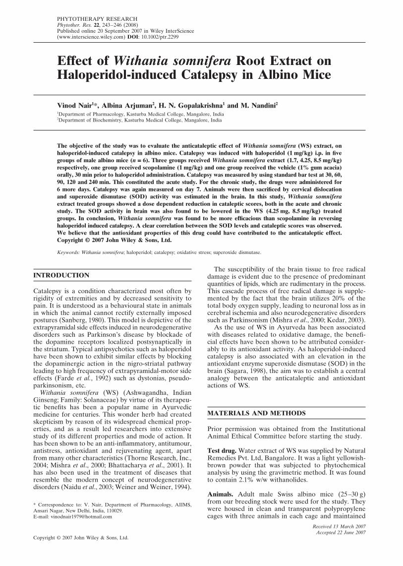
EFFECT OF WITHANIA SOMNIFERA ON HALOPERIDOL-INDUCED CATALEPSY 243
Copyright © 2007 John Wiley & Sons, Ltd. Phytother. Res. 22, 243–246 (2008)DOI: 10.1002/ptr
Copyright © 2007 John Wiley & Sons, Ltd.
PHYTOTHERAPY RESEARCHPhytother. Res. 22, 243–246 (2008)Published online 20 September 2007 in Wiley InterScience(www.interscience.wiley.com) DOI: 10.1002/ptr.2299
Effect of Withania somnifera Root Extract onHaloperidol-induced Catalepsy in Albino Mice
Vinod Nair1*, Albina Arjuman2, H. N. Gopalakrishna1 and M. Nandini2
1Department of Pharmacology, Kasturba Medical College, Mangalore, India2Department of Biochemistry, Kasturba Medical College, Mangalore, India
The objective of the study was to evaluate the anticataleptic effect of Withania somnifera (WS) extract, onhaloperidol-induced catalepsy in albino mice. Catalepsy was induced with haloperidol (1 mg/kg) i.p. in fivegroups of male albino mice (n ===== 6). Three groups received Withania somnifera extract (1.7, 4.25, 8.5 mg/kg)respectively, one group received scopolamine (1 mg/kg) and one group received the vehicle (1% gum acacia)orally, 30 min prior to haloperidol administration. Catalepsy was measured by using standard bar test at 30, 60,90, 120 and 240 min. This constituted the acute study. For the chronic study, the drugs were administered for6 more days. Catalepsy was again measured on day 7. Animals were then sacrificed by cervical dislocationand superoxide dismutase (SOD) activity was estimated in the brain. In this study, Withania somniferaextract treated groups showed a dose dependent reduction in cataleptic scores, both in the acute and chronicstudy. The SOD activity in brain was also found to be lowered in the WS (4.25 mg, 8.5 mg/kg) treatedgroups. In conclusion, Withania somnifera was found to be more efficacious than scopolamine in reversinghaloperidol induced catalepsy. A clear correlation between the SOD levels and cataleptic scores was observed.We believe that the antioxidant properties of this drug could have contributed to the anticataleptic effect.Copyright © 2007 John Wiley & Sons, Ltd.
Keywords: Withania somnifera; haloperidol; catalepsy; oxidative stress; superoxide dismutase.
Received 13 March 2007Accepted 22 June 2007
* Correspondence to: V. Nair, Department of Pharmacology, AIIMS,Ansari Nagar, New Delhi, India, 110029.E-mail: [email protected]
INTRODUCTION
Catalepsy is a condition characterized most often byrigidity of extremities and by decreased sensitivity topain. It is understood as a behavioural state in animalsin which the animal cannot rectify externally imposedpostures (Sanberg, 1980). This model is depictive of theextrapyramidal side effects induced in neurodegenerativedisorders such as Parkinson’s disease by blockade ofthe dopamine receptors localized postsynaptically inthe striatum. Typical antipsychotics such as haloperidolhave been shown to exhibit similar effects by blockingthe dopaminergic action in the nigro-striatal pathwayleading to high frequency of extrapyramidal-motor sideeffects (Farde et al., 1992) such as dystonias, pseudo-parkinsonism, etc.
Withania somnifera (WS) (Ashwagandha, IndianGinseng; Family: Solanaceae) by virtue of its therapeu-tic benefits has been a popular name in Ayurvedicmedicine for centuries. This wonder herb had createdskepticism by reason of its widespread chemical prop-erties, and as a result led researchers into extensivestudy of its different properties and mode of action. Ithas been shown to be an anti-inflammatory, antitumour,antistress, antioxidant and rejuvenating agent, apartfrom many other characteristics (Thorne Research, Inc.,2004; Mishra et al., 2000; Bhattacharya et al., 2001). Ithas also been used in the treatment of diseases thatresemble the modern concept of neurodegenerativedisorders (Naidu et al., 2003; Weiner and Weiner, 1994).
The susceptibility of the brain tissue to free radicaldamage is evident due to the presence of predominantquantities of lipids, which are rudimentary in the process.This cascade process of free radical damage is supple-mented by the fact that the brain utilizes 20% of thetotal body oxygen supply, leading to neuronal loss as incerebral ischemia and also neurodegenerative disorderssuch as Parkinsonism (Mishra et al., 2000; Kedar, 2003).
As the use of WS in Ayurveda has been associatedwith diseases related to oxidative damage, the benefi-cial effects have been shown to be attributed consider-ably to its antioxidant activity. As haloperidol-inducedcatalepsy is also associated with an elevation in theantioxidant enzyme superoxide dismutase (SOD) in thebrain (Sagara, 1998), the aim was to establish a centralanalogy between the anticataleptic and antioxidantactions of WS.
MATERIALS AND METHODS
Prior permission was obtained from the InstitutionalAnimal Ethical Committee before starting the study.
Test drug. Water extract of WS was supplied by NaturalRemedies Pvt. Ltd, Bangalore. It was a light yellowish-brown powder that was subjected to phytochemicalanalysis by using the gravimetric method. It was foundto contain 2.1% w/w withanolides.
Animals. Adult male Swiss albino mice (25–30 g)from our breeding stock were used for the study. Theywere housed in clean and transparent polypropylenecages with three animals in each cage and maintained

Copyright © 2007 John Wiley & Sons, Ltd. Phytother. Res. 22, 243–246 (2008)DOI: 10.1002/ptr
244 V. NAIR ET AL.
at 27 °C with 12 h: 12 h light–dark cycle for a period of7 days prior to the study. They were fed standard ratchow and water ad libitum. The animals were dividedinto five groups with six animals in each group. Allobservations were made between 10:00 hr and 16:00 hr.The test drug WS and scopolamine were suspended in1% gum acacia solution (vehicle). All drug solutionswere freshly prepared and administered orally using afeeding tube. The first group received vehicle and servedas the control, the second group received 1 mg/kg bodyweight of scopolamine and the third, fourth and fifthgroups received WS in doses of 1.7, 4.25 and 8.5 mg/kgbody weight respectively. Thirty minutes after adminis-tration of vehicle/drugs, haloperidol in a dose of 1 mg/kg body weight, in the form of an injectable solutionconstituted in normal saline was administered by thei.p. route to induce catalepsy. This dose of haloperidolwas chosen from our pilot study to produce a moderatedegree of catalepsy, so that attenuation or potentiationof the phenomenon could be detected.
Acute study. The degree of catalepsy was measuredat 30, 60, 90, 120 and 240 min after haloperidol admin-istration by using a method similar to the standard bartest (Ahtee and Buncombe, 1974). Briefly, catalepsywas measured as the time the animal maintained animposed position with both front limbs extended andresting on a 4 cm high wooden bar. The end point ofcatalepsy was considered to occur when both front pawswere removed from the bar or if the animal movedits head in an exploratory manner. If the animal main-tained the imposed posture for at least 20 s it was saidto be cataleptic and given one point. For every further20 s that the animal continued to maintain the cata-leptic posture one extra point was given. Based on thedata obtained in our pilot study, a cut-off catalepticscore of 60 (corresponding to 20 min) was used duringthe recording of observations. Between determinations,the animals were returned to their individual homecages.
Chronic study. WS, scopolamine and haloperidol wereadministered continuously for a period of 6 more daysin the manner described for the acute study. Catalepsywas again measured on day 7 at 30, 60, 90, 120 and240 min post-haloperidol administration. Soon afterthe behavioural study, the animals were sacrificed bycervical dislocation and the SOD (EC 1.15.1.1;
superoxide:superoxide oxidoreductase) activity in thewhole brain was estimated (Beauchamp and Fridovich,1971). Total protein (Peterson, 1979) in the brain wasestimated and the SOD activity was expressed in termsof units/mg total protein. To give the normal range forSOD activity in our testing conditions and to rule outthe involvement of the vehicle, three mice were admin-istered 10 mL/kg vehicle for 7 days. On day 7 they weresacrificed by cervical dislocation 270 min after vehicleadministration and the SOD activity and total proteinin the brain was determined.
Statistical analysis. The data were analysed by one-wayANOVA, followed by Dunnett’s multiple comparisontest. p < 0.05 was considered significant.
RESULTS
Acute study
In the acute phase of the study (Table 1), a dose de-pendent decrease in the cataleptic score was observedin the WS treated groups. When compared with thecontrol, a significant reduction in cataleptic scores wasobserved in the WS 4.25 and WS 8.5 mg/kg treatedgroups from the start of the study, whereas in the WS1.7 mg/kg treated groups there was an initial increasein the cataleptic score. Significant reduction in this groupwas observed only after 120 min into the study. Whencompared with scopolamine, only the WS 4.25 and WS8.5 mg/kg treated groups showed a greater reduction incataleptic scores throughout the study.
Chronic study
In the chronic study also (Table 2), there was a dosedependent decrease in the cataleptic scores in the WStreated groups. A significant reduction was observed inWS 4.25 and 8.5 mg/kg treated groups from the start ofthe study. A significant reduction in the WS 1.7 mg/kgand scopolamine treated groups was observed onlyafter 60 and 90 min, respectively. When compared withscopolamine, only the WS 8.5 mg/kg treated groupshowed a reduction in cataleptic score throughout theobservation period.
Table 1. Effect of WS on haloperidol-induced catalepsy: acute study
Time (min) after haloperidol administration
Group 30 60 90 120 240
Control 11.5 ± 1.25 24.5 ± 1.76 34 ± 3.67 47.5 ± 4.47 51.33 ± 2.21b
Scopolamine 1.0 12.66 ± 0.95 17 ± 1b 21.5 ± 1.14b 26.83 ± 0.98b 17.83 ± 1.37b
WS 1.7 15.33 ± 0.98 25.16 ± 0.70 28.16 ± 0.83 25.33 ± 2.39b 18.83 ± 2.15b
WS 4.25 5.66 ± 0.61b 9 ± 0.81b 14.5 ± 0.99b 16.33 ± 2.39b 15.33b
WS 8.5 4.42 ± 0.42b 7.76 ± 0.66b 10.66 ± 0.49b 16.33 ± 0.42b 11.33 ± 0.71b
One-way ANOVAF 27.577 59.804 27.489 24.942 109.54p <0.0001 <0.0001 <0.0001 <0.0001 <0.0001df 4,25 4,25 4,25 4,25 4,25
All values are mean ± SEM (n = 6 in each group). Statistical analysis by one-way ANOVA followed by Dunnett’s multiple comparisontest. b p < 0.01.

EFFECT OF WITHANIA SOMNIFERA ON HALOPERIDOL-INDUCED CATALEPSY 245
Copyright © 2007 John Wiley & Sons, Ltd. Phytother. Res. 22, 243–246 (2008)DOI: 10.1002/ptr
Table 2. Effect of WS on haloperidol-induced catalepsy: chronic study
Time (min) after haloperidol administration
Group 30 60 90 120 240
Control 13.33 ± 0.80 18.33 ± 1.16 25.33 ± 0.84 34.16 ± 1.40 37.33 ± 1.20Scopolamine 1.0 13.66 ± 0.49 15.33 ± 0.47 18.33 ± 0.40b 16.66 ± 0.49b 14.5 ± 0.50b
WS 1.7 11.16 ± 0.47 14.66 ± 0.91a 19.66 ± 0.88b 16.33 ± 0.66b 21.5 ± 1.58b
WS 4.25 9.83 ± 0.47b 13.16 ± 0.87b 17.83 ± 0.83b 14.16 ± 0.47b 16.16 ± 0.47b
WS 8.5 9.33 ± 0.71b 12.16 ± 0.65b 15.66 ± 0.49b 13.66 ± 0.49b 11.5 ± 0.56b
One way ANOVAF 10.693 7.832 25.702 118.82 111.11p <0.0001 0.0003 <0.0001 <0.0001 <0.0001df 4,25 4,25 4,25 4,25 4,25
All values are mean ± SEM (n = 6 in each group). Statistical analysis by one-way ANOVA followed by Dunnett’s multiple comparisontest. a p < 0.05, b p < 0.01.
SOD activity
A reduction in SOD activity was observed in scopo-lamine (Table 3), WS 4.25 and 8.5 mg/kg treated groups,but a significant reduction was observed only in the WS8.5 mg/kg treated group.
DISCUSSION
Haloperidol is a well known neuroleptic, primarily actingas a D2 receptor antagonist in the mesolimbic-mesocorticalpathway. Due to its non-selective action, it also producesblockade of post-synaptic D2 receptors in the nigro-striatal pathway leading to the development of extrapy-ramidal side effects in humans (Farde et al., 1992) andcatalepsy in animals (Sanberg, 1980). Haloperidol-induced catalepsy is also associated with an increase inoxidative stress in the brain (Sagara, 1998).
Studies have reported the beneficial effects of WS inhaloperidol-induced tardive dyskinesia (Bhattacharyaet al., 2002). In our study also a dose dependent decreasein catalepsy was observed in the WS treated groups inboth acute and chronic studies. When compared withthe standard drug scopolamine, WS in doses of 4.25and 8.5 mg/kg was found to be more effective in revers-ing haloperidol-induced catalepsy. Similarly a dosedependent decrease in SOD activity was observedin the WS treated groups. Even though the reductionwas significant only in the highest dose treated group,the SOD activity in the WS 4.25 mg/kg treated group
was closer to the normal brain SOD values, comparedwith the control which shows a decrease in oxidativestress. This reduction in brain SOD activity could beattributed to the antioxidant phytochemical constitu-ents of WS (Mishra et al., 2000; Bhattacharya et al.,2001). Paradoxically, WS 1.7 mg/kg showed a decreasein the cataleptic score but an increase in brain SODactivity compared with the control. The reason forthis increase cannot be explained by the present study.A plausible explanation could be the involvement ofdirect and indirect antioxidants in WS (Bhattacharyaet al., 2001; Naidu et al., 2003). A direct antioxidant isone which possesses the ability to quench free radicalsdirectly, whereas an indirect antioxidant is one whichinduces antioxidant enzymes. The net result of boththe processes is a decrease in the oxidative stress.To ascertain the actual mechanisms involved, furtherstudies are required.
In conclusion, WS in the highest dose tested wasfound to be more efficacious than the standard drugscopolamine in reversing haloperidol-induced catalepsy.A clear correlation between the SOD levels andcataleptic scores can be observed. Based on our findingsan analogy can be drawn between the antioxidantand anticataleptic effects of WS, with a possible appli-cation of the same as an adjuvant in the treatment indrug-induced Parkinsonism.
Acknowledgement
Natural Remedies Pvt. Ltd, Bangalore provided the test drug Withaniasomnifera.
Table 3. Effect of WS on brain SOD levels
SOD % reduction % increaseGroup (U/mg TP) from control from control
Normal 5.28 ± 0.28Control 8.81 ± 0.28Scopolamine 8.49 ± 0.66 3.63WS 1.7 9.88 ± 0.72 12.14WS 4.25 6.99 ± 0.28 20.66WS 8.5 6.18 ± 0.49a 29.85One way ANOVAF 8.086p 0.0035df 4,10
All values are mean ± SEM (n = 3 in each group). Statistical analysis by one-wayANOVA followed by Dunnett’s multiple comparison test. a p < 0.05.

Copyright © 2007 John Wiley & Sons, Ltd. Phytother. Res. 22, 243–246 (2008)DOI: 10.1002/ptr
246 V. NAIR ET AL.
REFERENCES
Ahtee L, Buncombe G. 1974. Metoclopramide induces catalepsyand increases striatal homovanillic acid content in mice.Acta Pharmacol Toxicol 35: 429–432.
Beauchamp C, Fridovich I. 1971. Superoxide dismutase:improved assays and an assay applicable to acrylamidegels. Anal Biochem 44: 276–287.
Bhattacharya A, Ghosal S, Bhattacharya SK. 2001. Antioxidanteffect of Withania somnifera glycowithanolides in chronicfootshock stress-induced perturbations of oxidative freeradical scavenging enzymes and lipid peroxidation in ratfrontal cortex and striatum. J Ethnopharmacol 74: 1–6.
Bhattacharya SK, Bhattacharya D, Sairam K, Ghosal S. 2002.Effect of Withania somnifera glycowithanolides on a ratmodel of tardive dyskinesia. Phytomedicine 9: 167–170.
Farde L, Nordstrom AL, Wiesel FA, Pauli S, Halldin C, Sedvall G.1992. Positron emission tomographic analysis of centralD1 and D2 dopamine receptor occupancy in patients treatedwith classical neuroleptics and clozapine: Relation toextrapyramidal side effects. Arch Gen Psychiatry 49: 538–544.
Kedar NP. 2003. Can we prevent Parkinson’s and Alzheimer’sdisease? J Postgrad Med 49: 236–245.
Mishra LC, Singh BB, Dagenais S. 2000. Scientific basis for thetherapeutic use of Withania somnifera (Ashwagandha):A review. Altern Med Rev 5: 334–346.
Naidu PS, Singh A, Kulkarni SK. 2003. Effect of Withaniasomnifera root extract on haloperidol induced orofacialdyskinesia: Possible mechanisms of action. J Med Food 6:107–114.
Peterson GL. 1979. Review of the folin phenol protein quantifi-cation method of Lowry, Rosebrough, Farr and Randall.Anal Biochem 100: 201–220.
Sagara Y. 1998. Induction of reactive oxygen species inneurons by haloperidol. J Neurochem 71: 1002–1012.
Sanberg PR. 1980. Haloperidol-induced catalepsy is mediatedby postsynaptic dopamine receptors. Nature 284: 472–473.
Thorne Research, Inc. 2004. Withania somnifera. Altern MedRev 9: 211–214.
Weiner MA, Weiner J. 1994. Ashwagandha (Indian ginseng). InHerbs that Heal. Quantum Books: Mill Valley, CA, 70–72.