Effect of treatment of cow’s urine “Gomutra” and antioxidants in alleviating the...
10
Effect of treatment of cow’s urine ‘‘Gomutra’’ and antioxidants in alleviating the lindane-induced oxidative stress in kidney of Swiss mice (Mus musculus) Girima Nagda • Devendra Kumar Bhatt Received: 5 December 2012 / Accepted: 4 January 2014 / Published online: 16 January 2014 Ó Springer Science+Business Media Dordrecht 2014 Abstract The study aimed to evaluate the effect of cow urine and combination of antioxidants against lindane induced oxidative stress in Swiss mice. Male healthy mice, 8–10 weeks old, weighing 30 ± 5 g were randomly selected and divided into eight groups, namely, control (C); lindane (L); antioxidant (A), antioxidant?lindane (A?L), cow urine (U), cow urine?lindane (U?L), cow urine?antioxidants (U?A) and cow urine?antioxidants?lindane (U?A?L). Group C animals were administered only the vehicle (olive oil); doses selected for other treatments were: lindane: 40 mg/kg b.w.; antioxidants: 125 mg/kg b.w. (vitamin C: 50 mg/kg b.w., vitamin E: 50 mg/kg b.w., a-lipoic acid: 25 mg/kg b.w.) and cow urine: 0.25 ml/kg b.w. In group A?L and U?L antioxidants and cow urine were adminis- tered 1 h prior to lindane administration and in group U?A and U?A?L cow urine was administered 10 min before antioxidants. All treatments were administered orally con- tinuously for 60 days. Lindane treated group showed increased lipid peroxidation, whereas glutathione, glutathi- one peroxidase, superoxide dismutase, catalase, protein and endogenous levels of vitamin C and E were significantly decreased compared to control. Administration of cow urine and antioxidants alleviated the levels of these biochemical parameters. Keywords Antioxidants Á Cow urine Á Kidney Á Lindane Á Oxidative stress Introduction Kidney is vital organ of the body responsible for segre- gating the metabolic waste products from the blood. Accumulation of metabolites of xenobiotics contributes significantly to its susceptibility to damage kidney [24]. Any nephrotoxic insult would result in accumulation of waste materials in the blood which in turn may lead to other toxic manifestations in the body. Toxic injury to the kidney is known to occur as a result of exposures to hal- ogenated hydrocarbons, such as lindane, carbon tetrachlo- ride and trichloroethylene, and the heavy metals cadmium and lead [3, 35, 36, 48, 59]. Some of these toxicants cause acute injury to the kidney, while others produce chronic changes that can lead to end-stage renal failure or cancer. Lindane, the gamma isomer of HCH possesses the prop- erty of persistence, bioaccumulation and long term toxicity [32] and fulfills the criteria of POPs i.e., persistent organo- chlorine pesticides. India has total installed capacity of lin- dane (technical) production of 1,300 tonnes per annum (tpa), with two companies producing: Kanoria Chemicals and Industries Ltd with a capacity of 1,000 tpa, and India Pes- ticides Limited with 300 tpa capacity. According to data available from Department of Chemicals and Petrochemi- cals, Ministry of Chemicals and Fertilizers, between 1995 and 2005, India has produced 5,387 tonnes of technical grade lindane. In India, lindane formulations are registered for use in pharmaceutical products for control of head lice and scabies on people, to control fly, flea, cockroach, mosquito, bed bug, and beetle populations and for the control of pests in cotton, sugarcane, pumpkin, cabbage, onion, apple, walnut, maize, okhra, potato, tomato, cauliflower, radish, cucumber and beans [15]. Lindane is highly lipophilic and absorbed by the respiratory, digestive or cutaneous pathways. Its accu- mulation depends on the duration of the exposure and affect G. Nagda (&) Á D. K. Bhatt Cancer Biology and Toxicology Research Laboratory, Department of Zoology, University College of Science, M L Sukhadia University, Udaipur 313 001, India e-mail: [email protected] 123 Mol Biol Rep (2014) 41:1967–1976 DOI 10.1007/s11033-014-3044-6
-
Upload
neem-plant -
Category
Documents
-
view
11 -
download
1
Transcript of Effect of treatment of cow’s urine “Gomutra” and antioxidants in alleviating the...

Effect of treatment of cow’s urine ‘‘Gomutra’’ and antioxidantsin alleviating the lindane-induced oxidative stress in kidneyof Swiss mice (Mus musculus)
Girima Nagda • Devendra Kumar Bhatt
Received: 5 December 2012 / Accepted: 4 January 2014 / Published online: 16 January 2014
� Springer Science+Business Media Dordrecht 2014
Abstract The study aimed to evaluate the effect of cow
urine and combination of antioxidants against lindane
induced oxidative stress in Swiss mice. Male healthy mice,
8–10 weeks old, weighing 30 ± 5 g were randomly selected
and divided into eight groups, namely, control (C); lindane
(L); antioxidant (A), antioxidant?lindane (A?L), cow urine
(U), cow urine?lindane (U?L), cow urine?antioxidants
(U?A) and cow urine?antioxidants?lindane (U?A?L).
Group C animals were administered only the vehicle (olive
oil); doses selected for other treatments were: lindane:
40 mg/kg b.w.; antioxidants: 125 mg/kg b.w. (vitamin C:
50 mg/kg b.w., vitamin E: 50 mg/kg b.w., a-lipoic acid:
25 mg/kg b.w.) and cow urine: 0.25 ml/kg b.w. In group
A?L and U?L antioxidants and cow urine were adminis-
tered 1 h prior to lindane administration and in group U?A
and U?A?L cow urine was administered 10 min before
antioxidants. All treatments were administered orally con-
tinuously for 60 days. Lindane treated group showed
increased lipid peroxidation, whereas glutathione, glutathi-
one peroxidase, superoxide dismutase, catalase, protein and
endogenous levels of vitamin C and E were significantly
decreased compared to control. Administration of cow urine
and antioxidants alleviated the levels of these biochemical
parameters.
Keywords Antioxidants � Cow urine � Kidney � Lindane �Oxidative stress
Introduction
Kidney is vital organ of the body responsible for segre-
gating the metabolic waste products from the blood.
Accumulation of metabolites of xenobiotics contributes
significantly to its susceptibility to damage kidney [24].
Any nephrotoxic insult would result in accumulation of
waste materials in the blood which in turn may lead to
other toxic manifestations in the body. Toxic injury to the
kidney is known to occur as a result of exposures to hal-
ogenated hydrocarbons, such as lindane, carbon tetrachlo-
ride and trichloroethylene, and the heavy metals cadmium
and lead [3, 35, 36, 48, 59]. Some of these toxicants cause
acute injury to the kidney, while others produce chronic
changes that can lead to end-stage renal failure or cancer.
Lindane, the gamma isomer of HCH possesses the prop-
erty of persistence, bioaccumulation and long term toxicity
[32] and fulfills the criteria of POPs i.e., persistent organo-
chlorine pesticides. India has total installed capacity of lin-
dane (technical) production of 1,300 tonnes per annum (tpa),
with two companies producing: Kanoria Chemicals and
Industries Ltd with a capacity of 1,000 tpa, and India Pes-
ticides Limited with 300 tpa capacity. According to data
available from Department of Chemicals and Petrochemi-
cals, Ministry of Chemicals and Fertilizers, between 1995
and 2005, India has produced 5,387 tonnes of technical grade
lindane. In India, lindane formulations are registered for use
in pharmaceutical products for control of head lice and
scabies on people, to control fly, flea, cockroach, mosquito,
bed bug, and beetle populations and for the control of pests in
cotton, sugarcane, pumpkin, cabbage, onion, apple, walnut,
maize, okhra, potato, tomato, cauliflower, radish, cucumber
and beans [15]. Lindane is highly lipophilic and absorbed by
the respiratory, digestive or cutaneous pathways. Its accu-
mulation depends on the duration of the exposure and affect
G. Nagda (&) � D. K. Bhatt
Cancer Biology and Toxicology Research Laboratory,
Department of Zoology, University College of Science,
M L Sukhadia University, Udaipur 313 001, India
e-mail: [email protected]
123
Mol Biol Rep (2014) 41:1967–1976
DOI 10.1007/s11033-014-3044-6
tissues in the following order: fat [ brain [ kidney [muscle[ lung[heart[ spleen[ liver[blood[56].Toxicity
induced by lindane is attributed to oxidative stress as it
induces the release of free radicals and generation of reactive
oxygen species (ROS) [67].
Oxidative stress is defined as a disruption of the pro-
oxidant–antioxidant balance in favor of the former, leading
to potential damage [37]. It is a result of one of three
factors: (i) an increase in ROS, (ii) an impairment of
antioxidant defence systems, or (iii) an incapacity to repair
oxidative damage.
A number of studies indicate the toxicity of lindane on
kidney [22, 48]. It has been considered that ROS play an
important role in the pathogenesis of renal injury. Since the
entire range of toxic metabolites in the body is excreted
mainly from the kidney, this organ is endowed with sig-
nificant antioxidant defense system next only to liver. This
is understandable because ROS play a key intermediary
role in the pathophysiologic processes of a wide variety of
clinical and experimental renal diseases [50, 65].
The human body has a strong inherent synergistic and
multilevel defense mechanism, to combat and counteract
the damage caused by free radicals [41]. This defense is
mediated by endogenous antioxidant system which either
prevent these reactive species from being formed, or cause
their removal before they can damage vital components of
the cell [17]. The excessive free radicals or the suppression
of antioxidant defense of body results into toxicity. In such
conditions, though the body tissue is endowed with enzy-
matic and non-enzymatic protective systems, but it seems
that the homeostasis of the body fails. In such situations,
the use of exogenous substances with antioxidative
potential becomes important [64].
Antioxidants have gained immense importance in recent
years. The potency of various antioxidants on different
organ systems has been investigated against lindane tox-
icity [8, 9, 11, 42, 63].
Cow urine or Gomutra is considered sacred in Hindu
mythology and from ancient times it has been used as a
medicine in India. The medicinal properties of cow’s urine
have been mentioned in Sushrut (45/221) and Charak
(sloka-100) where it is considered useful in treating renal
colic, jaundice, anemia, diarrhea, gastric infection, piles
and skin diseases including vitiligo. It is also considered as
an appetizer and is known to reverse inflammation, a
diuretic as well as a nephroprotective agent. It also acts at
cellular level and generates bioenergy [31]. The analysis of
cow urine has shown that it contains nitrogen, sulphur,
phosphate, sodium, manganese, carbolic acid, iron, silicon,
chlorine, magnesium, melci, citric, titric, succinic, calcium
salts, vitamin A, B, C, D, E, minerals, lactose, enzymes,
creatinine, hormones and gold acids. The cow urine con-
tains those substances, which are present in the human
body and thus its consumption maintains the balance of
these substances and cures incurable diseases [49]. Cow
urine is also used along with herbs to treat various diseases
like fever, epilepsy, anemia, abdominal pain, constipation
etc. by the traditional healers [34].
There is a paucity of information regarding the role of
fresh cow urine and combination of vitamin E, vitamin C
and alpha lipoic acid against lindane toxicity in kidney.
Therefore, in the present study their role in alleviating the
oxidative stress induced by lindane intoxication in kidney
of mice has been investigated. Moreover, the use of cow
urine from ancient times has shown to be quite effective
but its efficiency against lindane induced toxicity and its
combined effect along with the various antioxidants is
hitherto unreported. Hence, present study was undertaken
to fill the lacuna in this regard.
Materials and methods
Chemicals
Lindane (c-HCH) was obtained from Sigma chemicals St.
Louis, Mo, USA (CAS No. 58-89-9 and purity 97 %). vita-
min E, vitamin C, a-lipoic acid, sodium azide, thiobarbituric
acid, dinitrophenylhydrazine (DNPH), 2, 2 dipyridyl, and
phenazine methosulphate were obtained from Himedia,
India. Dithiobisnitro benzene (DTNB), reduced glutathione
and bovine serum albumin were purchased from Sisco
Research Laboratories, Mumbai, India. All other chemicals
and solvents used were of analytical grade. Cow urine: Urine
of young cow was collected from local cowshed and stored in
an air tight bottle for further use.
Animals and treatment
Male Swiss mice, weighing 30 ± 5 g and 8–10 weeks old
were procured from Cadila Health Care Institute, Ahme-
dabad. Animals were maintained on sterilized rice husk
bedding in polypropylene cages and kept at a temperature
of about 23 ± 3 �C with 12 ± 1 h L:D cycle. Animals
were fed on standard pelletal diet (Pranav Agro, Baroda).
Food and water were ad libitum. Experimental protocol
was approved by the Institutional Animal Ethics Commit-
tee. Handling of animals was according to the guidelines of
1968 Mol Biol Rep (2014) 41:1967–1976
123
Committee for the Purpose of Control and Supervision of
Experiments on Animals (CPCSEA), Ministry of Envi-
ronment and Forests, Govt. of India.
Dose selection
Dose for lindane was selected after conducting pilot
experiments in our laboratory. LD50 for lindane was found
to be at 60 mg/kg body wt. considering this aspect, a dose
level which may show adverse effect on kidney was
selected as the dose for the present study. Duration of
treatment was based on occupational exposure of workers
i.e., for 2 months during active malaria vector control
programme. Dose selected for lindane was 40 mg/kg body
wt. and duration of treatment was for 2 months i.e.,
60 days. There was no mortality in exposure group during
the study.
Doses for antioxidants were calculated keeping the
doses prescribed for humans and also in accordance with
the previous reports [8, 9, 42]. The combined dose of
antioxidants selected was 125 mg/kg body wt. which
included vitamin C 50 mg/kg body wt., vitamin E 50 mg/kg
body wt. and a-lipoic acid 25 mg/kg body wt. Cow urine
was administered at a dose equivalent to the corresponding
dose for human in ml/kg b.w. i.e., 0.25 ml/kg b.w.
Doses of lindane, vitamin E and lipoic acid were pre-
pared by dissolving in olive oil. Dose of vitamin C was
prepared in distilled water. Cow urine was administered
without any modification.
Experimental protocol
A sub chronic study was done for 60 days and oral route of
dose administration was chosen for all treatments. Mice
were divided into eight groups with minimum of 8–10
animals in each group.
In the group IV and VIII the antioxidants and cow
urine were administered 1 h prior to lindane administra-
tion. In group VI and VII cow urine was administered
10 min before the antioxidants administration. All the
treatments were given continuously for a period of
60 days.
Mice were sacrificed by cervical dislocation at the
end of the scheduled period of 60 days and 24 h after
the last dose treatment. Both the kidneys were blotted
free of blood, weighed and to maintain uniformity in
all groups the right kidney was used for biochemical
analysis. The right kidney (just to maintain uniformity
amongst animals of all groups) was washed with ice
cold physiological saline and a 10 % w/v homogenate
was prepared in 0.1 M phosphate buffer (pH 7.4). The
homogenate was centrifuged at 6,0009g for 10 min
to obtain the supernatant. Supernatant was diluted
five times and used for estimating the biochemical
parameters.
Biochemical analysis
The kidney tissue homogenate was used for the estimation
of lipid peroxidation (LPO) [68], superoxide dismutase
(SOD) [30], catalase (CAT) [16], glutathione peroxidase
(GPx) [54], glutathione (GSH) [40], protein [38], vitamin E
(a-tocopherol) [18] and vitamin C (ascorbic acid) [45].
Statistical evaluation
Values are mean ± SD and the results obtained were
analyzed using one way ANOVA. Inter group comparisons
were performed by using the least significance difference
(LSD) test. A probability value of P \ 0.05, 0.01 was
considered as statistically significant.
S. no. Group no. Group code Treatment Dose Duration
1 I C Control: vehicle only Olive oil only 60 days
2 II L Lindane 40 mg/kg b.w. 60 days
3 III A Antioxidants alone Combined dose of 125 mg/kg b.w. 60 days
4 IV A?L Antioxidants?lindane Antioxidant dose of 125 mg/kg b.w. followed
by lindane at 40 mg/kg b.w.
60 days
5 V U Cow urine alone 0.25 ml/kg b.w. cow urine 60 days
6 VI U?L Cow urine?lindane 0.25 ml/kg b.w. cow urine ?40 mg/kg b.w. lindane 60 days
7 VII U?A Cow urine?antioxidants 0.25 ml/kg b.w. cow urine ?125 mg/kg b.w. antioxidants 60 days
8 VIII U?A?L Cow urine?antioxidants?lindane 0.25 ml/kg b.w. cow urine ?125 mg/kg b.w.
antioxidants ?40 mg/kg b.w. lindane
60 days
Mol Biol Rep (2014) 41:1967–1976 1969
123
Results
The changes in various biochemical parameters in different
groups have been presented in Graphs 1, 2, 3, 4, 5, 6, 7, 8.
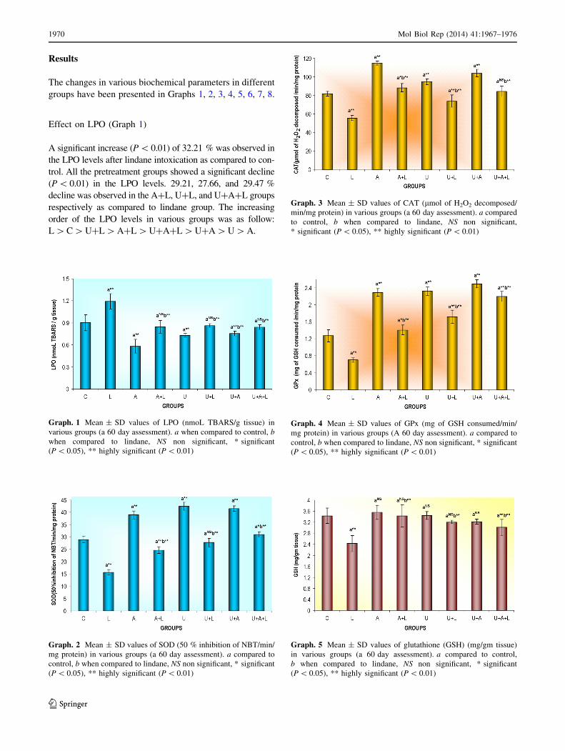
Effect on LPO (Graph 1)
A significant increase (P \ 0.01) of 32.21 % was observed in
the LPO levels after lindane intoxication as compared to con-
trol. All the pretreatment groups showed a significant decline
(P\ 0.01) in the LPO levels. 29.21, 27.66, and 29.47 %
decline was observed in the A?L, U?L, and U?A?L groups
respectively as compared to lindane group. The increasing
order of the LPO levels in various groups was as follow:
L [C [ U?L [ A?L [ U?A?L [ U?A [ U [ A.
Graph. 1 Mean ± SD values of LPO (nmoL TBARS/g tissue) in
various groups (a 60 day assessment). a when compared to control, b
when compared to lindane, NS non significant, * significant
(P \ 0.05), ** highly significant (P \ 0.01)
Graph. 2 Mean ± SD values of SOD (50 % inhibition of NBT/min/
mg protein) in various groups (a 60 day assessment). a compared to
control, b when compared to lindane, NS non significant, * significant
(P \ 0.05), ** highly significant (P \ 0.01)
Graph. 3 Mean ± SD values of CAT (lmol of H2O2 decomposed/
min/mg protein) in various groups (a 60 day assessment). a compared
to control, b when compared to lindane, NS non significant,
* significant (P \ 0.05), ** highly significant (P \ 0.01)
Graph. 4 Mean ± SD values of GPx (mg of GSH consumed/min/
mg protein) in various groups (A 60 day assessment). a compared to
control, b when compared to lindane, NS non significant, * significant
(P \ 0.05), ** highly significant (P \ 0.01)
Graph. 5 Mean ± SD values of glutathione (GSH) (mg/gm tissue)
in various groups (a 60 day assessment). a compared to control,
b when compared to lindane, NS non significant, * significant
(P \ 0.05), ** highly significant (P \ 0.01)
1970 Mol Biol Rep (2014) 41:1967–1976
123
Effect on SOD (Graph 2)
The SOD levels declined significantly (P \ 0.01) up to
45.96 % in lindane intoxicated mice as compared to
control group. The pretreatment groups A?L, U?L and
U?A?L showed a significant increase (P \ 0.01) of about
57.74, 77.64, and 98.26 % respectively, as compared to
lindane group. The increasing levels of SOD in various
groups were as follow: L \ A?L \ U?L \ C \ U?A?
L \ A \ U?A \ U.
Effect on CAT (Graph 3)
Lindane induced kidney toxicity showed a significant
decline (P \ 0.01) of 32.12 % in the CAT activity as
compared to control. A significant increase (P \ 0.01) of
58.91 % in A?L, 32.85 % in U?L and 51.97 % in
U?A?L was observed when compared to lindane. The
cow urine alone group also showed a decrease of 9.82 % as
compared to lindane but the decrease was not significant.
The maximum % of increase was observed in the A?L
group. The increasing order of the enzyme level in all the
eight groups was as follow: L \ U?L \ C \ U?A?L \A?L \ U \ U?A \ A.
Effect on GPx (Graph 4)
As compared to control animals, the lindane intoxicated
animals showed a significant decrease (P \ 0.01) of
45.19 % in the GPx levels. All the pretreatment groups
showed promising results and brought about a significant
rise of 101.89, 146.37, and 215.30 % in the GPx levels in
the groups A?L, U?L and U?A?L, respectively. The
increasing order of GPx levels in various groups was as
followed: L \ C \ A?L \ U?L \ U?A?L \ A \ U \U?A.
Effect on GSH (Graph 5)
A significant decline (P \ 0.01) of 29.24 % was observed
in the GSH levels after lindane intoxication as compared to
control animals. The pretreatment groups showed a non-
significant decrease in the levels of GSH as compared to
control which accounted to 0.10 % in the A?L group,
6.69 % in U?L group and 12.37 % in U?A?L group. But
when compared to lindane a significant increase (P \ 0.01)
of 41.18, 31.87, and 23.84 % was seen in the respective
groups A?L, U?L and U?A?L. The increasing order of
GSH levels in different groups was as follow: L \ U?
A?L \ U?L \ U?A \ A?L \ C \ U \ A.
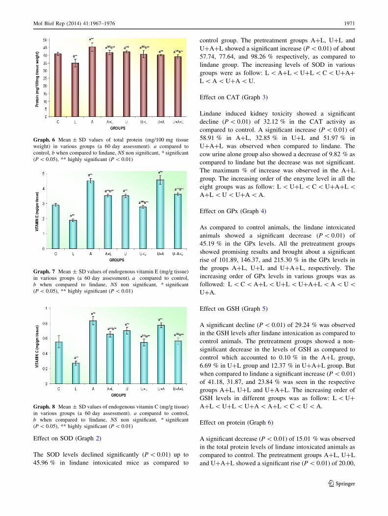
Effect on protein (Graph 6)
A significant decrease (P \ 0.01) of 15.01 % was observed
in the total protein levels of lindane intoxicated animals as
compared to control. The pretreatment groups A?L, U?L
and U?A?L showed a significant rise (P \ 0.01) of 20.00,
Graph. 6 Mean ± SD values of total protein (mg/100 mg tissue
weight) in various groups (a 60 day assessment). a compared to
control, b when compared to lindane, NS non significant, * significant
(P \ 0.05), ** highly significant (P \ 0.01)
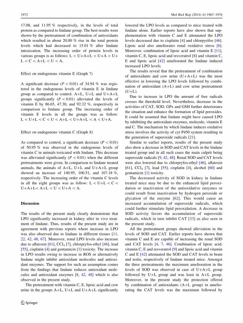
Graph. 7 Mean ± SD values of endogenous vitamin E (mg/g tissue)
in various groups (a 60 day assessment). a compared to control,
b when compared to lindane, NS non significant, * significant
(P \ 0.05), ** highly significant (P \ 0.01)
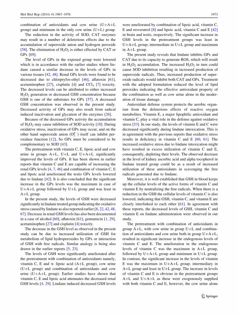
Graph. 8 Mean ± SD values of endogenous vitamin C (mg/g tissue)
in various groups (a 60 day assessment). a compared to control,
b when compared to lindane, NS non significant, * significant
(P \ 0.05), ** highly significant (P \ 0.01)
Mol Biol Rep (2014) 41:1967–1976 1971
123
17.08, and 11.95 % respectively, in the levels of total
protein as compared to lindane group. The best results were
shown by the pretreatment of combination of antioxidants
which resulted in about 20.00 % rise in the total protein
levels which had decreased to 15.01 % after lindane
intoxication. The increasing order of protein levels in
various groups is as follows: L \ U?A?L \ U?A \ U?
L \ C \ A?L \ U \ A.
Effect on endogenous vitamin E (Graph 7)
A significant decrease (P \ 0.01) of 34.94 % was regis-
tered in the endogenous levels of vitamin E in lindane
group as compared to control. A?L, U?L and U?A?L
groups significantly (P \ 0.01) alleviated the levels of
vitamin E by 86.65, 47.30, and 92.32 %, respectively in
comparison to lindane group. The increasing order of
vitamin E levels in all the groups was as follow:
L \ U?L \ C \ U \ A?L \ U?A?L \ A \ U?A.
Effect on endogenous vitamin C (Graph 8)
As compared to control, a significant decrease (P \ 0.01)
of 50.95 % was observed in the endogenous levels of
vitamin C in animals toxicated with lindane. This decrease
was alleviated significantly (P \ 0.01) when the different
pretreatments were given. In comparison to lindane treated
animals, the animals of A?L, U?L and U?A?L group
showed an increase of 140.95, 100.51, and 107.18 %,
respectively. The increasing order of the vitamin C levels
in all the eight groups was as follow: L \ U?L \ C \U?A?L\ A?L \ U \ U?A \ A.
Discussion
The results of the present study clearly demonstrate that
LPO significantly increased in kidney after in vivo treat-
ment of lindane. Thus, results of the present study are in
agreement with previous reports where increase in LPO
was also observed due to lindane in different tissues [11,
22, 42, 48, 67]. Moreover, renal LPO levels also increase
due to aflatoxin [61], CCl4 [7], chlorpryfos-ethyl [46], lead
[55], cisplatin [4] and gentamicin [1] toxicity. The increase
in LPO results owing to increase in ROS or alternatively
lindane might inhibit antioxidant molecules and antioxi-
dant enzymes. The support for such an assumption comes
from the findings that lindane reduces antioxidant mole-
cules and antioxidant enzymes [8, 42, 48] which is also
observed in the present study.
The pretreatment with vitamin C, E, lipoic acid and cow
urine in the groups A?L, U?L and U?A?L significantly
lowered the LPO levels as compared to mice treated with
lindane alone. Earlier reports have also shown that sup-
plementation with vitamin C and E attenuated the LPO
levels decreased due to cisplatin [4] and chlorpyrifos [46].
Lipoic acid also ameliorates renal oxidative stress [6].
Moreover, combination of lipoic acid and vitamin E [11],
vitamin C, E, lipoic acid and resveratrol [8] and vitamin C,
E and lipoic acid [42] ameliorated the lindane induced
increased LPO levels.
The results reveal that the pretreatment of combination
of antioxidants and cow urine (U?A?L) was the most
effective in lowering the LPO levels followed by combi-
nation of antioxidant (A?L) and cow urine pretreatment
(U?L).
Due to increase in LPO the amount of free radicals
crosses the threshold level. Nevertheless, decrease in the
activities of CAT, SOD, GPx and GSH further deteriorates
the situation and enhance the formation of lipid peroxides.
It could be assumed that lindane might have caused LPO
by inhibiting the antioxidant enzymes, molecule, vitamin E
and C. The mechanism by which lindane induces oxidative
stress involves the activity of cyt P450 system resulting in
the generation of superoxide radicals [21].
Similar to earlier reports, results of the present study
also show a decrease in SOD and CAT levels in the lindane
treated group and in all such cases the main culprit being
superoxide radicals [9, 42, 48]. Renal SOD and CAT levels
were also lowered due to chlorpryfos-ethyl [46], aflatoxin
[61], CCl4 [7], lead [55], cisplatin [4], alcohol [60] and
gentamicin [1] toxicity.
The decreased activity of SOD in kidney in lindane
treated mice may be due to the enhanced lipid peroxi-
dation or inactivation of the antioxidative enzymes or
could result from inactivation by hydrogen peroxide or
glycation of the enzyme [62]. This would cause an
increased accumulation of superoxide radicals, which
could further stimulate lipid peroxidation. A decrease in
SOD activity favors the accumulation of superoxide
radicals, which in turn inhibit CAT [33] as also seen in
the present study.
All the pretreatment groups showed alleviation in the
levels of SOD and CAT. Earlier reports have shown that
vitamin C and E are capable of increasing the renal SOD
and CAT levels [4, 7, 46]. Combination of lipoic acid,
vitamin C, E and resveratrol [9] and lipoic acid and vitamin
C and E [42] attenuated the SOD and CAT levels in brain
and testis, respectively of lindane treated mice. Amongst
the three pretreatments the maximum amelioration in the
levels of SOD was observed in case of U?A?L group
followed by U?L group and was least in A?L group.
Moreover, in the present study the protection offered
by combination of antioxidants (A?L group) in amelio-
rating the CAT levels was the maximum followed by
1972 Mol Biol Rep (2014) 41:1967–1976
123
combination of antioxidants and cow urine (U?A?L
group) and minimum in the only cow urine (U?L) group.
The reduction in the activity of SOD, CAT enzymes
may result in a number of deleterious effects due to the
accumulation of superoxide anion and hydrogen peroxide
[58]. The elimination of H2O2 is either effected by CAT or
GPx [69].
The level of GPx in the exposed group were lowered
which is in accordance with the earlier studies where lin-
dane caused a similar decrease in the levels of GPx in
various tissues [42, 48]. Renal GPx levels were found to be
decreased due to chlorpryfos-ethyl [46], aflatoxin [61],
acetaminophen [27], cisplatin [4] and CCl4 [7] toxicity.
The decreased levels can be attributed to either increased
H2O2 generation or decreased GSH concentration because
GSH is one of the substrates for GPx [57]. A decreased
GSH concentration was observed in the present study.
Decreased activity of GPx may also result from radical
induced inactivation and glycation of the enzymes [26].
Because of the decreased GPx activity the accumulation
of H2O2 may cause inhibition of SOD activity [10]. During
oxidative stress, inactivation of GPx may occur, and on the
other hand superoxide anion (O2•-) itself can inhibit per-
oxidase function [12]. So GPx must be considered to be
complementary to SOD [43].
The pretreatment with vitamin C, E, lipoic acid and cow
urine in groups A?L, U?L and U?A?L significantly
improved the levels of GPx. It has been shown in earlier
reports that vitamin C and E are capable of increasing the
renal GPx levels [4, 7, 46] and combination of vitamin C, E
and lipoic acid ameliorated the testis GPx levels lowered
due to lindane [42]. It is also concluded that the significant
increase in the GPx levels was the maximum in case of
U?A?L group followed by U?L group and was least in
A?L group.
In the present study, the levels of GSH were decreased
significantly in lindane treated group indicating the oxidative
stress caused by lindane as also reported earlier [8, 22, 42, 48,
67]. Decrease in renal GSH levels has also been documented
in a case of alcohol [60], aflatoxin [61], gentamicin [1, 29],
acetaminophen [27] and cisplatin [4] toxicity.
The decrease in the GSH level as observed in the present
study can be due to increased utilization of GSH for
metabolism of lipid hydroperoxides by GPx or interaction
of GSH with free radicals. Similar analogy is being also
drawn in the earlier reports [5, 23].
The levels of GSH were significantly ameliorated after
the pretreatment with combination of antioxidants namely,
vitamin C, E and a- lipoic acid (A?L group), cow urine
(U?L group) and combination of antioxidants and cow
urine (U?A?L group). Earlier studies have shown that
vitamin C, E and lipoic acid attenuates the decreased renal
GSH levels [4, 29]. Lindane induced decreased GSH levels
were ameliorated by combination of lipoic acid, vitamin C,
E and resveratrol [8] and lipoic acid, vitamin C and E [42]
in brain and testis, respectively. The significant increase in
GSH levels in the pretreatment groups was least in
U?A?L group; intermediate in U?L group and maximum
in A?L group.
The present study reveals that lindane inhibits GPx and
CAT due to its capacity to generate ROS, which will result
in H2O2 accumulation. The increased H2O2 in turn could
cause SOD inhibition resulting in increased production of
superoxide radicals. Thus, increased production of super-
oxide radicals would inhibit both CAT and GPx. Treatment
with the adopted formulation reduced the level of lipid
peroxides indicating the effective antioxidant property of
the combination as well as cow urine alone in the moder-
ation of tissue damage.
Antioxidant defense system protects the aerobic organ-
ism from the deleterious effects of reactive oxygen
metabolites. Vitamin E, a major lipophilic antioxidant and
vitamin C, play a vital role in the defense against oxidative
stress [53]. In our study, the levels of vitamin E and C were
decreased significantly during lindane intoxication. This is
in agreement with the previous reports that oxidative stress
results in deficiency in vitamin C and E [60, 61]. The
increased oxidative stress due to lindane intoxication might
have resulted in excess utilization of vitamin C and E,
consequently, depleting their levels. The observed decrease
in the level of kidney ascorbic acid and alpha tocopherol in
lindane treated group could be as a result of increased
utilization of these antioxidants in scavenging the free
radicals generated due to lindane.
Moreover, it is well established that GSH in blood keeps
up the cellular levels of the active forms of vitamin C and
vitamin E by neutralizing the free radicals. When there is a
reduction in the GSH the cellular levels of vitamin C is also
lowered, indicating that GSH, vitamin C, and vitamin E are
closely interlinked to each other [61]. In agreement with
these reports, the decreased levels of GSH, vitamin C and
vitamin E on lindane administration were observed in our
study.
The pretreatment with combination of antioxidants in
group A?L, with cow urine in group U?L and combina-
tion of antioxidants and cow urine both in group U?A?L,
resulted in significant increase in the endogenous levels of
vitamin C and E. The amelioration in the endogenous
levels of vitamin C was the maximum in A?L group,
followed by U?A?L group and minimum in U?L group.
In contrast, the significant increase in the levels of vitamin
E was the maximum in U?A?L group; intermediary in
A?L group and least in U?L group. The increase in levels
of vitamin C and E is obvious in the pretreatment groups
A?L and U?A?L as these were exogenously supplied
with both vitamin C and E, however, the cow urine alone
Mol Biol Rep (2014) 41:1967–1976 1973
123
(U?L) group also showed significant elevation in the
levels of the two vitamins. This could be attributed to the
composition of cow urine which is said to contain vitamins.
The analysis of total proteins is important for estimating
the degree of damage in the body. The protein profile of the
cells is indicative of the physiological status of animal and
there exists dynamic equilibrium between the synthetic and
degenerative pathways with these biomolecules. In the
present study a decline in the total proteins was observed
after lindane intoxication which can be due to decreased
protein synthesis or increased protein loss. Similar reduc-
tion in the levels of total proteins due to lindane have been
reported [9, 42, 48]. It is also reported that renal toxicity
due to CCl4 [7] and gentamicin [1] causes a similar
decrease in protein levels. Decreased protein levels could
be attributed to decreased feed consumption, maldigestion
or malabsorption, hepatic dysfunction [13, 47]. Reduced
protein levels can also be ascribed to increased urinary
excretion owing to kidney damage [66] and glomerular
apparatus or reduced protein synthesis [39]. Prabhakaran
and Devi [51] have proposed that a toxicant can affect the
protein content of the tissue either by inhibiting RNA
synthesis or inhibiting of amino acids into the polypeptide
chain.
The pretreatment groups showed an elevation in the
protein levels which is in accordance with earlier reports
where administration of either vitamin C, E or in combi-
nation and combination of vitamin C, E and lipoic acid and
resveratrol elevated the decreased protein levels [7, 9, 42].
The most effective pretreatment was that of combination of
antioxidants (A?L group); cow urine pretreatment group
(U?L) was moderately effective and least effective was
pretreatment with combination of antioxidants and cow
urine both (U?A?L group).
Hypoglycemic [44], cardio-respiratory effect [20],
immunomodulatory [14], antigenotoxic and antioxidant
properties in vitro [34], anticlastogenic [19] and chemo-
protective [52] effects of distillate and redistillate of cow
urine have been reported. Attenuation of CCl4 induced
hepatotoxicity by Panchagavya ghrita (prepared by cow
milk, cow urine, cow dung, ghee and curd) [2] and cow
urine distillate [25] have also been reported. Efficacy of
cow urine therapy has been evaluated on cancer patients
[28]. The amelioration of oxidative stress by fresh cow
urine has not been reported so far and this work seems to be
the first report.
Thus it is inferred that the combination of antioxidants
taken in the present study are quiet helpful in mitigating and
modulating the oxidative stress in the kidney caused due to
lindane. Moreover, cow urine treatment also modulated the
oxidative stress parameters caused by lindane. From the
results it is apparent that the given combination of antiox-
idants and cow urine act synergistically in reducing lindane
induced dysfunction. The analysis of the results of the
present study reveals the efficacy of cow urine against
oxidative stress. It can be safely concluded that the sug-
gested combination of vitamin C, vitamin E, alpha lipoic
acid and cow urine can prove to be beneficial in a number of
ailments. The highlight of the investigation is the efficiency
of cow urine against the pesticide toxicity which can open
new insights in the field of medicine. Cow urine can prove
to be an effective co-remedy for oxidative stress. This study
emphasizes the importance of antioxidants and cow urine
which could be beneficial in the therapeutic world for the
treatment of various disorders implicating oxidative stress.
References
1. Abdel-Raheem IT, Abdel-Ghany AA, Mohamed GA (2009)
Protective effect of quercetin against gentamicin induced neph-
rotoxicity in rats. Biol Pharm Bull 32(1):61–67
2. Achliya GS, Kotagale NR, Wadodkar SG, Dorle AK (2003)
Hepatoprotective activity of Panchagavya Ghrita against carbon
tetrachloride-induced hepatotoxicity in rats. Ind J Pharmacol
35:308–311
3. Adewole SO, Salako AA, Doherty OW, Naicker T (2007) Effect
of melatonin on carbon tetrachloride-induced kidney injury in
Wistar rats. Afr J Biomed Res 10:153–164
4. Ajith TA, Usha S, Nivith AV (2007) Ascorbic acid and a-
tocopherol protect anticancer drug cisplatin induced nephrotoxi-
city in mice: a comparative study. Clin Chim Acta 375:82–86
5. Anbarasi K, Vani G, Balakrishna K, Shyamala CSD (2006) Effect
of bacoside A on brain antioxidant status in cigarette smoke
exposed rats. Life Sci 78:1378–1384
6. Bae EH, Lee JU, Kwon Ma S, Kim IJ, Frokiaer J, Nielsen S, Kim
SY, Kim SW (2009) a-Lipoic acid prevents cisplatin-induced acute
kidney injury in rats. Nephrol Dial Transplant 24:2692–2700
7. Balahoroglu R, Dulger H, Ozbek H, Bayram I, Sekeroglu MR
(2008) Protective effects of antioxidants on the experimental liver
and kidney toxicity in mice. Eur J Gen Med 5(3):157–164
8. Bano M, Bhatt DK (2007) Neuroprotective role of a novel
combination of certain antioxidants on lindane (c-HCH) induced
toxicity in cerebrum of mice. Res J Agric Bio Sci 3:664–669
9. Bano M, Bhatt DK (2010) Ameliorative effect of a combination
of vitamin E, vitamin C, alpha-lipoic acid and stilbene resveratrol
on lindane induced toxicity in mice olfactory lobe and cerebrum.
Indian J Exp Biol 48:150–158
10. Bast A, Haenen GRMM, Doelman CJA (1991) Oxidants and
antioxidants: state of the art. Am J Med 91:25–135
11. Bist R, Bhatt DK (2009) The evaluation of effect of alpha- lipoic
acid and vitamin E on the lipid peroxidation, gamma-amino
butyric acid and serotonin level in the brain of mice (Mus mus-
culus) acutely intoxicated with lindane. J Neurol Sci 276:99–102
12. Blum J, Fridovich I (1985) Inactivation of glutathione peroxidase
by superoxide radical. Arch Biochem Biophys 240:500–508
13. Brzoska MM, Moniuszko-Jakoniuk J, Pilat-Marcinkiewicz B,
Sawicki B (2003) Liver and kidney function and histology in rats
exposed to cadmium and ethanol. Alcohol Alcohol 38(1):2–10
14. Chauhan RS, Singh BP, Singhal LK (2001) Immunomodulation
with kamdhenu Ark in mice. J Immunol Immunopathol 71:89–92
15. CIBRC (2005) Central Insecticide Board and Registration
Committee, Dept. Of Plant Protection and Quarantine, Ministry
of Agriculture, India. http://www.ipen.org/ipenweb/documents/
work%20documents/lindanes%20dirty%20secret.pdf
1974 Mol Biol Rep (2014) 41:1967–1976
123
16. Cohen G, Dembiec D, Marcus J (1970) Measurement of catalase
activity in tissue extracts. Anal Biochem 34:30–38
17. Davies K (1995) Oxidative stress: the paradox of aerobic life.
Biochem Soc Symp 61:1–31
18. Desai ID (1984) Vitamin E methods for animal tissues. Methods
Enzymol 105:138–143
19. Dutta D, Devi SS, Krishnamurthi K, Chakrabarti T (2006) An-
ticlastogenic effect of redistilled cow’s urine distillate in hu-
manperipheral lynphocytes challenged with manganese dioxide
and hexavalent chromium. Biomed and Environ Sci 19:487–494
20. Elegbe RA, Oyebola DDO (1976) Cow’s urine poisoning in
Nigeria: the cardiotoxic effects of cow’s urine in dogs. Trans R
Soc Trop Med Hyg 71:127–132
21. English D, Schell M, Siakotos A, Gabig TG (1986) Reversible
activation of the neutrophil superoxide generating system by
hexachlorocyclohexane: correlation with effects on a subcellular
superoxide-generating fraction. J Immunol 137:283–290
22. Fidan AF, Cigerci IH, Sozbilir NB, Kucukkurt I, Yuksel H, Keles
H (2008) The effects of the dose-dependent hexachlorocyclo-
hexane (Lindane) on blood and tissue antioxidant defense sys-
tems, lipid peroxidation and histopathological changes in rats.
J Anim Vet Adv 7:1480–1488
23. Flohe L (1982) Glutathione peroxidase brought into focus. In:
Pryor WA (ed) Free radicals in biology. Academic Press, New
York, p 233
24. Goldstein RS, Schnellmann RG (1996) Toxic responses of the
kidney. Cassrrett and Doull’s toxicology, 5th edn. McGraw Hill,
New York
25. Gururaja MP, Joshi AB, Joshi H, Sathyanarayana D, Subrah-
manyam EV, Chandrashekhar KS (2009) Attenuation of carbon
tetrachloride-induced hepatotoxicity by cow urine distillate in
rats. Biomed Environ Sci 22(4):345–347
26. Hodgson EK, Fridovich I (1975) The interaction of bovine
erythrocyte superoxide dismutase with hydrogen peroxide and
inactivation of the enzyme. Biochemistry 14:5294–5299
27. Isik B, Bayrak R, Akcay A, Sogut S (2006) Erdosteine against
acetaminophen induced renal toxicity. Mol Cell Biochem 287:
185–191
28. Jain NK, Gupta VB, Garg R, Silawat N (2010) Efficacy of cow
urine therapy on various cancer patients in Mandsaur district,
India—a survey. Int J Green Pharm 4(1):29–35
29. Kadkhodaee M, Khastar H, Faghihi M, Ghaznavi R, Zahmatkesh M
(2004) Effects of co-supplementation of vitamins E and C on gen-
tamicin-induced nephrotoxicity in rat. Exp Physiol 90(4):571–576
30. Kakkar P, Das B, Viswanathan PN (1984) A modified spectro-
photometric assay of superoxide dismutase. Indian J Biochem
Biophys 21:130–132
31. Kelly JF (1997) The urine cure and other curious medical treat-
ments. http://wfmu.org/LCD/_Articles/LCD_19/Urine.html
32. Khanna RN, Das M, Anand M (2002) Influence of phenobarbitol
and CCl4 on the modulation of tissue retention profile of hexa-
chlorocyclohexane in rats. Biomed Environ Sci 15:119–129
33. Kono Y, Fridovich I (1982) Superoxide radical inhibits catalase.
J Biol Chem 257:5751–5754
34. Krishnamurthi K, Dutta D, Devi SS, Chakrabarti T (2004) Pro-
tective effect of distillate and redistillate of cow’s urine in human
polymorphonuclear leukocytes challenged with established
genotoxic chemicals. Biomed Environ Sci 17:57–66
35. Kumar MR, Reddy KS, Reddy AG, Anjaneyulu Y, Kalakumar B,
Reddy GD (2009) Amelioration of lead induced nephrotoxicity by
certain adaptogens in broilers. Indian J Vet Pathol 33(2):19–190
36. Lash HL, Qian W, Putt DA, Hueni SE, Elfarra AA, Krause RJ,
Parker JC (2001) Renal and hepatic toxicity of trichloroethylene
and its glutathione derived in rats and mice: sex-, species-, and
tissue dependent differences. J Pharmacol Exp Ther 297(1):
155–164
37. Lightboy JH, Stevenson LM, Jackson F, Donaldson K, Jones DG
(2001) Comparative aspects of plasma antioxidant status in sheep
and goats and the influence of experimental abomasal nematode
infection. J Comp Pathol 124:192–199
38. Lowry OH, Roesborough MJ, Farr AL, Randall RJ (1951) Protein
measurement with Folin–Phenol reagent. J Biol Chem 193:265
39. Lynch MJ, Raphael SS, Miller LD, Spare DD, Inwood MJH (1969)
Medical laboratory technology and clinical pathology. W.B. Saun-
ders Co., Igaku shoin Ltd, Philadelphia, Tokyo, pp 2–629
40. Moron MA, Depierre JW, Mannervick B (1979) Levels of glu-
tathione, glutathione redutase, glutathione S-transferase activities
in rat lung and liver. Biochem Biophys Acta 582:67–78
41. Muzakova V, Kandar R, Vojtisek P, Skalicky J, Vankova R,
Cegan A, Cervinkova Z (2001) Antioxidant vitamin levels and
glutathione peroxidase activity during ischemia/reperfusion in
myocardial infarction. Physiol Res 50:389–396
42. Nagda G, Bhatt DK (2011) Alleviation of lindane induced tox-
icity in testis of Swiss mice (Mus musculus) by combined treat-
ment with vitamin C, vitamin E and a-Lipoic acid. Indian J Exp
Biol 49:191–199
43. Nouri M, Rahbani-Nobar M, Argani H, Rokhforooz F (1999)
Superoxide dismutase and glutathione peroxidase in hemodia-
lyzed patients and renal transplant recipients and their relation-
ship to osmotic fragility. Med J Islam Acad Sci 12(2):33–38
44. Ojewole JA, Olusi SO (1976) Effects of cow’s urine concoction
on plasma glucose concentration in fasted rats. Trans R Soc Trop
Med Hyg 71:241–245
45. Omaye ST, Turbull TP, Sauberchich HC (1979) Selected meth-
ods for determination of ascorbic acid in cells, tissues and fluids.
Methods Enzymol 6:3–11
46. Oncu M, Gultekin F, Karaoz E, Altuntas I, Delibas N (2002)
Nephrotoxicity in rats induced by chlorpyrifos-ethyl and ameliorat-
ing effects of antioxidants. Hum Exp Toxicol 21(4):223–230
47. Osfor MMH, Ibrahim HS, Mohamed YA, Ahmed SM, Abd El
Azeem AS, Hegazy AM (2010) Effect of alpha lipoic acid and
vitamin E on heavy metals intoxication in male albino rats. J Am
Sci 6(80):56–63
48. Padma VV, Sowmya P, Felix TA, Baskaran R, Poornima P
(2011) Protective effect of gallic acid against lindane induced
toxicity in experimental rats. Food Chem Toxicol 49:991–998
49. Pathak ML, Kumar A (2003) Gomutra a descriptive study.
Sachitra Ayurveda 7:81–84
50. Poirier B, L-Bournville M, Conti M (2000) Oxidative stress occur
in absence of hyperglycemia and inflammation in the onset of
kidney lesions in normotensive obese rats. Nephrol Dial Trans-
plant 15:467–476
51. Prabhakaran S, Devi KS (1993) Impact of protein deficiency and
exposure to HCH or malathione on lipid metabolism in pregnant
rats. Indian J Biochem Biophys 30:234–238
52. Raja W, Agarwal RC (2010) Chemoprotective potential of cow
urine against 7, 12-dimethylbenz(a) antracene-induced skin pap-
illomagenesis in mice. Acad J Can Res 3(1):7–10
53. Ray G, Hussain SA (2002) Oxidants, antioxidants and carcino-
genesis. Indian J Exp Biol 42:1213–1232
54. Rotruck JT, Pope AL, Ganther HE, Swanson AB (1973) Sele-
nium: biochemical roles as a component of glutathione peroxi-
dise. Science 179:588–590
55. Salawu EO, Adeleke AA, Oyewo OO, Ashamu EA, Ishola OO,
Afolabi AO, Adesanya TA (2009) Prevention of renal toxicity
from lead exposure by oral administration of Lycopersicon es-
culentum. J Toxicol Environ Health Sci 1(2):022–027
56. Sauviat MP, Pages N (2002) Cardiotoxicity of lindane: a gamma
isomer of hexachlorohexane. J Soc Biol 196:339–348
57. Scibior D, Skrzycki M, Podsiad M, Czeczot H (2008) Glutathione
level and glutathione-dependent enzyme activities in blood serum of
patients with gastrointestinal tract tumors. Clin Biochem 41:852–858
Mol Biol Rep (2014) 41:1967–1976 1975
123

58. Searle AJ, Wilson RL (1980) Glutathione peroxidase: effect of
superoxide, hydroxyl and bromine free radicals on enzyme
activity. Int J Radiant Biol 37:213–217
59. Shaikh ZA, Vu TT, Zaman K (1999) Oxidative stress as a mechanism
of chronic cadmium-induced hepatotoxicity and renal toxicity and
protection by antioxidants. Toxicol Appl Pharmacol 154(3):256–263
60. Shanmugam KR, Ramakrishna CH, Mallikarjuna K, Reddy KS
(2010) Protective effect of ginger against alcohol-induced renal
damage and antioxidant enzymes in male albino rats. Indian J
Exp Biol 48:143–149
61. Sivanesan D, Begum H (2007) Preventive role of Gyanandropsis
gynandra L., against aflatoxin B1 induced lipid peroxidation and
defense mechanism in rat. Indian J Exp Biol 45(3):299–303
62. Sozmen EY, Sozmen B, Delen Y, Onat T (2001) Catalase/super-
oxide dismutase (SOD) and catalase/paraoxonase (PON) ratios may
implicate poor glycemic control. Arch Med Res 32:283–287
63. Srivastava A, Shivanandappa T (2005) Hexachlorocyclohexane
differentially alters the antioxidant status of the brain regions in
rat. Toxicology 214:123–130
64. Tasaduq SA, Singh K, Sethi S, Sharma SC, Bedi KL, Singh J,
Jaggi BS, Johri RK (2003) Hepatocurative and antioxidant profile
of HP-1, a polyherbal phytomedicine. Hum Exp Toxicol 22(12):
639–645
65. Vela C, Cristol JP, Maggi MF (1999) Oxidative stress in renal
transplant recipients with chronic rejection: rationale for antiox-
idant supplementation. Transplant Proc 31:1310–1311
66. Verschurren RK, Engelina MD, Berkvens JB, Helleman PW,
Rauws AG, Schuller PL, Vanesch GJ (1976) Toxicity of methyl
mercuric chloride in rats (I) short term study. Toxicology 6(1):
85–96
67. Videla LA, Barros SBM, Junqueira VBC (1990) Lindane-induced
liver oxidative stress. Free Rad Biol Med 9:169–179
68. Wilber KM, Baerheim F, Shapiro OW (1949) The thiobarbituric
acid reagent as a test for the oxidation of unsaturated fatty acid by
various reagents. Arch Biochem Biophys 24:304–311
69. Zini A, Schlegel PN (1996) Catalase mRNA expression in the
male rat reproductive tract. J Androl 17:473–480
1976 Mol Biol Rep (2014) 41:1967–1976
123