Effect of tank colour on foraging capacity, growth and ...
33
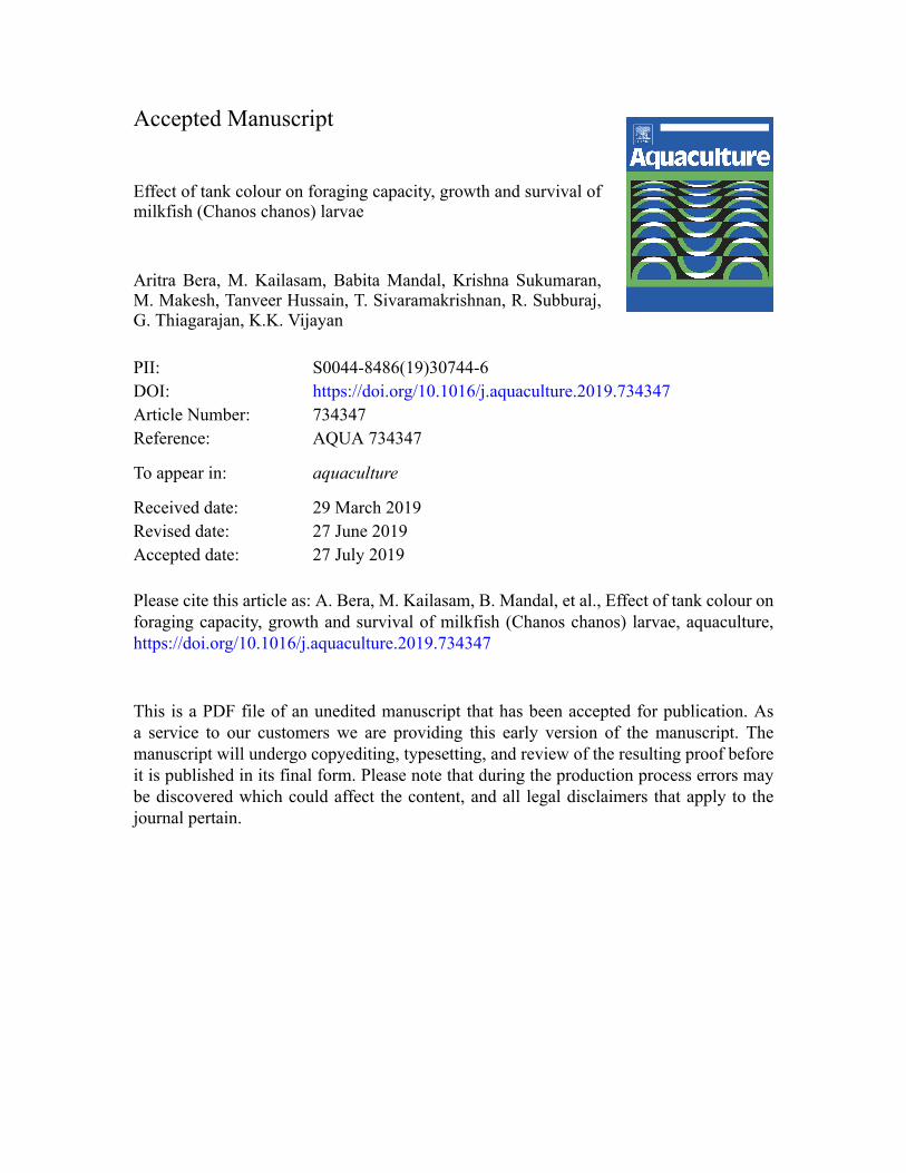
Accepted Manuscript Effect of tank colour on foraging capacity, growth and survival of milkfish (Chanos chanos) larvae Aritra Bera, M. Kailasam, Babita Mandal, Krishna Sukumaran, M. Makesh, Tanveer Hussain, T. Sivaramakrishnan, R. Subburaj, G. Thiagarajan, K.K. Vijayan PII: S0044-8486(19)30744-6 DOI: https://doi.org/10.1016/j.aquaculture.2019.734347 Article Number: 734347 Reference: AQUA 734347 To appear in: aquaculture Received date: 29 March 2019 Revised date: 27 June 2019 Accepted date: 27 July 2019 Please cite this article as: A. Bera, M. Kailasam, B. Mandal, et al., Effect of tank colour on foraging capacity, growth and survival of milkfish (Chanos chanos) larvae, aquaculture, https://doi.org/10.1016/j.aquaculture.2019.734347 This is a PDF file of an unedited manuscript that has been accepted for publication. As a service to our customers we are providing this early version of the manuscript. The manuscript will undergo copyediting, typesetting, and review of the resulting proof before it is published in its final form. Please note that during the production process errors may be discovered which could affect the content, and all legal disclaimers that apply to the journal pertain.
Transcript of Effect of tank colour on foraging capacity, growth and ...
Accepted Manuscript
Effect of tank colour on foraging capacity, growth and survival ofmilkfish (Chanos chanos) larvae
Aritra Bera, M. Kailasam, Babita Mandal, Krishna Sukumaran,M. Makesh, Tanveer Hussain, T. Sivaramakrishnan, R. Subburaj,G. Thiagarajan, K.K. Vijayan
PII: S0044-8486(19)30744-6DOI: https://doi.org/10.1016/j.aquaculture.2019.734347Article Number: 734347Reference: AQUA 734347
To appear in: aquaculture
Received date: 29 March 2019Revised date: 27 June 2019Accepted date: 27 July 2019
Please cite this article as: A. Bera, M. Kailasam, B. Mandal, et al., Effect of tank colour onforaging capacity, growth and survival of milkfish (Chanos chanos) larvae, aquaculture,https://doi.org/10.1016/j.aquaculture.2019.734347
This is a PDF file of an unedited manuscript that has been accepted for publication. Asa service to our customers we are providing this early version of the manuscript. Themanuscript will undergo copyediting, typesetting, and review of the resulting proof beforeit is published in its final form. Please note that during the production process errors maybe discovered which could affect the content, and all legal disclaimers that apply to thejournal pertain.
ACC
EPTE
D M
ANU
SCR
IPT
1
Effect of tank colour on foraging capacity, growth and survival of milkfish
(Chanos chanos) larvae
Aritra Bera*, M.Kailasam, Babita Mandal, Krishna Sukumaran, M. Makesh, Tanveer
Hussain, T. Sivaramakrishnan, R. Subburaj, G.Thiagarajan, K.K.Vijayan
ICAR-Central Institute of Brackishwater Aquaculture, 75-Santhome High Road, RA Puram,
Chennai-28, Tamil Nadu, India.
*Author for Correspondence: Present address: Scientist, ICAR-Central Institute of
Brackishwater Aquaculture, 75, Santhome High Road, Chennai, Tamil Nadu – 600 028,
India. Telephone: +91 44 24618817 (Office); Fax: +91 44 24610311.
E-mail address: [email protected]; [email protected]
Abstract
Larviculture of milkfish (Chanos chanos) associated with issues like larval deformity, mass
mortalities which contribute in variable seed production. Effect of abiotic factors as rearing
tank colour and illumination on larvae foraging behaviour, prey localization and ingestion
were investigated to improvise milkfish larval rearing system. Reinforced Cement Concrete
(RCC) tanks (capacity, 8 t; water salinity, 32 ppt) with three different background colors,
white, blue and yellow were divided in five (05) treatment groups with triplicates, i.e. indoor
white (T1/C), blue (T2), yellow (T3) color tanks with artificial illumination and semi outdoor
blue (T4), yellow (T5) color tanks with solar illumination. Newly hatched milkfish larvae (tl.,
3.4 mm) were stocked in the experimental tanks @ 2.5 no l-1 and apart from background
colour and source of illumination uniform water quality and feeding regime were maintained
in all treatments. Phytoplankton, Chlorella salina @ 103 – 104 cells ml-1 were maintained
from 2 dph to 20 dph; Chlorella grown rotifers, Brachionus plicatilis (enriched with
Nannochloropsis oculata paste) were provided @ 20-30 no ml-1 from 3 dph to 14 dph
ACCEPTED MANUSCRIPT

ACC
EPTE
D M
ANU
SCR
IPT
2
depending on the larval density. Artemia nauplii @ 0.5– 1.0 no ml-1 was introduced from 15
dph. At the end of the experiment- 20 dph, highest (p<0.05) larval survival (45 ± 5.63 %)
was achieved in tanks providing yellow background colour (T5) compared to control and
other treatments. Larval growth (tl., 17.1± 1.37mm) was also found to be highest (p< 0.05) in
T5. Increased survival and growth of milkfish in T5 was synchronized with significantly
higher (p< 0.05) specific growth rate (SGR), larval gut content relative to other treatments.
Milkfish larvae being a day feeder did maximum foraging during 0700h to 1600h evident
from decreeing prey abundance during that period and as a result positive correlation found
between larval standard length and gut content. Larval visibility enhancement in solar
illuminated yellow tank act synergistically to perform necessary foraging to acquire
nutritional energy for metamorphosis to fry. Above phenomenon may not have occurred in
other treatments except T5 and partially in T3. Solar illuminated yellow colour tanks
significantly contribute towards mass scale seed production of milkfish.
Key words: Milkfish, larval rearing, prey visibility, foraging capacity, yellow tank
1. Introduction:
Indoor finfish larval rearing system is completely different from natural environment where
marine fish larvae have to hunt for food in order to survive harsh environment. Improvisation
in indoor larval rearing technology increases growth and survival. Abiotic factors such as
light and colour plays active role in larval visual field, retinal development, prey selection,
foraging and survival (Shand et al., 2008 and Cobcroft et al., 2012). It is important to induce
the foraging behaviour of early larvae through the provision of abiotic factors such as light
and tank background colour for efficient live feed preying. Indoor larval rearing
simultaneously modifies the larval natural feeding behaviour (Butts, 2016). Under rearing
conditions marine larvae do not utilize their full capacity of highly adaptive sensory systems
ACCEPTED MANUSCRIPT

ACC
EPTE
D M
ANU
SCR
IPT
3
to detect and locate prey, but rather, they are able to obtain food using a limited range of their
sensory potential primarily visual capacity (Ullmann et al., 2011). Larval visual ability is
dependent on its spectral sensitivity of retina, visual capabilities, ambient light environment,
photoperiod and most importantly tank background colour. Tank background colour may
play a critical factorial role as it can change visual field of larvae. Early stage larvae having
primitive visual field can identify live prey against tank background if higher contrast is
provided (Shand et al., 2008).
There are of reports where effect of different tank background colour and light spectrum on
larval survival and growth has been studied in different finfish species but not sufficient to
understand how tank background colour affects foraging behaviour, visual field and prey
localization in tank. Effect of tank background colour in finfish larvae of Eurasian perch,
Perca fluviatilis (Tamazouzt et al., 2000), juvenile rainbow trout, Oncrhynchus mykiss
(Luchiari and Pirhonen, 2008) , yellow perch, Perca flavescents (Hinshaw, 1985 & Jentoft et
al., 2006), mahi mahi, Coryphaena hippurus (Ostrowski, 1989), striped bass, Morone
saxatilis (Martin- Robichaud and Peterson, 1998), grouper, Epinephelus suillus (Duray et al.,
1996), haddock, Melanogrammus aeglefinus (Downing and Litvak, 1999), guppies, Poecilia
reticulate (Ruchin, 2004) and Black bream (Shand et al., 2008), Acanthopagrus butcheri
were studied. Varied background colour develops different frequency of shorter or longer-
wavelength sensitive cones in retina which helped larvae to enhance its visual field for prey
localization ( Shand et al., 2008). In depth research into these factors will help to improve
feeding rates and therefore improve efficiency in the aquaculture industry.
Milkfish (Chanos chanos) is an acclaimed food fish widely cultured in countries such as
Philippines, Indonesia and Taiwan (Gapasin et al., 1998). During 2016 milkfish farming
contributed 1188 thousand t production which is 2% of total finfish produced globally from
ACCEPTED MANUSCRIPT

ACC
EPTE
D M
ANU
SCR
IPT
4
aquaculture (FAO, 2018). Indian milkfish aquaculture is traditional way of farming and
predominately dependent on wild caught seeds. In India prevalence of milkfish seed from
wild has been reported during two season viz., March – May and Sept – Oct from coastal
Andhra Pradesh and Rameswaram or Pamban areas of Tamil Nadu. The wild caught seeds
are not adequate and are often mixed with predatory species (Silas et al., 1982). Therefore,
research towards developing milkfish captive breeding and seed production technology is the
need of the hour to meet the seed demand for the farming community. To overcome this
problem ICAR-Central Institute of Brackishwater Aquaculture (ICAR-CIBA), Chennai,
India, has standardized comprehensive technology of induced breeding of milkfish during
June 2015 and since then severe need was felt to improve larval survival. In spite of milkfish
breeding (Lee et al., 1986) and seed production technology developed by Aquaculture
Department of Southeast Asian Fisheries Development Centre issues such as larval
deformity, mass mortalities, and variable production still exists. Reports are scanty on use of
different tank colour and strict feeding regime on milkfish larvae survival. Milkfish being
transparent larvae gives us an opportunity to understand its feeding preferences and capacity
in a vivid way. Vision in fish larvae is a neural process which starts with absorption of light
by retinal photoreceptors. Hence larval ability to detect a light stimulus is dependent on
spectral absorption properties of opsin proteins in photoreceptors of retina. It has been found
that in vertebrates there are four opsin classes in retinal cone photoreceptors i.e. short
wavelength-sensitive 1 (SWS1) pigments having peak sensitivities (λmax) in the UV–violet
region of the spectrum, short wavelength sensitive 2 (SWS2) pigments with λmax in the blue
region, middle wavelength-sensitive rod-like (Rh2) pigments with λmax in the green region,
and long wavelength-sensitive (LWS) pigments with λmax in the yellow–red region (Shand
et al., 2008). In this study we selected three different tank colours i.e. white, blue and yellow
from entire range of spectral sensitivity to understand how reflection type, scattering
ACCEPTED MANUSCRIPT

ACC
EPTE
D M
ANU
SCR
IPT
5
phenomenon influences spectral perceiving power of larvae ultimately influencing preying
capacity growth and survival. The objective of this experiment was to study the effect of
tank background colour viz. white, blue and yellow on the foraging capacity, growth and
survival of milkfish larvae.
2.Materials and methods
2.1. Milkfish Larvae
Fertilized eggs of milkfish (Chanos chanos) were obtained from hormone pellet implanted
more than 10 years old broodstocks (Total 16 numbers, 4.4 – 7.2 kg) maintained in 144 t
capacity open RCC tank at Muttukadu Experimental Station of ICAR-CIBA,Chennai, India.
Eggs (1.24 mm dia) were collected from egg collection tank during early morning (6 am) and
incubated in 500 l capacity conical FRP tanks with mild flow through (1.75 l/min) of filtered
seawater (Salinity 32 ppt, Temperature 27° C - 29° C) and constant aeration to facilitate
movement and floatation of eggs to hatch out within 25 h – 26 h after fertilization (Lee et al.,
1986a & 1986b).
2.2. Ethics Statement
The research undertaken complies with the current animal welfare laws in India. Even though
fish larvae have been used in the present study, no animals have been stressed or sacrificed
for the same. However, care and treatment of brood stock used in this study for procurement
of eggs and larvae, were in accordance with the guidelines of the CPCSEA [Committee for
the Purpose of Control and Supervision of Experiments on Animals, Ministry of Environment
& Forests (Animal Welfare Division), Govt. of India] on care and use of animals in scientific
research. The study was undertaken with approval of statutory authorities of the Central
ACCEPTED MANUSCRIPT

ACC
EPTE
D M
ANU
SCR
IPT
6
Institute of Brackishwater Aquaculture, Chennai, India. As the experimental fish, Chanos
chanos is not an endangered fish, the provisions of the Govt. of India’s Wildlife Protection
Act of 1972 are not applicable for experiments on this fish.
2.3. Experimental design and larval rearing
Fifteen RCC tanks (each capacity, 8 t; water salinity, 32 ppt) with three different background
colours, white, blue and yellow were divided in five (05) treatments groups i.e. indoor white
(T1/C), blue (T2), and yellow (T3) background color tanks with artificial illumination and
semi outdoor blue (T4), and yellow (T5) background color tanks with solar illumination in
triplicates. Tanks were painted with epoxy color having code yellow (7861), Blue (9206) and
white (L152) from Asian Paints, India following manufactures instruction. Treatments such
as T1, T2 and T3 were illuminated with non-heating white florescent light provided with
timer regulated 12 h: 12 h: L: D photoperiod. Treatments such as T4 and T5 were also
maintained 12 h: 12 h: L: D photoperiod by using collapsible black sheet during 18.00hr to
06.00 hr for 12 h interval over tank (Average day length was 12.38 hr). White background
tanks under solar illumination was prone to infest with algae on wall and lost white
background very fast and was not considered for experiment. Apart from background colour
and source of illumination other factors such as water quality, water exchange rate, feeding
frequency etc. were uniformly maintained in the larval rearing tank (LRT) in all the
treatments. Newly hatched milkfish larvae having a large yolk sac with initial mean total
length (TL) of 3.4 ± 0.06 mm were collected from incubation tank and stocked in RCC tanks
at the rate of 2.5 larvae L-1. Total of 0.9 million newly hatched larvae of three cohorts (each
0.3 million larvae from three different spawning) were equally distributed in triplicate in 15
RCC tanks @ 20000 numbers/tank over three larval rearing cycles (March-May 2017) of
each 21 days.
ACCEPTED MANUSCRIPT

ACC
EPTE
D M
ANU
SCR
IPT
7
Phytoplankton, Chlorella salina with mass culture cell density of 0.4-0.5 million cells ml-1
were introduced in LRTs from 2 dph to 20 dph to maintain the green water with a cell density
of 103 to 104 cells ml−1 . Mass cultured Brachionus plicatilis (140 – 210 µm) were collected
and enriched with frozen green algae paste Nannochloropsis oculata (Nanno 3600 TM , 68
billion cells/ml) (Thépot, 2016) and fresh Chlorella salina (1:1 ratio) in a 100 L volume
container overnight and were supplied as initial feed to the larvae @ 20-30 numbers ml-1
from 3 dph to 14 dph . Larvae were co-fed with rotifers until 15 dph during the rotifer–
Artemia transition (Woolley et al., 2012). Newly hatched Artemia nauplii@ 0.5– 1.0 numbers
ml-1 was introduced from 15 dph till 20 dph following initiation of weaning to artificial feed
(200-300 µm) from 21 dph. Water exchange was initiated from 6 dph, initially at 10 % once a
day, and increased to 50 % once a day by 20 dph. Rotifer and Artemia nauplii were added
two times a day only (0700 h & 1600 h) without adding extra prey in-between during study
period. Prey was counted every 3 h till 20 dph and any reduction recorded in the prey density
was adjusted by adding the required feed in the experimental tanks following the above
feeding schedule. Sufficient aeration was provided in LRTs to ensure homogenous
distribution of algae and live feeds throughout the water column. Sand filtered seawater at
28° C - 29° C was used for entire study period and important water quality parameters were
always in optimum range (APHA 2005; Bagarinao, 1986; Kailasam et al., 2002 & 2007;
Sorgeloos et al., 1986; Gapasin and Marte 1990).
2.4. Larval growth and survival rate analysis
A total of 30 larvae from each triplicate of all the treatments were sampled during 0, 5, 10, 14
and 20 dph for Specific Growth Rate (SGR), length and weight analysis and survival rate
estimation. Milkfish larvae were categorized into pre-flexion larvae (TL 5.0-6.2 mm), flexion
ACCEPTED MANUSCRIPT
ACC
EPTE
D M
ANU
SCR
IPT
8
larvae (TL 5.4-10 mm), post flexion larvae (TL 10-17 mm) and metamorphosed larvae (TL
15-20 mm) during sampling according to Bagarinao, T.U., 1999. Samples of larvae were
anaesthetized in 0.6% 2-phenoxyethanol (Himedia, India) solution (Cobcroft et al., 2012) and
morphometric characteristics were measured in Motic BA210 trinocular microscope using
software to measure morphometric parameters. Weight was taken (pooled data of 30 larvae)
using an electronic digital balance after blotting the larvae with water absorbent paper
(Biswas et al., 2010)
Following parameters were calculated using the formula given bellow
1. Specific Growth Rate (SGR) =
Weight recorded in milligram (mg) after hatching and 20 dph.
2. The mathematical relationship between length and weight was calculated at 20 dph
using the conventional formula, W = aLb, by regression after log transformation
(Pauly, 1993; Le Cren, 1951). Where, W=Weight of fish (mg), L is observed total
length (mm), ‘a’ is the regression intercept and ‘b’ is the regression slope. The
logarithmic transformation of the above formula is-
Log W = Log a + b Log L
3. Survival rate (%) =
Log (Final Weight) – Log (Initial Weight)
Rearing duration (days)
X 100
Final number of larvae
Initial number of larvae
X 100
ACCEPTED MANUSCRIPT
ACC
EPTE
D M
ANU
SCR
IPT
9
Survival rate was estimated using above equation in representative 30 l nylon cage (300 µm
mesh siz) with 50 larvae inside respective LRTs and other larval rearing condition was
similar.
2.5. Estimation of feed consumption and gut content analysis
Enriched rotifers were harvested from bin by syphoning the rotifer culture over a sub-merged
55 µm screen to ensure survival. Harvested rotifers were washed with UV treated, filtered
seawater and fed to the larvae. To understand the diurnal abundance of rotifers in different
treatments, systematic alternate day sampling were done during 3 dph – 14 dph (before
introduction of artemia) and 15 dph – 18 dph (after introduction of artemia) phase at 0600 h,
0700 h (feeding point), 1000 h, 1300 h, 1600 h (feeding point), and 1900 h. Number of
rotifers was estimated by counting 1 ml samples (n = 4) using a Sedgwick rafter counter and
a Motic BA210 trinocular microscope. Artemia sp count was also estimated in similar
procedure only during 15dph – 21 dph (Thépot et al., 2016). Estimation of larval gut content
procedure was modified from Butt (2016) and Blanco et al. (2017). Milkfish larvae are
transparent and gut content is clearly visible under light microscope. Ingested rotifers can be
identified and counted by presence of lorica under microscope within 1 h of feeding. Artemia
is visible inside gut due to contrast in colour and shape. Gut rotifer content was estimated
between 5 dph and 14 dph in all the treatments while gut artemia content was estimated
between 15 dph and 21 dph. Light microscope pictures of 5 dph and 10 dph larvae were
analyzed for qualitative assessment of gut content in different treatments. Prey was counted
in entire length of gut in larvae and expressed as number/gut (n = 30). Gut content was
denoted as EG: empty gut, RT: rotifer, RT+: more rotifer, RT-/ EG+: less rotifer- more empty
gut, RT+/ EG- : more rotifer – less empty gut (Fig.4C-4L). Correlation study was made with
gut rotifer content and larval standard length at 14 dph to understand interdependency of both
the factors.
ACCEPTED MANUSCRIPT

ACC
EPTE
D M
ANU
SCR
IPT
10
2.6. Statistical analysis
Statistical significance of different parameters was analysed using one-way analysis of
variance (ANOVA) and Student’s paired t test via SPSS 19.0 for Windows. Tukey method
was used for post hoc comparison of mean (P < 0.05) between different tank background
colour. All data presented in the text, figures and tables are means ± standard error and
statistical significance for all statistical tests were set at P < 0.05. Asterisks were used to
indicate significant differences between two different treatments while comparing at same
time point and within same treatment at different time point (* p < 0.05, ** p < 0.01, *** p <
0.001)
3.Result
3.1. Development, growth and survival response of milkfish larvae
The growth of milkfish was determined by increase in standard length (SL), SGR, L-W
relationship i.e. b value and overall survival. Maximum (p < 0.05) larval SL (17.1 ± 1.37
mm) was recorded in T5 at 20 dph with complete metamorphosis (Fig. 1B). Concomitant
with this result it was observed that solar illuminated yellow tank (T5) had improved (p <
0.01) SL in larvae compared to artificially illuminated yellow tank (T3) as well as control
(T1) ( p < 0.001) with 12.1± 0.86 mm and 9.5 ± 0.50 mm length respectively. In all the
treatments significant (p < 0.05) increase in SL was noticed during 10 dph to 20 dph but T1
larvae reached only flexion stage (SL: 5.4 -10 mm) whereas larvae in T2, T3 and T4 were
able to reach post flexion (SL: 10 -17 mm) stages at 20 dph. Flexion larvae were transparent
and without forked caudal fin where as in of post flexion stage larvae were showing pigment
deposition with characteristic forked caudal fin. After 20 day of rearing specific growth rate
of larvae also followed the similar trend and maximum (p < 0.05) SGR was recorded in T5
with 3.2 ± 0.7 %/day compared to C, T2 , T3, T4 with SGR of 1.21 ± 0.14 %/day , 1.64 ±
ACCEPTED MANUSCRIPT
ACC
EPTE
D M
ANU
SCR
IPT
11
0.03 %/day, 2.7 ± 0.02%/day and 2.48 ± 0.06 %/day, respectively. Length weight
relationship of larvae in different tank background clearly indicates allometric growth pattern
with b ≠ 3. Larvae grown in T5 showed allometric growth with b value of 3.66 near to 3 with
r2 value around 0.93 (Table 1). Survival of milkfish larvae gradually decreased (p < 0.05)
from 5 dph to 20 dph in all the treatments and significantly (p<0.05) high survival was
achieved in T5 with 45±5.63 % compared to T4 and C with survival of 32± 3.2% and 10 ±
0.98% respectively. It was evident that in all the treatment group significant reduction in
survival happened during 5 dph – 10 dph. Survival during 10 dph – 20 dph was stable in all
the treatments. (Fig. 1A)
3.2. Consumption rate and abundance of rotifer and artemia in larval rearing tank
There is a clear variation of rotifer and artemia in all the larval rearing tanks of different
treatments. Prey abundance was checked in LRTs every 3 hours and prey was introduced two
times during 0700 h and 1600 h. Relative abundance of live prey in tank was proportionate
with rate of prey ingestion by milkfish larvae. During 3 dph to 14 dph rotifer abundance
pattern in all the groups except C, indicates lowest (p < 0.05) prey abundance during 1600 h
before feeding and subsequently increase at night during 1900 h with a reduction at morning
0600 h. Rotifer abundance pattern rest of the day also followed similar trend with a peak at
0700 hr after feeding, with gradual decrease at 1000 h, 1300 h and 1600 h. In spite of
following general abundance pattern absolute rotifer density in tanks varied significantly (p <
0.05) within treatments at different time points, except control. Rotifer abundance was found
lowest at T5 in all sampling points showing minimum (9.00 ± 0.72 number/ml) at 1600 h
compared to control with 21.00 ± 2.45 number/ml at same time from initial average prey
density of 20.00 ± 0.01 number/ml and 29.00 ± 2.50 number/ml respectively. It is remarkable
to note that rotifer preying rate in control was very slow compared to T5 (Table 2) throughout
ACCEPTED MANUSCRIPT
ACC
EPTE
D M
ANU
SCR
IPT
12
the day. Similar trend of rotifer abundance is true during 15 dph to 18 dph of larval rearing
when rotifer co-feeding is in practice with artemia. Post 14 dph of larval rearing lowest rotifer
abundance during 1600h in T5 and control was found to be 02.00 ± 0.14 number/ml and
14.00 ± 1.51 number/ml respectively. Residual rotifer contributing to relative abundance was
always high in T2, T3 and T4 during 3 dph to 14 dph as well as during 15 dph to 18 dph.
Artemia abundance since 15 dph was recorded highest (p < 0.05) in control with maximum
and minimum values of 1.52 ± 0.09 number/ml and 1.10 ± 0.10 no/ml during 0700 h and
1600 h respectively whereas with significant reduction (p <0.05) at same time point in T5
maximum and minimum artemia abundance were recorded as 0.50 ±0 .03 number/ml and
0.30 ± 0.04 numbers/ml respectively. Residual artemia contributing to relative abundance
was always high in T2, T3 and T4 compared to T5 during 15 dph to 21 dph (Fig 2. A-C).
3.3. Gut rotifer and artemia content
Relative prey abundance and gut content of larvae in same tank is inversely proportional i.e.
higher the relative abundance of rotifer or artemia in tank, lesser the count inside the gut and
vice versa. It was found that gut rotifer content in milkfish larvae was significantly higher (p
< 0.01) in T5 both at 5 dph and 14 dph with 10.5±0.04 number/gut and 14.78 ± 0.13 numbers
/gut respectively, compared to control having 4.11±0.04 numbers/gut and 6.66±0.01
numbers/gut during same point of time (Fig. 3A). Gut rotifer content in 5 dph larvae (Fig
3.C-G) were analysed and T5 is showing gut full of rotifer (RT+ / EG-) but T1/C, T2, T3 have
substantially empty gut (EG) containing unidentified masses and sporadically occurred
rotifers. Similar trend was found during 10 dph where T1,T2 and T3 showed comparatively
higher occurrence of rotifer although higher percentage of empty gut (EG+) prevailed as
larvae has grown in five days but larvae in T5 were found with gut full of rotifers (RT+ / EG-)
(Fig 3. H-L). As higher gut content has positive bearing on larval growth, it was found that
ACCEPTED MANUSCRIPT
ACC
EPTE
D M
ANU
SCR
IPT
13
during 14 dph larval length is positively correlated with gut content with a r2 value of 0.97
(Fig.2D). Similar to gut rotifer content artemia was also found to be significantly higher (p
<0.001) in T5 with 4.12±0.01 number/gut and 7.23±0.06 number/gut during 15 dph and 21
dph respectively, compared to control having 2.9 ±0.00 numbers/gut and 4.22±0.01
numbers/gut during same point of time (Fig. 3B).
4. Discussion
Larval nutrition is one of the key factors in growth and survival during initial phase of larval
rearing in most of the marine finfish hatchery. It was found that milkfish larval nutrition is
dependent on larvae’s ability to prey upon live feeds. Tank background colour/visual
environment has profound effect on larval growth, survival, malformation, prey ingestion
capacity etc. Background of tank colour significantly affects the feeding intensity when other
abiotic and biotic factors are uniform. Proper larval nutrition gives balanced amount of
dietary micronutrients, HUFA, phospholipids etc. which are physiological requirements for
metamorphosis (Koven et al., 2018).
4.1. Semi-outdoor tank with yellow colour background improves growth and survival
The reflection of incident light from larval rearing tank can be roughly categorized into two
types of reflection: specular reflection where light reflected from surface at a definite angle,
and diffuse reflection, where light reflects in all directions. Reflection type, intensity of
spectrums of incident light from tank wall is dependent on background colour and associated
with phototactic responses of larvae (Cobcroft et al., 2012). It is reported that white tanks
reflects more light than other tank background and induces a strong phototactic response of
larvae towards the wall causing larval injury and distraction from prey. It is documented that
larvae do maximum walling behaviour in white tanks and induce early appearance of jaw
malformation (Cobcroft et al., 2009 and 2012). Scattering is a form of diffuse reflection of
ACCEPTED MANUSCRIPT
ACC
EPTE
D M
ANU
SCR
IPT
14
light and higher the scattering higher is the contrast of prey within tank. Tank other than
white and blue colour increases the scattering phenomenon and eventually prey visibility.
Larval nutrition must have compromised in T1, T2 and T4 where lower prey intake during
rotifer and artemia feeding phase due to poor scattering phenomenon within tank which
further lowered the prey visibility to milkfish larvae. In artificially illuminated tanks (T3)
with yellow background have higher light scattering than T1, T2, T4 but lower than T5, as it
was illuminated by sunlight. As solar illuminated yellow colour tanks have maximum
scattering (Shand et al., 2008) along with light scattered by algal cells or other suspended
particles in the culture water significantly reduced specular reflection from tank walls
compared to white and blue tanks and encouraged a uniform distribution of milkfish larvae
across the tank. Milkfish larvae in nature lives as part of small zooplankton community in
spectrally poor turbid environment such as estuaries, backwaters, lagoons. Planktonic larvae
must feed continuously during day time, survive by avoiding predators and travel to preferred
location for larval development. Fish larval vision is very important factor in daytime feeding
unlike shrimp larvae which depends mainly on chemoreception for night time feeding.
Colour vision requires at least two types of photoreceptors with different spectral sensitivities
in blue, green and yellow spectral regions. Different colours have different contrasts against
background colour and influence the efficiency of detecting and catching the prey or feeds by
sight. A high contrast leads to higher visibility and more prey ingestion. In nature water
turbidity can affect preying rate by increasing or decreasing contrast between prey and
background due to the scattering of incident light (Kawamura, G et al., 2016). Similarly in
our experiment tank background colour played role same as turbidity by increasing or
decreasing contrast between prey and background due to scattering of light. As yellow colour
tank did maximum scattering must have given better contrast for rotifer and artemia for
milkfish larvae and contributed in potentially high prey ingestion and survival as high as
ACCEPTED MANUSCRIPT
ACC
EPTE
D M
ANU
SCR
IPT
15
45±5.63 %. Consistent growth in T5 may be due to initial significant growth contributed in
increased visual field with age and development till 20 dph. Lower prey contrast in white and
blue tank due to lesser contrast contributed in lower survival due to insufficient feeding. In
terms of tank background colour, higher standard length, specific growth rate and survival in
T5 can be explained by a potentially higher prey contrast vision of larvae to rotifers and
artemia against a yellow background compared with the white and blue wall background
(Browman and Marcotte, 1987; Ostrowski, 1989; Utne-Palm, 1999).
4.2. Scattering in yellow background tank increases prey visibility
Yellow environment provides best contrast for food which is brown (rotifer) in colour. Best
growth in barramundi was found in red and yellow environment (Ullman et al., 2011). In
trichromatic human eye perceived yellow as most bright colour as it stimulates both green
and red photoreceptors. Different tank backgrounds generate differing spectral irradiance in
tank. Milkfish being a pelagic teleost tend to possess wide range of visual pigments inside
photoreceptor in retina as they lived in a varied spectral irradiance in varied salinities during
evolution in nature since there appearance 40-50 billion years ago (Bagarinao et al., 1999).
As milkfish larvae finds it easy to prey upon in yellow background tank, it may be due to the
retina having long wavelength sensitive (LWS) pigments in opsin protein mosaic showing
peak sensitivity (λmax) in the yellow – red region of spectra which is bouncing back from
yellow wall (Shand et al., 2008). These pigments are vitamin A derived and algae enriched
rotifers are good source of Vit A for larvae. As larval nutrition improves in T5 it further
contributes adaption during metamorphosis for improved survival. Rearing of black bream,
Acanthopagrus butcheri in yellow environment significantly increased LWS pigments in
retina. Under yellow environment milkfish larvae also must have adopted above strategy. In
our experiment tanks were illuminated with artificial fluorescent light or sunlight both having
ACCEPTED MANUSCRIPT
ACC
EPTE
D M
ANU
SCR
IPT
16
entire range of spectral wavelengths. It is well documented that lower the wavelength higher
is the scattering event. As source light contains blue spectra having shorter wavelength, it
must have contributed to the required scattering (blue light causes maximum scattering)
phenomenon while bouncing back from yellow background tanks, in contrary which must
have absorbed by blue colour tanks. Majority of teleost larvae naturally have shorter
wavelength sensitivity towards blue spectrum and it may be concluded that naturally present
SWS pigments helped milkfish to prey with less effort in T5 as scattering from yellow wall
act as an advantage for milkfish larvae with a vision evolutionarily biased to use it for prey
locationing (Shand et al., 2008, Ullmann et al., 2011). Additional expression of LWS
pigments in retina helped to perceive yellow background which is otherwise absent in natural
environment. In solar illuminated yellow colour tank (T5) (Fig. 4) milkfish larvae enhanced
their preying capacity with enhanced vision of prey which were uniformly scattering from
yellow background and perceived by LWS pigment in retina.
4.3. Milkfish larvae is a daytime feeder
Taking clue from above discussion it may be explained that milkfish is a sight feeder i.e.
preying capacity is external light dependent. Benitez, L. V. et al. (1989) already found that
intestinal amylase activity consistently reached the peak at about noon when milkfish gut was
full. This confirms that milkfish is a daytime feeder. Our experiment also shows similar result
as residual rotifer and artemia in larval tank starts decreasing since 1000 h and found
minimum during 1600 h. As discussed earlier T5 improved larval capacity to prey upon live
feeds, it is also encouraging to notice that different degree of preying capacity has developed
a diurnal pattern of rotifer and artemia abundance inside tank. Larvae with enhanced vision in
T5 and T3 has showed increased preying capacity with less residual prey in every time point
whereas in T1, T2 , T4 larvae with inadequate vision to prey upon live rotifers or artemia
ACCEPTED MANUSCRIPT
ACC
EPTE
D M
ANU
SCR
IPT
17
significantly increased residual numbers per mililiter of water compared to T5 and T4.
Carnivorous fish like Asian seabass (Lates calcarifer) larvae contains abundant rods and
large cone cells as well as lipid tapetum lucidum at back of the eye which reflects light back
into the retina causing the pupil to glow in the dark and ultimately gives ability to see in the
dark (Iigo et al., 1997). Unlike seabass, milkfish don’t have night vision and mainly feeds on
day time. During initial 21 days of larval rearing milkfish need to metamorphose majorly
from daytime feeding unlike seabass which have the luxury to prey upon during night. This
explains why milkfish larvae in experiment needed more than 16-20 numbers (Fig. 3L) of
rotifer/ml of water compared to species like Asian seabass, snapper and rabbitfish where
below 20 numbers of rotifer/ml of water is sufficient during day time (Marte et al. 2003).
Comparatively high abundance of residual prey in all the treatment groups after 1900 h can
be explained from above phenomenon. Gut rotifer and artemia content is inversely
proportional to the amount of residual prey per millilitre of water. Larvae from T5 and T3
showed higher rotifer and artemia gut content which is marker of enhanced foraging capacity.
Maximum gut rotifer content in larvae from solar illuminated yellow tank (T5) post yolk
absorption since 5 dph to 10 dph (Fig. 3G & 3L) easily explains required reduction of rotifer
in tank water compared to control and this sequential feeding strategy is highly correlated
(Fig. 2D) with final larval growth (Koven et al., 2018)
5. Conclusion
Milkfish (Chanos chanos) larval rearing technology was not available for Indian condition.
As the fish breed under captive conditions first time in India during June 2015 at ICAR-
CIBA muttukadu experimental station, need has been felt to fine tune the existing milkfish
larval rearing technology available in literature. It has been observed that milkfish larval
rearing is successful in yellow background tanks which are illuminated by solar or artificial
ACCEPTED MANUSCRIPT
ACC
EPTE
D M
ANU
SCR
IPT
18
florescent light. We have understood that yellow background tank do maximum scattering of
light from its wall which in turn helps in enhancing visibility and contrast of prey in yellow
background for milkfish larvae. Milkfish larvae being a day feeder do maximum foraging
during 0700 h to 1600 h and visibility enhancement during that period act synergistically to
perform required foraging to harness nutritional energy for metamorphosis to fry in next 21
days. Yellow tank rearing method for mass milkfish seed production is giving significantly
better result than other tank colours.
Acknowledgement
The authors thank Indian Council of Agricultural Research for providing fund support to
carry out this research programme. We also appreciate fish hatchery staff at Muttukadu
Experimental Station for their help in conducting the experiment.
References
American Public Health Association, APHA. 2005. Standard Methods for the Examination of
Water and Wastewater. 21st ed. American Public Health Association, Washington DC,
1220p.
Bagarinao, T., 1986. Yolk resorption, onset of feeding and survivalpotential of larvae of three
tropical marine fish species reared in the hatchery. Mar. Biol. 91, 449–459.
Bagarinao, T.U., 1999. Ecology and farming of milkfish. Ecology and farming of milkfish.
Tigbauan, Iloilo, Philippines: Southeast Asian Fisheries Development Centre, Aquaculture
Department.
ACCEPTED MANUSCRIPT

ACC
EPTE
D M
ANU
SCR
IPT
19
Benitez, L. V., 1989. Milkfish nutrition: a review. In R. D. Fortes, L. C. Darvin, & D. L. de
Guzman (Eds.), Fish and crustacean feeds and nutrition: Proceedings of the seminar-
workshop on fish and crustacean feeds and nutrition held on 25-26 February 1985 at UPV,
Iloilo City, pp.31-34.
Biswas, G., Thirunavukkarasu, A.R., Sundaray, J.K. and Kailasam, M., 2010. Optimization
of feeding frequency of Asian seabass (Lates calcarifer) fry reared in net cages under
brackishwater environment. Aquaculture, 305(1-4), 26-31.
Blanco, E., Reglero, P., Ortega, A., de la Gándara, F., Fiksen, Ø. and Folkvord, A., 2017. The
effects of light, darkness and intermittent feeding on the growth and survival of reared
Atlantic bonito and Atlantic bluefin tuna larvae. Aquaculture, 479, 233-239.
Browman, H.I., Marcotte, B.M., 1987. Effects of prey color and background color on feeding
by Atlantic salmon alevins. Progressive Fish Culturist 49, 141–143
Butts, I.A.E., Sørensen, S.R., Politis, S.N. and Tomkiewicz, J., 2016. First-feeding by
European eel larvae: A step towards closing the life cycle in captivity. Aquaculture, 464, 451-
458.
Cobcroft, J.M. and Battaglene, S.C., 2009. Jaw malformation in striped trumpeter Latris
lineata larvae linked to walling behaviour and tank colour. Aquaculture, 289(3-4), 274-282.
ACCEPTED MANUSCRIPT
ACC
EPTE
D M
ANU
SCR
IPT
20
Cobcroft, J.M., Shu-Chien, A.C., Kuah, M.K., Jaya-Ram, A. and Battaglene, S.C., 2012. The
effects of tank colour, live food enrichment and greenwater on the early onset of jaw
malformation in striped trumpeter larvae. Aquaculture, 356, 61-72.
Downing, G., Litvak, M.K., 1999. The effect of photoperiod, tank colour and light intensity
on growth of larval haddock. Aquaculture International 7, 369–382.
Duray, M.N., Estudillo, C.B., Alpasan, L.G., 1996. The effect of background colour and
rotiferdensity on rotifer intake, growth and survival of the grouper (Epinephelus suillus)
larvae. Aquaculture 146, 217–224
FAO. 2018. The State of World Fisheries and Aquaculture 2018 - Meeting the sustainable
development goals. Rome.
Gapasin, R. S. J., & Marte, C. L, 1990. Milkfish hatchery operations. Tigbauan, Iloilo,
Philippines: SEAFDEC Aquaculture Department.
Gapasin, R.S.J., Bombeo, R., Lavens, P., Sorgeloos, P. and Nelis, H., 1998. Enrichment of
live food with essential fatty acids and vitamin C: effects on milkfish (Chanos chanos) larval
performance. Aquaculture, 162(3-4), 269-286.
Hinshaw, J.M., 1985. Effects of illumination and prey contrast on survival and growth of
larval yellow perch Perca flavescens. Transactions of the American Fisheries Society 114,
540–545.
ACCEPTED MANUSCRIPT

ACC
EPTE
D M
ANU
SCR
IPT
21
Iigo, M., Furukawa, K., Nishi, G., Tabata, M. and Aida, K., 2007. Ocular melatonin rhythms
in teleost fish. Brain, behavior and evolution, 69(2), 114-121.
Jentoft, S., Øxnevad, S., Aastveit, A.H., Andersen, Ø., 2006. Effects of tank wall color and
up-welling water flow on growth and survival of Eurasian perch larvae (Perca fluviatilis).
Journal of the World Aquaculture Society 37, 313–317.
Kailasam, M., Arasu, A.R.T., Abraham, M., Chandra, P.K. and Subburaj, R., 2002. Influence
of size variation and feeding on cannibalism of Asian sea bass Lates calcarifier (Bloch)
during hatchery rearing. Indian J. of Fish., 49(2): 107-113.
Kailasam, M., Thirunavukkarasu, A.R., Selvaraj, S. and Stalin, P., 2007. Effect of delayed
initial feeding on growth and survival of Asian sea bass Lates calcarifer (Bloch)
larvae. Aquaculture, 271(1-4), 298-306.
Kawamura, G., Bagarinao, T., Yong, A.S.K., Jeganathan, I.M.X. and Lim, L.S., 2016. Colour
preference and colour vision of the larvae of the giant freshwater prawn Macrobrachium
rosenbergii. Journal of experimental marine biology and ecology, 474, 67-72.
Koven, W., Nixon, O., Allon, G., Gaon, A., El Sadin, S., Falcon, J., Besseau, L., Escande,
M., Agius, R.V., Gordin, H. and Tandler, A., 2018. The effect of dietary DHA and taurine on
rotifer capture success, growth, survival and vision in the larvae of Atlantic bluefin tuna
(Thunnus thynnus). Aquaculture, 482, 137-145.
ACCEPTED MANUSCRIPT

ACC
EPTE
D M
ANU
SCR
IPT
22
Le Cren ED (1951) The length-weight relationships and seasonal cycle in gonad weight and
condition in the perch (Perca fluviatilis). J Anim Ecol 20:201–219.
Lee, C.S., Tamaru, C.S., Banno, J.E., Kelley, C.D., Bocek, A. and Wyban, J.A., 1986a.
Induced maturation and spawning of milkfish, Chanos chanos Forssakal, by hormone
implantation. Aquaculture, 52(3), 199-205.
Lee, C.S., Tamaru, C.S., Kelley, C.D. and Banno, J.E., 1986b. Induced spawning of milkfish,
Chanos chanos, by a single application of LHRH-analogue. Aquaculture, 58(1), 87-98.
Luchiari, A.C., Pirhonen, J., 2008. Effects of ambient colour on colour preference and growth
of juvenile rainbow trout Oncorhynchus mykiss (Walbaum). Journal of Fish Biology, 72,
1504–1514.
Marte, C.L., 2003. Larviculture of marine species in Southeast Asia: current research and
industry prospects. Aquaculture, 227(1-4), pp.293-304.
Martin-Robichaud, D.J., Peterson, R.H., 1998. Effects of light intensity, tank colour and
photoperiod on swimbladder inflation success in larval striped bass, Morone saxatilis
(Walbaum). Aquaculture Research 29, 539–547
Ostrowski, A.C., 1989. Effect of rearing tank background color on early survival of dolphin
larvae. Progressive Fish Culturist 51, 161–163.
Pauly, D. 1993. Editorial, Fish byte. NAGA, ICLARM Q.,16-26.
ACCEPTED MANUSCRIPT

ACC
EPTE
D M
ANU
SCR
IPT
23
Ruchin, A.B., 2004. Influence of colored light on growth rate of juveniles of fish. Fish
Physiology and Biochemistry 30, 175–178.
Shand, J., Davies, W.L., Thomas, N., Balmer, L., Cowing, J.A., Pointer, M., Carvalho,
L.S.,Trezise, A.E.O., Collin, S.P., Beazley, L.D., Hunt, D.M., 2008. The influence of
ontogenyand light environment on the expression of visual pigment opsins in the retina of the
black bream, Acanthopagrus butcheri. Journal of Experimental Biology, 211, 1495–1503.
Silas, E.G., Mohanraj, G., Gandhi, V. and Thirunavukkarasu, A.R., 1982. Spawning grounds
of the milkfish and seasonal abundance of the fry along the east and southwest coasts of
India. In: Proceedings of the Symposium on Coastal Aquaculture, Part 3; MBAI, 12-18
January 1980, Cochin.
Sorgeloos, P.; Lavens, P.; Leger, P.; Tackaert,W.and Versichele,D.,1986. -Manual for the
culture and use of brine shrimp Artemia in aquaculture. State University of Ghent, Belgium,
319p
Tamazouzt, L., Chatain, B., Fontaine, P., 2000. Tank wall colour and light level affect growth
and survival of Eurasian perch larvae (Perca fluviatilis L.). Aquaculture 182, 85–90.
Thépot, V., Mangott, A. and Pirozzi, I., 2016. Rotifers enriched with a mixed algal diet
promote survival, growth and development of barramundi larvae, Lates calcarifer (Bloch).
Aquaculture Reports, 3, pp.147-158.
ACCEPTED MANUSCRIPT

ACC
EPTE
D M
ANU
SCR
IPT
24
Ullmann, J.F., Gallagher, T., Hart, N.S., Barnes, A.C., Smullen, R.P., Collin, S.P. and
Temple, S.E., 2011. Tank color increases growth, and alters color preference and spectral
sensitivity, in barramundi (Lates calcarifer). Aquaculture, 322, 235-240.
Utne-Palm, A.C., 1999. The effect of prey mobility, prey contrast, turbidity and spectral
composition on the reaction distance of Gobiusculus flavescens to its planktonic prey. Journal
of Fish Biology 54, 1244–1258.
Woolley, L.D., Partridge, G.J. and Qin, J.G., 2012. Mortality reduction in yellowtail kingfish
(Seriola lalandi) larval rearing by optimising Artemia feeding regimes. Aquaculture, 344,
161-167.
ACCEPTED MANUSCRIPT

ACCEPTED MANUSCRIPT
25
Table 1: Growth responses of milkfish larvae
Different superscripts in the same column indicate significant difference (P<0.05) amongst different treatments (Tukey test, α = 0.05).Values are
expressed as mean ± SE (n=30). Unit: Specific growth rate (%/day) , b = regression slope , r2
= coefficient of determination, L= Length , W =
Weight
Treatments SGR (%/day) b r2
L-W Relationship (Log W = Log a + b Log L)
T1 (control) 1.21 ±0.14a
1.0627 0.3535 Log W = 1.0627Log L – 1.017
T2 1.64 ±0.03b
3.2889 0.7609 Log W = 3.2889Log L – 3.255
T3 2.71 ±0.02c
4.7088 0.7442 Log W = 4.0788Log L – 4.9736
T4 2.48 ±0.06c
1.1956 0.8871 Log W = 1.1956Log L – 1.1066
T5 3.21 ±0.07d
3.6634 0.9364 Log W = 3.6634Log L – 4.3328
P < 0.05
ACCEPTED MANUSCRIPT
ACCEPTED MANUSCRIPT
26
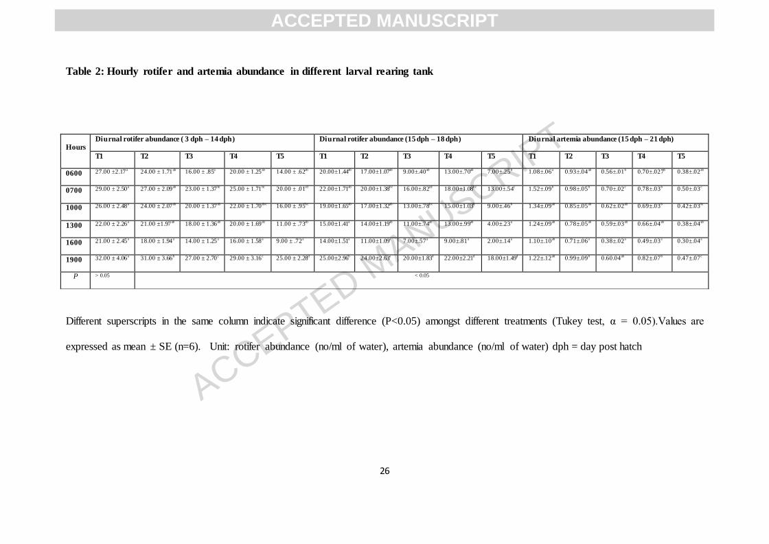
Table 2: Hourly rotifer and artemia abundance in different larval rearing tank
Different superscripts in the same column indicate significant difference (P<0.05) amongst different treatments (Tukey test, α = 0.05).Values are
expressed as mean ± SE (n=6). Unit: rotifer abundance (no/ml of water), artemia abundance (no/ml of water) dph = day post hatch
Hours Diurnal rotifer abundance ( 3 dph – 14 dph) Diurnal rotifer abundance (15 dph – 18 dph) Diurnal artemia abundance (15 dph – 21 dph)
T1 T2 T3 T4 T5 T1 T2 T3 T4 T5 T1 T2 T3 T4 T5
0600 27.00 ±2.17a 24.00 ± 1.71ab 16.00 ± .85a 20.00 ± 1.25ab 14.00 ± .62ab 20.00±1.44ab 17.00±1.07ab 9.00±.40ab 13.00±.70ab 7.00±.25b 1.08±.06a 0.93±.04ab 0.56±.01b 0.70±.027b 0.38±.02ab
0700 29.00 ± 2.50a 27.00 ± 2.09ab 23.00 ± 1.37bc 25.00 ± 1.71bc 20.00 ± .01cd 22.00±1.71ab 20.00±1.38bc 16.00±.82cd 18.00±1.08bc 13.00±.54c 1.52±.09b 0.98±.05b 0.70±.02c 0.78±.03b 0.50±.03c
1000 26.00 ± 2.48a 24.00 ± 2.07ab 20.00 ± 1.37ab 22.00 ± 1.70abc 16.00 ± .95bc 19.00±1.65ab 17.00±1.32ab 13.00±.78bc 15.00±1.03b 9.00±.46b 1.34±.09ab 0.85±.05ab 0.62±.02ab 0.69±.03b 0.42±.03bc
1300 22.00 ± 2.26a 21.00 ±1.97ab 18.00 ± 1.36ab 20.00 ± 1.69ab 11.00 ± .73ab 15.00±1.41a 14.00±1.19ab 11.00±.74ab 13.00±.99ab 4.00±.23a 1.24±.09ab 0.78±.05ab 0.59±.03ab 0.66±.04ab 0.38±.04ab
1600 21.00 ± 2.45a 18.00 ± 1.94a 14.00 ± 1.25a 16.00 ± 1.58a 9.00 ± .72a 14.00±1.51a 11.00±1.09a 7.00±.57a 9.00±.81a 2.00±.14a 1.10±.10ab 0.71±.06a 0.38±.02a 0.49±.03a 0.30±.04a
1900 32.00 ± 4.06a 31.00 ± 3.66b 27.00 ± 2.70c 29.00 ± 3.16c 25.00 ± 2.28d 25.00±2.96b 24.00±2.63e 20.00±1.83d 22.00±2.21d 18.00±1.49d 1.22±.12ab 0.99±.09b 0.60.04ab 0.82±.07b 0.47±.07c
P > 0.05 < 0.05
ACCEPTED MANUSCRIPT

ACCEPTED MANUSCRIPT
27
Figure 1: Survival (A) and standard length (B) of milkfish larvae in different tank background colour. Different superscripts indicate
significant difference (P < 0.05) amongst different treatments (Tukey test, α = 0.05. Values are expressed as mean ± SEM [n=30(standard
length), n = 50 (survival)]. Asterisks were used to indicate significant differences between T1, T3 andT5 (* p < 0.05, ** p < 0.01, *** p <
0.001).
Figure 2: Hourly rotifer and artemia abundance in different larval rearing tank (A-C). Unit: no/ml of water. Values are expressed in
Table 1 as mean ± SE (n=30). Correlation of SL and gut rotifer content (D).
Figure 3: Milkfish larvae gut rotifer (A) and artemia (B) content. Unit: numbers/gut. Asterisks were used to indicate significant differences
between different rearing days and within the treatments (* p < 0.05, ** p < 0.01, *** p < 0.001). Values are expressed as mean ± SEM (n=30).
Gut content in different larval rearing tanks during 5 dph (C, D, E, F,G) and 10 dph (H, I, J, K, L). EG= empty gut, RT = rotifer , + = enhanced ,
- = decreased
Figure 4: Solar illuminated semi outdoor yellow colour tank .
ACCEPTED MANUSCRIPT

ACCEPTED MANUSCRIPT
28
Highlights of present study are as follows:
Milkfish (Chanos chanos) larval rearing technology was not available for Indian condition. As the fish bred under captive conditions first time in
India during June 2015 at ICAR-CIBA fish hatchery, need has been felt to fine tune the existing milkfish larval rearing technology available in
literature. It has been observed that milkfish larval rearing is successful in yellow background tanks which are illuminated by solar or artificial
florescent light. First report on effect of tank colour and solar illumination on milkfish larval rearing.
1. Rearing in semi-outdoor tank with yellow colour background improves milkfish larval growth and survival.
2. Prey ingestion rate of milkfish larvae is highest in yellow colour background tanks due to effective contrast against live feeds.
3. Milkfish larvae are daytime feeder and this feeding habit synergistically improves preying capacity when reared in yellow coloured
tank.
ACCEPTED MANUSCRIPT
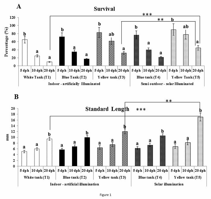
Figure 1

Figure 2

Figure 3

Figure 4