Effect of Pelvic Tilt on Standing Posture...Effect of Pelvic Tilt on Standing Posture JAMES W. DAY,...
7
Effect of Pelvic Tilt on Standing Posture JAMES W. DAY, GARY L. SMIDT, and THOMAS LEHMANN Low back dysfunction is associated in many cases with lumbar lordosis, and tilting the pelvis posteriorly is often recommended for therapeutic purposes. The influence of pelvic tilt on the spinal curves has not been studied. The purpose of this study was to use an objective noninvasive method to determine the effect of the pelvic tilt on the spinal curves in the sagittal plane. Thirty-two healthy subjects and 15 patients with chronic low back dysfunction (CLBD) were studied. Patients with CLBD and healthy subjects were instructed in performing active anterior and posterior pelvic tilt maneuvers, first in the supine and then in the standing position. Comparisons between the Patient Group and the Healthy Group were made for several variables representing the severity of spinal curves, pelvic orientation, hip orientation, and knee orientation. A computerized system, the Iowa Anatom- ical Position System, was used to obtain coordinates of external body surface landmarks from which pelvic tilt measurements were determined. The results showed that the voluntary pelvic tilt did not alter the thoracic spinal curve. For both the Healthy Group and the Patient Group, the lumbar curve was altered by the pelvic tilt: anterior tilt increased the depth of the lumbar curve and posterior tilt decreased the depth of the lumbar curve. The amount of pelvic tilt was the same whether knees were extended or flexed approximately 10 degrees. Pelvic tilt also tended to influence the orientation of the head and other parts of the body. Key Words: Backache, Pelvis, Physical therapy, Spine. Many investigators have assessed the mobility of the lumbar spine and the effect that different trunk positions or lower extremity positions or both have on lumbar lordosis. Little information is available, however, about the ability to correct lumbar lordosis voluntarily as a person stands or sits. Ironically, a com- monly prescribed treatment regimen for the millions of people with low back dysfunction includes a pelvic tilt exer- cise to decrease lumbar lordosis volun- tarily. 1-12 The correction is ostensibly accomplished by rotating the upper por- tion of the pelvis posteriorly in the sag- ittal plane. The main goals for using pelvic tilt are to strengthen the abdom- inal muscles and to decrease lumbar lordosis. 10, 13, 14 Typically, the patient is taught to practice tilting the pelvis posteriorly first, in the supine position; then, in the standing position; and finally, during walking. 4, 15-18 When clinicians give in- structions to patients for the posterior pelvic tilt in the supine position, the patient is usually told to flex the knees and hips so that the maneuver is more easily accomplished. Using this line of reasoning, the flexed leg position in standing should also be more effective in reducing the lordotic curve. Available objective evidence fails to answer the question of how much pelvic rotation is possible while standing. Is the amount of lumbar lordotic curvature of a person with low back dysfunction greater than that of one without low back dysfunc- tion? What effect does standing posture have on the ability to rotate the pelvis? Objective information to these ques- tions is needed to assess the effect, ad- vantages, and limitations of using pelvic tilt as a therapeutic procedure for pos- tural correction. The purpose of this study was 1) to examine differences among anterior, neutral, and posterior tilt of healthy sub- jects and patients with chronic low back dysfunction (CLBD) for extended and flexed knee positions and 2) to measure the variables representing severity of spinal curves, pelvic orientation, and hip orientation. METHOD Subjects Thirty-two male subjects who had no complaints of back dysfunction within six months preceding data collection and who had not undergone back sur- gery composed the Healthy Group. The Healthy Group was matched in age by decade to a Patient Group made up of 15 men with an age range of 25 years to 55 years. The mean age for the Healthy Group was 39 years, and the mean age for the Patient Group was 44 years. The Patient Group was composed of those with CLBD who had at least a three- year history of low back pain and who had experienced low back pain within three months of the laboratory assess- ment. We excluded patients with spinal fusions, herniated intervertebral disks, lateral curvatures of spine, or known muscle atrophic diseases. The Patient Group was selected randomly from the patients scheduled in the orthopedic clinic. Patients who then consented to participate were scheduled for the labo- ratory assessment. METHOD Subjects Thirty-two male subjects who had no complaints of back dysfunction within six months preceding data collection and who had not undergone back sur- gery composed the Healthy Group. The Healthy Group was matched in age by decade to a Patient Group made up of 15 men with an age range of 25 years to 55 years. The mean age for the Healthy Group was 39 years, and the mean age for the Patient Group was 44 years. The Patient Group was composed of those with CLBD who had at least a three- year history of low back pain and who had experienced low back pain within three months of the laboratory assess- ment. We excluded patients with spinal fusions, herniated intervertebral disks, lateral curvatures of spine, or known muscle atrophic diseases. The Patient Group was selected randomly from the patients scheduled in the orthopedic clinic. Patients who then consented to participate were scheduled for the labo- ratory assessment. Mr. Day is Director of Physical Therapy, Vir- ginia Baptist Hospital, Lynchburg, VA 24503. Dr. Smidt is Professor and Director, Physical Therapy Education, College of Medicine, Univer- sity of Iowa, Iowa City, IA 52242 (USA). Dr. Lehmann is Assistant Professor of Or- thopedic Surgery, College of Medicine, University of Iowa. This study was completed in partial fulfillment of Mr. Day's Master of Arts Degree in Physical Therapy at the University of Iowa. Address all correspondence to Dr. Smidt. This article was submitted July 21, 1983; was with the authors eight weeks for revision; and was accepted November 22, 1983. 510 PHYSICAL THERAPY
Transcript of Effect of Pelvic Tilt on Standing Posture...Effect of Pelvic Tilt on Standing Posture JAMES W. DAY,...

Effect of Pelvic Tilt on Standing Posture
JAMES W. DAY, GARY L. SMIDT, and THOMAS LEHMANN
Low back dysfunction is associated in many cases with lumbar lordosis, and tilting the pelvis posteriorly is often recommended for therapeutic purposes. The influence of pelvic tilt on the spinal curves has not been studied. The purpose of this study was to use an objective noninvasive method to determine the effect of the pelvic tilt on the spinal curves in the sagittal plane. Thirty-two healthy subjects and 15 patients with chronic low back dysfunction (CLBD) were studied. Patients with CLBD and healthy subjects were instructed in performing active anterior and posterior pelvic tilt maneuvers, first in the supine and then in the standing position. Comparisons between the Patient Group and the Healthy Group were made for several variables representing the severity of spinal curves, pelvic orientation, hip orientation, and knee orientation. A computerized system, the Iowa Anatomical Position System, was used to obtain coordinates of external body surface landmarks from which pelvic tilt measurements were determined. The results showed that the voluntary pelvic tilt did not alter the thoracic spinal curve. For both the Healthy Group and the Patient Group, the lumbar curve was altered by the pelvic tilt: anterior tilt increased the depth of the lumbar curve and posterior tilt decreased the depth of the lumbar curve. The amount of pelvic tilt was the same whether knees were extended or flexed approximately 10 degrees. Pelvic tilt also tended to influence the orientation of the head and other parts of the body.
Key Words: Backache, Pelvis, Physical therapy, Spine.
Many investigators have assessed the mobility of the lumbar spine and the effect that different trunk positions or lower extremity positions or both have on lumbar lordosis. Little information is available, however, about the ability to correct lumbar lordosis voluntarily as a person stands or sits. Ironically, a commonly prescribed treatment regimen for the millions of people with low back dysfunction includes a pelvic tilt exercise to decrease lumbar lordosis voluntarily.1-12 The correction is ostensibly accomplished by rotating the upper portion of the pelvis posteriorly in the sagittal plane. The main goals for using pelvic tilt are to strengthen the abdominal muscles and to decrease lumbar lordosis.10, 13, 14
Typically, the patient is taught to practice tilting the pelvis posteriorly first, in the supine position; then, in the standing position; and finally, during walking.4, 15-18 When clinicians give instructions to patients for the posterior pelvic tilt in the supine position, the patient is usually told to flex the knees and hips so that the maneuver is more easily accomplished. Using this line of reasoning, the flexed leg position in standing should also be more effective in reducing the lordotic curve. Available objective evidence fails to answer the question of how much pelvic rotation is possible while standing. Is the amount of lumbar lordotic curvature of a person with low back dysfunction greater than that of one without low back dysfunction? What effect does standing posture have on the ability to rotate the pelvis? Objective information to these questions is needed to assess the effect, advantages, and limitations of using pelvic tilt as a therapeutic procedure for postural correction.
The purpose of this study was 1) to examine differences among anterior, neutral, and posterior tilt of healthy subjects and patients with chronic low back dysfunction (CLBD) for extended and
flexed knee positions and 2) to measure the variables representing severity of spinal curves, pelvic orientation, and hip orientation. METHOD Subjects
Thirty-two male subjects who had no complaints of back dysfunction within six months preceding data collection and who had not undergone back surgery composed the Healthy Group. The Healthy Group was matched in age by decade to a Patient Group made up of 15 men with an age range of 25 years to 55 years. The mean age for the Healthy Group was 39 years, and the mean age for the Patient Group was 44 years. The Patient Group was composed of those with CLBD who had at least a three-year history of low back pain and who had experienced low back pain within three months of the laboratory assessment. We excluded patients with spinal fusions, herniated intervertebral disks, lateral curvatures of spine, or known muscle atrophic diseases. The Patient Group was selected randomly from the patients scheduled in the orthopedic clinic. Patients who then consented to participate were scheduled for the laboratory assessment.
METHOD Subjects
Thirty-two male subjects who had no complaints of back dysfunction within six months preceding data collection and who had not undergone back surgery composed the Healthy Group. The Healthy Group was matched in age by decade to a Patient Group made up of 15 men with an age range of 25 years to 55 years. The mean age for the Healthy Group was 39 years, and the mean age for the Patient Group was 44 years. The Patient Group was composed of those with CLBD who had at least a three-year history of low back pain and who had experienced low back pain within three months of the laboratory assessment. We excluded patients with spinal fusions, herniated intervertebral disks, lateral curvatures of spine, or known muscle atrophic diseases. The Patient Group was selected randomly from the patients scheduled in the orthopedic clinic. Patients who then consented to participate were scheduled for the laboratory assessment.
Mr. Day is Director of Physical Therapy, Virginia Baptist Hospital, Lynchburg, VA 24503.
Dr. Smidt is Professor and Director, Physical Therapy Education, College of Medicine, University of Iowa, Iowa City, IA 52242 (USA).
Dr. Lehmann is Assistant Professor of Orthopedic Surgery, College of Medicine, University of Iowa.
This study was completed in partial fulfillment of Mr. Day's Master of Arts Degree in Physical Therapy at the University of Iowa.
Address all correspondence to Dr. Smidt. This article was submitted July 21, 1983; was
with the authors eight weeks for revision; and was accepted November 22, 1983.
510 PHYSICAL THERAPY
RESEARCH
Anatomical Position System We used a noninvasive computerized
method, Iowa Anatomical Position System (IAPS), to obtain coordinates for known external body landmarks. The system is made up of three electromechanical units fastened to an inverted T-shape steel frame supported by a heavy-duty, adjustable drill press base. Each electromechanical unit is composed of a 10-turn precision (hybrid) potentiometer connected to a drum posterior to an eyelet for the passage of a .03-cm diameter stainless steel cable 330 cm in length. The IAPS probe is a 31-cm steel rod with a plastic handle at one end and a small plastic sphere at the other. Three 74-cm stainless steel cables extend from the probe's tip to each of the electromechanical units. The handle of the probe contained a switch, which signaled the computer for acquisition of coordinates. When the switch was depressed for less than two seconds, a single coordinate was determined.
Pelvic Tilt Instructions We positioned the subject supine on
a plinth with hips and knees flexed and both feet flat on the plinth. We instructed the subject to tighten abdominal and gluteal muscles simultaneously and to press the lumbar spine firmly against the plinth. We judged the subject as correctly completing the posterior pelvic tilt when the tester could not place his hand between the subject's lumbar spine and the surface of the support during three consecutive posterior pelvic tilt maneuvers.
We considered the neutral tilt of the pelvis to be the orientation of the pelvis when the subject assumed the relaxed standing posture. While standing, the subject practiced the posterior pelvic tilt movement. The subject was instructed to tighten his abdominal muscles while squeezing his buttocks together to complete a posterior pelvic tilt in a standing position. The subject was considered able to complete a standing posterior pelvic tilt when the tester observed the subject rotate the anterior part of his pelvis upward three consecutive times on command.
We also instructed the subject in the anterior pelvic tilt. He was told to relax his abdominal muscles and allow his pelvis to rotate in the anterior direction. We judged the subject to complete an anterior pelvic tilt when the tester ob
served the subject rotate the anterior part of his pelvis downward three consecutive times. We requested the subject to practice the anterior and posterior pelvic tilt movements in a standing position between the first testing session and the second testing session. The same investigator instructed all subjects in the pelvic tilt movements.
Data Acquisition Process We positioned a small platform in the
middle of the IAPS measuring field. The subject stood on the platform with his back to the IAPS. After the investigator aligned the navicular tuberosities with a frontal plane line equidistant from the platform axis of rotation, the subject maintained a neutral standing position with knees extended while the IAPS probe was placed on the following points in the stated sequence: 1) edge of platform to establish point P(0,0,0), 2) point on platform midway between the navicular tuberosities and in back of the feet, 3) left tenocalcaneus insertion, 4) right tenocalcaneus insertion, 5) midpoint left popliteal fossa, 6) right popliteal fossa, 7) extreme posterior sacral protrusion, 8) left posterior superior iliac spine, 9) right posterior superior iliac spine, 10) S2, 11) T12, 12) posterior lateral corner of the left acromion process, 13) posterior lateral corner of the right acromion process, 14) C7, and 15) inion.
We obtained a stream of coordinates for the outline of the spinal curves by maintaining the probe on the inion, depressing a control switch, and moving the probe along the cervical spine to C7 before releasing the switch; depressing the switch at C7 and moving the probe along the dorsal spine to T12 before releasing the switch; and depressing the switch at T12 and moving the probe along the lumbar spine to S2 before releasing the switch.
With the subject maintaining position, we unlocked the platform, rotated it 90 degrees clockwise, and relocked it in position. We reestablished the coordinates for the reference point to allow the computer to determine the angle of rotation necessary to transform the body orientation from the coronal to the sagittal plane. We determined coordinates for the anatomical landmarks using the IAPS probe in the following sequence: 1) fifth metatarsal head, 2) lateral malleolus, 3) lateral femoral epicondyle, 4) greater trochanter, 5) anterior superior
iliac spine, 6) lateral posterior corner of the acromion process, 7) tragus, and 8) superior lateral edge of the orbita.
The same order of obtaining coordinates for anatomical landmarks was
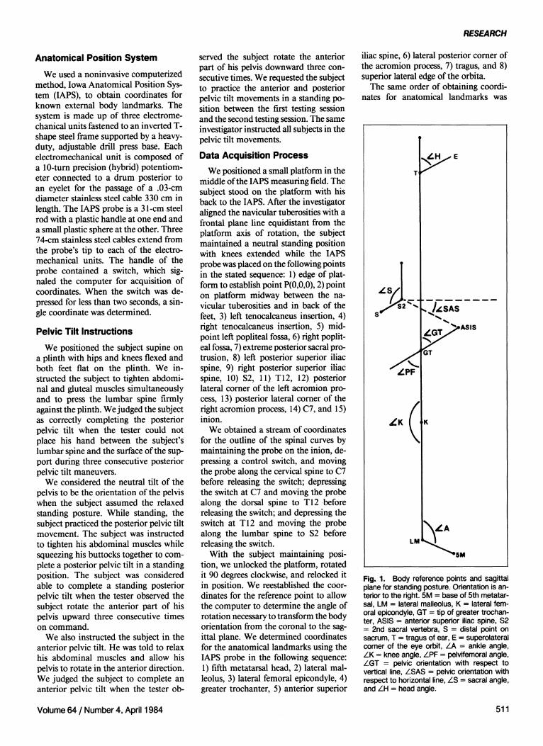
Fig. 1. Body reference points and sagittal plane for standing posture. Orientation is anterior to the right. 5M = base of 5th metatarsal, LM = lateral malleolus, K = lateral femoral epicondyle, GT = tip of greater trochanter, ASIS = anterior superior iliac spine, S2 = 2nd sacral vertebra, S = distal point on sacrum, T = tragus of ear, E = superolateral corner of the eye orbit, A = ankle angle,
K = knee angle, PF = pelvifemoral angle, GT = pelvic orientation with respect to
vertical line, SAS = pelvic orientation with respect to horizontal line, S = sacral angle, and H = head angle.
Volume 64 / Number 4, April 1984 511

Fig. 2. Sagittal plane measurements for spinal curves. I = inion; C7 = spinous process of C7; T12 = spinous process of T12; S2 = spine at the level of the S2; DC, DT, DL = maximum depth of cervical, thoracic, and lumbar curves, respectively; d1-c7, dc7-T12, dT12-s2 = distance between the designated spinous processes; and Lc, LT, LL = location of the deepest portion of the cervical, thoracic, and lumbar curves, respectively.
completed tor each of three pelvic positions (neutral tilt, anterior tilt, and posterior tilt) for both extended knee and flexed knee standing postures. The subject assumed either a 10-degree flexed knee position or a completely extended knee position with the aid of instructions from the investigator who aligned a goniometer axis with the right lateral femoral epicondyle; one arm was coincident with the lateral malleolus and the other arm was coincident with the greater trochanter. We randomized the order of the measures.
Variables Measured We obtained angular measurements
for seven anatomical angles in the three pelvic positions for both standing postures to assess pelvic motion and its influence on body posture (Fig. 1).
To express changes in cervical, thoracic, and lumbar spinal curve segments with respect to postural and pelvic position changes, we determined depth of lordotic curvatures. Straight lines passing from the inion (I) to C7 (dI-C7), C7 to T12 (dc7-T12), and T12 to S2 (dT12-s2)
represented size of spinal curve segments and were constructed by an In-terdata minicomputer (Fig. 2). From each straight line connecting the designated spinal landmarks, a perpendicular line to the deepest portion of each corresponding spinal curve also was determined by the Interdata minicomputer.
These perpendicular distances represented depth of lordotic curvatures for cervical (Dc), thoracic (DT), and lumbar (DL) spinal-curve segments. Depth of lordotic curvature measures were normalized to size of spinal-curve segments in the form of a ratio to account for variances in size of spinal segments among subjects. The ratios were expressed as depth of lordotic curvature to total distance between cephalad and caudal points on each spinal curve, ie, DC:dI-C7, DT:dC7-T12, and DL:dT12-S2, respectively for the cervical, thoracic, and lumbar levels. Also, the distance from the perpendicular line to the cephalad landmark was expressed as a ratio of the total distance between the cephalad and caudal points on the straight line, ie, LC:dI-C7, LT:dC7-T12, and LL:dT12-S2, to permit an estimate of the anatomical location of the deepest portion of each spinal curve.
Data Analysis We analyzed the ratios and angles
using an analysis of variance (ANOVA) test with split level, three-factor, randomized block design (Fig. 3). We used a p value of ≤.05 in the analysis and performed the Duncan's Multiple Range test on the data to determine if the differences were large enough to be statistically significant.
Accuracy of Measurement System
The accuracy of the IAPb is expressed as a ratio of the algebraic difference between the measured IAPS coordinate and the known coordinate to the full scale output, expressed as a percentage. We determined the accuracy of a measurement field large enough to accommodate a person in the standing position. The dimensions of the field were 91 cm x 91 cm x 200 cm. We placed a 61-cm x 61-cm x 9-cm pegboard mounted on a plywood platform in the middle of the measuring field with its center 150 cm from the base of the IAPS. Using a micrometer caliper for measurement, the mean and standard
Fig. 3. Diagram of research design.
512 PHYSICAL THERAPY
RESEARCH
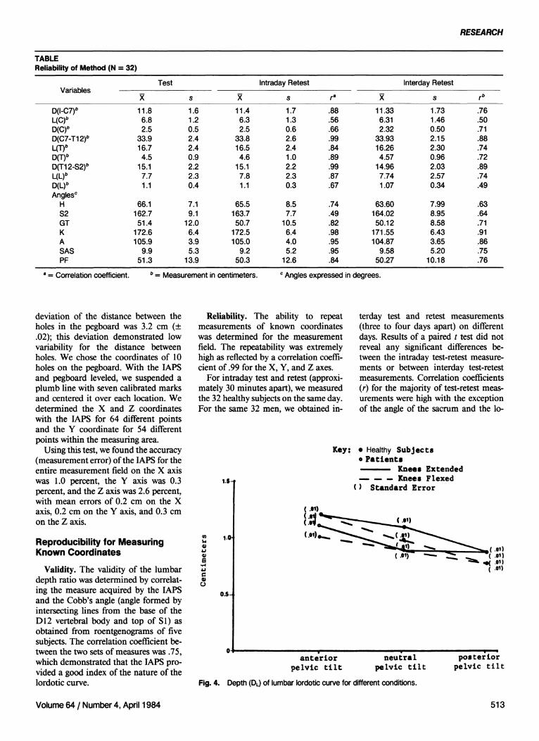
TABLE Reliability of Method (N = 32)
Variables
D(I-C7)b
L(C)b
D(C)b
D(C7-T12)b
L(T)b
D(T)b
D(T12-S2)b
L(L)b
D(L)b
Anglesc
H S2 GT K A SAS PF
Test
11.8 6.8 2.5
33.9 16.7 4.5
15.1 7.7 1.1
66.1 162.7 51.4
172.6 105.9
9.9 51.3
s 1.6 1.2 0.5 2.4 2.4 0.9 2.2 2.3 0.4
7.1 9.1
12.0 6.4 3.9 5.3
13.9
11.4 6.3 2.5
33.8 16.5 4.6
15.1 7.8 1.1
65.5 163.7 50.7
172.5 105.0
9.2 50.3
Intraday Retest
s 1.7 1.3 0.6 2.6 2.4 1.0 2.2 2.3 0.3
8.5 7.7
10.5 6.4 4.0 5.2
12.6
ra
.88
.56
.66
.99
.84
.89
.99
.87
.67
.74
.49
.82
.98
.95
.95
.84
11.33 6.31 2.32
33.93 16.26 4.57
14.96 7.74 1.07
63.60 164.02 50.12
171.55 104.87
9.58 50.27
Interday Retest
s 1.73 1.46 0.50 2.15 2.30 0.96 2.03 2.57 0.34
7.99 8.95 8.58 6.43 3.65 5.20
10.18
rb
.76
.50
.71
.88
.74
.72
.89
.74
.49
.63
.64
.71
.91
.86
.75
.76
deviation of the distance between the holes in the pegboard was 3.2 cm (± .02); this deviation demonstrated low variability for the distance between holes. We chose the coordinates of 10 holes on the pegboard. With the IAPS and pegboard leveled, we suspended a plumb line with seven calibrated marks and centered it over each location. We determined the X and Z coordinates with the IAPS for 64 different points and the Y coordinate for 54 different points within the measuring area.
Using this test, we found the accuracy (measurement error) of the IAPS for the entire measurement field on the X axis was 1.0 percent, the Y axis was 0.3 percent, and the Z axis was 2.6 percent, with mean errors of 0.2 cm on the X axis, 0.2 cm on the Y axis, and 0.3 cm on the Z axis.
Reproducibility for Measuring Known Coordinates
Validity. The validity of the lumbar depth ratio was determined by correlating the measure acquired by the IAPS and the Cobb's angle (angle formed by intersecting lines from the base of the D12 vertebral body and top of S1) as obtained from roentgenograms of five subjects. The correlation coefficient between the two sets of measures was .75, which demonstrated that the IAPS provided a good index of the nature of the lordotic curve.
Reliability. The ability to repeat measurements of known coordinates was determined for the measurement field. The repeatability was extremely high as reflected by a correlation coefficient of .99 for the X, Y, and Z axes.
For intraday test and retest (approximately 30 minutes apart), we measured the 32 healthy subjects on the same day. For the same 32 men, we obtained in
terday test and retest measurements (three to four days apart) on different days. Results of a paired t test did not reveal any significant differences between the intraday test-retest measurements or between interday test-retest measurements. Correlation coefficients (r) for the majority of test-retest measurements were high with the exception of the angle of the sacrum and the lo-
Fig. 4. Depth (DL) of lumbar lordotic curve for different conditions.
a = Correlation coefficient. b = Measurement in centimeters. c Angles expressed in degrees.
Volume 64 / Number 4, April 1984 513

Fig. 5. Relative depth (DL:dT12-s2) of lumbar curve for different conditions.
cation to the deepest portion of the cervical curve. The intraday and interday reliability data appear in the Table.
With six out of seven variables having an intraday correlation coefficient above .80 and the same six variables having an interday correlation above .70, we believed the IAPS method for determining postural changes was dependable. Depth of thoracolumbar curve was the one variable that demonstrated the lowest correlation coefficient for intraday and interday reliability. Because of the small range of DL variability among healthy subjects, this low r might be expected. The magnitude of the change in the absolute depth value was small but within the accuracy of the IAPS.
RESULTS We found no significant differences
between the DL for the Healthy Group
and Patient Group (Figs. 4 and 5). The flexed knee position tended to flatten the lordotic curve. Both the Healthy Group and Patient Group were able to rotate their pelvis a sufficient amount to change the thoracolumbar curve (Fig. 6). Pelvic rotation or pelvic tilt did not alter the configuration of the thoracic spinal curve. For the extended knee position, the postures of anterior and neutral pelvic tilt were not significantly different. The posture assumed during posterior pelvic tilt was significantly different (p < .05) from the neutral and anterior pelvic tilt postures. With the knees flexed 10 degrees, the posture assumed during neutral pelvic tilt was not significantly different from those assumed during either anterior or posterior pelvic tilt, but anterior pelvic tilt produced a posture significantly different (p < .05) from that associated with posterior pel
vic tilt. The orientation of the pelvis during the neutral, posterior, and anterior pelvic tilts was not significantly affected by flexing the knee 10 degrees (Fig. 6).
When we compared the results of the Healthy Group with those of the Patient Group, the only significantly different measurement (p < .05) was the mean pelvifemoral angle or amount of hip flexion while standing. The Patient Group stood with hips in a greater amount of flexion (Fig. 7).
Other body segments seemed to be affected by the pelvic tilt maneuvers, indicating that muscles and limb segments do not act in isolation. For example, the average sagittal plane rotation of the head was 4 degrees when the pelvic tilt was voluntarily performed. The head and neck flexed (about 4°) during the anterior pelvic tilt and extended (about 4°) during the posterior pelvic tilt.
DISCUSSION Tilting the pelvis posteriorly de
creased the absolute depth of the lumbar curve, and tilting the pelvis anteriorly increased the absolute depth of the lumbar curve. (This relationship supported the basic principle that rotating the pelvis posteriorly can decrease the lumbar curve.) The results also demonstrated that a person properly trained in a pelvic tilt maneuver can voluntarily rotate his pelvis a sufficient amount to alter the lumbar lordotic curve. Instructing the subjects to flex their knees 10 degrees while completing the posterior pelvic tilt may not have increased the effect of the pelvic tilt. We did not assess the ability of a subject to maintain a posterior pelvic tilt during walking. Boynton reported average depth ratios for the lumbar curve ranging from 0.192 for full active extension to 0.048 for full active flexion of the trunk in 50 women (unpublished thesis, 1934). The subjects were in a natural standing position ready for roentgenograms. In our study, the average ratios were 0.079 for subjects performing the anterior pelvic tilt and 0.52 when they performed the posterior pelvic tilt (extended knees postion). Al though association between the two studies may be confounded by the gender differences, for upright standing the lumbar curve appears to be oriented more toward the maximal trunk flexed position than toward the maximal extended position.
514 PHYSICAL THERAPY

RESEARCH
Fig. 6. (Left) Sagittal plane orientation (Angle SAS) during standing for different conditions. Fig. 7. (Right) Influence of pelvic tilt on hip (pelvifemoral angle) measurements in standing for different conditions.
The change in the absolute depth of the lumbar curve in this study was probably not reflected in equal angular changes among each of the vertebrae that comprise the curve. Lindh reported marked reduction of the lordotic lumbar curve (Cobb's angle) with forced posterior rotation of the pelvis in women with scoliosis.19 Using roentgenograms, the angular change of the lumbar curve has not been determined for men completing a posterior pelvic tilt. We may safely assume, however, that the individual vertebral rotation would follow the trends described by Lindh, whereby the majority of the lumbar curve motion occurred at L4-5 and L5-S1 segments. From the cephalad point to L4, the lumbar vertebrae tended to maintain their basic relationship to one another.
The data exhibited an inverse relationship with the lumbar curve length (dT12-s2) and the thoracic curve length (dc7-T12) during the posterior pelvic tilt. As the pelvis rotated posteriorly, the thoracic area was rotated in the anterior direction by the abdominal muscles. Therefore, the length of the lumbar segment increased, and the cord length be
tween C7 and T12 decreased. In our study, minimal thoracic spine movement in flexion and extension occurred during anterior and posterior pelvic tilt. For both healthy subjects and subjects with CLBD, the maximum change in thoracic curve depth was less than 0.3 cm when the anterior and posterior pelvic tilt were executed. This minimal movement suggests that physical therapists may need to consider alternative exercises to effect postural changes in the thoracic curve.
Kapandji,20 White and Panjabi,21 and Beal and Beckwith22 reported a range of 60 degrees to 95 degrees of flexion and extension motion in the lumbar spine. These authors agreed that the L5-S1 and L4-5 intervertebral motion accounted for approximately 40 degrees of the lumbar spine motion, and Beal and Beckwith stated, in addition, that motion of the spine was always initiated in the caudal segments. With the rotating influence of the posterior pelvic tilt on the lumbar curve occurring at the L4-5 and L5-S1 levels initially, the pelvis reached an end point of posterior rotation before the lumbar curve obtained its end point
of flexion. In our study, the lumbar spine was apparently not limiting pelvic rotation. Other factors that might have limited the posterior pelvic tilt were shortened length of the abdominal muscles (rectus abdominus and external and internal obliques) during contraction and the presence of connective tissue about the anterior aspect of the hip joint. Anterior rotation of the pelvis might be restricted by the structural inability of the superincumbent spinal structures to accommodate further changes and still maintain the upright position.
Our results showed that flexing the knees 10 degrees does not allow a greater range of pelvic tilt motion to occur than if the knees are maintained extended. Perhaps patients find it easier to learn or maintain the pelvic tilt by standing with lower extremities flexed in such a way that the knees are flexed more than 10 degrees.
The ANOVA did not show a significant difference between the pelvic tilt angle (neutral pelvic tilt) of the Healthy Group and Patient Group for relaxed standing. Therefore, the data of our study do not support the idea that pa-
Volume 64 / Number 4, April 1984 515
tients with CLBD have a greater anterior pelvic tilt in posture than those without CLBD. Nor does this study support the concept that male patients with CLBD have a greater lumbar lordosis than healthy men. One clinical implication for this result is that indiscriminate training of patients with CLBD in the posterior pelvic tilt exercise to decrease lumbar lordosis is not justified. Rotating the pelvis posteriorly may provide other benefits that are more important than the need to decrease the amount of lumbar lordosis. For example, Nachemson23
reported that tilting the pelvis posteriorly increased intra-abdominal pressure, which provided structural stability to the spine.
We found a significant difference between the means for the pelvifemoral angle of the Healthy Group (52.5°) and the Patient Group (59.3°). The implication is that the Patient Group had significantly more hip flexion for relaxed standing posture than the Healthy Group. The mean clinical measurement of the pelvifemoral angle was similar for both groups and corresponded to the IAPS pelvifemoral measurement in standing. Because of the pain, the patient with CLBD might be flexing at the
hip to maintain his spine in a pain-free position. Whatever the case, we believe examination of the hip joints in patients with low back dysfunction would be prudent.
SUMMARY
While standing, all study subjects were able to rotate the pelvis voluntarily in the sagittal plane, which affected the lumbar lordotic curve, hip position, and head position. The thoracic curve was not affected by the pelvic rotation. The only significant difference between the results of the Healthy Group and the Patient Group was that the Patient Group stood with a greater amount of hip flexion. REFERENCES
1. Denniston HD: Physical treatment in postural defect. Arch Phys Ther, X-ray, Radium 16:525-527,1935
2. Forrester-Brown MF: Improvement of posture. Lancet 2:115-117, 1930
3. Hansson KG: Body mechanics in geriatrics. J Am Geriatr Soc 2:429-433, 1954.
4. House F, O'Connor S: Specific management for lumbar and sacral radiculitis. JAMA 66:1285-1290,1958
5. La Freniere J: The Low Back Pain Patient. New York, NY, Masson Publishing USA Inc, 1979
6. La Place LB, Nicholson JT: Physiologic effects of the correction of faulty posture. JAMA 107:1009-1012, 1936
7. Lidstrom A, Zachrisson M: Physical therapy on low back pain and sciatica. Scand J Rehabil Med 2:37-42, 1970
8. Micheli L: Low back pain in the adolescent: Differential diagnosis. Am J Sports Med 7:362-364, 1979
9. Montgomery RP: Etiology and treatment for low-back and related complaints. Wis Med J 44:1076-1083,1945
10. Pheasant H: Practical posture building. Clin Orthop 25:83-91, 1962
11. Stanish W: Low back pain in middle-aged athletes. Am J Sports Med 7:367-369, 1979
12. Whitman A: Proper posture. Hygeia 7:1205-1207,1929
13. Cyriax FE: Antero-posterior tilt of the pelvis. British Journal of Children's Disease 21:279-283,1924
14. Macnab I: Backache. Baltimore, MD, Williams &Wilkins, 1977, pp 133-169
15. Rahrni H: Backache Relieved. Springfield, IL, Charles C Thomas, Publisher, 1966, p 50
16. Farfan H: The biomechanical advantage of lordosis and hip extension for upright activity. Spine 3:336-342, 1978
17. Finneson B: Low Back Pain. Philadelphia, PA, JB Lippincott Co, 1973, pp 78-138
18. Williams P: The Lumbosacral Spine. New York, NY, McGraw-Hill Inc, 1965
19. Lindh M: The effect of sagittal curve changes on the brace correction of idiopathic scoliosis. Spine 5:26-36, 1980
20. Kapandji I: The lumbar vertebral column. In Physiology of Joints, ed 2. New York, NY, Churchill Livingstone Inc, 1974, vol 3, pp 7 2 -126
21. White A, Panjabi M: The basic kinematics of the human spine. Spine 3:12-20, 1978
22. Beal M, Beckwith C: Studies of vertebral motion: I. Cineradiographic studies on the Halla-day spine. JAOA 63:319-325, 1963
23. Nachemson A: The lumbar spine an orthopaedic challenge. Spine 1:59-71, 1976
516 PHYSICAL THERAPY