Effect of mild hypothermia preconditioning against low ... file10.1371/journal.pone.0176652 Editor:...
15
RESEARCH ARTICLE Effect of mild hypothermia preconditioning against low temperature (4˚C) induced rat liver cell injury in vitro Jiasheng Qin 1,2☯ , Yanxing Mai 3☯ , Yang Li 1,2 , Zesheng Jiang 1,2 , Yi Gao 1,2 * 1 Second Department of Hepatobiliary Surgery, Zhujiang Hospital, Southern Medical University, Guangzhou, Guangdong, P.R. China, 2 Institute of Regenerative Medicine, Southern Medical University, Guangzhou, Guangdong, P.R. China, 3 Department of Geriatrics, Zhujiang Hospital, Southern Medical University, Guangzhou, Guangdong, P.R. China ☯ These authors contributed equally to this work. * [email protected] Abstract Bioartificial liver holds special position in the field of regenerative medicine, and cold envi- ronment at 4℃ is widely used for the short storage of both organ and liver cell for later appli- cation. However, the disadvantages of such cold storage could influence cell viability and lead to cell apoptosis in different degrees. In this study, we mainly explore the pre-protective effect of mild hypothermia against low temperature (4℃)-induced rat liver cell injury in vitro. Our results indicated that the precondition with mild hypothermia could increase cell viability, such as cell proliferation, LDH regulation and glycogen synthesis ability of liver cell. The pre- condition also decreased the ROS production and relieved cell apoptosis in liver cells. Com- pared with the model group, the mitochondrial membrane potential was restored in the mild hypothermia group, as well as the mitochondrial membrane permeability transition pore opening, indicating that the therapeutic mechanism was related to mitochondrial protection. Further analysis showed that PI3K-Akt-GSK3β signal pathway might be associated with the pre-protective effect of mild hypothermia. Thus, our study suggested that the precondition with mild hypothermia hold the protective effect for liver cell in cold environment, and further developed a novel strategy for the storage of liver seed cells, even bioartificial liver. Introduction With the development of regenerative medicine, bioartificial liver has attracted scientists’ attention in recent years, for its promising application in disease treatment. Somatic liver cells are also widely used for pharmacological and toxicological research, as well as for cell trans- plantation. In present, cold environment at 2–8℃ (usually at 4℃) is used for the short storage of both the organ and liver cell for later application, and the storage time can vary from several minutes to several hours for different researches. However, scientists have demonstrated the disadvantages of such cold storage could influence cell viability and lead to oxidative damage, mitochondrial dysfunction and cell apoptosis in different degrees [1, 2]. Therefore, the PLOS ONE | https://doi.org/10.1371/journal.pone.0176652 April 28, 2017 1 / 15 a1111111111 a1111111111 a1111111111 a1111111111 a1111111111 OPEN ACCESS Citation: Qin J, Mai Y, Li Y, Jiang Z, Gao Y (2017) Effect of mild hypothermia preconditioning against low temperature (4˚C) induced rat liver cell injury in vitro. PLoS ONE 12(4): e0176652. https://doi.org/ 10.1371/journal.pone.0176652 Editor: Beicheng Sun, The First Affiliated Hospital of Nanjing Medical University, CHINA Received: January 1, 2017 Accepted: April 13, 2017 Published: April 28, 2017 Copyright: © 2017 Qin et al. This is an open access article distributed under the terms of the Creative Commons Attribution License, which permits unrestricted use, distribution, and reproduction in any medium, provided the original author and source are credited. Data Availability Statement: All relevant data are within the paper and its Supporting Information files. Funding: This study was supported by the grants from the National High Technology Research and Development Program of China (“863” Program 2012AA020505); National Natural Science Foundation of China (81470875); Science and Technology Planning Project of Guangdong Province (2014B020227002,2015B020229002, 2015B090903069); Natural Science Foundation of Guangdong Province (2014A030312013);
Transcript of Effect of mild hypothermia preconditioning against low ... file10.1371/journal.pone.0176652 Editor:...

RESEARCH ARTICLE
Effect of mild hypothermia preconditioning
against low temperature (4˚C) induced rat
liver cell injury in vitro
Jiasheng Qin1,2☯, Yanxing Mai3☯, Yang Li1,2, Zesheng Jiang1,2, Yi Gao1,2*
1 Second Department of Hepatobiliary Surgery, Zhujiang Hospital, Southern Medical University, Guangzhou,
Guangdong, P.R. China, 2 Institute of Regenerative Medicine, Southern Medical University, Guangzhou,
Guangdong, P.R. China, 3 Department of Geriatrics, Zhujiang Hospital, Southern Medical University,
Guangzhou, Guangdong, P.R. China
☯ These authors contributed equally to this work.
Abstract
Bioartificial liver holds special position in the field of regenerative medicine, and cold envi-
ronment at 4℃ is widely used for the short storage of both organ and liver cell for later appli-
cation. However, the disadvantages of such cold storage could influence cell viability and
lead to cell apoptosis in different degrees. In this study, we mainly explore the pre-protective
effect of mild hypothermia against low temperature (4℃)-induced rat liver cell injury in vitro.
Our results indicated that the precondition with mild hypothermia could increase cell viability,
such as cell proliferation, LDH regulation and glycogen synthesis ability of liver cell. The pre-
condition also decreased the ROS production and relieved cell apoptosis in liver cells. Com-
pared with the model group, the mitochondrial membrane potential was restored in the mild
hypothermia group, as well as the mitochondrial membrane permeability transition pore
opening, indicating that the therapeutic mechanism was related to mitochondrial protection.
Further analysis showed that PI3K-Akt-GSK3β signal pathway might be associated with the
pre-protective effect of mild hypothermia. Thus, our study suggested that the precondition
with mild hypothermia hold the protective effect for liver cell in cold environment, and further
developed a novel strategy for the storage of liver seed cells, even bioartificial liver.
Introduction
With the development of regenerative medicine, bioartificial liver has attracted scientists’
attention in recent years, for its promising application in disease treatment. Somatic liver cells
are also widely used for pharmacological and toxicological research, as well as for cell trans-
plantation. In present, cold environment at 2–8℃ (usually at 4℃) is used for the short storage
of both the organ and liver cell for later application, and the storage time can vary from several
minutes to several hours for different researches. However, scientists have demonstrated the
disadvantages of such cold storage could influence cell viability and lead to oxidative damage,
mitochondrial dysfunction and cell apoptosis in different degrees [1, 2]. Therefore, the
PLOS ONE | https://doi.org/10.1371/journal.pone.0176652 April 28, 2017 1 / 15
a1111111111
a1111111111
a1111111111
a1111111111
a1111111111
OPENACCESS
Citation: Qin J, Mai Y, Li Y, Jiang Z, Gao Y (2017)
Effect of mild hypothermia preconditioning against
low temperature (4˚C) induced rat liver cell injury in
vitro. PLoS ONE 12(4): e0176652. https://doi.org/
10.1371/journal.pone.0176652
Editor: Beicheng Sun, The First Affiliated Hospital
of Nanjing Medical University, CHINA
Received: January 1, 2017
Accepted: April 13, 2017
Published: April 28, 2017
Copyright: © 2017 Qin et al. This is an open access
article distributed under the terms of the Creative
Commons Attribution License, which permits
unrestricted use, distribution, and reproduction in
any medium, provided the original author and
source are credited.
Data Availability Statement: All relevant data are
within the paper and its Supporting Information
files.
Funding: This study was supported by the grants
from the National High Technology Research and
Development Program of China (“863” Program
2012AA020505); National Natural Science
Foundation of China (81470875); Science and
Technology Planning Project of Guangdong
Province (2014B020227002,2015B020229002,
2015B090903069); Natural Science Foundation of
Guangdong Province (2014A030312013);

optimized storage method is essential for the application of bioartificial liver and other bioen-
gineering technologies in clinical studies.
Mild hypothermia has been reported to be a very promising neuroprotective therapeutic
strategy for patients with brain injury [3–8]. Some groups also demonstrated that the hypo-
thermia could protect liver cell from cell death or apoptosis in some hepatic diseases [9–11]. In
recent years, the clinical application of the hypothermia has showed the therapeutic action for
the patients with those diseases, and researchers also try to explain the mechanism for the
action [12]. For example, mild hypothermia holds the ability to up-regulate the expression of
anti-apoptotic gene Bcl-2, and decrease the levels of some inflammatory chemokines (such as
IL-8, MCP-1 and COX-2) in endothelial cells [13]. Some scientists also indicated that hypo-
thermia could induce the expression of cold-inducible RNA-binding protein to inhibit cell
apoptosis induced by tumor necrosis factor-α via the activation of extracellular signal-regu-
lated kinase [14]. However, most reports describe the complex mechanism about the effect of
mild hypothermia on the brain, the detailed molecular mechanisms of underlying potential
beneficial effects of hypothermia treatment on the liver cell injury or liver failure may be still
far away from our understanding.
As known, cold storage (usually at 4℃) can lead to oxidative damage and cell apoptosis, and
liver cell apoptosis is always regarded as an important component of some liver diseases, and
the apoptotic signaling pathways mediated by Fas and other apoptotic genes hold significant
potential during the process [15–19]. In 2004, Fu et al first evaluated the hepatocyte apoptosis
with the treatment of mild hypothermia, and their research indicated that mild hypothermia
(26℃) could suppress Fas-mediated apoptotic signaling pathways in liver cells. This function
mainly depended on the inhibition of some signaling events, such as cytochrome c release,
effector caspase activation, and so on [10]. In recent years, the protective effect of mild hypo-
thermia was also evaluated in some other kinds of liver cell injury models to clarify more
detailed mechanisms. Sakurai et al. suggested that hypothermia could protect hepatic cell from
cell death through the reduction of ROS production in fulminant hepatitis directly. In their
study, concanavalin A-induced hepatitis models were established in mice, and the hypothermia
group were kept at 25℃. Their results indicated that hypothermia treatment hold the ability to
attenuated liver injury and prolong survival through the activation of c-Jun N-terminal kinase
as well as Akt. Similar to their further study about the function of hypothermia in brain injury,
the expression of cold-inducible RNA-binding protein was also up-regulated, leading to the
decreased hepatocyte apoptosis in the group with mild hypothermia treatment [11, 14].
Therefore, we can conclude that some researchers have demonstrated the protective effect
of mild hypothermia from liver cell apoptosis or injury and explored the associated mechanism
preliminarily. However, whether the hypothermia still holds the pre-protective effect against
liver cell damage and cell apoptisis is still unclear. In this study, low temperature-induced liver
cell injury model was established to evaluate the pre-protective effect of mild hypothermia and
further establish a novel strategy for the storage of liver seed cells, even bioartificial liver.
Materials and methods
This study was carried out in strict accordance with the recommendations in the Guide for the
Care and Use of Laboratory Animals of the National Institutes of Health. The protocol was
approved by the Institutional Animal Care and Use Committee of Southern Medical University.
Cell culture and treatment
Rat liver cell line, Brl-3A, was used in our study, which was purchased from the Cell Bank of
Type Culture Collection of Chinese Academy of Sciences (Shanghai, China). The cell was
Pre-protection of mild hypothermia against 4˚C induced liver cell injury
PLOS ONE | https://doi.org/10.1371/journal.pone.0176652 April 28, 2017 2 / 15
Educational Commission of Guangdong Province
(C10319040).
Competing interests: The authors have declared
that no competing interests exist.

cultured in DMEM (Hyclone, glucose concentration = 11.1 mM) supplemented with 10% FBS
(Hyclone), 100 U/mL penicillin and 0.1g/mL streptomycin (Hyclone) at 37℃ under 5% CO2
condition. Brl-3A cells cultured in 6-well plate were treated with the hypothermia when the
cells were 70–80% confluent, and only 1ml medium was left in each well to ensure the suffi-
cient heat exchange during the treatment process. Five groups were designed as follow (Fig
1A):
1. Control group: Cells were cultured normally in a humidified atmosphere at 37℃ with 5%
CO2;
2. Model group: Cells were treated at 4℃for 6 h, and further come back to 37℃ for another 3
h;
3. Mild hypothermia group: Cells were treated with mild hypothermia preconditioning (cells
underwent three cycles of incubation at 26℃ for 10min followed with incubation at 37℃for 15min as the reference [20]), and the following process was the same as the model
group;
4. Mild hypothermia+LY294002: All of the operations were the same as the mild hypothermia
group. Differently, the medium was further supplemented with 20 μM LY294002 (PI3K
inhibitor);
5. Mild hypothermia+LiCl: All of the operations were the same as the mild hypothermia
group. Differently, the medium was further supplemented with 20 mM LiCl (GSK3βinhibitor).
Cell viability assay
Cell viability assay was performed using CCK-8 Kit (Dojindo). Briefly, the cells which were
seeded into 96-well plates at a density of 1×10⁴ cells/well, were detected with 10 μL CCK-8,
and incubated at 37℃ for 4h. The absorbance was measured at 450 nm. In this study, the pro-
liferation index = the absorbance of experimental group—the absorbance of blank group, was
used to evaluate the cell viability of each group. Besides, cell proliferation ability was evaluated
with Click-iT1 Plus EdU Proliferation Kit (Alexa Fluor1 555, Thermo Fisher Scientific), and
the assay was mainly performed according to the manufacturer’s instructions.
To further evaluate cell viability, the extracellular/intracellular LDH, intracellular glycogen
and glucose in medium in each group were measured with LDH-Cytotoxicity Colorimetric
Assay KitⅡ(BioVision), Glycogen Colorimetric/Fluorometric Assay Kit (BioVision) and
Amplex1 Red Glucose Assay Kit (BioVision) according to the manufacturer’s instruction
respectively.
Transmission electron microscope (TEM) assay
The cell samples were fixed in glutaraldehyde first, and further dehydrated in graded ethyl
alcohols, and embedded with Epon812 finally. In this study, the ultrathin section was cut with
an Ultracut E ultramicrotome, and the section was stained with uranyl acetate and lead citrate,
and further observed under TEM as previously described [21–23].
Evaluation of reactive oxygen species (ROS)
To evaluate the oxidative stress in each group, the ROS level of each group was measured
using CellROX Orange Reagent (Life Technologies) according to the manufacturer’s instruc-
tion. Briefly, Brl-3A cells in each group were collected with trypsin, and washed with PBS for
Pre-protection of mild hypothermia against 4˚C induced liver cell injury
PLOS ONE | https://doi.org/10.1371/journal.pone.0176652 April 28, 2017 3 / 15

once, then the cells were further incubated with 50 μM CellROX Reagent at 37℃ for 30 min,
then washed with PBB for three times. The cells incubated with nothing were used as the nega-
tive group. Finally, the cell samples were analyzed using a FACSCalibur cytometer (BD).
Fig 1. Effect of precondition with mild hypothermia on cell viability. A. Schematic diagram of the approach for treatment of each group. Red orange: the
treatment with 37℃; Purple: the treatment with 26℃; Blue: the treatment with 4℃. B-E. Evaluation of cell viability, cell proliferation ability, LDH regulation,
glycogen synthesis ability and glucose absorption of each group respectively. Similar results were obtained in three independent experiments and results
were expressed as mean ± SEM. A t-test was used to compare the various groups, and P<0.05 was considered statistically significant. *: P<0.05 between
the two groups. F. Ultrastructures of Brl-3A cells in each group. Scale bar corresponds to 1 μm.
https://doi.org/10.1371/journal.pone.0176652.g001
Pre-protection of mild hypothermia against 4˚C induced liver cell injury
PLOS ONE | https://doi.org/10.1371/journal.pone.0176652 April 28, 2017 4 / 15

Cell apoptosis assay
In this study, cell apoptosis assay was performed using Vybrant apoptosis Assay Kit (Life Tech-
nologies) according to the manufacturer’s instruction. In brief, Brl-3A cells in each group were
dissociated into single cells with 0.25% trypsin, and further washed with PBS, then incubated
with Hoechst 33342 and PI solution for 30 min. The cells incubated with nothing were used as
the negative group. Finally, the cell samples were analyzed using a FACSCalibur cytometer
(BD).
Evaluation of mitochondrial membrane potential (MMP) and
mitochondrial membrane permeability transition pore (MPTP)
JC-1 probe (Life Technologies) was used for the evaluation of MMP in each group. After the
treatment of each group, the cells were washed with PBS, following by adding 1ml DMEM
without FBS, then 1ml JC-1 solution (10 μg/mL) was added, and the system was further incu-
bated at 37℃ for 20 min. The Images of each group were observed under a confocal laser scan-
ning microscopy (Zeiss 710 NLO).
For the detection of MPTP in each group, Living Cell MPTP Fluorescence Detection kit
(Genmed) was used in our study, and the operation was mainly performed according to the
manufacturer’s instruction. Finally, Samples of all groups were analyzed using a FACSCalibur
cytometer (BD).
Real-Time Quantitative PCR
Total RNA was isolated using Trizol (Invitrogen) according to the manufacturer’s instructions,
followed by converting to cDNA using Reverse Transcription System (A3500, Promega).
Then, the cDNA samples were used for Real-Time Quantitative PCR (qPCR) with SYBR
Green qPCR SuperMix (Invitrogen) on the ABI PRISM 7500 Sequence Detection System. The
relative amount of gene transcriptions was normalized to β-actin. The gene expression level of
the normal group was regarded as “1.0”, and the relative expression level of the other groups
was evaluated. All primer sequences (5’-3’) are as follows:
caspase-3-F: 5’-AATTCAAGGGACGGGTCATG-3’
caspase-3-R: 5’-TGACACAATACACGGGATCTG-3’
caspase-9-F: 5’-CAACAACGTGAACTTCTGCC-3’
caspase-9-R: 5’-GTCAGGTCGTTCTTCACCTC-3’
PI3K -F: 5’-AGTCTGCAGGGACAAAGGAT-3’
PI3K -R: 5’-TGACATGCTGGTTTGAAAGC-3’
AKT -F: 5’-AACCGTGTCCTGCAGAACTC-3’
AKT-R: 5’-CACAATCTCCGCACCGTAGA-3’
GSK-3β-F: 5’- CCACCATCCTTATCCCTCCT-3’
GSK-3β-R: 5’-GTTATTGGTCTGTCCACGGT-3’
β-actin-F: 5’-AGGGAAATCGTGCGTGACAT-3’
β-actin-R: 5’-GAACCGCTCATTGCCGATAG-3’
Pre-protection of mild hypothermia against 4˚C induced liver cell injury
PLOS ONE | https://doi.org/10.1371/journal.pone.0176652 April 28, 2017 5 / 15
Western-blot
The cell samples were harvested with RIPA lysis buffer and the protein content of cell lysates
in each group was further determined using BCA protein estimation kit (Pierce, USA). Equal
amounts (20 mg) of protein were loaded per lane and electrophoresed in a 10% acrylamide gel
(120 V for 1 h). The protein transfer was further performed using nitrocellulose for 1 h at 100
V. The primary antibodies used were anti-Cytochrome (1:500; Santa), anti-PI3K (1:400;
Santa), anti-Akt (1:200; Santa), anti-p-Akt (1:200; Santa), anti-GSK-3β(1:200; Santa) and anti-
p-GSK-3β (1:300; Santa). Anti-mouse, rabbit or goat HRP and an Amersham ECL kit (GE
Healthcare) were further used to detect protein. The band densities were quantified by densi-
tometry (Quantity One v4.62). GAPDH was used for normalization and the relative intensity
level of the normal group was regarded as “1.0”, and the relative intensity level of the other
groups was evaluated.
Immunofluorescence staining
The cells were fixed in 4% paraformaldehyde in PBS. For the Immunofluorescence staining,
the primary antibodies used were anti-rat voltage dependent anion channel (VDAC; 1:100,
Santa), anti-rat hexokinase 2 (HK2, 1:50, Santa). After 12 hours of incubation at 4˚C, the sam-
ples were washed 3 times with PBS and processed using second antibodies and DAPI, then
observed using Axio Scope A1 as the reference [24, 25].
Statistical analysis
The results were presented as means ± SEM, and the statistical analysis was performed with
SPSS16.0. The differences among each group were analyzed by one-way ANOVA and followed
by t-test. P<0.05 was considered statistically significant.
Results
The pre-protective effect of mild hypothermia in liver cell injury model
The viability of each group was evaluated with CCK-8 method, and the results indicated that
the control group hold best potential in cell proliferation compared with other groups. After
culture for 2 days, we found that the mild hypothermia group showed better proliferation abil-
ity than the model group (P<0.05), even though still worse than the control group. After add-
ing LY294002 (PI3K inhibitor) or LiCl (GSK3β inhibitor) in the culture system, the viability of
mild hypothermia group was further inhibited obviously (P<0.05), indicating that PI3K-Akt-
GSK3β signal pathway might play an important role in the pre-protective effect of mild hypo-
thermia (Fig 1B). Besides, to evaluate the proliferation ability of each group, EdU assay was
performed herein. The results indicated that nearly all of cells in the normal group showed
positive staining of EdU, while the positive rate was reduced obviously in model group. The
mild hypothermia preconditioning could enhance the positive rate significantly, indicating the
protection of mild hypothermia from low temperature damage. Similar to CCK-8 result, the
treatment of LY294002 or LiCl also could inhibit the pre-protective effect of mild hypothermia,
suggesting the significance of PI3K-Akt-GSK3β signal pathway for the pre-protection (Fig
1C).
To further evaluate the cell viability and function, the extracellular/intracellular LDH and
intracellular glycogen in each group were measured in our study. Both extracellular LDH and
intracellular LDH showed a similar tendency in each group, while the difference of the intra-
cellular LDH among each group was more obvious than that of extracellular LDH. The model
group hold higher level of LDH than the control group and mild hypothermia group (P<0.05),
Pre-protection of mild hypothermia against 4˚C induced liver cell injury
PLOS ONE | https://doi.org/10.1371/journal.pone.0176652 April 28, 2017 6 / 15

indicating the pre-protective effect of mild hypothermia in the liver cell injury model (Fig 1D).
On the other side, capability of glycogenesis was also inhibited in the model group, and the
inhibition could be relieved by the precondition with mild hypothermia (P<0.05). In addition,
compared with mild hypothermia group, higher level of LDH and less glycogen could be de-
tected in the groups of mild hypothermia + LY294002 and mild hypothermia + LiCl (P<0.05,
Fig 1D and 1E). We also detected the glucose concentration of each group in medium, and the
results indicated that the glucose concentration in each group was down-regulated compared
with normal medium group. The mild hypothermia group showed better glucose absorption
than the model group (P<0.05, Fig 1E).
Cell samples from each group were further observed under TEM to evaluate the ultrastruc-
tures. The normal Brl-3A cells had clear ultrastructures with minimal heterochromatin and
numerous organelles in the cytoplasm. In the model group, lots of vacuoles existed in the cyto-
plasm, as well as fewer organelles. This damage could be rescued through the precondition
with mild hypothermia, and more endocytoplasmic reticulum and mitochondria could be
observed in the mild hypothermia group, compared with model group. However, the rescue
function also could be disturbed by LY294002 and LiCl in different degree (Fig 1F).
ROS and cell apoptosis assay
Scientists have indicated that hypothermia could protect hepatocytes from cell death through
the reduction of ROS production in some liver disease [11]. Therefore, the ROS production in
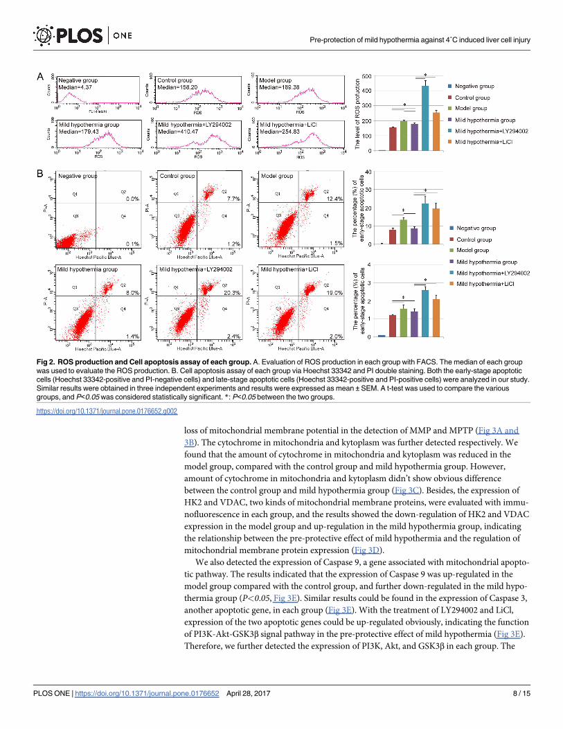
each group was evaluated in our study. In the FACS detection, the median of each group was
used to evaluate the ROS production. Compared with control group (154.76±2.35), the higher
level of ROS could be observed in the model group (196.44±6.57), and a little relief existed in
the mild hypothermia group (178.53±6.42). As expected, the treatment of LY294002 and LiCl
could further increase the ROS production in the damaged liver cells (Mild hypothermia+-
LY294002: 430.20±36.76, Mild hypothermia+LiCl: 254.61±15.5, Fig 2A).
Besides, the cell apoptosis of each group was also analyzed through Hoechst 33342 and PI
double staining. The staining assay revealed that low temperature-induced damage could lead
to an increase in the percentage of early-stage apoptotic cells (Hoechst 33342-positive and PI-
negative cells), as well as late-stage apoptotic cells (Hoechst 33342-positive and PI-positive
cells) in Brl-3A cells, and further analysis indicated that this apoptosis-induced effect could be
inhibited with the precondition with mild hypothermia. Moreover, the groups of mild hypo-
thermia + LY294002 and mild hypothermia + LiCl showed more apoptotic cells in both early
stage and late stage than the mild hypothermia group (Fig 2B).
Evaluation of mitochondrial mechanism
To further analyze the pre-protective effect of mild hypothermia from liver cells damage on
the level of subcellular organelle, both MMP and MPTP were analyzed in each group. Herein,
JC-1 staining assay was carried out to determine the alteration of MMP. All of groups showed
positive staining of JC-1 aggregates (red), while the staining of JC-1 monomers (green) was dif-
ferent in each group. Few JC-1 monomers could be observed in the control group, and the
amount of JC-1 monomers was increased in the damaged liver cells. The mild hypothermia
also exhibited the pre-protective effect and the number of cells with loss of mitochondrial
membrane potential was decreased with the precondition (Fig 3A). Similar results were
achieved in the MPTP detection with FACS, in which the median was also used to evaluate the
MPTP in each group. Compared with control group (19.20±0.93), the MPTP of the model
group (25.80±1.21) was increased in some degree. A little relief could be observed in the mild
hypothermia group (22.41±0.78) (Fig 3B). In addition, LY294002 and LiCl could promote the
Pre-protection of mild hypothermia against 4˚C induced liver cell injury
PLOS ONE | https://doi.org/10.1371/journal.pone.0176652 April 28, 2017 7 / 15
loss of mitochondrial membrane potential in the detection of MMP and MPTP (Fig 3A and
3B). The cytochrome in mitochondria and kytoplasm was further detected respectively. We
found that the amount of cytochrome in mitochondria and kytoplasm was reduced in the
model group, compared with the control group and mild hypothermia group. However,
amount of cytochrome in mitochondria and kytoplasm didn’t show obvious difference
between the control group and mild hypothermia group (Fig 3C). Besides, the expression of
HK2 and VDAC, two kinds of mitochondrial membrane proteins, were evaluated with immu-
nofluorescence in each group, and the results showed the down-regulation of HK2 and VDAC
expression in the model group and up-regulation in the mild hypothermia group, indicating
the relationship between the pre-protective effect of mild hypothermia and the regulation of
mitochondrial membrane protein expression (Fig 3D).
We also detected the expression of Caspase 9, a gene associated with mitochondrial apopto-
tic pathway. The results indicated that the expression of Caspase 9 was up-regulated in the
model group compared with the control group, and further down-regulated in the mild hypo-
thermia group (P<0.05, Fig 3E). Similar results could be found in the expression of Caspase 3,
another apoptotic gene, in each group (Fig 3E). With the treatment of LY294002 and LiCl,
expression of the two apoptotic genes could be up-regulated obviously, indicating the function
of PI3K-Akt-GSK3β signal pathway in the pre-protective effect of mild hypothermia (Fig 3E).
Therefore, we further detected the expression of PI3K, Akt, and GSK3β in each group. The
Fig 2. ROS production and Cell apoptosis assay of each group. A. Evaluation of ROS production in each group with FACS. The median of each group
was used to evaluate the ROS production. B. Cell apoptosis assay of each group via Hoechst 33342 and PI double staining. Both the early-stage apoptotic
cells (Hoechst 33342-positive and PI-negative cells) and late-stage apoptotic cells (Hoechst 33342-positive and PI-positive cells) were analyzed in our study.
Similar results were obtained in three independent experiments and results were expressed as mean ± SEM. A t-test was used to compare the various
groups, and P<0.05 was considered statistically significant. *: P<0.05 between the two groups.
https://doi.org/10.1371/journal.pone.0176652.g002
Pre-protection of mild hypothermia against 4˚C induced liver cell injury
PLOS ONE | https://doi.org/10.1371/journal.pone.0176652 April 28, 2017 8 / 15

qPCR results suggested that the precondition with mild hypothermia could increase the
expression of those genes compared with model group (P<0.05) (Fig 3E). In the level of pro-
tein, modest up-regulation of the three genes expression could be observed in the mild hypo-
thermia group compared with model group. In addition, we also detected the functional
proteins in PI3K-Akt-GSK3β signal pathway, p-Akt, and p-GSK3β, and the result showed that
the expression of p-Akt and p-GSK3β were decreased in the model group obviously, and the
precondition with mild hypothermia also could increase the expression of p-Akt and p-
GSK3β, suggesting that the pre-protective effect of mild hypothermia depended on the func-
tion of PI3K-Akt-GSK3β signal pathway in some degree (Fig 3E). With the treatment of inhib-
itors, qPCR results showed that the expression of GSK3β was down-regulated in the group of
mild hypothermia + LY294002 and mild hypothermia + LiCl, while the expression levels of
PI3K and Akt were up-regulated in those groups. Western-blot results also indicated the up-
regulation of p-Akt and p-GSK3β in those groups (Fig 3E). This phenomenon might be due to
the negative feedback control mechanisms in the culture system.
Discussion
Although several groups have indicated that mild hypothermia had a protective effect against
liver cell injury, the pre-protective effect of hypothermia on the liver cell has not been deter-
mined so far [9–11]. In the present study, our results indicated that the precondition with mild
Fig 3. Mitochondrial mechanism of the pre-protective effect of mild hypothermia. A. Evaluation of MMP of each group. Top: Staining of JC-1
aggregates (red); Middle: Staining of JC-1 monomers (green); Bottom: Marge of Top and Middle. Scale bar corresponds to 10 μm. B. Analysis of MPTP
opening of each group with FACS. The median of each group was used to evaluate MPTP opening. C. Expression of cytochrome in mitochondria and
kytoplasm. D. Evaluation of HK2 (red) and VDAC (green) expression with immunofluorescence. Scale bar corresponds to 20 μm. E. Gene expression assay
with qPCR and western-blot. Similar results were obtained in three independent experiments and results were expressed as mean ± SEM. A t-test was used
to compare the various groups, and P<0.05 was considered statistically significant. *: P<0.05 between the two groups.
https://doi.org/10.1371/journal.pone.0176652.g003
Pre-protection of mild hypothermia against 4˚C induced liver cell injury
PLOS ONE | https://doi.org/10.1371/journal.pone.0176652 April 28, 2017 9 / 15
hypothermia could inhibit ROS production and cell apoptosis and increase cell viability of Brl-
3A cell line, and develop a novel strategy for the storage of liver cell. Scientists have demon-
strated that ROS are considered as a major mediator of inflammation and the reduction of
ROS levels can lead to the attenuation of liver cell injury and apoptosis directly [26–30]. There-
fore, the mild hypothermia group holds better ability on cell proliferation, LDH regulation
and glycogen synthesis. However, Brl-3A cell line used in our study is different from primary
hepatocype, which are always applied in clinic. For example, the Brl-3A cells hold strong pro-
liferative ability and stable hepatic function, while primary hepatocype could only maintain
their hepatic function and proliferative ability for a short period in vitro. Even though Brl-3A
cell line is regarded as a good hepatic cell model and used to evaluate the liver cell function in
vitro widely [31–34], the exploration with primary hepatocyte should hold more clinical signif-
icance. Our group isolated rat primary hepatocyte as the reference [35] and further analyzed
the effect of hypothermia on cell viability and cell proliferation ability. Similar to Brl-3A cell
line, our results indicated that precondition with mild hypothermia still hold protective func-
tion on rat primary hepatocyte (S1 Fig). However, the detailed mechanism about such phe-
nomenon still need our further investigation.
Besides, we detected the temperature changing during the hypothermia preconditioning
process (S2 Fig), and the result also indicated that 10 min or 15 min was enough for the heat
exchange in our culture system. However, more than 109 cells are needed for bioartificial liver
normally [36–38]. Both the cell number and medium are much more than that used in our
study. Therefore, the either 10min or 15min treatment indeed is not suitable for such large-
scale liver cell storage or real bioartificial liver. Therefore, the accurate time of mild hypother-
mia preconditioning still needs further preclinical exploration, even clinical study.
In addition, some scientists also indicated that the therapeutic mechanism of mild hypo-
thermia may be related to mitochondrial protection. Mild hypothermia treatment could
decrease MPTP opening and restore the MMP. Furthermore, hypothermia also could decrease
mitochondrial malondialdehyde and elevate mitochondrial-reduced glutathione, consistent
with the restored mitochondrial function [39]. However, the mechanisms about the reduction
in MMP by mild hypothermia are complex [40]. For example, ischemia-reperfusion could
increase the level of MPTP activators (Ca2þ, Pi and ROS,) and reduce the level of MPTP inhib-
itors (ATP/ADP and low PH) [41]. Both the activator and inhibitors of MPTP opening could
be impacted by the mild hypothermia in different degree, and the balance between the activa-
tor and inhibitors determined the duration of MPTP opening [39, 40, 42]. In this study, the
reduction of MPTP opening and the restoration of MMP were also observed in the mild hypo-
thermia group, as well as the inhibition of ROS production, a MPTP activator. The expression
of Caspase 9, a gene associated with mitochondrial apoptotic pathway, was also down-regu-
lated in the mild hypothermia group, compared with model group. Further detection showed
that the expression of both HK2 and VDAC, two kinds of membrane proteins of mitochon-
dria, could be down-regulated with low temperature treatment, and mild hypothermia pre-
conditioning could up-regulated their expression in damaged hepatic cells. Thus, the pre-
protective effect of mild hypothermia should also be related to mitochondrial protection, and
HK2 and VDAC might be the target proteins during the protection process. However, whether
the detailed mechanism of MMP and MPTP regulation is similar to the previous reports, still
needs our further exploration.
In our research, we found that the effect of precondition with mild hypothermia could be
inhibited through adding LY294002 (PI3K inhibitor) or LiCl (GSK3β inhibitor) in the culture
system. The results suggested that PI3K-Akt-GSK3β signal pathway might play an important
role in the pre-protective effect of mild hypothermia. PI3K-Akt-GSK3β signal pathway has
been demonstrated to be essential for the recovery of liver cell injury by some groups [43–47].
Pre-protection of mild hypothermia against 4˚C induced liver cell injury
PLOS ONE | https://doi.org/10.1371/journal.pone.0176652 April 28, 2017 10 / 15

The pathway also has been indicated to hold the ability to regulate the protective effect of mild
hypothermia against ischaemia/reperfusion injury. The expression levels of p-Akt, and p-
GSK3β in the mild hypothermia treated group were higher than those in the ischaemia/reper-
fusion group, and the therapeutic function also could be influenced by LY294002 [48, 49]. In
our study, the results showed that the cell viability, MMP and other characteristics were much
worse in the group of mild hypothermia + LY294002 and mild hypothermia + LiCl than those
in the control group, the mild hypothermia group, even the model group, indicating that the
PI3K-Akt-GSK3β signal pathway might be associated with not only pre-protective effect of
mild hypothermia, but also other essential functions in liver cell. Interestingly, with the treat-
ment of LY294002 and LiCl, the expression levels of some key genes in the pathway were up-
regulated in those groups. Maybe this phenomenon was due to the negative feedback control
mechanisms in the low temperature-induced liver cell injury model. Anyway, this conclusion
still needs our further confirmation. In addition, our study only evaluated the expression of
PI3K, Akt/p-Akt and GSK3β/p-GSK3β in the signal pathway with qPCR and western-blot pre-
liminary, more detailed and accurate analysis about the pathway, such as the expression of
upstream or downstream genes, should also be investigated intensively.
Conclusion
In conclusion, this study mainly investigated the pre-protective effect of mild hypothermia on
low temperature-induced liver cell injury, and provided a novel strategy for the storage of liver
cell in bioengineering. The results indicated that the precondition with mild hypothermia
could enhance cell proliferation, LDH regulation and glycogen synthesis ability, and relief the
ROS production and cell apoptosis in Brl-3A cells. Further analysis showed that the precondi-
tion also could decrease MPTP opening and restore MMP, and PI3K-Akt-GSK3β signal path-
way might be associated with the pre-protective effect of mild hypothermia. However, the
detailed mechanism of this effect still needs our further exploration.
Supporting information
S1 Fig. Effect of precondition with mild hypothermia on rat primary hepatocyte. A. Evalu-
ation of cell viability with CCK-8. B. Analysis of cell proliferation ability with EDU staining.
Similar results were obtained in three independent experiments and results were expressed as
mean ± SEM. A t-test was used to compare the various groups, and P<0.05 was considered sta-
tistically significant. �: P<0.05 between the two groups.
(PDF)
S2 Fig. The temperature changing during the hypothermia preconditioning process.
(PDF)
Author Contributions
Conceptualization: JQ YG.
Data curation: JQ YM YG.
Formal analysis: ZJ YG.
Funding acquisition: YG.
Investigation: JQ YG.
Methodology: JQ YM YL ZJ.
Pre-protection of mild hypothermia against 4˚C induced liver cell injury
PLOS ONE | https://doi.org/10.1371/journal.pone.0176652 April 28, 2017 11 / 15

Project administration: JQ YG.
Resources: JQ YG.
Software: JQ YM.
Supervision: JQ YG.
Validation: JQ YM.
Visualization: JQ YG.
Writing – original draft: JQ YG.
Writing – review & editing: JQ YG.
References
1. Puts CF, Berendsen TA, Bruinsma BG, Ozer S, Luitje M, Usta OB, et al. Polyethylene glycol protects
primary hepatocytes during supercooling preservation. Cryobiology. 2015; 71(1):125–9. PubMed Cen-
tral PMCID: PMC4506890. https://doi.org/10.1016/j.cryobiol.2015.04.010 PMID: 25936340
2. Ferrigno A, Rizzo V, Boncompagni E, Bianchi A, Gringeri E, Neri D, et al. Machine perfusion at 20
degrees C reduces preservation damage to livers from non-heart beating donors. Cryobiology. 2011;
62(2):152–8. https://doi.org/10.1016/j.cryobiol.2011.02.004 PMID: 21315707
3. Jacobs SE. Selective head cooling with mild systemic hypothermia after neonatal encephalopathy: Mul-
ticentre randomised trial. The Journal of pediatrics. 2005; 147(1):122–3. https://doi.org/10.1016/j.jpeds.
2005.04.047 PMID: 16027712
4. Gluckman PD, Wyatt JS, Azzopardi D, Ballard R, Edwards AD, Ferriero DM, et al. Selective head cool-
ing with mild systemic hypothermia after neonatal encephalopathy: multicentre randomised trial. Lan-
cet. 2005; 365(9460):663–70. https://doi.org/10.1016/S0140-6736(05)17946-X PMID: 15721471
5. Wu TC, Grotta JC. Hypothermia for acute ischaemic stroke. The Lancet Neurology. 2013; 12(3):275–
84. https://doi.org/10.1016/S1474-4422(13)70013-9 PMID: 23415567
6. Azzopardi DV, Strohm B, Edwards AD, Dyet L, Halliday HL, Juszczak E, et al. Moderate hypothermia to
treat perinatal asphyxial encephalopathy. The New England journal of medicine. 2009; 361(14):1349–
58. https://doi.org/10.1056/NEJMoa0900854 PMID: 19797281
7. Gao XY, Huang JO, Hu YF, Gu Y, Zhu SZ, Huang KB, et al. Combination of mild hypothermia with neu-
roprotectants has greater neuroprotective effects during oxygen-glucose deprivation and reoxygena-
tion-mediated neuronal injury. Scientific reports. 2014; 4:7091. https://doi.org/10.1038/srep07091
PMID: 25404538
8. Sabir H, Walloe L, Dingley J, Smit E, Liu X, Thoresen M. Combined Treatment of Xenon and Hypother-
mia in Newborn Rats—Additive or Synergistic Effect? PloS one. 2014; 9(10):e109845. https://doi.org/
10.1371/journal.pone.0109845 PMID: 25286345
9. Blei A. Hypothermia for fulminant hepatic failure: a cool approach to a burning problem. Liver transplan-
tation: official publication of the American Association for the Study of Liver Diseases and the Interna-
tional Liver Transplantation Society. 2000; 6(2):245–7.
10. Fu T, Blei AT, Takamura N, Lin T, Guo D, Li H, et al. Hypothermia inhibits Fas-mediated apoptosis of
primary mouse hepatocytes in culture. Cell transplantation. 2004; 13(6):667–76. PMID: 15648737
11. Sakurai T, Kudo M, Watanabe T, Itoh K, Higashitsuji H, Arizumi T, et al. Hypothermia protects against
fulminant hepatitis in mice by reducing reactive oxygen species production. Digestive diseases. 2013;
31(5–6):440–6. https://doi.org/10.1159/000355242 PMID: 24281018
12. Hong MF, Dorian P. Update on advanced life support and resuscitation techniques. Current opinion in
cardiology. 2005; 20(1):1–6. PMID: 15596952
13. Diestel A, Roessler J, Berger F, Schmitt KR. Hypothermia downregulates inflammation but enhances
IL-6 secretion by stimulated endothelial cells. Cryobiology. 2008; 57(3):216–22. https://doi.org/10.1016/
j.cryobiol.2008.08.005 PMID: 18790695
14. Sakurai T, Itoh K, Higashitsuji H, Nonoguchi K, Liu Y, Watanabe H, et al. Cirp protects against tumor
necrosis factor-alpha-induced apoptosis via activation of extracellular signal-regulated kinase. Biochi-
mica et biophysica acta. 2006; 1763(3):290–5. https://doi.org/10.1016/j.bbamcr.2006.02.007 PMID:
16569452
Pre-protection of mild hypothermia against 4˚C induced liver cell injury
PLOS ONE | https://doi.org/10.1371/journal.pone.0176652 April 28, 2017 12 / 15

15. Qadir XV, Chen W, Han C, Song K, Zhang J, Wu T. miR-223 Deficiency Protects against Fas-Induced
Hepatocyte Apoptosis and Liver Injury through Targeting Insulin-Like Growth Factor 1 Receptor. The
American journal of pathology. 2015; 185(12):3141–51. https://doi.org/10.1016/j.ajpath.2015.08.020
PMID: 26598234
16. Jacobs S, Jager S, Jansen E, Peter A, Stefan N, Boeing H, et al. Associations of Erythrocyte Fatty
Acids in the De Novo Lipogenesis Pathway with Proxies of Liver Fat Accumulation in the EPIC-Potsdam
Study. PloS one. 2015; 10(5):e0127368. PubMed Central PMCID: PMC4435749. https://doi.org/10.
1371/journal.pone.0127368 PMID: 25984792
17. Chen WN, Han C, Zhang JQ, Song K, Wang Y, Wu T. Deletion of Mir155 Prevents Fas-Induced Liver
Injury through Up-Regulation of Mcl-1. American Journal Of Pathology. 2015; 185(4):1033–44. https://
doi.org/10.1016/j.ajpath.2014.12.020 PMID: 25794705
18. Tanoi T, Tamura T, Sano N, Nakayama K, Fukunaga K, Zheng YW, et al. Protecting liver sinusoidal
endothelial cells suppresses apoptosis in acute liver damage. Hepatology research. 2016; 46(7):697–
706. https://doi.org/10.1111/hepr.12607 PMID: 26490536
19. Lian F, Wang Y, Xiao Y, Wu X, Xu H, Liang L, et al. Activated farnesoid X receptor attenuates apoptosis
and liver injury in autoimmune hepatitis. Molecular medicine reports. 2015; 12(4):5821–7. PubMed Cen-
tral PMCID: PMC4581797. https://doi.org/10.3892/mmr.2015.4159 PMID: 26238153
20. Khaliulin I, Clarke SJ, Lin H, Parker J, Suleiman MS, Halestrap AP. Temperature preconditioning of iso-
lated rat hearts—a potent cardioprotective mechanism involving a reduction in oxidative stress and inhi-
bition of the mitochondrial permeability transition pore. The Journal of physiology. 2007; 581(Pt
3):1147–61. PubMed Central PMCID: PMC1976396. https://doi.org/10.1113/jphysiol.2007.130369
PMID: 17395631
21. Cai J, Zhang Y, Liu P, Chen S, Wu X, Sun Y, et al. Generation of tooth-like structures from integration-
free human urine induced pluripotent stem cells. Cell regeneration. 2013; 2(1):6. PubMed Central
PMCID: PMC4230506. https://doi.org/10.1186/2045-9769-2-6 PMID: 25408878
22. Liu P, Feng Y, Dong C, Yang D, Li B, Chen X, et al. Administration of BMSCs with Muscone in Rats with
Gentamicin-Induced AKI Improves Their Therapeutic Efficacy. PloS one. 2014; 9(5):e97123. https://
doi.org/10.1371/journal.pone.0097123 PMID: 24824427
23. Liu P, Feng Y, Dong C, Liu D, Wu X, Wu H, et al. Study on therapeutic action of bone marrow derived
mesenchymal stem cell combined with vitamin E against acute kidney injury in rats. Life sciences. 2013;
92(14–16):829–37. https://doi.org/10.1016/j.lfs.2013.02.016 PMID: 23499556
24. Liu P, Chen S, Li X, Qin L, Huang K, Wang L, et al. Low immunogenicity of neural progenitor cells differ-
entiated from induced pluripotent stem cells derived from less immunogenic somatic cells. PloS one.
2013; 8(7):e69617. PubMed Central PMCID: PMC3724937. https://doi.org/10.1371/journal.pone.
0069617 PMID: 23922758
25. Huang K, Liu P, Li X, Chen S, Wang L, Qin L, et al. Neural progenitor cells from human induced pluripo-
tent stem cells generated less autogenous immune response. Science China Life sciences. 2014; 57
(2):162–70. https://doi.org/10.1007/s11427-013-4598-6 PMID: 24443177
26. Sakurai T, Kudo M, Umemura A, He G, Elsharkawy AM, Seki E, et al. p38alpha inhibits liver fibrogen-
esis and consequent hepatocarcinogenesis by curtailing accumulation of reactive oxygen species. Can-
cer research. 2013; 73(1):215–24. PubMed Central PMCID: PMC3605785. https://doi.org/10.1158/
0008-5472.CAN-12-1602 PMID: 23271722
27. Xiao J, Lv Y, Lin B, Tipoe GL, Youdim MB, Xing F, et al. A novel antioxidant multitarget iron chelator
M30 protects hepatocytes against ethanol-induced injury. Oxidative medicine and cellular longevity.
2015; 2015:607271. PubMed Central PMCID: PMC4334871. https://doi.org/10.1155/2015/607271
PMID: 25722794
28. Yu Y, Bai F, Liu Y, Yang Y, Yuan Q, Zou D, et al. Fibroblast growth factor (FGF21) protects mouse liver
against D-galactose-induced oxidative stress and apoptosis via activating Nrf2 and PI3K/Akt pathways.
Molecular and cellular biochemistry. 2015; 403(1–2):287–99. https://doi.org/10.1007/s11010-015-
2358-6 PMID: 25701356
29. Zhang F, Wang X, Qiu X, Wang J, Fang H, Wang Z, et al. The protective effect of esculentoside a on
experimental acute liver injury in mice. PloS one. 2014; 9(11):e113107. PubMed Central PMCID:
PMC4236201. https://doi.org/10.1371/journal.pone.0113107 PMID: 25405982
30. Hosseini MJ, Shahraki J, Tafreshian S, Salimi A, Kamalinejad M, Pourahmad J. Protective effects of
Sesamum indicum extract against oxidative stress induced by vanadium on isolated rat hepatocytes.
Environmental toxicology. 2016; 31(8):979–85. https://doi.org/10.1002/tox.22107 PMID: 25727928
31. Guo X, Cui R, Zhao J, Mo R, Peng L, Yan M. Corosolic acid protects hepatocytes against ethanol-
induced damage by modulating mitogen-activated protein kinases and activating autophagy. European
journal of pharmacology. 2016; 791:578–88. https://doi.org/10.1016/j.ejphar.2016.09.031 PMID:
27663281
Pre-protection of mild hypothermia against 4˚C induced liver cell injury
PLOS ONE | https://doi.org/10.1371/journal.pone.0176652 April 28, 2017 13 / 15

32. Seidah NG, Benjannet S, Wickham L, Marcinkiewicz J, Jasmin SB, Stifani S, et al. The secretory pro-
protein convertase neural apoptosis-regulated convertase 1 (NARC-1): liver regeneration and neuronal
differentiation. Proceedings of the National Academy of Sciences of the United States of America.
2003; 100(3):928–33. PubMed Central PMCID: PMC298703. https://doi.org/10.1073/pnas.
0335507100 PMID: 12552133
33. Zuo D, Duan Z, Jia Y, Chu T, He Q, Yuan J, et al. Amphipathic silica nanoparticles induce cytotoxicity
through oxidative stress mediated and p53 dependent apoptosis pathway in human liver cell line HL-
7702 and rat liver cell line BRL-3A. Colloids and surfaces B, Biointerfaces. 2016; 145:232–40. https://
doi.org/10.1016/j.colsurfb.2016.05.006 PMID: 27187187
34. Pan C, Chen H, Wang L, Yang S, Fu H, Zheng Y, et al. Down-regulation of MiR-127 facilitates hepato-
cyte proliferation during rat liver regeneration. PloS one. 2012; 7(6):e39151. PubMed Central PMCID:
PMC3376093. https://doi.org/10.1371/journal.pone.0039151 PMID: 22720056
35. Takahara I, Akazawa Y, Tabuchi M, Matsuda K, Miyaaki H, Kido Y, et al. Toyocamycin attenuates free
fatty acid-induced hepatic steatosis and apoptosis in cultured hepatocytes and ameliorates nonalco-
holic fatty liver disease in mice. PloS one. 2017; 12(3):e0170591. https://doi.org/10.1371/journal.pone.
0170591 PMID: 28278289
36. Enosawa S, Miyashita T, Saito T, Omasa T, Matsumura T. The significant improvement of survival
times and pathological parameters by bioartificial liver with recombinant HepG2 in porcine liver failure
model. Cell transplantation. 2006; 15(10):873–80. PMID: 17299991
37. Selden C, Spearman CW, Kahn D, Miller M, Figaji A, Erro E, et al. Evaluation of encapsulated liver cell
spheroids in a fluidised-bed bioartificial liver for treatment of ischaemic acute liver failure in pigs in a
translational setting. PloS one. 2013; 8(12):e82312. PubMed Central PMCID: PMC3867376. https://
doi.org/10.1371/journal.pone.0082312 PMID: 24367515
38. Enosawa S, Miyashita T, Fujita Y, Suzuki S, Amemiya H, Omasa T, et al. In vivo estimation of bioartifi-
cial liver with recombinant HepG2 cells using pigs with ischemic liver failure. Cell transplantation. 2001;
10(4–5):429–33. PMID: 11549067
39. Xiong W, Xu S, Li Md H, Liang K. Moderate hypothermia ameliorates enterocyte mitochondrial dysfunc-
tion in severe shock and reperfusion. The Journal of surgical research. 2016; 200(1):250–9. https://doi.
org/10.1016/j.jss.2015.06.068 PMID: 26227675
40. Gong P, Hua R, Zhang Y, Zhao H, Tang Z, Mei X, et al. Hypothermia-induced neuroprotection is associ-
ated with reduced mitochondrial membrane permeability in a swine model of cardiac arrest. Journal of
cerebral blood flow and metabolism: official journal of the International Society of Cerebral Blood Flow
and Metabolism. 2013; 33(6):928–34. PubMed Central PMCID: PMC3677114.
41. Baines CP. The mitochondrial permeability transition pore and ischemia-reperfusion injury. Basic
research in cardiology. 2009; 104(2):181–8. PubMed Central PMCID: PMC2671061. https://doi.org/10.
1007/s00395-009-0004-8 PMID: 19242640
42. Petronilli V, Penzo D, Scorrano L, Bernardi P, Di Lisa F. The mitochondrial permeability transition,
release of cytochrome c and cell death. Correlation with the duration of pore openings in situ. The Jour-
nal of biological chemistry. 2001; 276(15):12030–4. https://doi.org/10.1074/jbc.M010604200 PMID:
11134038
43. Lai SS, Zhao DD, Cao P, Lu K, Luo OY, Chen WB, et al. PP2Acalpha positively regulates the termina-
tion of liver regeneration in mice through the AKT/GSK3beta/Cyclin D1 pathway. Journal of hepatology.
2016; 64(2):352–60. https://doi.org/10.1016/j.jhep.2015.09.025 PMID: 26456844
44. Zheng T, Yang X, Wu D, Xing S, Bian F, Li W, et al. Salidroside ameliorates insulin resistance through
activation of a mitochondria-associated AMPK/PI3K/Akt/GSK3beta pathway. British journal of pharma-
cology. 2015; 172(13):3284–301. PubMed Central PMCID: PMC4500366. https://doi.org/10.1111/bph.
13120 PMID: 25754463
45. Kim HJ, Joe Y, Kong JS, Jeong SO, Cho GJ, Ryter SW, et al. Carbon monoxide protects against
hepatic ischemia/reperfusion injury via ROS-dependent Akt signaling and inhibition of glycogen
synthase kinase 3beta. Oxidative medicine and cellular longevity. 2013; 2013:306421. PubMed Central
PMCID: PMC3880761. https://doi.org/10.1155/2013/306421 PMID: 24454979
46. Lakshmanan J, Zhang B, Nweze IC, Du Y, Harbrecht BG. Glycogen synthase kinase 3 regulates IL-
1beta mediated iNOS expression in hepatocytes by down-regulating c-Jun. Journal of cellular biochem-
istry. 2015; 116(1):133–41. https://doi.org/10.1002/jcb.24951 PMID: 25160751
47. Iqbal J, McRae S, Banaudha K, Mai T, Waris G. Mechanism of hepatitis C virus (HCV)-induced osteo-
pontin and its role in epithelial to mesenchymal transition of hepatocytes. The Journal of biological
chemistry. 2013; 288(52):36994–7009. PubMed Central PMCID: PMC3873557. https://doi.org/10.
1074/jbc.M113.492314 PMID: 24240095
48. Dai HB, Xu MM, Lv J, Ji XJ, Zhu SH, Ma RM, et al. Mild Hypothermia Combined with Hydrogen Sulfide
Treatment During Resuscitation Reduces Hippocampal Neuron Apoptosis Via NR2A, NR2B, and PI3K-
Pre-protection of mild hypothermia against 4˚C induced liver cell injury
PLOS ONE | https://doi.org/10.1371/journal.pone.0176652 April 28, 2017 14 / 15
Akt Signaling in a Rat Model of Cerebral Ischemia-Reperfusion Injury. Molecular neurobiology. 2016;
53(7):4865–73. https://doi.org/10.1007/s12035-015-9391-z PMID: 26350917
49. Mochizuki T, Yu S, Katoh T, Aoki K, Sato S. Cardioprotective effect of therapeutic hypothermia at 34
degrees C against ischaemia/reperfusion injury mediated by PI3K and nitric oxide in a rat isolated heart
model. Resuscitation. 2012; 83(2):238–42. https://doi.org/10.1016/j.resuscitation.2011.08.013 PMID:
21875500
Pre-protection of mild hypothermia against 4˚C induced liver cell injury
PLOS ONE | https://doi.org/10.1371/journal.pone.0176652 April 28, 2017 15 / 15