
Influence of NP size and concentration on the physical properties of Glycerol/Cu 2 O nanofluid

U.P.B. Sci. Bull., Series B, Vol. 74, Iss. 4, 2012 ISSN 1454-2331
EFFECT OF INITIATOR’S CONCENTRATION ON PROPERTIES OF GELATIN – HEMA HYDROGELS
Diana-Maria DRĂGUŞIN1, Dan Sorin VASILESCU2, Sandra Van VLIERBERGHE3, Peter DUBRUEL4, Izabela-Cristina STANCU5
Acest studiu descrie sinteza de sisteme polimerice bicomponente, pe bază de gelatină şi polimeri 2-hidroxietil metacrilat. Sintezele s-au realizat prin procese combinate de polimerizare şi reticulare folosind fotoiniţierea. S-au sintetizat trei tipuri de hidrogeluri, folosind diferite concentraţii de fotoiniţiator. Succesul sintezei şi influenţa compoziţiei asupra proprietăţilor metarialelor au fost evaluate prin diferite metode de caracterizare; a fost determinată compoziţia optimă. Ulterior au fost realizate structuri poroase prin tratament criogenic şi liofilizare. S-a realizat o caracterizare morfologică a materialelor poroase, care a demonstrat faptul că arhitectura materialelor este puternic influenţată de compoziia lor.
The present work describes the synthesis of bicomponent polymer systems based on gelatin and also on 2-hydroxyethyl methacrylate polymers. The syntheses were performed through a combined process of cross-linking polymerization while using photoinitiation. Three types of hydrogels have been synthesised using different photoinitiator concentrations. The success of the syntheses and the influence of the compositions on properties of the materials, were assessed through various characterization methods; optimal composition was determined. Subsequently, porous structures have been obtained by cryogenic treatment and freeze-drying. A morphological characterization of the scaffolds was subsequently performed, showing that the architecture of the scaffolds was strongly influenced by the composition of the materials.
Keywords: gelatin, PHEMA, bicomponent polymeric system, rheology, porous
scaffolds
1 PhD Student, Faculty of Applied Chemistry and Materials Science, University POLITEHNICA
of Bucharest, e-mail: [email protected] 2 Prof., Faculty of Applied Chemistry and Materials Science, University POLITEHNICA of
Bucharest, e-mail: [email protected] 3 PhD, Department of Organic Chemistry, Faculty of Sciences, GHENT University, Belgium, e-
mail: [email protected] 4 Prof., Department of Organic Chemistry, Faculty of Sciences, GHENT University, Belgium, e-
mail: [email protected] 5 Lecturer, Faculty of Applied Chemistry and Materials Science, University POLITEHNICA of
Bucharest, e-mail: [email protected]

32 Diana Draguşin, Dan Vasilescu, Sandra Van Vlierberghe, Peter Dubruel, Izabela Stancu
1. Introduction During the recent years, hydrogels have become popular scaffold
platforms in the field of tissue engineering due to their capacity to retain large amounts of water, while remaining insoluble; another important feature consists in the fact that they can be synthesised under cyto-compatible conditions. In this framework, a large variety of hydrogels from both synthetic as well as natural polymers is continuously developed [1-4]. An ideal hydrogel should be able to mimic the mechanical and biological requirements for the tissues to be replicated [1]. Considering the preparation method, hydrogels can be classified either as homopolymer and copolymer hydrogels [5]. In the recent years, gelatin has become one of the most studied materials for a wide variety of applications, ranging from food-related ones to pharmaceutical and photographic or technical products. Due to the fact that gelatin is a natural protein with many desirable properties as a biomaterial it has been used as a material for biomedical applications. Gelatin is composed from a large variety of side chains that offer the possibility of a great variety of chemical modification methods in order to introduce cross-linkable groups [5, 6, 9].
2-Hydroxyethyl methacrylate (HEMA) is a synthetic monomer which, as such, is very toxic for the cells. However, in its polymerized form as poly(2-hydroxyethyl methacrylate) (PHEMA) it is a highly compatible polymer with the living tissues [7, 8].
In the present study we have developed photo-cross-linkable bicomponent systems based on methacrylamide-modified gelatin (MAG) and 2-hydroxyethyl methacrylate as synthetic monomer, using as photoinitiator 1-[4-(2-hydroxyethoxy)-phenyl]-2-hydroxy-2-methyl-1-propane-1-one (Irgacure® 2959). The initial idea of the study was that multicomponent hydrogels systems would present superior properties, when compared with the properties of each component. There is evidence that these materials exhibit improved mechanical properties with respect to their individual crosslinked components [6]. The novelty of this work is the choice of the two materials for generating new types of hydrogels, but also the type of initiation used. To the best of our knowledge, Irgacure® 2959 has not been used yet as initiator for HEMA polymerization.
2. Materials and methods
2.1. Materials Gelatin (type B), isolated from bovine skin by alkaline process, was
supplied by Rousselot (Ghent, Belgium). Methacrylic anhydride (MA), ethanol absolute, n-butylamine 99,5% and 2-hydroxyethyl methacrylate 97% were purchased from Sigma-Aldrich and used as such. 1,2-Phthalic dicarboxaldehyde

Effect of initiator’s concentration on properties of gelatin – hema hydrogels 33
98% was purchased from Acros Organic and mercaptoethanol from Merk. Dialysis membranes Spectra/Por® 4 (MWCA 12,000 – 14,000 Da) have been obtained from Polylab. Likewise, 1-[4-(2-hydroxyethoxy)-phenyl]-2-hydroxy-2-methyl-1-propane-1-one (Irgacure®2959) was purchased from Ciba Speciality Chemicals.
2.2. Methods
Synthesis of MAG MAG was prepared using the procedure described by Van Den Bulcke in
[10]. Briefly, after the swelling of 100 g of gelatin (35 mmol ε-amine side groups of lysine and hydroxylysine) in 1 L of phosphate buffer (pH 7.8) for 1 h, the solution was heated to 40°C. One equivalent of MA (5.66 mL, or 0.038 mol) was added to the homogeneous gelatin solution and the mixture stirred vigorously for 1 h at 40°C. The reaction mixture was then diluted with 1 L of double distilled water (ddw) and dialyzed (Spectra/Por® 4, MWCO 12,000–14,000 Da). After dialysis in water at 40°C for 24 h, the derivative was freeze-dried.
Characterization of MAG In order to further use the methacrylamide groups from MAG in
polymerization and crosslinking reactions, it is compulsory to determine a key feature of MAG, namely the degree of substitution (DS) resulted after the reaction. DS is defined as the ratio of the amount of incorporated methacrylamide functionalities to the amount of free amine groups available for modification. This parameter was calculated using two methods: 1) proton nuclear magnetic resonance (1H-NMR) and 2) UV-Vis spectroscopy.
DS obtained by 1H-NMR 1H-NMR was used to obtain DS, following the method reported in [6].
Briefly, 1H-NMR spectra of the modified gelatin were recorded at 40°C in deuterated water (D2O) (Fig. 1) using a Bruker WH 500 MHz instrument; the chemical shift was expressed in ppm as a function of tetramethylsilane as internal standard. DS was obtained by comparing the integrals of the characteristic peaks of the methacrylamide moieties (i.e., I5.62 ppm and I5.85 ppm) and the integration peak of the amino acids not involved in the modification (Val + Leu + Ile at 1.12 ppm). Based on the known amino acid composition of the gelatin applied, the DS was calculated using the equation (1):
mol) 5(100/0.038 )/I(I mol 0.3836 (%) DS ppm 1.1 ppm 5.7 ××= (1)
This way, gelatin that was chemically modified with methacrylamide side groups has shown a DS of 61%.

34 Diana Draguşin, Dan Vasilescu, Sandra Van Vlierberghe, Peter Dubruel, Izabela Stancu
Fig. 1. 1H-NMR spectrum of gel-MOD recorded in D2O at 40°C.
DS by UV-Vis spectroscopy
DS was estimated from the quantitative determination of –NH2 groups in the raw gelatin and in MAG, respectively. O-phtalic dialdehyde (OPA) was used. UV-VIS spectroscopy was performed on a CINTRA 101 (GBC Scientific Equipment Pty Ltd) double-beam spectrometer, using the fixed wavelength interrogation (in water). Briefly, 50 x 10-6 L gelatin / MAG -containing aliquot was mixed with 950 x 10-6 L distilled water and then 1.5 x 10⁻³ L solution A and 0.5 x 10⁻³ L solution B were added. Solution A consisted of 0.05 L borate buffer pH 10 and 25 x 10-6 L mercaptoethanol and solution B was prepared using 20 x 10-3 g OPA dissolved in 0.01 L ethanol and 0.04 L distilled water. The experiment was performed in triplicate, under fixed wavelength interrogation at 340 nm (specific absorption for the complex OPA-amine groups), at room temperature. The specific absorbance was measured after 15 minutes of reaction. Control samples were prepared in triplicate, following the same procedure, except that the sample was replaced with distilled water. The molar absorptivity of n-butylamine and the concentration of amines were calculated from a standard curve obtained for n-butylamine solutions of known concentrations. These values were further converted into amount of reacted –NH2 and, further, in DS. The obtained value was 60.7±1, well correlated with the value obtained by 1H-NMR.
Effect of initiator’s concentration on properties of gelatin – hema hydrogels 35
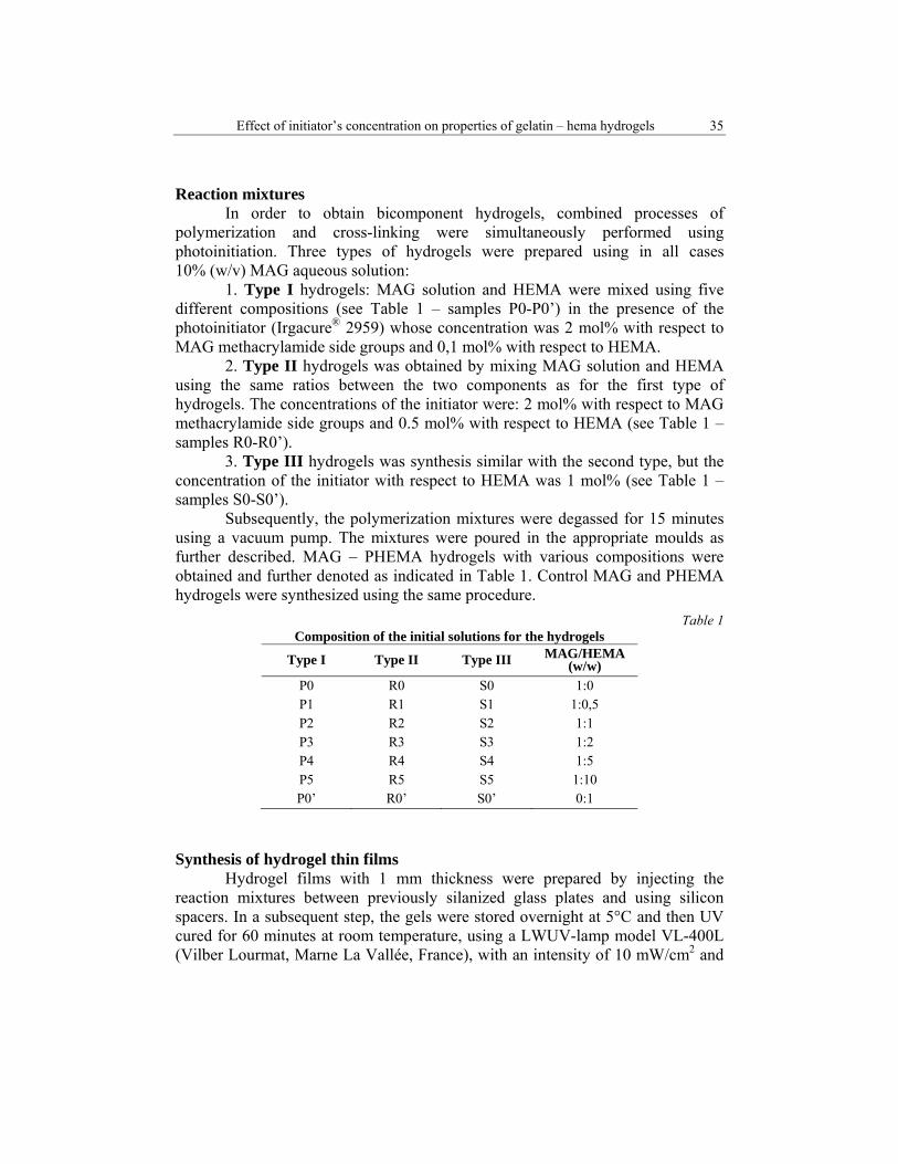
Reaction mixtures In order to obtain bicomponent hydrogels, combined processes of
polymerization and cross-linking were simultaneously performed using photoinitiation. Three types of hydrogels were prepared using in all cases 10% (w/v) MAG aqueous solution:
1. Type I hydrogels: MAG solution and HEMA were mixed using five different compositions (see Table 1 – samples P0-P0’) in the presence of the photoinitiator (Irgacure® 2959) whose concentration was 2 mol% with respect to MAG methacrylamide side groups and 0,1 mol% with respect to HEMA.
2. Type II hydrogels was obtained by mixing MAG solution and HEMA using the same ratios between the two components as for the first type of hydrogels. The concentrations of the initiator were: 2 mol% with respect to MAG methacrylamide side groups and 0.5 mol% with respect to HEMA (see Table 1 – samples R0-R0’).
3. Type III hydrogels was synthesis similar with the second type, but the concentration of the initiator with respect to HEMA was 1 mol% (see Table 1 – samples S0-S0’).
Subsequently, the polymerization mixtures were degassed for 15 minutes using a vacuum pump. The mixtures were poured in the appropriate moulds as further described. MAG – PHEMA hydrogels with various compositions were obtained and further denoted as indicated in Table 1. Control MAG and PHEMA hydrogels were synthesized using the same procedure.
Table 1 Composition of the initial solutions for the hydrogels
Type I Type II Type III MAG/HEMA (w/w)
P0 R0 S0 1:0 P1 R1 S1 1:0,5 P2 R2 S2 1:1 P3 R3 S3 1:2 P4 R4 S4 1:5 P5 R5 S5 1:10 P0’ R0’ S0’ 0:1
Synthesis of hydrogel thin films Hydrogel films with 1 mm thickness were prepared by injecting the
reaction mixtures between previously silanized glass plates and using silicon spacers. In a subsequent step, the gels were stored overnight at 5°C and then UV cured for 60 minutes at room temperature, using a LWUV-lamp model VL-400L (Vilber Lourmat, Marne La Vallée, France), with an intensity of 10 mW/cm2 and

36 Diana Draguşin, Dan Vasilescu, Sandra Van Vlierberghe, Peter Dubruel, Izabela Stancu
a wavelength range of 365nm. The resulting materials were intensively washed with ddw, at 400C, for 48 hours to assure the complete removal of the unreacted species.
Preparation of porous scaffolds In order to obtain porous samples, the polymerization has been performed
in glass Petri dishes using the same polymerization procedure as previously described. The hydrogels resulted have been subsequently submitted to a cryo-treatment (the procedure and the experimental set up have been described in [9]). Briefly, the hydrogels at maximum swelling degree (MSD) were placed in the cryo-unit and they were cooled from 21°C to -31°C at a slow cooling rate of 0.15°C/min. During the freezing step, a temperature gradient of 30°C was applied between the top and the bottom of the scaffolds. The temperature of the freezing as well as the cooling rate was programmed with an external cooling circuit (type FP40-ME, Julabo). The mould was equipped with a Peltier element (thermo electric cooler – TEC – DuraTec DT12 type from Marloe Industries). The aluminium heat exchangers and the electronic TEC controller were designed and assembled by the technical workshop (CWFW) of the Faculty of Sciences, Ghent University. After 5½ hours, the frozen hydrogels were transferred for freeze-drying (using a Christ freeze-dryer alpha I-5), resulting into porous scaffolds.
Gel fraction analysis Gel fraction (GF, %) was gravimetrically appreciated, and it was
determined using the following equation (2) :
GF (%)= mf /m0 ×100%, (2)
where m0 is the mass of the dried materials, as they result from the reaction without purification and mf is the mass of the dried materials after the extraction in ddw at 400C for 48 hours.
Fourier Transform Infrared Spectroscopy (FT-IR) The bicomponent hydrogels were characterized using a JASCO 4200
spectrometer equipped with a Specac Golden Gate attenuated total reflectance (ATR) device in the 4000-600 cm-1 wave number region.
Water uptake behaviour The swelling behaviour was assessed in ddw, at 40°C. The gravimetric
method was used to estimate the swelling degree (SD) at predetermined time intervals, t, using the well known equation (3):
SD (%) = (mt-m0)/m0×100 (3)
where mt is the mass of the wet sample at moment t while m0 represents the mass of the dried sample before incubation in water. All data represented as points are

Effect of initiator’s concentration on properties of gelatin – hema hydrogels 37
the mean ± standard deviation from three separate measurements. The maximum swelling degree (MSD) was estimated as the equilibrium value of SD.
Rheology The mechanical properties of the hydrogel films were evaluated using a
rheometer type Physica MCR-301 (Anton Paar, Sint-Martens-Latem, Belgium). The oscillation measurements have been performed using two parallel plates, with an upper plate diameter of 50 mm. The mechanical spectra were obtained using oscillation rheology to measure the storage modulus (G’) with a normal force (FN) of 0.2 N and a gap of 0.7 mm. The storage modulus gives an estimation of the strain energy which is reversibly stored in a material.
Scanning Electron Microscopy Analysis (SEM) Morphological information with respect to the porosity, interconnection of
pores and other features were revealed through SEM analysis of the gold-coated freeze-dried hydrogels. Longitudinal as well as cross-sections were analyzed. The study was performed using a QUANTA INSPECT F SEM device equipped with a field emission gun (FEG) with 1.2 nm resolution and with an X-ray energy dispersive spectrometer (EDS).
3. Results and discussion
3.1. Characterization of the hydrogel films
Gel fraction analysis The investigation of the complex MAG-PHEMA hydrogels developed in
this study was started with the estimation of the gel fraction (GF). This quantifies the success of the reaction while indicating the migration of the hydrosoluble species in water. Therefore, the materials have been incubated at 40°C in ddw for 48 hours. The results were estimated using the equation (2) and the obtained GF values are shown in Fig. 2. All the synthesized hydrogels are characterized by remarkably high GF values, even for the smallest concentration of the initiator. For type I the values of GF are ranging between 93 ± 2.21% and 97 ± 2.04%, for the second type of hydrogels GF presents values between 95 ± 1.93 % and 98 ± 1.66% and for the third type the values are ranging between 94 ± 2.14% and 98 ± 1.76%. An analysis over the values of the GF concludes that the hydrogels present an excellent mass conservation during the chemical treatment and that the bicomponent systems are water insoluble.
The findings regarding the gel fraction represent the first proof that the synthesis of the hydrogels is a successful one and also demonstrate that the materials present a great stability. Another important finding is that the concentration of the photoinitiator does not significantly affect the values of the GF, in other words all three types of hydrogels present similar values for GF.

38 Diana Draguşin, Dan Vasilescu, Sandra Van Vlierberghe, Peter Dubruel, Izabela Stancu
Fig. 2. GF average values for the new types of hydrogels.
FT-IR analysis The next step in our study was represented by FT-IR analyses. In order to
confirm the presence of the two components in the materials, all the samples were screened with respect to their functional groups typical to each component. The results obtained were compared to MAG and PHEMA control samples. First, MAG presents as specific vibrations: a broad spike at 3294 cm-1 known as common signal for O-H and N-H stretching, a vibration at 3081 cm-1 assigned to N-H, typical saturated C-H vibrations stretch at 2935 cm-1, amide I at 1630 cm-1, and amide II at 1537 cm-1; the last two vibrations are characteristic to the amide groups of the protein and distinguish from PHEMA signals. PHEMA displays the typical signals for O-H stretching vibration at approximately 3394 cm-1, the C-H stretching vibrations at 2942 cm-1 and 2886 cm-1, respectively, and the C=O vibration at 1710 cm-1. FT-IR spectra of the synthesized materials have shown combinations of these vibrations.

Effect of initiator’s concentration on properties of gelatin – hema hydrogels 39
Fig. 3. FT-IR spectra recorded on (a): MAG control sample, (b): PHEMA control sample, (c):
MAG-PHEMA (1:0.5 w/w) hydrogel, (d): MAG-PHEMA (1:5 w/w) hydrogel
The modification of MAG/HEMA ratios is noticed in the corresponding spectral changes. (e.g.: a left shift of the spike from 3294 cm-1 to 3304 cm-1 (see Figs. 3c, 3d) with respect to MAG spectrum). Also, combination of C=O vibration from PHEMA with amide I and amide II from MAG can be observed. For the sample richer in PHEMA, a strong decrease of the intensity of the specific MAG vibrations has been noticed; the spectrum of the material was almost identical with that of the control PHEMA. However, small signals for amide I and amide II are still present, but they are strongly left shifted. Finally it can be concluded that all the materials present both components into their structure, this being another proof of a successful synthesis. Water uptake capacity
As the new materials are intended to be used in the tissue engineering field, the capacity to absorb body fluids and transfer nutrients represents a key element. Therefore, we have studied the water uptake capacity of all types of developed materials through incubation in ddw at 40°C, while monitoring the

40 Diana Draguşin, Dan Vasilescu, Sandra Van Vlierberghe, Peter Dubruel, Izabela Stancu
water uptake at regular time points. SD was calculated using equation (3). The effect of the PHEMA content was, as expected, a strong one. It should be noticed (see Fig. 4) that the MAG is approximately 20 times more hydrophilic when compared to PHEMA homopolymer. Generally, as expected, increasing the PHEMA content lead to a decrease of the equilibrium swelling (MSD) for all hydrogels types. Also, it can be noticed that the water uptake capacity of the hydrogels decreases with decreasing MAG/HEMA ratio for all types of hydrogels.
Fig. 4. Maximum liquid uptakes for the investigated materials
This behaviour reflects in the dimensional stability of the scaffolds with
higher PHEMA concentrations. For instance, increasing two times the amount of PHEMA in samples P2, R2 and S2 with respect to samples P1, R1 and S1 has resulted in a reduction with 25%, 26% and 18% of the MSD, while increasing twenty times the amount of PHEMA in P5, S5 and R5 with respect to samples P1, R1 and S1 leads to a decrease with approximately 90% of the MSD. Even in this situation, the samples with the highest PHEMA content corresponding to each hydrogel type, present an almost two times higher affinity for water than the PHEMA homopolymer.
An obvious observation is that for all three types of hydrogels, the swelling behaviour is similar, meaning that all samples present a significant decrease of the swelling rate in time (see Figs. 5, 6 and 7). Also, as shown in Fig. 4, the values of the MSD are approximately the same for the samples with

Effect of initiator’s concentration on properties of gelatin – hema hydrogels 41
identical ratios of MAG/PHEMA. Just a minor decrease of the MSD of the type II and type III hydrogels can be noticed when compared with type I. This fact demonstrates that the concentration of the initiator has not a strong influence over the behaviour of the hydrogels, fact that could be also noticed in case of GF.
Fig. 5. Swelling behaviour as a function of time for type I hydrogels
Fig. 6. Swelling behaviour as a function of time for type II hydrogels

42 Diana Draguşin, Dan Vasilescu, Sandra Van Vlierberghe, Peter Dubruel, Izabela Stancu
Fig. 7. Swelling behaviour as a function of time for type III hydrogels
To conclude, the swelling behaviour of the bicomponent materials is not
significantly influenced by the concentration of the initiator in the initial solutions but is strongly influenced by the different PHEMA loadings.
Rheological evaluation of hydrogel films
During the rheological assessments, the influence of the Irgacure® 2959 on the rheological properties of the hydrogels has been evidenced. It was expected to obtain increasing values for G’ with increasing the initiator’s concentration. However, as it can be noticed from Fig. 8 the hydrogels do not present this rheological behaviour. Moreover, according to the measurements recorded, the highest mechanical properties belong to type I hydrogels, namely the materials with the lowest initiator concentration. The lowest values of G’ belong to sample R1. By comparison with the values G’ for P1 they are approximately 2.6 times smaller. Sample S1 also presents lower mechanical properties when compared to P1, G’ of the first sample being 1.3 times smaller than G’ of latter one. Similar results have been found for the other hydrogels with different MAG/PHEMA ratios. In conclusion, even the highest concentrations of photoinitiator do not improve the rheological behaviour of the hydrogels; the samples presenting the highest values for G’ are the ones belonging to type I bicomponent materials.
From the characterization of the hydrogels films, we have found that the
optimal compositions for the new materials are the ones used for the synthesis of type I hydrogels, namely the compositions with the lowest initiator concentration with respect to the double bonds from the synthetic monomer, HEMA.

Effect of initiator’s concentration on properties of gelatin – hema hydrogels 43
Fig. 8. Mechanical spectra showing the influence of the concentration of photoinitiator on G’
Due to the fact that the primary goal of the new materials is their usage in
the tissue engineering field, it was necessary to obtain 3D structures with a convenient architecture. 3.2. Morphology of the porous hydrogels Porous hydrogels have been synthesized as previously described, using a cryogenic treatment followed by freeze-drying only for type I hydrogels. This protocol offers the possibility to obtain porous structures for the following compositions: P0, P1, P2 and P3. For the last three compositions compact hydrogel blocks without any pores were obtained. SEM analyses were used to assess the morphology of the materials after freeze-drying. The freeze-drying further generates pores with different morphology due to different contents of MAG and PHEMA.
Sample P0 present the typical morphology for MAG cryogels. Fig. 9 depicts the specific architecture with top-bottom longitudinal channels, oriented in the direction of the heat transfer, while the cryogenic treatment was applied. The average pore diameter of P0 is about 433 µm. However, the channels are individual ones, with a very low interconnectivity; interconnection holes between the vertical walls of the pores were not noticeable. Sample P1, the sample with the lowest PHEMA content, preserves the main features of the morphology of MAG homopolymer sample with the top-bottom oriented channels, but a slightly increase of the interconnectivity between the channels can be observed. Also the average pore diameter slowly decreases to approximately 330 µm.

44 Diana Draguşin, Dan Vasilescu, Sandra Van Vlierberghe, Peter Dubruel, Izabela Stancu
If further the natural/synthetic polymers ratio exceeds 1 (w/w), the materials tend to lose the top-bottom orientation of the pores. Moreover, the materials do not present tubular channels but more ovoid pores and higher interconnectivity degrees.
Fig. 9. SEM images presenting the cross-sectional morphology and the pore size in
scaffolds P0 and P3
The average pore diameter of sample P2 is 250 µm. Sample P3 present an interesting micro-architecture with combinations between top-bottom orientated channels filled with ovoid pores, with a small average diameter ranging between 30 to 70 µm and bigger ovoid pores with diameters of approximately 320 µm. To conclude, SEM investigations over the architecture of the scaffolds support the hypothesis of a different internal organization of the polymers; this depends on the initial ratios between the synthetic and natural components.
4. Conclusions New types of bicomponent materials based on MAG and HEMA were
synthesised in combined processes of polymerization and crosslinking which were performed simultaneously using photoinitiation. Initially, three types of hydrogels were obtained using different initiator concentration. GF, FT-IR, swelling and rheological assessments were performed in order to demonstrate the success of the synthesis and also the influence of the photoinitiator amount over the characteristics of the materials. From all the analyses performed it can be concluded that higher initiator concentrations do not improve the characteristics of the new hydrogels when referring to GF and SD. Moreover, whatever the ratio between the two components of the hydrogels, the best mechanical properties are

Effect of initiator’s concentration on properties of gelatin – hema hydrogels 45
exhibited by the materials with the lowest photoinitiator content. Thus, G’ for type I hydrogel is 2.6 times higher when compared to G’ of the type III hydrogel.
In what concerns the micro-architecture of the porous hydrogels, the main observation is that the synthetic polymer loading strongly influences the morphology of the scaffolds.
Acknowledgement
The work has been funded by the Sectorial Operational Programme Human Resources Development 2007-2013 of the Romanian Ministry of Labour, Family and Social Protection through the Financial Agreement POSDRU/88/1.5/S/61178.
R E F E R E N C E S
[1] C. Hutson, J. Nichol, H. Aubin, “Synthesis and characterization of tunable poly(ethylene glycol): gelatin methacrylate composite hydrogels”, Tissue Engineering: Part A, vol. 17, no. 14 and 14, 2011, pp. 1713-1723
[2] S. Van Vlierberghe, E. Schacht, P.Dubruel, “Reversible gelatin-based hydrogels: Finetuning of material properties”, European Polymer Journal, vol. 47, no. 5, May 2011, pp. 1039-1047
[3] I.C. Stancu, D.M. Dragusin, E. Vasile, R. Trusca, I. Antoniac, D.S. Vasilescu, “Porous calcium alginate-gelatin interpenetrated matrix and its biomineralization potential”, Journal of Materials Science: Materials in Medicine, vol. 22, no. 3, 2011, pp.451-460
[4] E. Hesse, T.E. Hefferan , J.E. Tarara , C. Haasper, “Collagen type I hydrogel allows migration proliferation and osteogenic differentiation of rat bone marrow stromal cells” Journal of Biomedical Materials Research Part A, vol.9A, no.2, August 2011, pp. 442-449
[5] S. Van Vlierberghe, P. Dubruel, E. Schacht, “Biopolymer-based hydrogels as scaffolds for tissue engineering applications: A review”, Biomacromolecules, vol. 12, no. 5, March 2011, pp. 1387-1408
[6] S. Van Vlierberghe, P. Dubruel, E. Schacht, “Effect of cryogenic treatment on the rheological properties of gelatin hydrogels”, Journal of bioactive and compatible polymers, vol. 25, no. 5, September 2010, pp. 498-512

46 Diana Draguşin, Dan Vasilescu, Sandra Van Vlierberghe, Peter Dubruel, Izabela Stancu
[7] J.J. Grodzinski, ”Polymeric gels and hydrogels for biomedical and pharmaceutical applications”, Polymers for Advanced Technologies, vol. 21, no.1, January 2010, pp. 27-47
[8] S.L. Tomić, M.M. Mićić, S.N. Dobić, et al. “Smart poly(2-hydroxyethyl methacrylate/itaconic acid) hydrogels for biomedical application”, Radiation Physics and Chemistry, vol. 79, no. 5, May 2010, pp. 643-649
[9] S. Van Vlierberghe , V. Cnudde , P. Dubruel, et al., “Porous gelatin hydrogels: 1. Cryogenic formation and structure analysis”, Biomacromolecules, vol. 8, no. 2, January 2005, pp. 331-337
[10] An I. Van Den Bulcke, B. Bogdanov, N. De Rooze, E.H. Schacht, M. Cornelissen, H. Berghmans, “Structural and rheological properties of methacrylamide modified gelatin hydrogels”, Biomacromolecules, vol. 1, no. 1, February 2000, pp. 31-38


![The Effects of Ore Properties on the Characterization of ... · concentration. Paste properties are produced by the relatively high suspended solids concentrations [10-14]. Several](https://static.fdocuments.in/doc/165x107/5eb8d4875c9281304d1a9504/the-effects-of-ore-properties-on-the-characterization-of-concentration-paste.jpg)


