Effect of hypertension and captopril treatment on the ... of hypertension and captopril treatment...
5
Histol Histopathol (1999) 14: 45-49 001 : 10.14670/HH-14.45 http://www.hh.um.es Histology and Histopathology Effect of hypertension and captopril treatment on the vasopressin in the rat median eminence and posterior lobe of the hypophysis. An immunohistochemical study A. Castaneyra-Perdomo, M. del M. Perez Delgado, E. Carmona-Calero, H. Perez-Gonzalez, N. Marrero-Gordillo and R. Ferres-Torres Department of Anatomy, Faculty of Medicine, University of La Laguna, Tenerife, Spain Summary. The present study analyses the effects of hypertension and/ or its oral treatment with captopril (angiotensine-converting enzyme inhibitor) on the rat median eminence (ME) and the posterior lobe of the hypophysis (PL). After an immunohistochemical reaction using an antibody against arginine-vasopressin, we compared by densitometry the amount of vasopressin immunoreactive material (vasopressin-ir) of these centers in 4 groups of animals: control Wistar Kyoto rats (WKY), spontaneously hypertensive rats (SHR), WKY rats treated with captopril (WKY-T) and SHR rats also treated with the same drug (SHR -T). Captopril was administrated at a dosage of 0.1 mg/ml in the drinking water from the 8th to the 15th weeks. We have found that the rats showing the lowest level of vasopressin-ir, in both ME and PL, were those from the SHR group, the concentration increasing after oral captopril treatment (SHR-T), although without reaching the values of WKY rats. Then, ACE inhibition by captopril influences vasopressin content in brain areas where the hormone is concentrated before being released, which supports the hypothesis that suggests a central modulatory effect of ACE inhibitors, contributing to their therapeutic action on hypertension. Key words: Hypertension, Captopril, Median eminence, Posterior lobe, Hypophysis, Vasopressin Introduction The hormone vasopressin is primarily synthesized in the magnocellular neurons of the hypothalamus, and the axons originated in these neurons pass through the internal zone of the median eminence (ME) and reach the posterior lobe of the hypophysial stalk (PL) (Holmes et aI., 1991). This hormone is well known for its pressor Offprint requests to: Dr. Agustin Castaneyra-Perdomo, Departamento de Anatomfa, Facultad de Medicina, Universidad de La Laguna, 38320 Tenerife, Spain. Fax: 34-922-660253. e-mail: [email protected] and antidiuretic effects and its secretion is controlled, at least in part, by angiotensinergic imputs that come from the subfornical organ (SFO) and reach the para- ventricular (PVN) and the supraoptic nucleus (SPO) of the hypothalamus (Sgro et aI., 1984; Tanaka, 1989). In this sense, central administration of angiotensine II has been reported to stimulate vasopressin release from the posterior pituitary gland (Phillips, 1987). The spontaneously hypertensive rat (SHR) is the most widely studied animal model of genetic hyper- tension and it has been described that plasma vasopressin concentration is elevated in SHR rats (Crofton et aI., 1978), and vasopressin contribution in the hypertensive process has been examined in different animal models of hypertension (Share and Crofton, 1982). Captopril (an angiotensin-converting enzyme inhibitor) is a commonly used drug in the treatment of hypertension. The hypotensive effect of captopril could be due not only to the blockade of formation of angiotensine Il in plasma, but also to a local blockade of the brain renin-angiotensine system (Dzau, 1987; Fabris et aI., 1990). The aim of the present study was to analyze possible changes induced by hypertension and its oral treatment with captopril in the amount of vasopressin immuno- reactivity in the rat median eminence and pituitary posterior lobe. Material and methods We have studied 20 male rats, that were all sacrificed at the 15th week of life. Ten control rats (Wistar-Kyoto rats, WKY) were divided in two groups: one control group of 5 animals (WKY) and another group that was treated with oral captopril from the 8th to the 15th week of life (WKY-T). Another group of ten spontaneously hypertensive rats (SHR) (Leica S/A Barcelona, Spain), were also subdivided into a subgroup of 5 animals without treatment (SHR) and another 5 animals (SHR-T) that also received captopril during the
Transcript of Effect of hypertension and captopril treatment on the ... of hypertension and captopril treatment...
Histol Histopathol (1999) 14: 45-49
001 : 10.14670/HH-14.45
http://www.hh.um.es
Histology and Histopathology
Effect of hypertension and captopril treatment on the vasopressin in the rat median eminence and posterior lobe of the hypophysis. An immunohistochemical study A. Castaneyra-Perdomo, M. del M. Perez Delgado, E. Carmona-Calero, H. Perez-Gonzalez, N. Marrero-Gordillo and R. Ferres-Torres Department of Anatomy, Faculty of Medicine, University of La Laguna, Tenerife, Spain
Summary. The present study analyses the effects of hypertension and/or its oral treatment with captopril (angiotensine-converting enzyme inhibitor) on the rat median eminence (ME) and the posterior lobe of the hypophysis (PL). After an immunohistochemical reaction using an antibody against arginine-vasopressin, we compared by densitometry the amount of vasopressin immunoreactive material (vasopressin-ir) of these centers in 4 groups of animals: control Wistar Kyoto rats (WKY), spontaneously hypertensive rats (SHR), WKY rats treated with captopril (WKY-T) and SHR rats also treated with the same drug (SHR -T). Captopril was administrated at a dosage of 0.1 mg/ml in the drinking water from the 8th to the 15th weeks. We have found that the rats showing the lowest level of vasopressin-ir, in both ME and PL, were those from the SHR group, the concentration increasing after oral captopril treatment (SHR-T), although without reaching the values of WKY rats. Then, ACE inhibition by captopril influences vasopressin content in brain areas where the hormone is concentrated before being released, which supports the hypothesis that suggests a central modulatory effect of ACE inhibitors, contributing to their therapeutic action on hypertension.
Key words: Hypertension, Captopril, Median eminence, Posterior lobe, Hypophysis, Vasopressin
Introduction
The hormone vasopressin is primarily synthesized in the magnocellular neurons of the hypothalamus, and the axons originated in these neurons pass through the internal zone of the median eminence (ME) and reach the posterior lobe of the hypophysial stalk (PL) (Holmes et aI., 1991). This hormone is well known for its pressor
Offprint requests to: Dr. Agustin Castaneyra-Perdomo, Departamento
de Anatomfa, Facultad de Medicina, Universidad de La Laguna, 38320 Tenerife, Spain. Fax: 34-922-660253. e-mail: [email protected]
and antidiuretic effects and its secretion is controlled, at least in part, by angiotensinergic imputs that come from the subfornical organ (SFO) and reach the paraventricular (PVN) and the supraoptic nucleus (SPO) of the hypothalamus (Sgro et aI., 1984; Tanaka, 1989). In this sense, central administration of angiotensine II has been reported to stimulate vasopressin release from the posterior pituitary gland (Phillips, 1987).
The spontaneously hypertensive rat (SHR) is the most widely studied animal model of genetic hypertension and it has been described that plasma vasopressin concentration is elevated in SHR rats (Crofton et aI., 1978), and vasopressin contribution in the hypertensive process has been examined in different animal models of hypertension (Share and Crofton, 1982).
Captopril (an angiotensin-converting enzyme inhibitor) is a commonly used drug in the treatment of hypertension. The hypotensive effect of captopril could be due not only to the blockade of formation of angiotensine Il in plasma, but also to a local blockade of the brain renin-angiotensine system (Dzau, 1987; Fabris et aI., 1990).
The aim of the present study was to analyze possible changes induced by hypertension and its oral treatment with captopril in the amount of vasopressin immunoreactivity in the rat median eminence and pituitary posterior lobe.
Material and methods
We have studied 20 male rats, that were all sacrificed at the 15th week of life. Ten control rats (Wistar-Kyoto rats, WKY) were divided in two groups: one control group of 5 animals (WKY) and another group that was treated with oral captopril from the 8th to the 15th week of life (WKY-T). Another group of ten spontaneously hypertensive rats (SHR) (Leica S/A Barcelona, Spain), were also subdivided into a subgroup of 5 animals without treatment (SHR) and another 5 animals (SHR-T) that also received captopril during the
46
Hypertension and captopril effects on the ME and PL
same period as the WKY-T group. All animals received water and food ad libitum.
Captopril was added to the drinking water of the treated groups (WKY-T and SHR-T rats) after the 8th week of life until sacrifice (15th week), at a dosage of 0.1 mg/ml (Thunhorst et aI., 1987) .We weekly measured the blood pressure by an indirect tail-cuff method, that allowed us to determine systolic and diastolic blood pressure. We also registered the daily fluid ingestion.
Animals were fixed by perfusion with Bouin's fluid, postfixed during 24 hours in the same fixative, dehydrated and embedded in paraffin under standard conditions. Brains were cut in two alternative serial coronal sections. One of the serial sections was stained with the Kluver-Barrera method . In the other series of sections we selected the slides corresponding to the regions in which we were interested, i.e. , the ME and the PL of the hypophysis. All sections corresponding to the areas of our interest from the different groups of animals (WKY, SHR, WKY-T and SHR-T) were mounted on the same slide and incubated simultaneously in the same coplin jar containing rabbit anti-arginine-vasopressin antibody (INC Immunobiologicals) for 24 hours at a dilution of 1:2000 in a solution of phosphate-buffered saline O,OlM, triton X-lOO 0.2% and sodium azide 0.1 %, pH 7.4. Anti-rabbit IgG (whole molecule) peroxidase conjugate (Sigma) at a dilution of 1:100 in the same solution as the primary antisera was used as a second antibody for 2 hours. The peroxidase reaction product was visualized through the diaminobenzidine reaction.
The content of immunoreactive material (vasopressin-ir) was determined by the optical densitometry, using the Magiscan Analysis System and the Genias Program (Newcastle, UK). We also meas ured the volume of the posterior lobe of the hypophysis in the
Kluver-Barrera stained serial using the serial reconstruction system "SRSS" (Eutectic Electronic Inc. , North Carolina, USA). The values of 5 rats of each group were statistically analyzed by a two-way ANOVA and a post hoc Bonferroni test.
Results
Drinking
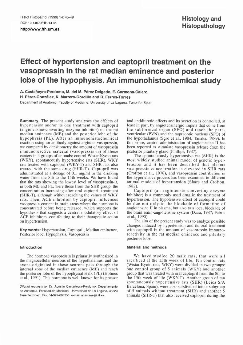
The amount of water intake in hypertensive groups was significantly higher than in control animals (p<0.05). Control animals treated with captopril (WKY-T) drank more water than WKY animals (p<0.05). Although hypertensive animals treated with captopril (SHR-T) tended to drink a little more water than SHR rats, the difference was not significant in these hypertens ive animals (Fig. 1A).
Systolic blood pressure
SHR rats showed higher systolic blood pressure (SBP) values than WKY rats (p<O,Ol) and SHR-T rats (p<0,02). No significant differences in SBP was detected between WKY, WKY-T and SHR-T rats.(Fig. 1B).
Global volume of the posterior lobe of the hypophysis
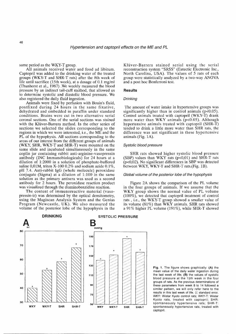
Figure 2A shows the comparison of the PL volume in the four groups of animals. If we assume that the WKY group shows the normal value of PL volume (100%), we detected that captopril treatment of control rats , i.e., the WKY-T group showed a smaller value of its volume (81 %) than WKY animals. SHR rats showed a 91 % higher PL volume (191 %), while SHR-T showed
DRINKING SYSTOLIC PRESSURE
A ml
80 .----------------,
70 o
WKY WKY-T SHR SHR-T
B mmHg
190
170
150
• c-:~::::---
130 .--""/
110
WKY WKY-T SHR
• 7 /
SHR-T
Fig. 1. The figure shows graph ically: (A) the mean value of the daily water ingestion during the last week of life; (8) the values of systolic blood pressure at the 15th week in the four groups of rats. As the previous determinations of these parameters from week 8 to 14 followed a similar pattern, we will only refer here to the results in this last week of life. 0: standard error. WI<Y : Wistar Kyoto control rats ; WI<Y-T: Wi star Kyoto rats, treated with captopril; SHR : spontaneously hypertensive rats; SHR -T : spontaneously hypertensive rats , treated with caplopril.
47
Hypertension and captopril effects on the ME and PL
a 41 % higher one (141 %) than control rats.
Immunohistochemistry (densitometry)
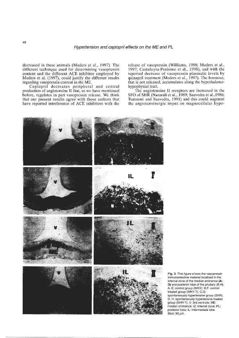
The SHR group showed the lowest amount of vasopressin-ir along the analyzed structures of the hypothalamo hypophysial axis (Figs. 2B,C, 3C,G). The highest concentration of vasopressin-ir was shown in the WKY-T and WKY groups (Figs. 2B,C, 3A,B,E,F). The treatment of the hypertensive rats with captopril (SHRT) produced an increase in IRM in both structures (Figs. 2B,C, 3D,H).
Discussion
In this study we have found that blood pressure in WKY rats does not change with captopril treatment, while in SHR-T rats, blood pressure decreases to normal levels, according to the results described in hypertensive mice by Webb et a1. (1986).
Several studies have revealed pituitary hypertrophy in the hypertension and that this hypertrophy is aggravated with emotional stress (Horie et aI. , 1991). We have also detected an increase in the volume of the posterior lobe of the hypophysis in the hypertensive animals which is followed by a partial reduction after captopril treatment.
It has been suggested that endogenous vasopressin plays a role in the central control of blood pressure (Pittman et aI., 1982) and in the present study, the concentration of vasopressin-ir has been shown to be
VOLUME OF PL
lower in the SHR rats than in the control WKY rats. This could indicate that in the development of the hypertension of the SHR rats , additionally to the reported implication of the renin-angiotensine system (Harrap et aI. , 1990), the decrease of vasopressin-ir as expression of an alteration in vasopressin release and/or production could be another factor conditioning the high values of blood pressure, since vasopressin is well known as a hormone-producing pressor and antidiuretic effects. In fact , plasma vasopressin levels have been described to be increased in these genetic hypertensive animals (Crofton et al.,1978). Also, the control system of vasopressin release of the SHR rats has been reported to be more sensible than that of WKY rats (Crofton et aI. , 1981).
In a previous work, performed in male normotensive mice, we described an increase in immunoreactive material for arginine-vasopressin in the ME, after chronic oral captopril treatment (Castafleyra-Perdomo et aI., 1998). In the present study, captopril treatment for seven weeks also produced an increase in the concentration of vasopressin-ir in ME and PL of the SHR rats, while in the normotensive WKY rats there was a small increase in vasopressin content which was not statistically significant. However, Muders et a1. (1997) have recently reported that chronic ACE inhibition by quinapril in normotensive rats reduced brain ACE activity, but also reduced vasopressin content (determined by micropunch technique) in several areas involved in central cardiovascular regulation, including the ME. Plasma levels of vasopressin were significantly
ME PL A vasopressin density vasopressin density
B C "of Yolume
250~----------------------,
%ofiRM %ofiRM
120~----------------------~ 140.-----------------------~
200 120
100 ~
100 ~ 150 110
110
60 100 60
40 40
50 20 20
WKY WKY-T SHR SHR-T WKY WKY-T SHR SHR-T WKY WKY-T SHR SHR-T
Fig. 2. This figure shows graphically the differences (in %) in the volume of the posterior lobe of the pituitary (A), the proportion of immunoreactive material (IRM) for vasopressin in the median eminence (ME) (B) and the posterior lobe of the pituitary (PL) (e) in the four groups of rats. IRM: immunoreactive material (vasopressin-ir); WKY: Wistar Kyoto rats; WKY-T: Wistar Kyoto rats , treated with captopril ; SHR: spontaneously hypertensive rats; SHR·T: spontaneously hypertensive rats , treated with captopril.
48
Hypertension and captopril effects on the ME and PL
decreased in these animals (Muders et al., 1997). The different technique used for determining vasopressin content and the different ACE inhibitor employed by Muders et al. (1997), could justify the different results regarding vasopressin content in the ME.
Captopril decreases peripheral and central production of angiotensine II that, as we have mentioned before, regulates in part vasopressin release. We think that our present results agree with those authors that have reported interference of ACE inhibitors with the
release of vasopressin (Williams, 1988; Muders et al., 1997; Castaiieyra-Perdomo et al., 1998), and with the reported decrease of vasopressin plasmatic levels by quinapril treatment (Muders et al., 1997). The hormone, that is not released, accumulates along the hypothalamohypophysial tract.
The angiotensine II receptors are increased in the SFO of SHR (Nazarali et al., 1989; Saavedra et al.,1986; Tsutsumi and Saavedra, 1991) and this could augment the angiotensinergic imput on magnocellular hypo-
Fig. 3. This figure shows the vasopressin immunoreactive material localized in the internal zone of the median eminence (AD) and posterior lobe of the pituitary (E-H). A, E: control group (WKY); B,F: control treated group (WKY-T); e,G: spontaneously hypertensive group (SHR). D, H: spontaneously hypertensive treated group (SHR-T); V: 3rd ventricle; ME: median eminence; IZ: internal zone; PL: posterior lobe; IL: intermediate lobe. Bars: 90 pm.
49
Hypertension and captopril effects on the ME and PL
thalamic neurons that would the n induce a higher vasopressin release from the neurohypophysis. EnaJapril (another ACEI) treatment has been reported to decrease th e angiotensine II receptors in the SFO of SHR (Nazarali et aI. , 1989; Saavedra et aI. , 1986; Tsutsumi and Saavedra, 1991). Captopril could then also interfere with th e central angiotensinergic s timulus , and consequently could reverse the effect of hypertension on vasopressin-ir shown in our results.
In conclusion: 1) hypertension decreases the vasopressin-ir of the two studied components of the hypothalamus-hypophysial axis (ME and PL), probably in relation to alterations of the central angiotensinergic inputs that control the production and/or release of this hormone in the hypertensive animals. We suggest that the currently described decrease in vasopressin-ir could be produced by an increase in vasopressin release by the neurohypophysis in the SHR rats, since an increase in pla s matic va sopressin level s has been previously reported in these animals (Crofton et aI. , 1978). 2) The angiotensinergic control is influenced by the action of captopril , which decreases the peripheral and central production of angiotensine II , and this decrease is one possible reason for the increase in vasopressin-ir detected in both ME and PL.
Acknowledgements. This work was supported by: Gobierno Aut6nomo
de Canarias. proyecto no. 93/051
References
Castaneyra-Perdomo A., Carmona-Calero E., Perez-Delgado M.M .,
Perez-Gonzalez H., Marrero-Gordillo N. and Ferres-Torres R.
(1998) . The effects of chronic administration of captopril on the
mouse median eminence. Histol. Histopathol. 13, 301 -305.
Crofton J.T., Share L., Shade R.E. , Allen C. and Tarbowski D. (1978) .
Vasopressin in the rat with spontaneous hypertension . Am. J.
Physiol. 235, H361-H366.
Crofton J .T ., Rockhold R.W. , Share L., Wang B.B., Horovitz Z .P.,
Manning M. and Sawyer W.H. (1981) . Effect of intracerebro
ventricular captopril on vasopressin and blood pressure in
spontaneously hypertensive rats. Hypertension 3, 11-11714.
Dzau V .J . (1987) . Implications of local angiotensin production in
cardiovascular physiology and pharmacology. Am. J. Cardiol. 59,
59A-65A.
Fabris B., Chen B., Pipic V ., Perich R. and Johnston C.L. (1990) .
Inhibition of angiotensin-converting enzyme in plasma and tissue. J.
Cardivasc. Pharmacol. 15, S6-S13
Harrap S.B., Van der Merwe W.M., Griffin SA Macpherson F. and
Lever A.F. (1990). Brief angiotensin converting enzyme inhibitor
treatment in young spontaneous hypertenSive rats reduces blood
pressure long-term. Hypertension 16, 603-614.
Holmes M.C., Antoni FA, Aguilera G. and Calt K.J. (1986) . Magno
cellular axons in passage through the median eminence release
vasopressin. Nature 319,326-329.
Horie R., Yamori Y., Nara Y. , Sawamura M. and Mizushima S. (1991) .
Aggravating effects of isolated caging on the development of
hypertenSion and its complications in stroke-prone spontaneously
hypertensive rats (SHRSP) and Wistar-Kyoto rats (WKY) . Clin. EXp.
Hypertens. 13, 858-864.
Murders F. , Elsner D., Jandeleit K. , Bahner U. , Kromer E.P. , Riegger
G.A. and Palkovits M. (1997) . Chronic ACE inhibition by quinapril
modulates central vasopressinergic system. Cardiovasc. Res. 34,
575-581 .
Nazarali A.J., Gutkind J.S., Correa F.M.A. and Saavedra J.M. (1989).
Enalapril decreases angiotensin II receptors in subfornical organ of
SHR Am . J. Physiol. 256, H1609- H1614.
Phillips M.1. (1987) . Functions of angiotensin in the central nervous
system. Annu. Rev. Physiol. 49, 413-435.
Pittman Q.J ., Lawrence D. and McLean L. (1982) . Central effects of
arginine vasopressin on blood pressure in rats . Endocrinology 110,
1058-1060.
Saavedra J.M., Israel A. , Plunkett L.M., Kurihara M.M., Shigematsu K.
and Correa F.M.A. (1986). Quantitative distribution of angiotensin II
binding sites in rat brain by autoradiography. Peptides 7, 679-687.
Share L. and Crofton J.T. (1982) . Contribution of vasopressin to
hypertenSion. HypertenSion 4, 11185-11192
Sgro S., Ferguson A.V. and Renaul L.P. (1984) . Subfornical organ
supraoptic nucleus connections: An electrophysiological study in the
rat. Brain Res. 303, 7-13
Tanaka J. (1989) . Involvement of the median preoptiC nucleus in the
regulation of paraventricular vasopressin neurons by the subfornical
organ in the rat. Exp. Brain Res. 76, 47-54.
Thunhorst RL. , Fitts D.A. and Simpson J .B. (1987) . Separation of
captopril effects on salt and water intake by subfornical organ
lesions. Am. J. Physiol. 252, R409-R418.
Tsutsumi K. and Saavedra J.M. (1991) . Quantitative autoradiography
reveals different angiotensin II receptor subtypes in selected rat
brain nuclei. J. Neurochem. 56, 348-351 .
Webb RC., Hamlin M.N., Henry J.P., Stephens P.M., and Vander JA
(1986) . Captopril , blood pressure , and vascular reactivity in
psychosocial hypertensive mice. Hypertension 8 (Suppl. I). 1-119-1-
122.
Williams G.H. (1988) . Converting-enzyme inhibitors in the treatment of
hypertension. N. Engl. J. Med. 319, 1517-1525.
Accepted April 22, 1 998


![Captopril - media.dav-medien.demedia.dav-medien.de/sample/9783804737426_p.pdf · Pharmakokinetik: Captopril PB [%] 25–30 BV [%] 70–75 HWZ [h] 2 tmax [h] 1–1,5 WE [min] 15–30](https://static.fdocuments.in/doc/165x107/5d502b5788c993f62d8b4eff/captopril-mediadav-pharmakokinetik-captopril-pb-2530-bv-7075.jpg)

