Effect Of Folate Deficiency On Mtor Signaling Network On ...
46
Wayne State University Wayne State University eses 1-1-2014 Effect Of Folate Deficiency On Mtor Signaling Network On e Liver Of Wild Type Mice Essra Moussawi Wayne State University, Follow this and additional works at: hp://digitalcommons.wayne.edu/oa_theses Part of the Nutrition Commons is Open Access esis is brought to you for free and open access by DigitalCommons@WayneState. It has been accepted for inclusion in Wayne State University eses by an authorized administrator of DigitalCommons@WayneState. Recommended Citation Moussawi, Essra, "Effect Of Folate Deficiency On Mtor Signaling Network On e Liver Of Wild Type Mice" (2014). Wayne State University eses. Paper 349.
Transcript of Effect Of Folate Deficiency On Mtor Signaling Network On ...

Wayne State University
Wayne State University Theses
1-1-2014
Effect Of Folate Deficiency On Mtor SignalingNetwork On The Liver Of Wild Type MiceEssra MoussawiWayne State University,
Follow this and additional works at: http://digitalcommons.wayne.edu/oa_theses
Part of the Nutrition Commons
This Open Access Thesis is brought to you for free and open access by DigitalCommons@WayneState. It has been accepted for inclusion in WayneState University Theses by an authorized administrator of DigitalCommons@WayneState.
Recommended CitationMoussawi, Essra, "Effect Of Folate Deficiency On Mtor Signaling Network On The Liver Of Wild Type Mice" (2014). Wayne StateUniversity Theses. Paper 349.

EFFECT OF FOLATE DEFICIENCY ON mTOR SIGNALING NETWO RK ON THE LIVER OF WILD TYPE MICE
by
ESSRA MOUSSAWI
THESIS
Submitted to the Graduate School
of Wayne State University,
Detroit, Michigan
in partial fulfillment of the requirements
for the degree of
MASTERS OF SCIENCE
2014
MAJOR: NUTRITION & FOOD SCIENCE
Approved By:
_________________________________
Advisor Date

COPYRIGHT BY
ESSRA MOUSSAWI
2014
All Rights Reserved

ii
DEDICATION
I would like to dedicate my thesis to my family who has continuously
supported me in my endeavors. A special thanks to my Mother, I would not be
where I am today had I not had my mother’s continuous support and mentorship.

iii
ACKNOWLEDGMENTS
It is with immense gratitude that I acknowledge the support and help of my
mentor Dr. Ahmad R. Heydari who has supported me since my undergraduate
studies. He has provided me with support and resources in order to complete the
work that was needed for my thesis. In addition I would like to sincerely thank Dr.
Archana Unnikrishnan as she has also been a mentor, supporter and has given
me much of her valuable time and efforts in order to guide me throughout the
process; it has been an honor to get to know and be able to work with both Dr.
Heydari and Dr. Unnikrishnan. Lastly, I would like to extend appreciation to my
lab members: Ali, Amanda, Rawya, Safa, Sukayina and Tom.

iv
TABLE OF CONTENTS
Dedication_________________________________________ _____________iii
Acknowledgments____________________________________ ___________iv
List of Figures____________________________________ ______________ivi Chapter 1-Introduction_____________________________ _______________1
A. Folate__________________________________________ ________1
B. Role of Folate in One-Carbon Metabolism_________ ___________2
C. Folate Deficiency Leading to Health Concerns____ ____________4
1. Vitamin B 12 Deficiency and Anemia____________________5
2. Neural Tube Defects_____________________________ ___6
3. Cancer and Epigenetics _________________________ ____7
D. Roles of Autophagy and mTOR Signaling Pathway___ _________8
Chapter 2-Specific Aims____________________________ ______________12
Chapter 3-Method___________________________________ ____________13
A. Animals_________________________________________ ______13
B. Diet and Carcinogenic Treatment_________________ _________13
C. Aberrant Colonic Crypt (ACF) Analysis___________ __________14
D. Western Blot Analysis___________________________ ________14
E. Statistical Analysis____________________________ __________15
Chapter 4-Figures__________________________________ _____________16
Chapter 5-Results and Discussion___________________ ______________30
Chapter 6-Conclusion_______________________________ _____________35

v
References_________________________________________ ____________36
Abstract___________________________________________ ____________44
Autobiographical Statement_________________________ _____________45

vi
LIST OF FIGURES
Figure 1-1: Folate and its Metabolism_________________________________3
Figure 1-2: Illustration of mTOR Pathway_____________________________11
Figure 4-1: Experimental Design____________________________________17
Figure 4-2 : ACF Formation________________________________________19
Figure 4-3: Effect of folate deficiency and DMH treatment on protein levels of
Beclin in young WT mice__________________________________________21
Figure 4-4: Effect of folate deficiency and DMH treatment on protein levels of
ATG5 in young WT mice___________________________________________23
Figure 4-5: Effect of folate deficiency and DMH treatment on protein levels of
LC3 in young WT mice____________________________________________25
Figure 4-6: Effect of folate deficiency and DMH treatment on protein levels of
ATG12 in young WT mice__________________________________________27
Figure 4-7 : Effect of folate deficiency and DMH treatment on protein levels of
ATG3 in young WT mice___________________________________________29

1
CHAPTER 1
INTRODUCTION
Nutritional health has always been of important concern to humanity. We are
a society built around the preservation of life. For centuries we have been a part
of the “Battle of the Fittest”. With modern technology, and research we are apart
of an era in which we are able to understand and/or observe the effects of
bioactive food components, such as folate, on the human genome. Such insight
is accomplished through the study of nutrigenomics. It is of great importance that
we understand the components of the foods consumed effect the human body on
the surface, deep within deep within our physiological begins, and on a molecular
biology level. Recent studies have indicated an inverse relationship between
dietary intake of folate, and the development of cancer [1]. As one increases their
intake level of folate, one is inadvertently decreasing the risk of cancers such as
liver, pancreatic, stomach, colon, and breast cancer [1]. Studies have suggested
that the activation and inactivation of autophagy may benefit against the
progression of cancer cells [41]. Thus, it is important for researchers to
understand the mechanism that surrounds folate restriction and the maturation of
cancer, in essence to the effects of autophagy by observing the mTOR signaling
pathway.
A. Folate
Folate is a naturally occurring water-soluble vitamin, belonging to the vitamin
B-complex group. Folate is found in plant-based foods such as, fruits, legumes,
lentils, beans, green leafy vegetables, and other foods [3,14]. Folic acid is the

2
synthetically produced form of folate used in fortified foods. Individuals
dependent on receiving nutrients from fast foods, or diets low in fruits and
vegetables are under consuming folate, and thus they are putting themselves at
risk for developing diseases and/or dangerous health conditions [3]. Additionally
folate is thought to be the most common vitamin deficiency not only in the United
States but worldwide [4]. Folate is a cofactor involved in several mechanistic
pathways, such as synthesis of DNA, repairs of DNA and methylation of DNA.
However the details of the folate pathway and its effects are still unknown. Folate
has been associated with the etiology of birth defects, and many chronic
diseases such as cardiovascular, neurological degeneration and various cancers.
B. Role of Folate in One-Carbon Metabolism
Discovered in the 1940s, folate is an essential cofactor for cell growth in living
organisms. Folic acid is a cofactor in one-carbon metabolism [5]. Folate behaves
as a carrier in one-carbon metabolism, and it is essential for cellular functions
such as cell division, DNA repair, and the regulation of amino acid homocysteine
[11]. It is of utter importance to understand the mechanism behind folic acid’s
ability to alter the one-carbon metabolism. Folic acid is found at different
oxidative states in a cell. The different oxidative states are known as folate,
dihydrofolate (DHF) and tetrahydrofolate (THF) [6]. The cofactor known as 5-
methylenetetrahydrofolate (5-methyl THF) is the main factor that is involved in
one-carbon metabolism. The enzyme known as methylenetetrahydrofolate
reducatase (MTHER) is the enzyme responsible for causing an irreversible
conversion of 5,10-methylene THF to 5-methyl THF. The main functions that are

a result of folate metabolism are: one
synthesis of thymidylate and purines (adenine and guanine) and DNA
methylation [7,9,10].
In the methionine pathway the function of 5
the amino acid homocysteine
Methionine is then broken down to
(SAM).
3
metabolism are: one-carbon transfer in the methionine cycle,
synthesis of thymidylate and purines (adenine and guanine) and DNA
In the methionine pathway the function of 5-methyl THF is to
the amino acid homocysteine to its essential amino acid for “methionine
Methionine is then broken down to the methyl group donor s-adenosylmethionine
carbon transfer in the methionine cycle,
synthesis of thymidylate and purines (adenine and guanine) and DNA
remethylate
methionine”.
adenosylmethionine

4
Levels of purines adenosine and guanosine are relatively adequate, as 5-
methyl THF is the dominant source used as a methyl donor. Through the
pyrimidine biosynthesis pathway, the production of thymidine is established
involving an exchange of deoxyuridine monophosphate (dUMP) to thymidine
monophosphate (TMP). The enzyme thymidylate synthase (TS) catalyzes the
reaction. All of these reactions are essential for a continued production of
nucleotides, and further more to ensure that DNA proliferation, as well as repair
activities are appropriately maintained. A folate restricted diet will have a
negative impact on these reactions causing altering the reasons that need to take
place and ultimately affecting the well being of individuals [9,11].
C. Folate Deficiency Leading to Health Concerns
There are many factors to take into consideration when in the process of
understanding the underlying cause of a disease of health concern. It is the battle
between cause and effect, which may be due to genetic, environmental or dietary
susceptibility. Nutrition has been considered to play a vital role on an individual’s
health [4]. The correlation between an individual’s degenerating health may not
always be a straight path of cause and effect. Many times there are a series of
indirect reactions and occurrences that lead up to a person’s development of
their disease or condition. Through careful epidemiological studies, researchers
have suggested that folate restriction is the indirect cause of poor health. Folate
restriction has shown to increase homocystine (tHcy) levels, which in turn has
increased the risk for abortion, birth defects, low birth weights and cardiovascular
disease. Folate is responsible for DNA and RNA synthesis, growth, cognitive
5
functions and proliferation [14]. Understanding the health concerns associated
with folate restriction is important because it plays a serious role is diseases such
as Alzheimers, various cancers, cardiovascular diseases and neural tube
defects. These are amongst just some of the possible diseases and conditions
that come from folate restriction [15]. Folate restriction is considered to be the
most common vitamin deficiency worldwide that can severely alter an individual’s
health condition because of the molecular role it plays within the body [4,13].
1. Vitamin B 12 Deficiency and Anemia
Anemia is the inadequacy of red blood cell production due to the inability of
hemoglobin generation, which in return leaves the organism with insufficient
levels. The primary function for red blood cells is to provide nourishment of
oxygen to the cells, through the transportation of hemoglobin [18]. When
hemoglobin levels are altered, the oxygen delivery to cells is altered as well. An
inadequacy of folate levels may severely impact the de novo pathway of DNA
and RNA synthesis, thus causing a disruption in cell division and/or may cause
deformed red blood cells [18].
An alarming concern is the ability of folic acid to mask anemia caused by
vitamin B12 deficiency as past studies have suggested. Case reports have
suggested that a consumption of ≥5000 µg of folic acid daily will mask the
anemia caused by vitamin B12 [22]. This is important finding, because anemia
caused by vitamin B12 deficiency if not detected early on-set may lead to
peripheral neuropathy, which is the disease or damage of nerves [22]. Studies in
India have reported anemia to be the seventh most common death amongst

6
Indian women working in factories, according to the factory medical officer’s
reports [20].
2. Neural Tube Defects
Neural tube defects (NTDs) occur when the neural tube of the brain and/or
the spinal cord does not successfully close during the early development stages
of an embryo resulting in damage of the embryo neural tissues. This may result
in death depending on the severity and location of the lesion [22,24].
Insufficient amounts of dietary folate are known to be the cause of
neural tube defects, miscarriages, and premature births. Thus after many studies
conducted by various researchers the CDC has recommended that women with
prior history of NTD consume a daily amount of 4000 µg prior to conceiving.
Consuming folate is essential prior to conception, since the neural tubes close
early during the growth of the embryo (28 days after conception). Additionally the
U.S Public Health has recommended that all women consume a daily dose of
400 µg of folate [22,25].
3. Cancer and Epigenetics
Through the studies of epidemiology, researchers have found a strong
correlation between folate deficiency and the risk of developing cancerous
diseases [22,25]. As previously mentioned, studies have shown that folate
restriction may increase the risk of cancer development in the liver, pancreas,
stomach, colon and breast regions [1]. Whilst suggesting that folate consumption
through regular diet of fruits and vegetables will have auspicious effects. Most
studies have shown that there is a strong link between folate consumption and

7
colorectal cancer [22]. Studies relating to folate consumption and epigenetic
patterns are being conducted, in order to better understand the link between the
two.
Understanding folate’s relation to epigenetics is important because of the
metabolic roles folate plays in both the DNA synthesis and DNA methylation.
Researches believe that an adequate consumption of folic acid may prevent
development of tumor cells by providing an adequate amount of methyl groups to
enhance DNA repair during the occurrence of DNA damage. On the other hand
other researches have suggested, that high consumption of folic acid may
accelerate the development of pre-existing tumors thus suggestion that early
exposure to folic acid should be observed in order to maintain normal
physiological activities of DNA synthesis and DNA methylation. If we gain a
better understanding of epigenetics we will have a better understanding of the
health concerns that come along with folate deficiency [25].
D. Roles of Autophagy and mTOR Signaling Pathway
The mTOR (mammalian target of rapamycin) is a serine/threonine protein
kinase that plays a very important role in signaling pathways which in return
controls cells growth, cell proliferation, cell motility, cell survival, protein synthesis
and transcription [31]. mTOR activity is supervised by a series of amino acids,
mainly leucine, as well as growth factors and AMP-activated protein kinase as an
overall energy supplier [30]. mTOR senses cellular nutrient, oxygen and energy
levels.

8
The mTOR signaling pathway can easily be affected by various factors,
including factors that may cause an alteration in the nutrient transport, energy
metabolism, protein availability and purine biosynthesis. In the state of a disease,
the mTOR signallng pathway is deregulated, causing the homeostatsis of an
organism to be compromised [31]. Such diseases include the following: cancer,
metabolic disorder such as diabetes, and ageing. When the mTOR pathway is
deregulated proliferation of cancer cells are stimulated and further damage is
inflected upon the organism [31]. During meticulous stages of tumor and cancer
development, the signaling mechanism in upstream and downstream of the
substrates have a great affect on the mTOR-signaling pathway [30].
Autophagy is the cell degradation of unnecessary and dysfunctional
intracellular components through the process of lysosome in the eukaryotic
system. During autophagy, the cytoplasmic cellular components are engulfed by
lysosomes. Reports have indicated that autophagy plays a very important role in
pathology, leading to the belief that both activation and inactivation of autophagy
may benefit cancer cells [40]. If a cell is unable to activate autophagy, protein
synthesis rises over degradation of protein and tumor growth will continue.
However, if autophagy is being activated this could lead to an effective method of
eliminating infectious components that gain access into the cytosol of the cell [39,
40].
The protein Beclin, a tumor suppressor, is required during the initial
regulation of autophagy [36]. Autophagy related protein 5 (ATG5) is an E3
ubiquitin ligase protein that is necessary for autophagy and is activated by ATG7.

9
ATG5 is additionally involved in preservation of the quality of the mitochondria
after oxidative damage has been inflected, as well as cellular longevity [37, 38].
The light-chain 3 (LC3) can be used to monitor autophagy activity and two
distinct bands are shown: LC3-I and LC3-II. The amount of autophagy is
correlated with LC3-II [40]. During autophagy, the cytoplasmic form LC3-I is
drafted to the autophagososmes, where LC3-II is produced by site-specific
proteins proteolysis and lipidation, which remains associated with
autophagososmes until it is fused with lysosome then LC3 is degraded [39, 40].
Autophagy related protein 12 (ATG12) is a protein involved in promotion of
apoptosis. It is involved in vesicle formation of autophagy, and ATG12-ATG5 act
as E3-like enzymes, which are required for lipidation [38]. Autophagy related
protein 3 (ATG3) is a protein involved in autophagy, degradation and turnover
and recycling of the cytoplasmic materials in eukaryotic cells. Additionally ATG3
regulates autophagy during the death of a cell [43].
There have been many suggestions which link autophagy and cancer [39].
Figure 1-2 is an illustration of the autophagy pathway in the mTOR signaling
pathway. Through the mTOR pathway autophagy can be induced, helping to fight
against cancer [42]. In specific tumor and cancer stages, certain autophagy-
related proteins have a major effect on the mTOR signaling pathway. It is
important to identify the substrates and proteins along with how they significantly
contribute to the regulation of the mTOR signaling pathway, during a folate
deficient status.

10
Figure 1-2: Illustration of mTOR Signaling Pathway [42].

11
CHAPTER 2
SPECIFIC AIMS
In order to gain an understanding of the mechanism behind the effects of
folate restriction on tumorigenesis, it is crucial we examine the mechanism
behind folate, and the autophagy related proteins in the mTOR signaling
pathway. Upon deregulation of the mTOR signaling pathway, proliferation of
cancer cells are stimulated causing extensive damage. Specific proteins involved
in the autophagy of the mTOR pathway affect the growth and development of
cancer and tumors. We hypothesize that dietary folate alters the mTOR signaling
pathway in the liver of wild type mice protecting them against tumorigenesis,
which have been treated with a carcinogenic substance.
The following are the specific aims of the research:
Specific Aim 1: To examine the effects of folate deficiency against tumor growth
in the liver of wild type mice through measurement of ACF counts per mouse.
Specific Aim 2: To examine the effects of folate deficiency on the signaling
pathway of the substrates involved in the regulation of autophagy in mTOR
signaling pathway in wild type mice that have been treated with DMH, a
cariogenic substance.

12
CHAPTER 3
METHODS
A. Animals
The experiments were performed on young male wild type mice 6 weeks of
age. Mice came from a C57BL/6-specfic pathogen-free strain. All procedures
were performed in accordance with the National Institutes of Health guidelines for
the use and care of laboratory mice. All mice survived, and no retardation was
developed as a result of dietary intake. Mice used during this experimental trial
were backcrossed into the C57BL/6 strain for a minimum of 20 generations. In
order to determine the genotype of all mice Southern blot analysis was
performed as described by Cabelof et. al. Animal protocol was given approval
through the Wayne State University Animal Investigation Committee [15]. All
mice during this experiment were maintained on a 12-hr light/dark cycle and
given water ad libitum [15].
B. Diet and Carcinogenic Treatment
After a weeklong acclimation process, wild type (WT) mice were randomly
delegated to the following dietary groups: folate adequate (FA), and folate
deficient (FD) AIN93G-purified isoenergetic diet (Dyets, Inc., Lehigh Valley, PA).
The FA dietary group received a folate adequate diet composed of 2 mg/kg of
folic acid, while the folate deficient group received a 0 mg/kg of folic acid. 1%
succinyl sulfathiazole was used in all diets for experimental purposes, and all
diets were stored at -20 °C [15]. One week proceeding to dietary ingestion, mice
were randomly selected from both FA and FD groups and injected

13
intraperitoneally with the carcinogenic 1,2-dimethylhydrazine HCL (DMH, 30
mg/kg body weight) in 10 mmol/liter of NaHCO3 (Fisher Scientific) once a week
for the duration of 6 weeks. Both dietary intake and body weight were recorded
twice a week to monitor for signs of toxicity and/or weight loss, and diets
continued for the duration of 12 weeks [15].
C. Aberrant Colonic Crypt (ACF) Analysis
Animals were anesthetized under CO2 asphyxiation the abdominal cavity was
exposed and the colon was extracted, cleansed with cold phosphate-buffered
saline, cut longitudinally, and fixed flat overnight in 10% neutral buffered formalin.
The colonic crypts were stained with 2 g/liter of methylene blue in phosphate-
buffered saline for 5 min. The number of ACF and aberrant crypts per foci were
observed by light microscopy at a magnification of X10 in a blinded manner [15].
D. Western Blot Analysis
Western blot analysis was performed during this experiment, using 200 µg of
nuclear protein. Upon completion of SDS-PAGE, the region containing the
protein(s) of interest was extracted and primed for Western blot analysis.
Remaining portion of the gel was dyed with a GelCode blue stain reagent (Pierce
Biotechnology) to establish equivalent protein loading in each of wells. As an
internal control for protein loading, membranes were re-probed with anti-Lamin B
antibody (Santa Cruz Biotechnology, Santa Cruz, CA). The bands were
visualized and quantified using a ChemilmagerTM System (Alphalnnotech, San
Leandro, CA) proceeding incubation in SuperSignal® West Pico

14
Chemiluminescent Substrate (Pierce Biotechnology). Data are shown as the
integrated density value of band per µg of protein loaded [15].
E. Statistical Analysis
Statistical significance between means was determined using a “unpaired t-
test”, after quantification of protein data. P values representing a value less than
0.05 were considered statistically significant. Significant values will be marked by
“*” on graphs, where significant p value is present.

15
CHAPTER 4
FIGURES
Figure 4-1: Experimental Design. Wild type (WT) mice were fed either a folate adequate (2 mg/kg, FA) diet or a folate deficient (0 mg/kg, FD) diet for the duration of 12 weeks. All WT mice in this experiment were treated with a carcinogenic at 6 weeks old (young mice). Proceeding the first week of dietary intake, all groups then followed the same treatment and were injected with 30 mg/kg body weight of 1,2-dimethylhydrazine (DMH), a colon and liver carcinogen, for a period of 6 weeks. After 6 weeks of consecutive treatment, the animals were sacrificed using a CO2 asphyxiation chamber on week 12.

16
Figure 4-2: ACF Formation. Wild Type (WT) mice received either folate adequate (FA) or folate deficient (FD) diet. Additionally both diet groups FA and FD were subjected to intraperitoneal treatment with 1,2-dimethylhydrazine (DMH) for 6 weeks at 30 mg/kg body weight. After sacrifice, colons were processed as described under “Methods”. Colons were analyzed under light microscopy to visualize the number of ACF per mouse colon (ACF/mouse).

17
Figure 4-3: Effect of folate deficiency and DMH tre atment on protein levels of Beclin in young WT mice. This figure is a representation of the analysis of the Beclin protein levels in mucosa of young (Y) WT mice that received either folate adequate (FA) or folate deficient (FD) diet, and were subjected to intraperitoneal treatment with DMH for 6 weeks at 30 mg/kg body weight (DMH). The protein levels were quantified using Western blot analysis. Values represent an average (±SEM) of data obtained from 3 mice of each group and are representative of separate identical experiments. Values with different letter superscripts indicate significant differences at p>0.05.

18

19
Figure 4-4: Effect of folate deficiency and DMH tre atment on protein levels of ATG5 in young WT mice. This figure is a representation of the analysis of the ATG5 protein levels in mucosa of young (Y) WT mice that received either folate adequate (FA) or folate deficient (FD) diet, and were subjected to intraperitoneal treatment with DMH for 6 weeks at 30 mg/kg body weight (DMH). The protein levels were quantified using Western blot analysis. Values represent an average (±SEM) of data obtained from 3 mice of each group and are representative of separate identical experiments. Values with different letter superscripts indicate significant differences at p>0.05.

20
Figure 4-5: Effect of folate deficiency and DMH tre atment on protein levels of LC3 in young WT mice. This figure is a representation of the analysis of the LC3 protein levels in mucosa of young (Y) WT mice that received either folate adequate (FA) or folate deficient (FD) diet, and were subjected to intraperitoneal treatment with DMH for 6 weeks at 30 mg/kg body weight (DMH). The protein levels were quantified using Western blot analysis. Values represent an average (±SEM) of data obtained from 3 mice of each group and are representative of separate identical experiments. Values with different letter superscripts indicate significant differences at p>0.05.
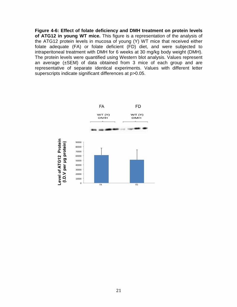
21
Figure 4-6: Effect of folate deficiency and DMH tre atment on protein levels of ATG12 in young WT mice. This figure is a representation of the analysis of the ATG12 protein levels in mucosa of young (Y) WT mice that received either folate adequate (FA) or folate deficient (FD) diet, and were subjected to intraperitoneal treatment with DMH for 6 weeks at 30 mg/kg body weight (DMH). The protein levels were quantified using Western blot analysis. Values represent an average (±SEM) of data obtained from 3 mice of each group and are representative of separate identical experiments. Values with different letter superscripts indicate significant differences at p>0.05.

22
Figure 4-7: Effect of folate deficiency and DMH tre atment on protein levels of ATG3 in young WT mice. This figure is a representation of the analysis of the ATG3 protein levels in mucosa of young (Y) WT mice that received either folate adequate (FA) or folate deficient (FD) diet, and were subjected to intraperitoneal treatment with DMH for 6 weeks at 30 mg/kg body weight (DMH). The protein levels were quantified using Western blot analysis. Values represent an average (±SEM) of data obtained from 3 mice of each group and are representative of separate identical experiments. Values with different letter superscripts indicate significant differences at p>0.05.

23
CHAPTER 5
RESULTS AND DISCUSSION
The purpose of our study was to determine the effects of folate deficiency
against tumor growth in wild type mice, which were treated with colon and liver
carcinogen, through the measurement of ACF counts per mouse, in addition too
the effects of folate deficiency on autophagy regulation in the mTOR signaling
pathway. This was done through the observation autopohagy related proteins
Becline, ATG5, LC3, ATG12 and ATG3, which all play a crucial role in the
autophagy pathway. Through the knowledge of previous studies, a correlation
has linked folate deficiency to carcinogenesis. Previous studies have also linked
autophagy to cancer, using the mTOR pathway to analyze autophagy’s effects
on cancer we have seen that in specific tumor and cancer stages, certain
autophagy related proteins help prevent cancer. It is important to identify the key
components and observe their specific roles contributing to the regulation of the
mTOR signaling pathway during a state of folate restriction and turmogenesis.
Data collected from our laboratory, and microarray analysis have
observed the effects folate deficiency displaces upon the expression of the
mammalian target of rapamycin (mTOR) signaling pathway in the colon of young
wild type mice, which were treated with carcinogen 1,2-dimethyhydrazine (DMH).
The mTOR signaling pathway, as seen in figure 1-4, plays a crucial role cell
growth, proliferation and survival incorporating both extracellular and intracellular
signals. Studies have shown that the mTOR pathway has been trigged by
various cellular activities such as: tumor growth, insulin resistance (IR) and/or

24
adipogenesis. Additionally the mTOR pathway can be deregulated during the
development of cancer, causing stimulation to cancer cells, which further causes
damage. An experimental design, as illustrated by figure 4-1, was developed by
our laboratory to determine whether folate deficiency would protect against the
progression of tumorigenesis, with young wild type (WT) mice that were treated
with a carcinogen beginning at 6 weeks of age. Mice were divided into two
dietary groups folate adequate (FA) and folate deficient (FD), for the duration of
12 weeks. The FA group were fed 2 mg/kg of dietary folate, while the FD group
was fed 0 mg/kg dietary folate. All WT mice were injected with 30 mg/kg body
weight of 1,2-dimethylhydrazine (DMH), a colon and liver carcinogen, for a period
of 6 weeks. After 6 weeks of consecutive treatment, the animals were sacrificed
using a CO2 asphyxiation chamber on week 12.
After scarification of the animals the abdominal cavity was exposed and
the colon was carefully and properly extracted from the body in order to analyze
the number of Aberrant Colonic Crypt (ACF). Mice colon was analyzed under
light microscopy, as illustrated by figure 4-2, an image explaining the ACF count
per mouse in colon (ACF/mouse). Previous studies showed that untreated wild
type mice showed no sign of ACF formation in their colon. In our studies, our
ACF findings, as illustrated by figure 4-2, the young WT mice in the folate
deficiency (FD) group, whom were treated with DMH, showed a significant
increase in ACF (~50%) as compared to animals that were on folate adequate
(FA) diets. This was an indication that folate deficiency leads to a significant
increase in colon carcinogen in response to DMH. As both the FA and FD groups

25
were young wild type mice, this finding indicated that age was not a factor in the
development of colon carcinogenesis.
Using the data collected from our experimental study, we will provide a
comparison amongst the two dietary groups in order to show a better perception
of the upstream and downstream signaling pathway of these different substrates,
which are involved in the autophagy of the mTOR pathway. Figures 4-3 to 4-7
will draw a comparison between the different dietary groups. No other
comparisons will be shown as all mice used in this experiment were of wild type
(WT) strain and young (“Y”) mice in age, and all mice were administered with the
DMH treatment for the duration of 6 weeks.
Macroautophagy is the primary pathway that occurs in damaged cell
organelles and unused proteins. Macroautophagy uses the involvement of a
double membrane formation around the cytoplasm of the substrate resulting in
autophagosome, which is induced by class III PIK3 (phosphoinsitide-3-kinase).
PIK3 is the autophagy related gene that includes Becline1 (ATG6), ATG4,
ATG12, ATG5, ATG12 and ATG16 are additionally involved in the autophagy
pathway in mTOR. Autophagosome voyages through the cytoplasm of the cell to
a lysosome and both the organelles and amalgamate. Due to the acidic nature of
lysosome the organelles go through hydrolases breaking down the waste
material.
The autophagy protein Beclin is a tumor suppressor, which is required
during the initial regulation of autophagy [36]. Folate deficiency and DMH
treatment on the autophagy related protein Beclin is illustrated by figure 4-3 using

26
young WT mice. The figure for Beclin shows an insignificant difference of protein
levels with treated young WT mice between the FA and FD diet. Figure 4-3
shows a p > 0.05, Beclin is reported at 0.46.
Autophagy related protein 5 (ATG5) is a E3 ubiquitin ligase protein that is
necessary for autophagosome elongation. ATG5 is additionally involved in the
maintenance of the quality of the mitochondria after oxidative damage has been
inflected upon it, as well as cellular longevity. Folate deficiency and DMH
treatment on the autophagy related protein ATG5 is illustrated by figure 4-3 using
young WT mice did not show a significant difference. The p-value for ATG5 is
reported at 0.62.
The light-chain 3 (LC3), autophagy protein is used to monitor the activities
of autophagy, using two bands LC3-I and LC3-II for distinction. LC3-I band is
used for autophagososmes, while the second band LC3-II to indicated the
amount of autophagy within the protein that is drafted for proteolysis and
lipidation, which is fused with lysosome before it is degraded. Folate deficiency
and DMH treatment on the autophagy related protein LC3 is illustrated by figure
4-3 using young WT mice. The autophagy protein LC3 is p value was 0.83 which,
indicated there is no significant difference between the dietary groups in treated
young WT mice.
Autophagy related protein 12 (ATG12) is involved in the promotion of
apoptosis. It is also involved the vesicle formation of autophagy. Folate
deficiency and DMH treatment on the autophagy related protein ATG12 is
illustrated by figure 4-3 using young WT mice. There was an insignificant

27
difference between the dietary groups that were illustrated by figure 4-3, as the p-
value was at a 0.67.
Autophagy-related protein 3 (ATG3) is involved in the autophagy,
degradation and turnover and recycling of the cytoplasmic materials in eukaryotic
cells. In addition ATG3 protein regulates autophagy during the death of a cell.
Folate deficiency and DMH treatment on the autophagy related protein ATG3 is
illustrated by figure 4-3 using young WT mice, after running an unpaired t-test the
p-value was at a 0.53 indicating an insignificant difference between the treated
FA and FD dietary groups.

28
CHAPTER 6
CONCLUSION
A respective some of studies linked a correlation between folate
restriction, aging and carcinogenesis, suggesting that folate restriction can
reduce the progression of cancer with the aid of apoptosis through autophagy in
the mTOR signaling pathway. It is important that we understand the folate
pathway, the mTOR signaling pathway and the role autophagy plays in cancer.
Previous studies have found folate deficiency in young WT mice resulted in a
significant increase in ACF formation in comparison to the FA counter-group.
This finding suggested that folate deficiency resulted in a significant increase in
colon tumorogenesis in response to DMH treatment. Our data agrees with these
findings, as they have suggested that folate deficiency in DMH treated young WT
mice resulted in a significant increase (~50%) in ACF formation as compared to
the FA counter-group, illustrated by figure 4-2. These findings suggested that
apoptosis my play a significant role in anti-cancer progression. However, when
we conducted our western blot analysis on autophagy related proteins comparing
the two dietary groups (FA ad FD) no significant differences were observed,
according to the p-values obtained. This finding may suggest these specific
autophagy proteins are not affected by dietary folate. Our studies are insufficient
and need to conduct further studies to further understand the mechanism behind
folate deficiency’s affect on autophagy in the mTOR pathway and how it plays a
role in cancer prevention.

29
REFERENCE
1. Unnikrishnan, Archana, Ahmad R. Heydari, and Tom M. Prychitko. Folate
Deficiency Regulates Expression of DNA Polymerase β In Response to Oxidative
Stress. Free Radic Bio Med, 5 Jan. 2011.
http://www.ncbi.nlm.nih.gov/pmc/articles/PMC3018545 /
2. Y I Kim, R N Salomon, F Graeme-Cook, S W Choi, D E Smith, G E Dallal,
and J B Mason. "Dietary Folate Protects against the Development of
Macroscopic Colonic Neoplasia in a Dose Responsive Manner in Rats." GUT,
Nov. 1996.
http://www.ncbi.nlm.nih.gov/pmc/articles/PMC1383400
3. Combs GF. The Vitamins, Fundamental aspects in Nutrition and Health.
Second edition. Academic Press: 1998
4. Marilia L. Cravo, Joel B. Mason, Yogeshwar Dayal, Et Al. "Folate Deficiency
Enhances the Development of Colonic Neoplasia in Dimethylhydrazine-treated
Rats." Folate Deficiency Enhances the Development of Colonic Neoplasia in
Dimethylhydrazine-treated Rats. Cancer Research, 1992.
http://cancerres.aacrjournals.org/content/52/18/5002.long
5. Leonie G. Mikael, Jill Pancer, Qing Wu, and Rima Rozen*. "Journal of
Nutrition." Disturbed One-Carbon Metabolism Causing Adverse Reproductive
Outcomes in Mice Is Associated with Altered Expression of Apolipoprotein AI and
Inflammatory Mediators PPARα, Interferon-γ, and Interleukin-10. The Journal of
Nutrition, Dec.-Jan. 2011.
http://jn.nutrition.org/content/142/3/411.long

30
6. Meier, Christoph, Lester G. Carter, Graeme Winter, Ray J. Owens, David I.
Stuart, and Robert M. Esnouf. "Abstract." National Center for Biotechnology
Information. U.S. National Library of Medicine, 23 Feb. 2007.
http://www.ncbi.nlm.nih.gov/pmc/articles/PMC2330188 /#!po=8.33333
7. Duthie SJ. Folate and cancer: how DNA damage, repair, and methylation
impact on colon carcinogenesis. Springer.2010:34:101-109.
8. Kim Y. Folate and colorectal cancer: An evidence- based critical review. Mol.
Nutr. Food Res.2007:51:267-292.
9. Young-In K. Folate and colorectal cancer: An evidence-based critical review.
Mol. Nutr. Food Res.2007;51:267-292.
10. Ueland PM. et al. Biological and clinical implications of the MTHFR C677T
polymorphism. Trends Pharmacol. Sci. 2001;22:195-201.
11. Duthie, SJ. Folate and Cancer: how DNA damage, repair, and methylation
impact on colon carcinogenesis. J. Inherit Metab Dis.2010.
12. Wille A ,W. C., Stampfer, M. J., Colditz, G. A., Rosner, B. A., and Speizer, F.
E. Relation of meat, fat, and fiber intake to the risk of colon cancer in a
prospective study among women. N. Engl. J. Med., 323: 1664-1672, 1990.
13. Potter, J. D., and McMichael, A. J. Diet and cancer of the colon and rectum: a
case control study. J. Nati. Cancer Inst., 76: 557-569, 1986.

31
14. Manjeswori Ulak Mail, Ram K. Chandyo, Ramesh K. Adhikari, Pushpa R.
Sharma, Halvor Sommerfelt, Helga Refsum, Tor A. Strand. "Cobalamin and
Folate Status in 6 to 35 Months Old Children Presenting with Acute Diarrhea in
Bhaktapur, Nepal." PLOS ONE:. N.p.
http://www.plosone.org/article/info%3Adoi%2F10.1371%2Fjournal.pone.0090079
#s1
15. Ventrella-Lucente LF1, Unnikrishnan A, Pilling AB, Patel HV, Kushwaha D,
Dombkowski AA, Schmelz EM, Cabelof DC, Heydari AR. National Center for
Biotechnology Information. U.S. National Library of Medicine, 19 Apr. 2010.
http://www.ncbi.nlm.nih.gov/pubmed/20404327
16. Ysun Karabulut, Osman Şevket, and Ayhan Acun. "Iron, Folate and Vitamin
B12 Levels in First Trimester Pregnancies in the Southwest Region of Turkey."
Turk Ger Gynecol Assoc, 2011.
http://www.ncbi.nlm.nih.gov/pmc/articles/PMC3939272/
17. Siddharth Agarwal and Vani Sethi. " Nutritional disparities among women in
urban India." National Center for Biotechnology Information. U.S. National Library
of Medicine, n.d.
18. Iyer R, Tomer S.K. Folate: A Functional Food Constituent. Journal of Food
Science 2009;74:114-122.

32
19. Murray M. 1996. Vitamins. In: Murray M, editor. Encyclopedia of nutritional
supplements: the essential guide for improving your health naturally. 1st
ed.
Rocklin, Calif.: Prima Publishing. p. 119-26.
20. Joseph, Bobby, and Naveen Ramesh. "Abstract." National Center for
Biotechnology Information. U.S. National Library of Medicine, 03 July 0005.
21. Brugnara C1, Chambers LA, Malynn E, Goldberg MA, Kruskall MS. "Altered
Erythrocyte Membrane Protein Composition in Chronic Kidney Disease Stage 5
Patients under Haemodialysis and Recombinant Human Erythropoietin Therapy."
National Center for Biotechnology Information. U.S. National Library of Medicine,
1993.
http://www.ncbi.nlm.nih.gov/pubmed/?term=folate+effects+on+RBC+production
22. Crider KS, Bailey LB, Berry RJ. Folic Acid Food Fortification- Its History,
Effect, Concerns, and future Directions. Nutrients. 2011:3:370-384.
23. Shao L1, Wang Y2, Chang J1, Luo Y1, Meng A3, Zhou D1. National Center
for Biotechnology Information. U.S. National Library of Medicine, 2013.
24. Czeizel, Andrew E., Istvan Dudás, Attila Vereczkey, and Ferenc Bánhidy.
"Abstract." National Center for Biotechnology Information. U.S. National Library
of Medicine, 21 Nov. 2013.

33
25. Rosati, Rita, Hongzhi Ma, and Diane C. Cabelof. "Abstract." National Center
for Biotechnology Information. U.S. National Library of Medicine, 09 Oct. 2012.
http://www.ncbi.nlm.nih.gov/pmc/articles/PMC3474250/
26. Hickson ID. Base excision Repair of DNA Damage. Landes Bioscience and
Chapman & Hall:1997.
27. Li, Hui, Rafal Swiercz, and Ella W. Englander. "Abstract." National Center for
Biotechnology Information. U.S. National Library of Medicine, 08 July 2009.
http://www.ncbi.nlm.nih.gov/pmc/articles/PMC2759109/
28. Base Excision Repair of Tandem Modifications in a Methylated CpG
Dinucleotide." Base Excision Repair of Tandem Modifications in a Methylated
CpG Dinucleotide. N.p., n.d.
http://www.jbc.org/content/early/2014/04/02/jbc.M114.557769.long
29, "MTOR Signaling Pathway." Cell Signaling Technology (CST): Antibodies,
Reagents, Proteomics, Kits and Consumables. N.p., n.d.
30. "MTOR Signaling Pathway." MTOR Signaling Pathway. N.p., n.d.
31. Zoncu R, Efeyan A, Sabatini DM. mTOR: from growth signal integration to
cancer, diabetes, and aging. Molecular Cell Biology.2011
32. Wellen K E, Thompson C B. Cellular Metabolic Stress: Considering How
Cells Respond to Nutrient Excess. Cell Press. 2010:40: 323-332.

34
33. Guertin DA and Sabatini D. An expanding role for mTOR in cancer.
Journal.2005:11:353-361.
34.http://www.qiagen.com/products/genes%20and%20pathways/pathway%20det
ails?pwid=304
35. Levine A J, Hu W, Feng Z. The p53 pathway: what questions remain to be
explored?Cell Death Differ. 2006:13:1027-1036.
36. Sophie Pattingrea, B, Lucile Espertc, Martine Biard-Piechaczykc, Patrice
Codognoa, B,. "Regulation of Macroautophagy by MTOR and Beclin 1
Complexes." Regulation of Macroautophagy by MTOR and Beclin 1 Complexes.
N.p., Feb. 2008.
37. He Liu1, Zhaoyue He1, Thomas Von Rütte1, Shida Yousefi1, Robert E.
Hunger2 and Hans-Uwe Simon1,*. "Down-Regulation of Autophagy-Related
Protein 5 (ATG5) Contributes to the Pathogenesis of Early-Stage Cutaneous
Melanoma." Down-Regulation of Autophagy-Related Protein 5 (ATG5)
Contributes to the Pathogenesis of Early-Stage Cutaneous Melanoma. N.p., n.d.
http://stm.sciencemag.org/content/5/202/202ra123.abstract
38. "Autophagy Protein 5 - ATG5 - Homo Sapiens (Human)." Autophagy Protein
5 - ATG5 - Homo Sapiens (Human). N.p., n.d.
http://www.uniprot.org/uniprot/Q9H1Y0
39. Ruth Scherz-Shouvala, Hilla Weidbergb, Chagay Gonena, Sylvia Wildera,

35
Zvulun Elazarb, and Moshe Orena,1. "P53-dependent Regulation of Autophagy
Protein LC3 Supports Cancer Cell Survival under Prolonged Starvation." P53-
dependent Regulation of Autophagy Protein LC3 Supports Cancer Cell Survival
under Prolonged Starvation. N.p., n.d.
http://www.pnas.org/content/early/2010/10/08/1006124107.full.pdf
40. "Immunoblot Analysis of LC-3." Autophagic Marker LC3 Microtubule-
associated Protein1 Light Chain 3. NanoTools Antikoerpertechnik, n.d.
http://www.nanotools.de/flyer/Flyer_LC-3.pdf
41. Clelia Miracco, MDa, Corresponding Author Contact Information, E-mail the
Corresponding Author, Gabriele Cevenini, MEngb, Alessandro Franchi, MDc,
Pietro Luzi, MDa, Elena Cosci, BSca, Vasileios Mourmouras, MDa, Irene
Monciatti, BSca, Susanna Mannucci, BSca, Maurizio Biagioli, MDd, Marzia
Toscano, MSce, Daniele Moretti, PhDe, Roberto Lio, MDa, Daniela Massi, MDc.
"Beclin 1 and LC3 Autophagic Gene Expression in Cutaneous Melanocytic
Lesions ☆." Human Pathology. Elsevier, 2014.
http://www.sciencedirect.com/science/article/pii/S0046817709003323
42. "Figure 2." Figure. N.p., n.d. Web.
http://www.molecularneurodegeneration.com/content/4/1/16/figure/F2?highres=y
43. "Autophagy Related 3." Gene Cards. Weizmann Institute of Science., n.d.
http://www.genecards.org/cgi-bin/carddisp.pl?gene=ATG3

36
44. ATG13: Just a Companion, or an Executor of the Autophagic Program?"
Autophagy: Review. Institute of Molecular Medicine; Heinrich-Heine-University;
Düsseldorf, Germany, n.d.
https://www.landesbioscience.com/journals/autophagy/2014AUTO0021R1.pdf

37
ABSTRACT
EFFECT OF FOLATE DEFICIENCY ON mTOR SIGNALING NETWO RK ON THE LIVER OF WILD TYPE MICE
by
ESSRA MOUSSAWI
August 2014
Advisor: Dr. Ahmad R. Heydari
Major: Nutrition and Food Science
Degree: Masters of Science
Through various studies of dietary nutrients on the affects of the
physiological and molecular pathways are a key study in understanding the
interaction between dietary nutrients and the human genome that may severely
impact the development of various cancers. A primary approach is observing the
dietary nutrients, that we consume and how it plays a role in the cellular pathway
of our bodies through experimental methods. Folate deficiency (FD) has shown,
through studies, to play a role anti-cancer progression. Our goal was to observe
the autophagy related proteins in the mTOR signaling pathway as it was
suggested that mTOR may induce autophagy, which in return could help reduce
the progression of cancer by apoptosis of cancer cells. We anticipated to see an
increase in the protein levels of autophagy related proteins, with our DMH treated
FD dietary experimental group. We hypothesize that dietary restriction will
increase the protein levels of autophagy proteins in the mTOR signaling pathway,
acting as an anti-cancer aid.

AUTOBIOGRAPHICAL STATEMENT
Education
Graduating- August 2014
December 2011
Awards, Memberships & Volunteer Wor
Member of Golden Future for South AfricaUniversity 2014 to PresentVolunteer HUDA Clinic Detroit, 2014 to Present Volunteer American Red Cross, 2010 to Member of American Student Dental Association, 2009 and 2010Volunteer Detroit Partnership, 2009 and 2010
38
AUTOBIOGRAPHICAL STATEMENT
ESSRA MOUSSAWI
Master of Science in Nutrition and Food Science
December 2011 Bachelor of Science in Nutrition and Food Science
Awards, Memberships & Volunteer Wor k
Golden Future for South Africa Graduate Students, Wayne State to Present
Volunteer HUDA Clinic Detroit, 2014 to Present Volunteer American Red Cross, 2010 to Present Member of American Student Dental Association, 2009 and 2010 Volunteer Detroit Partnership, 2009 and 2010
Master of Science in Nutrition and Food Science
and Food Science
, Wayne State