Effect of Environmental and Host Factors on Biological ...
124
University of New England Effect of Environmental and Host Factors on Biological Control of Fusarium wilt by Non- pathogenic Fusarium oxysporum in Tomato Submitted by: Hayder Abdulhasan Ali B.Sc. (Agriculture-Plant protection) University of Basra, Agricultural College, Iraq M.Sc. (Plant Pathology) University of Basra, Agricultural College, Iraq A thesis submitted for the degree of Doctor of Philosophy, December 2017
Transcript of Effect of Environmental and Host Factors on Biological ...

University of New England
Effect of Environmental and Host Factors on
Biological Control of Fusarium wilt by Non-
pathogenic Fusarium oxysporum in Tomato
Submitted by:
Hayder Abdulhasan Ali
B.Sc. (Agriculture-Plant protection)
University of Basra, Agricultural College, Iraq
M.Sc. (Plant Pathology)
University of Basra, Agricultural College, Iraq
A thesis submitted for the degree of Doctor of Philosophy,
December 2017

i
Abstract
This study was focused on the effect of environmental and host factors on the antagonism of
Fusarium wilt of tomato, caused by Fusarium oxysporum f. sp. lycopersici (Fol), by non-
pathogenic strains of F. oxysporum. Seven non-pathogenic strains of F. oxysporum were
isolated and screened for antagonism of Fusarium wilt. Strains F1 and F4 were chosen for
further experiments as they reduced disease severity more than other non-pathogens. The best
method for applying non-pathogens and pathogens was to inoculate soil with conidial
suspension. The non-pathogens reduced seed germination and growth of tomato plants in the
absence of the pathogen. In a split root system, non-pathogens F1 and F4 induced resistance
of tomato plant against Fusarium wilt although there was no direct contact between the
pathogen and non-pathogens. Iron at high and standard concentration in the nutrient solution
stimulated induced resistance. However, direct antagonism of Fol by F4 was greatest at low
level of iron.
Tomato root exudates increased in the vitro antifungal activity of non-pathogens toward
pathogens and also increased spore germination of both non-pathogens and pathogens. The
components of root exudates including sugars and organic acids influenced the antagonism of
non-pathogens against pathogen in vitro. However sugars and organic acids had little effect
on disease suppression in pot trials.
In dual culture, using NaNO3 as source of N, the inhibition of Fol by F1 and F4 was
decreased at high level of N, whereas at high level of NaNO3 as source of N the antibiotic
production increased. Using NH4Cl as source of N at high and low level, the inhibition of Fol
by F1 and F4 was increased and the antibiotic production of non-pathogens also increased. In
pot trials, the disease severity was less at low N compared with high level of N.
In dual culture, at low level of K the antagonistic activity of s F1 and F4 against Fol was
improved. However, at high level of K, the antibiotic production of non-pathogens increased.
In glasshouse pot trials, non-pathogens improved plant health at low level of K, whereas the
growth of non-pathogens was decreased at high level of K.
At high level of Ca, the inhibition of growth of Fol by F1 and F4 was decreased. Also at low
level of Ca the inhibition of growth of Fol by antibiotic production of F4 was increased.
ii
However, the inhibition of growth of Fol by antibiotic production of F1 was increased at high
level of K.
The inhibition of growth of Fol by F1 and F4 was decreased with high level of iron. The
antibiotic production of F1 and F4 inhibited growth of Fol at low level of iron more than at
high level. Biological control did not work well at high levels of iron.
Further work is needed on the effect of non-pathogens on the growth of plants. More tests
should be done on the effect of root exudate on antagonism. Biocontrol agents should be
found that can work well at low nutrient levels.

iii
Acknowledgments
I would like to express my sincere appreciation to my principle supervisor, Dr David
Backhouse for his excellence continuous support and guidance during my candidature. I am
very grateful to him for assistance and advice during the writing up of this thesis. The fact he
was there for me whenever I need assistance. I could not have imagined having better advisor
and mentor for my PhD study. I highly appreciated his assistance to achieve my dream.
I thank Higher Committee for Education Development in Iraq scholarship grant and financial
support for undertaking PhD program.
I also thank technical team at the Agronomy and Soil Science department especially Mick
Faint, Leanne Lisle and Elizabeth Marshall, were extremely helpful. I would highly appreciate
the effort of the administrator of Agronomy and Soil Science Roz Mortimer.
Last not least, special thanks to my parents for their encouragement and constant prayers, my
brothers and sisters for their encouragement. I would like to thank my beloved wife, Inaam,
my son Hussein and my little daughter Lana without their full love and support I would never
have realized my full potential. Finally, I would like to thank bountiful grace of the Almighty
ALLAH for granting me health, strength to undertake this research task.

iv
Certification
I certify that the ideas, analyses, results, and conclusions reported in this thesis are entirely
my own effort, except where otherwise acknowledged. This work has not previously been
submitted for a degree or diploma in any university.
To the best of my knowledge and belief, the thesis contains no material previously published
or written by another person except where due reference is made in the thesis itself.
Signature of Candidate Date 05/ 12/ 2017
v
Table of Contents
Abstract ................................................................................................................................. i
Acknowledgments ................................................................................................................ iii
Certification ......................................................................................................................... iv
List of Tables ...................................................................................................................... vii
Table of Figures ................................................................................................................. viii
Chapter 1. General Introduction ........................................................................................ 1
Chapter 2. Literature Review ............................................................................................ 4
2.1. Tomato wilt ............................................................................................................ 4
2.1.1. Disease cycle ................................................................................................... 4
2.1.2. Symptoms ........................................................................................................ 5
2.1.3. Factors that affect disease ................................................................................ 6
2.2. Biological control: .................................................................................................. 7
2.2.1. Definitions ....................................................................................................... 7
2.2.2. Competition for space ...................................................................................... 7
2.2.3. Competition for nutrients ............................................................................... 10
2.2.4. Antibiosis ...................................................................................................... 12
2.2.5. Induced resistance .......................................................................................... 14
2.2.6. Effect of beneficial microorganisms ............................................................... 16
2.3. Making biocontrol work ........................................................................................ 17
2.3.1. Difference between field soil and sterilized soil ............................................. 17
2.3.2. Application methods ...................................................................................... 18
2.3.3. Timing of application ..................................................................................... 19
2.3.4. Combination of non-pathogenic fungi with organic amendment ..................... 20
2.4. Root exudates ....................................................................................................... 20
2.5. Conclusion ............................................................................................................ 23
Chapter 3. Isolation and screening of non-pathogenic Fusarium oxysporum .................... 24
3.1. Introduction .......................................................................................................... 24
3.2. Material and Methods ........................................................................................... 25
3.2.1. Isolation and identification of isolates ............................................................ 25
3.2.2. Pathogens ...................................................................................................... 25
3.2.3. Molecular identification ................................................................................. 26
3.2.4. Inoculum preparation ..................................................................................... 26
3.2.5. Comparison of inoculation methods for pathogen ........................................... 26
3.2.6. Antagonism of mycelial growth of pathogen in vitro ...................................... 27
3.2.7. Effect of antagonists on Fusarium wilt and growth of tomato ......................... 28

vi
3.2.8. Effect of antagonists on seed germination ...................................................... 28
3.2.9. Statistical analysis .......................................................................................... 29
3.3. Results .................................................................................................................. 29
3.3.1. Isolation and identification of isolates ............................................................ 29
3.3.2. Comparison of inoculation methods for pathogen ........................................... 30
3.3.3. Antagonism of mycelial growth of pathogens in vitro .................................... 32
3.3.4. Effect of antagonists on Fusarium wilt and growth of tomato ......................... 33
3.3.5. Effects of antagonists on the seed germination ............................................... 35
3.4. Discussion............................................................................................................. 37
Chapter 4. Induced resistance .......................................................................................... 40
4.1. Introduction .......................................................................................................... 40
4.2. Materials and methods .......................................................................................... 41
4.2.1. Plant materials ............................................................................................... 41
4.2.2. Tomato plant preparation and treatment ......................................................... 41
4.2.3. Effect of iron on the induction of systemic resistance ..................................... 42
4.2.4. Effect of iron on tomato interaction between pathogen and non-pathogen ...... 42
4.2.5. Statistical analysis .......................................................................................... 43
4.3. Result.................................................................................................................... 43
4.3.1. Effect of F1 and F4 on induced resistance to Fusarium wilt ............................ 43
4.3.2. Effect of iron on induction of systemic resistance ........................................... 44
4.3.3. Effect of iron on the interaction between pathogen and non-pathogen F4
together 47
4.5. Discussion............................................................................................................. 49
Chapter 5. Effect of root exudate on antagonism ............................................................. 51
5.1. Introduction .......................................................................................................... 51
5.2. Materials and Methods .......................................................................................... 52
5.2.1. Roots exudates production ............................................................................. 52
5.2.2. Effect of root exudate on the germination of fungal spores ............................. 52
5.2.3. Effect of root exudates on antagonism in vitro................................................ 53
5.2.4. Effect of root exudate on fungal growth ......................................................... 53
5.2.5. Effect of organic acid and sugar on antagonism in vitro ................................. 53
5.2.6. Effect of organic acids and sugars on antibiotic production of non-pathogen .. 54
5.2.7. Effect of root exudate on antagonism in planta ............................................... 54
5.3. Results .................................................................................................................. 55
5.3.1. Influence of root exudate on spore germination .............................................. 55
5.3.2. Effect of root exudates on antagonism in vitro................................................ 56
5.3.3. Effect of root exudates on fungal growth ........................................................ 56

vii
5.3.4. Effect of organic acid on antagonism in vitro ................................................. 57
5.3.5. Effect of sugar on antagonism in vito ............................................................. 58
5.3.6. Effect of organic acids and sugars on antibiotic production of non-pathogen .. 60
5.3.7. Effect of root exudate on antagonism in planta ............................................... 64
5.4. Discussion............................................................................................................. 66
Chapter 6. Effect of nutrients on antagonism ................................................................... 69
6.1. Introduction .......................................................................................................... 69
6.2. Materials and Methods .......................................................................................... 70
6.2.1. Effect of nutrients on antagonism in dual culture ............................................ 70
6.2.2. Effect of nutrients on antibiotic production .................................................... 71
6.2.3. Effect of iron on antagonism in dual culture ................................................... 72
6.2.4. Effect of N and K on antagonism in planta ..................................................... 72
6.2.5. Statistical analyses ......................................................................................... 72
6.3. Results .................................................................................................................. 73
6.3.1. Effect of nutrients on antagonism in dual culture ............................................ 73
6.3.2. Effect of iron on antagonism in dual culture ................................................... 76
6.3.3. Effect of nutrients on antibiotic production .................................................... 77
6.3.4. Effect of nitrogen on antagonism in planta ..................................................... 80
6.3.5. Effect of potassium on antagonism in planta .................................................. 81
6.4. Discussion............................................................................................................. 83
Chapter 7. General Discussion ........................................................................................ 88
7.1. Suggestions to improve antagonism by non-pathogens .......................................... 92
7.1.1. Suggestions for future work ........................................................................... 93
References .......................................................................................................................... 95
List of Tables
Table 5-1 Percentage inhibition of radial growth of pathogen F5769 in dual culture with
antagonist on Hoagland solution plus agar. The least significant difference for comparing
data within a row is 3.44. .................................................................................................... 56
Table 5-2 Effect of three liquid medium (root exudate plus Hoagland nutrient solation,
Hoagland nutrient solation and Hoagland nutrient solation plus sugar on the dry weight (mg)
of Fusarium spp. The least significant difference for comparing data within a row is 2.04 ... 57

viii
Table of Figures
Figure 3-1 Maximum likelihood tree showing relationships between partial translation
elongation factor 1α sequences from pathogens (F3445, F5766, F5768, F5769) and non-
pathogens (F1-F7) used in this study and representative sequences from the analysis of
O’Donnell et al. (1998). ...................................................................................................... 30
Figure 3-2 Effect of pathogens F3445 and F5768 on the height of tomato plants after
inoculation by root dipping (F3445d, F5678d) or adding conidial suspension to soil. Error
bars show standard errors (n=3)........................................................................................... 31
Figure 3-3 Effect of pathogens F3445 and F5768 on the shoot dry weight of tomato plants
after inoculation by root dipping (F3445d, F5678d) or adding conidial suspension to soil.
Error bars show standard errors (n=3) ................................................................................. 31
Figure 3-4 Effect of pathogens F3445 and F5768 on the root dry weight of tomato plants after
inoculation by root dipping (F3445d, F5678d) or adding conidial suspension to soil. Error
bars show standard errors (n=3)........................................................................................... 32
Figure 3-5 Effect of pathogens F3445 and F5768 on the tomato plant by using root dipping 32
Figure 3-6 Inhibition of growth of F. oxysporum f. sp. lycopersici F5679 by antagonistic
fungi in dual culture. Error bars show standard errors (n=3) ................................................ 33
Figure 3-7 Effect of antagonists on disease severity of Fusarium wilt by measuring the length
of brown discoloration in the tomato stem caused by pathogen F5769. Error bars show
standard errors (n=6) ........................................................................................................... 34
Figure 3-8 Effect of antagonists F1 and F4 on height of tomato plant when introduced into
conductive soil infested with pathogen F5768, F5766 and F3445 (200ml of approximately
106 conidia/ml). Error bars show standard errors (n=4) ........................................................ 34
Figure 3.9 Effect of antagonists F1 and F4 on shoot dry weight of tomato plant when
introduced into conductive soil infested with pathogen F5768, F5766 and F3445 (200ml of
approximately 106 conidia/ml). Error bars show standard errors (n=4)................................. 35

ix
Figure 3-10 . Effect of antagonists F1 and F4 on root dry weight of tomato plant when
introduced into conductive soil infested with pathogen F5768, F5766 and F3445 (200ml of
approximately 106 conidia/ml). Error bars show standard errors (n=4)................................. 35
Figure 3-11 Effect of antagonists F1 and F4 on seed germination of tomato when introduced
into soil (200ml of approximately 106 conidia/ml). Error bars show standard errors. ........... 36
Figure 3-12 Effect of antibiotic production of F1 and F4 on seed germination of tomato in
Petri dishes. Error bars show standard errors. ...................................................................... 36
Figure 4-1 Effect of nonpathogenic F. oxysporum F1 and F4 on Fusarium wilt of tomato
(height) caused by Fol (F3445, F5766 and F5768) in the split-root system. Error bars show
standard errors..................................................................................................................... 43
Figure 4-2 Effect of nonpathogenic F. oxysporum F1 and F4 on Fusarium wilt of tomato
(shoot dry weight) caused by Fol (F3445, F5766 and F5768) in the split-root system. Error
bars show standard errors. ................................................................................................... 44
Figure 4-3 Effect of nonpathogenic F. oxysporum F1 and F4 on Fusarium wilt of tomato (root
dry weight) caused by Fol (F3445, F5766 and F5768) in the split-root system. Error bars
show standard errors. .......................................................................................................... 44
Figure 4-4 Effect of interaction between non-pathogenic F. oxysporum F1 and F4, and
concentration of iron, on height of tomato plants infected with Fol, F3445 and F5768 in a
split-root system. Pathogen and non-pathogen were inoculated into separate compartments.
Error bars show standard errors (n=4) ................................................................................. 45
Figure 4-5 . Effect of interaction between non-pathogenic F. oxysporum F1 and F4, and
concentration of iron, on shoot dry weight of tomato plants infected with Fol, F3445 and
F5768 in a split-root system. Pathogen and non-pathogen were inoculated into separate
compartments. Error bars show standard errors (n=4) .......................................................... 46
Figure 4-6 Effect of interaction between non-pathogenic F. oxysporum F1 and F4, and
concentration of iron, on root dry weight of tomato plants infected with Fol, F3445 and
F5768 in a split-root system. Pathogen and non-pathogen were inoculated into separate
compartments. Error bars show standard errors (n=4) .......................................................... 46

x
Figure 4-7 Effect of nonpathogenic F. oxysporum F4 with different levels of iron on
Fusarium wilt of tomato (height) caused by Fol (F3445). Error bars show standard errors. 0 =
Low Fe, 1 = Standard Fe, 2 = High Fe ................................................................................. 47
Figure 4-8 Effect of nonpathogenic F. oxysporum F4 with different levels of iron on
Fusarium wilt of tomato (shoot dry weight) caused by Fol (F3445). Error bars show standard
errors. 0 = Low Fe, 1 = Standard Fe, 2 = High Fe ................................................................ 48
Figure 4-9 Effect of nonpathogenic F. oxysporum F4 with different levels of iron on
Fusarium wilt of tomato (root dry weight) caused by Fol (F3445). Error bars show standard
errors. 0 = Low Fe, 1 = Standard Fe, 2 = High Fe ................................................................ 48
Figure 5-1 Spore germination of pathogen and non-pathogen Fusarium spp. Data are mean
absorbance of germling suspensions in four media. Error bars show standard errors (n = 12).
........................................................................................................................................... 55
Figure 5-2 Effect of root exudate, Hoagland nutrient solution, Hoagland nutrient solution plus
sugar and water on germination of spores of Fusarium spp. Error bars show standard errors (n
= 12). .................................................................................................................................. 56
Figure 5-3 Inhibition of growth of Fol isolate F5679 by antagonists F1 and F4 on Czapek-
Dox agar with a range of concentrations of malic acid as sole organic source. Error bar shows
standard error for comparing any two data points (n=3). ...................................................... 57
Figure 5-4 Inhibition of growth of Fol isolate F5679 by antagonists F1 and F4 on Czapek-
Dox agar with a range of concentrations of succinic acid as sole organic source. Error bar
shows standard error for comparing any two data points (n=3). ........................................... 58
Figure 5-5 Inhibition of growth of Fol isolate F5679 by antagonists F1 and F4 on Czapek-
Dox agar with a range of concentrations of citric acid as sole organic source. Error bar shows
standard error for comparing any two data points (n=3). ...................................................... 58
Figure 5-6 Inhibition of growth of Fol isolate F5679 by antagonists F1 and F4 on Czapek-
Dox agar with a range of concentrations of lactose as sole carbon source. Standard error is
too small to show (n=3). ...................................................................................................... 59

xi
Figure 5-7 Inhibition of growth of Fol isolate F5679 by antagonists F1 and F4 on Czapek-
Dox agar with a range of concentrations of fructose as sole carbon source. Standard error is
too small to show (n=3) ....................................................................................................... 59
Figure 5-8 Inhibition of growth of Fol isolate F5679 by antagonists F1 and F4 on Czapek-
Dox agar with a range of concentrations of glucose as sole carbon source. Error bar shows
standard error for comparing any two data points (n=3). ...................................................... 60
Figure 5-9 Inhibition of growth of Fol isolate F5679 by antagonists F1 and F4 on Czapek-
Dox agar with a range of concentrations of xylose as sole carbon source. Error bar shows
standard error for comparing any two data points (n=3). ...................................................... 60
Figure 5-10 Inhibition of growth of Fol isolate F5679 by filtrate of F1 and F4 on ¼ PDA with
a range of concentrations of frutose as sole carbon source. Standard error is too small to show
(n=3). .................................................................................................................................. 61
Figure 5-11 Inhibition of growth of Fol isolate F5679 by filtrate of F1 and F4 on ¼ PDA with
a range of concentrations of lactose as sole carbon source. Standard error is too small to show
(n=3). .................................................................................................................................. 61
Figure 5-12 Inhibition of growth of Fol isolate F5679 by filtarte of F1 and F4 on ¼ PDA with
a range of concentrations of glucose as sole carbon source. Standard error is too small to
show (n=3). ......................................................................................................................... 62
Figure 5-13 Inhibition of growth of Fol isolate F5679 by filtrate of F1 and F4 on ¼ PDA with
a range of concentrations of xylose as sole carbon source. Error bar shows standard error for
comparing any two data points (n=3)................................................................................... 62
Figure 5-14 Inhibition of growth of Fol isolate F5679 by filtrate of F1 and F4 on ¼ PDA with
a range of concentrations of malic as sole orgainc source. Error bar shows standard error for
comparing any two data points (n=3)................................................................................... 63
Figure 5-15 Inhibition of growth of Fol isolate F5679 by filtrate of F1 and F4 on ¼ PDA with
a range of concentrations of succinic as sole orgainc source. Error bar shows standard error
for comparing any two data points (n=3). ............................................................................ 63

xii
Figure 5-16 Inhibition of growth of Fol isolate F5679 by filtrate of F1 and F4 on ¼ PDA with
a range of concentrations of citric as sole orgainc source. Error bar shows standard error for
comparing any two data points (n=3)................................................................................... 64
Figure 5-17 Effect of sugars and organic acids (300ml/2kg soil) on height of tomato plants
when introduced into soil infested with pathogen F5768 with or without F1 and F4 (200ml of
approximately 106 conidia/ml). Error bars show standard errors (n=4). ................................ 64
Figure 5-18 Effect of sugars and organic acids (300ml/2kg soil) on shoot dry weight of
tomato plants when introduced into soil infested with pathogen F5768 with or without F1 and
F4 (200ml of approximately 106 conidia/ml). Error bars show standard errors (n=4). .......... 65
Figure 5-19 Effect of sugars and organic acids (300ml/2kg soil) on root dry weight of tomato
plants when introduced into soil infested with pathogen F5768 with or without F1 and F4
(200ml of approximately 106 conidia/ml). Error bars show standard errors (n=4). ............... 65
Figure 6-1 Inhibition of growth of Fol isolate F5679 by antagonists F1 and F4 on Czapek-
Dox agar with a range of concentrations of NaNO3 as sole nitrogen source in two
experiments (a and b). Error bar shows standard error for comparing any two data points
(n=3). .................................................................................................................................. 74
Figure 6-2 Inhibition of growth of Fol isolate F5679 by antagonists F1 and F4 on Czapek-
Dox agar with a range of concentrations of NH4Cl as sole nitrogen source. Error bar shows
standard error for comparing any two data points (n=3). ...................................................... 74
Figure 6-3 Inhibition of growth of Fol isolate F5679 by antagonists F1 and F4 on Czapek-
Dox agar with a range of concentrations of KCl in two experiments (a and b). Error bar
shows standard error for comparing any two data points (n=3). ........................................... 75
Figure 6-4 Inhibition of growth of Fol isolate F5679 by antagonists F1 and F4 on Czapek-
Dox agar with a range of concentrations of CaNO3. Error bar shows standard error for
comparing any two data points (n=3)................................................................................... 76
Figure 6-5 Inhibition of growth of Fol isolate F5679 by antagonists F1 and F4 on Czapek-
Dox agar with highest and lowest concentration of N ( 3.6g/L and 0.4 g/L) and K ( 0.64g/L
and 0.32 g/L). Error bars show standard errors (n=3). .......................................................... 76

xiii
Figure 6-6 Inhibition of growth of Fol isolate F5679 by antagonists on Czapek-Dox agar with
a range of concentrations of FeSO4. There was no significant interaction between isolate and
concentration of FeSO4. Error bars show standard errors (n=3). .......................................... 77
Figure 6-7 Inhibition of growth of Fol isolate F5679 on Czapek-Dox agar with a range of
concentrations of FeSO4. Error bars show standard errors (n=3). ......................................... 77
Figure 6-8 Inhibition of growth of Fol isolate F5679 by filtrate of F1 and F4 on ¼ PDA with
a range of concentrations of NaNO3 as sole nitrogen source. Error bar shows standard error
for comparing any two data points (n=3). ............................................................................ 78
Figure 6-9 Inhibition of growth of Fol isolate F5679 by filtrate of F1 and F4 on ¼ PDA with
a range of concentrations of NH4Cl as sole nitrogen source. Error bar shows standard error
for comparing any two data points(n=3). ............................................................................. 78
Figure 6-10 Inhibition of growth of Fol isolate F5679 by filtrate of F1 and F4 on ¼ PDA with
a range of concentrations of KCl. Error bar shows standard error for comparing any two data
points(n=3).......................................................................................................................... 79
Figure 6-11 Inhibition of growth of Fol isolate F5679 by filtrate of F1 and F4 on ¼ PDA with
a range of concentrations of CaNO3. Error bar shows standard error for comparing any two
data points (n=3). ................................................................................................................ 79
Figure 6-12 Inhibition growth of Fol isolate F5679 by filtrate of F1 and F4 on ¼ PDA with a
range of concentrations of iron. Error bar shows standard error for comparing any two data
points (n=3)......................................................................................................................... 80
Figure 6-13 Effect of different concentrations of N (2.1, 8.7 and 17 g N/ 10L) on shoot dry
weight of tomato plant when introduced into conductive soil infested with pathogens F5768,
F3445 and with or without F1 and F4 (200ml of approximately 106 conidia/ml). Error bars
show standard errors (n=3). ................................................................................................. 80
Figure 6-14 Effect of different concentrations of N ( 2.1, 8.7 and 17 g N/ 10L) on root dry
weigh of tomato plant when introduced into conductive soil infested with pathogens F5768,
F3445 and with or without F1 and F4 (200ml of approximately 106 conidia/ml). Error bars
show standard errors (n=3). ................................................................................................. 81

xiv
Figure 6-15 Effect of different concentrations of N ( 2.1, 8.7 and 17 g N/ 10L) on height of
tomato plant when introduced into conductive soil infested with pathogens F5768, F3445
and with or without F1 and F4 (200ml of approximately 106 conidia/ml). Error bars show
standard errors (n=3). .......................................................................................................... 81
Figure 6-16 Effect of different concentrations of K (0.65, 2.6 and 10 g K/ 10L) on shoot dry
weight of tomato plant when introduced into conductive soil infested with pathogens F5768,
F3445 and with or without F1 and F4 (200ml of approximately 106 conidia/ml) Error bars
show standard errors (n=3). ................................................................................................. 82
Figure 6-17 . Effect of different concentrations of K (0.65, 2.6 and 10 g K/ 10L)on root dry
weigh of tomato plant when introduced into conductive soil infested with pathogens F5768,
F3445 and with or without F1 and F4 (200ml of approximately 106 conidia/ml) Error bars
show standard errors (n=3). ................................................................................................. 82
Figure 6-18 . Effect of different concentrations of K (0.65, 2.6 and 10 g K/ 10L) on height of
tomato plant when introduced into conductive soil infested with pathogens F5768, F3445
and with or without F1 and F4 (200ml of approximately 106 conidia/ml). Error bars show
standard errors (n=3). .......................................................................................................... 83

1
Chapter 1. General Introduction
Tomato (Solanum lycopersicum L., syn. Lycopersicon esculentum Mill.) is an important crop
that is cultivated widely in the world. Tomato has its origin in the South American Andes
(Naika et al., 2005). Tomato plants are infested by several diseases caused by bacteria,
viruses and fungi. Tomato wilt diseases are caused by several pathogenic fungi including
Verticillium and Fusarium species. Fusarium oxysporum is a soilborne fungal pathogen
which has the ability to infect plants at all stages of plant growth through roots, causing
necrosis and wilting symptoms in many crop plants which leads to major economic losses
(El- Khallal, 2007). F. oxysporum f. sp. lycopersici (Fol) is the cause of Fusarium wilt of
tomato which is considered a major restrictive factor in the production of tomato (Ignjatov et
al., 2012). It causes serious damage to both field and greenhouse-grown tomato plants. The
main evidence of Fusarium wilt is browning of the vascular system (Wong, 2003).
Controlling this pathogen by rotational cropping is not useful, because the pathogen is
soilborne and it has the ability to persist in the soil for a long period time without a host. Also
infected dead plants cause the spread of the pathogen, so different control methods should be
used for F. oxysporum (Ignjatov et al., 2012; Validov et al., 2007).
Biological control of soilborne plant pathogens can be achieved by application of natural
antagonists of the pathogen. Using Fusarium-resistant tomato plant cultivars could provide
protection against this disease, however new races of the pathogen can defeat resistance to
the cultivar which creates a problem. Using methyl bromide fumigation was the most
effective method to control Fusarium wilt of tomato. However methyl bromide was outlawed
in 2005 because it caused serious environmental problems. Many antagonists used as
biological control reduced the incidence of Fusarium wilt of tomato, such as Penicillium
oxalicum (Cal, 1999). Most of the microorganisms have the ability to consume nutrients
secreted by plants and occupy niches on the root surface faster than the pathogen which leads
to the competition between them (Compant et al., 2005). Nonpathogenic strains of F.
oxysporum have the ability to induce suppressiveness to pathogenic strains in the soil and
play an important role as biological control in the soil. Biological control of soilborne plant
pathogens can occur by competition for nutrient, primarily carbon, nitrogen, and iron
(Benson and Baker, 1970; Cook and Schroth, 1965). However, the activity of biological
control differs depending on the culture conditions of tomato plants.

2
Induced resistance is one of the factors in biological disease control. Fuchs et al. (1997)
reported that in split root systems, the non-pathogen F. oxysporum strain Fo47 induced
resistance in tomato plants against pathogenic Fol by increasing chitinase and β-1,4-
glucosidase activity in the plant. Pseudomonas fluorescens WCS417r and non-pathogen Fo47
had the ability to induce resistance of tomato plants possibly by accumulation of chitinases
against Fusarium wilt (Duijff et al., 1998). The disease severity of Fusarium oxysporum f.
sp. asparagi was reduced for Asparagus officinalis plants treated with non-pathogenic F.
oxysporum (npFo) compared with plants untreated with the non-pathogen (He et al., 2002).
Also, understanding the role of root exudate is very important to control disease. Kravchenko
et al. (2003) reported that root exudate of tomato plant plays important role in the growth and
antifungal activity of plant growth-promoting Pseudomonas strains showing that the
antifungal activity of plant growth-promoting rhizobacteria in the rhizosphere could depend
on the composition of root exudates such as sugar and organic acid. There was a relationship
between usage of sugar and amino acids in the root exudate and the diazotrophic population,
while the root exudate from different rice cultivars may have different effects on the
population of diazotrophs (Naher et al., 2009).
Nutrients also affect the biological control of diseases. The competition for Fe is able to
induce suppressiveness to Fusarium wilt pathogens (Scher and Baker, 1982). At low
concentration of iron, strains WCS374 of P. fluorescens and its pseudobactin-minus mutants
gave higher protection than at high concentration of iron against Fusarium wilt of radish
caused by Fusarium oxysporum f. sp. raphani (Leeman et al., 1996). At 10mM Fe the effect
of Trichoderma asperellum strain T34 on disease severity of Fol on tomato plants was greater
than at 1000mM Fe (Segarra et al., 2010).
Jabnoun-Khiareddine et al. (2016) found that potassium sorbate had antifungal activity on
fungal tomato pathogens Fol, Fusarium oxysporum f. sp. radicis-lycopersici (FORL) and
Verticillium dahliae (VD) as a direct effect, and indirect effect by decreasing tomato wilt
severity incited by Fol, FORL and VD by improving growth of tomato plants. The nitrogen
availability restricted the activities of antagonists of Fusarium pseudograminearum in straw,
so the displacement of F. pseudograminearum from straw pieces was increased when
Trichoderma harzianum was combined with nitrogen (Singh Lakhesar et al., 2010). Previous
studies indicated the role of calcium chloride to improve the activity of strain 138 of

3
Kloeckera apiculata toward gray and blue molds of apple and Rhizopus rot of peach
(Mclauchlin et al., 1992).
The aim of this study was to understand the role of environmental and host factors on the
antagonism of Fol on tomato by non-pathogenic strains of F. oxysporum. Antagonistic strains
of F. oxysporum that can reduce symptoms of Fusarium wilt of tomato were isolated and
identified. Then the ability of these antagonists to induce resistance of tomato plants against
the pathogen was tested. The role of root exudate of tomato plant in growth of pathogen and
non-pathogen was studied and the effect of root exudate on the efficacy of antagonists against
pathogen was tested. Also, the effect of major plant nutrients on the interaction of antagonists
against the pathogen and on biological control was examined.

4
Chapter 2. Literature Review
2.1. Tomato wilt
Tomato (S. lycopersicum) is one of the most popular vegetables crops in the world. It
contributes to a healthy, good balanced diet. Tomato is a perfect source of vitamins, and
minerals such as iron and phosphorus. The tomato crop is susceptible to different sorts of
diseases and disorders. There are many possible causes of tomato diseases, such as bacteria,
viruses, nematodes, oomycetes and fungi. Fusarium wilt caused by the fungus Fol is
considered one of the most important diseases in the world. This fungus is a soil borne
pathogen that infects tomato plants at all stages of growth through the roots. This disease
causes excessive losses, particularly on the susceptible varieties of tomato (Cotxarrera et al.,
2002; Gilreath et al., 1994; Srinon et al., 2006). There are similarities of symptoms for many
plant diseases. It is very necessary to study the symptoms and signs of pathogens to diagnose
the condition of the plant and to identify the causative pathogen definitely and accurately
(Bost, 2011).
2.1.1. Disease cycle
Three kinds of spores are produced by F. oxysporum. The colourless and slightly curved
macroconidia usually have four cells. Microconidia are unicellular, small and without any
colour. Chlamydospores are round, with thick walls and they can develop on or in mycelium
or from cells of the macroconidia (University of Illinois Extension, 1990). The most
important source of inoculum in soil is chlamydospores. Couteaudier and Alabouvette
(1990b) confirmed that the percentage of germination of chlamydospores were higher than
microconidia in the rhizosphere of flax seedlings. In addition with low inoculum density of
the chlamydospores, it had the ability to produce more disease on flax than the microconidia.
There are several ways the fungus could transfer from soil to soil. The fungus maybe brought
by seeds and transplants from wilt carrying soil, or by windborne or water spread of soil, or
by garden tools, field machinery, infested tomato stakes and cages or any other way of
bringing a small quantity of infested soil in the normal condition (University of Illinois
Extension, 1990). Then when seedlings are transplanted in the infested soil, the fungus starts
to grow in the soil adhering to the root. After that the fungus creates a small hyphal network
on the root surface. Then the hyphal networks on the root surface grow denser and start to

5
merge. After that, the fungus appears at the base of the lateral roots and after different times
the fungus was noticed penetrating epidermal cells (Olivain et al., 2006). The fungus arrived
at the stele after invasion of the cortex then fungus was noticed growing from one cell to
another by digestion of the cell walls. The fungus penetrates into the apex and leads to rapid
destruction of the apical cells then the fungus enters the hypodermis and reaches the xylem
(Olivain and Alabouvette, 1997). The fungus then grows through the xylem of roots and
stems. After this, these tissues are blocked which leads to wilt of plants. Wilting can also
result from fungal toxins.
The fungus may live for an indefinite length of time after its arrival, particularly in hot soils
like in the greenhouse or when there are no tomato plants. It can live in the fibrous root
systems of many plants including weeds like species of crabgrass and mallow (University of
Illinois Extension, 1990).
2.1.2. Symptoms
Symptoms are reactions expressed by the host plant as a result of injury by the pathogen or
are external or internal changes that appear on the plant after an infection by any kind of
disease. Symptoms depend on the type of the pathogen and kind of the plant. The tomato
plants are infected by F. oxysporum in greenhouses and field. It affects the plant at all stages
of growth. Symptoms appear in the form of wilting and drooping of the lower leaves
accompanied by the loss of the green colour of the main veins of the leaves. Often plants die
before reaching the stage of flowering or fruiting. In old plants, the symptoms appear in the
form of yellowing and wilting of plant. These plants may get wilt on one side and that further
spreads to all parts of the plant. Thus it stops plant growth and eventually gets permanent
wilt, causing change in the colour of the leaves from normal green to brown. The plants
produce small amounts of fruit or zero production when attacked by the pathogen F.
oxysporum under suitable environmental conditions in early stages of plant development
(University of Illinois Extension, 1990).
There are many characteristics that could identify the disease. When the skin and bark are cut
for a fraction of the main stem near the base of the plant, there will be noted discoloration
(brown colour) of the vessels in the form of brown lines. In case of wet weather, one can note
the pink white mycelium on the injured stem or its remains in the dead part of the plant. The
spores of the pathogenic fungus badly infest these sites. Then, the affected site of the plant

6
speeds up the wilting and death of leaves of the plant. The seeds that are planted in the soil
containing pathogenic fungus lose their ability to grow and cause significant economic losses.
That is why many farmers suffer economically from this disease (University of Illinois
Extension, 1990).
2.1.3. Factors that affect disease
2.1.3.1. Temperature
Larkin and Fravel (2002) claimed that the temperature has an effect on the wilt disease. They
found wilt disease incidence was low (7.8%) at cool temperature of 22°C, was 77 to 82% at
warm temperature of 27°C, and at high temperature of 32°C the disease incidence was 45%.
Fusarium wilt occurred at a range of temperature from 20-34°C, however 27°C was the best
temperature for the pathogen and wilting of the host plant happened most quickly and
severely.
2.1.3.2. Light
Larkin and Fravel (2002) claimed that light condition has an effect on the disease occurrence.
Plants exposed to shade had lower disease incidence, compared with full light. Foster and
Walker, 1947 reported that Fusarium wilt at light of low intensity (100 ft-c) or of short
duration developed quickly in the susceptible Bonny Best cultivar, but more slowly in the
multiple-gene resistant Marglobe.
2.1.3.3. Soil moisture
In a susceptible cultivar wilt disease was found to be highest at a moderate moisture level
(20-32% of water- holding capacity), which was most suitable for growth (Clayton, 1923). In
addition, high soil moisture levels lead to high severity of wilt disease. Disease was more
severe in two cultivars of plants held at nearly 89, 95, and more than 100% of water- holding
capacity(Endo et al., 1975). Plants grown in soil with high moisture availability and poor
aeration had high mortality, because they are considered important factors for diseases
development. But, when the plant gets enough water and good aeration plants remained
tolerant to the disease. Also, with approximately- saturation levels of moisture in the soil as
well restricted aeration can inhibit the ability of the host to mount a successful resistance to
infection (Frank and Bakker, 1975).

7
2.2. Biological control
2.2.1. Definitions
Biological control is defined as the use of micro-organisms or their products to resist or
eliminate pathogenic micro-organisms and their effects on plants. Biological control can use
the microorganisms from the environment directly, or bring about a change in their
characteristics, which leads to the spread and increase in effectiveness, or it can use one of
their products (Pal and Gardener, 2006).
There are various methods to control pathogens. Soil fumigants lead to almost elimination of
most microbial residents, so that leads to low microbial activity in the soil which often
improves pathogen activity. Therefore, this leads to increased population of pathogens which
often cause more damage than those initially targeted for control (Gamliel et al., 2000). Use
of resistant cultivars is one of the most acceptable and economic systems of control of
pathogens. However, sometimes new races of pathogen can destroy the resistant cultivars
after a few years of commercial use (Alabouvette and Couteaudier, 1992). Therefore, the
main idea concentrates on using Integrated Pest Management (IPM) to control pathogens and
to avoid damaging the environment. Biological control is one of the components of Integrated
Pest Management programs that highly focus on the protection of plants from pathogen. So,
this study will work on the biological control of Fusarium wilt. There are many possible ways
for biological control to work.
2.2.2. Competition for space
One of the biological control methods depends on the ability of biocontrol agents to colonize
large parts of the root system and prevent pathogens from infecting the plant. Colonization of
microorganisms on the roots can be affected by several circumstances such as environmental,
nutrients, plant species and others. For example, Hadar et al. (1984) tested two isolates of
Trichoderma, T. harzianum T- 18 from a Columbian soil, and T. harzianum T-12 from New
York soil against Pythium spp. to protect pea seeds. They found both isolates had the ability
to colonize all parts of the root system, and grew well on pea seeds which led to preventing
infection of seeds by pathogenic Pythium spp. Papavizas (1985) studied the ability of

8
Trichoderma spp. to control pathogens. He found Trichoderma spp. can exclude pathogens,
such as Fusarium spp. because they are very fast growing and rapidly colonize substrates.
Treatment of fruit of cherry tomato with Trichoderma spore suspension (106, 107 and 108
cells per ml) played an important role to decrease incidence of rot symptoms at high
concentration by inhibiting pathogen spore germination on the surface of tomato fruits (El-
Katatny and Emam, 2012).
There are many strains of fungi can that colonize the roots of plants without causing any
evident symptoms, these strains are defined as non-pathogen parasites. On the other hand
there are strains that can induce evident symptoms which may be called pathogenic
(Armstrong and Armstrong, 1981). There has been a lot of work on non-pathogenic FO,
especially Fo47, in the soil. F. oxysporum is considered one of the most common fungi in the
soil. Non-pathogenic F. oxysporum strain Fo47 was isolated from a soil suppressive to
Fusarium wilt at Châteaurenard, France (Alabouvette, 1986). Its efficacy against pathogenic
Fusarium has been established in tomato and other plants (Alabouvette et al., 2009; Fravel et
al., 2003). It has the ability to stay for a long time in the soil as chlamydospores, or as active
saprophytes on the organic residues (Burgess, 1981) In the past, there was no way to
distinguish between non-pathogenic and pathogenic strains of F. oxysporum. Therefore, there
was just one way which was inoculation of the host plant. So, if it induced symptoms it was
called pathogenic fungi or if it cannot induce symptoms it was called non-pathogenic (Katan
et al., 1991; Kistler et al., 1991).
Studies in soil naturally suppressive for Fusarium wilt led to the use of non-pathogenic
Fusarium against Fusarium wilt. These studies found most suppressive soils had a huge
population of non-pathogenic Fusaria (Smith and Snyder, 1971a; Smith and Snyder, 1971b;
Toussoun, 1975). On the other hand, not all Fusarium spp. isolated from suppressive soil had
the ability to induce suppressiveness in disinfected soil. The percentage of population of the
species F. oxysporum that caused suppression was higher than for the species Fusarium
solani (Tamietti and Alabouvette, 1986; Tamietti and Pramotton, 1990). Previous studies
refer mostly to strains of F. oxysporum which have the ability to restrict the incidence of
Fusarium wilt in the disinfected soil, but they were not able to distinguish which strain was
more active (Croman et al., 1986). There is a relationship between the population density of
the non-pathogenic strain and biological control of Fusarium wilt. With increased population
density of non-pathogenic Fo47, the disease severity is reduced. After these results, they
9
started to use non-pathogenic strains to control Fusarium wilt and choosing non-pathogenic
strains which have a good efficacy by knowing the mode of action (Duijff et al., 1998;
Lemanceau et al., 1993).
There is evidence that non-pathogenic strains of F. oxysporum are better competitors for
space on the root surface than pathogens. Olivain et al. (2006) studied the interactions
between non-pathogenic Fo47 and pathogenic Fo18 at the root surface of tomato. After three
days from transplantation of tomato seeds, the density of colonization of F. oxysporum
reduced from the upper part of the root towards the elongation zone. The colonization of
Fo47 entered the root surface and started to form chlamydospores. Also, after 2 days when
Fo47 and Fo18 were inoculated at the same concentration, Fo47 was discovered alone on
younger parts of the root. The density of colonization of Fo47 was more than Fo18 on the
root surface after three days of culture. Mandeel and Baker (1991) reported that F. oxysporum
C5 when introduced into the soil prevented F. o. cucumerinum from colonization of the root
of 4 to 5 days old cucumber plants Nahalkova et al. (2008) studied the inoculation of tomato
roots at the concentration of 1 x10³ and 1x 10⁵ spores/ml by Fo47 and Fo18 respectively.
After 5, 6 days, the hyphae of Fo47 grew, collected and produced an intense network of
hyphae and it penetrated into the upper and middle part of the root, while growth of Fo18 was
restricted to a few hyphae growing between the mycelial network of Fo47 in the upper part of
the taproot and some of lateral roots.
Non-pathogens appear to induce less defence reaction from the host but are more susceptible
to it. The pathogenic Fol can quickly colonize the root surface at 24 h after inoculation, and at
this time the pathogen penetrated into the epidermis of the root. It continued growth towards
the stele, although there was a barrier formed in the hypodermis and cortex by the plant
which the pathogen passed to reach the xylem. However, the barrier in the cortex prevented a
non-pathogenic strain from reaching to the stele (Olivain and Alabouvette, 1997).
Olivain et al. (2003) were working on flax wilt. Flax was inoculated with Fo47 and Foln3 by
dipping radicles in a microconidial suspension of 106 microconidia / ml for 1 h. They
observed that when Fo47 colonized the outer cortex of the root the plant reaction was
formation of wall appositions, collapsed cells and osmiophilic material, whereas these were
less frequent and less intense in the root colonized by Foln3 and the dead cells in the flax root
were more with Fo47 than Foln3.

10
The first stage, H2O2 production in the flax plant was formed by germinated microconidia of
the pathogenic Fo1n3. When flax plants were inoculated by Fo1n3, the Ca2+ influx was
higher than in the control cells, whereas, it was lesser than that induced by Fo47. The
germinated microconidia for non-pathogenic Fo47 and pathogenic Fo1n3 stimulated the
death of the flax cells at the same rate till 12h postinoculation. However, the percentage of
cell death as result of inoculation with Fo47 was higher than the percentage of cell death
when inoculations with Fo1n3 after 18 h post inoculation. It was 93%, 56% respectively
(Olivain et al., 2003).
Fo47 was faster than pathogen Fo18 in the saprophytic development and development of
Fo47 was also greater than Fo18 after 18 h. In addition, after three days of transplantation
Fo47 had more evenly colonized through the entire root surface of tomato plants and begun
to form chlamydospores whereas the density of colonization for Fo18 was decreased from the
upper part of the root toward the elongation zone (Olivain et al., 2006).
2.2.3. Competition for nutrients
Competition for a nutrient can be defined as indirect interaction between microorganisms.
This could be especially very important where a resource is in very limited supply, and one
fungus has a high demand for the resource such as, carbon, nitrogen, iron or potassium.
Therefore, one of the competitors would be present more strongly than others without
apparent interaction. Previous study indicated that competition for nutrient could play an
important role in the biological control of Penicillium digitatum by Debaryomyces
hansenii on grapefruit (Droby et al., 1989). Also, the competition for nutrient in greenhouse
between the pathogen of Pythium damping off and bacteria led to reduce oospore
germination in rhizospheres of wheat, tomato, cucumber, melon, bean and cotton (Elad and
Chet, 1987). Khattabi et al. (2004) reported that adding fertilizer to the soil such as urea,
ammonium sulphate, potassium nitrate or manure showed increased antagonistic action of T.
harzianum against sclerotia of Sclerotium rolfsii.
So, each nutrient has a different effect on biological control. Competition for carbon in
disinfested soil and in the nutrient solution between non-pathogenic F. oxysporum and
pathogenic was studied by Couteaudier and Alabouvette (1990a), as one of the mechanisms
of biological control. Larkin and Fravel (1999) studied the effect of non-pathogenic Fo47 and
different concentrations of glucose on the germination of chlamydospores for pathogenic

11
Fusarium wilt for many crops. There was no effect for other non- pathogens Fusarium spp.
CS-1 and CS-20 on the germination or germ tube growth. These results showed the ability of
Fo47 to compete for the nutrient, but Fusarium spp. CS-1 and CS-20 had lesser or no activity
for competition for nutrition. Germination of chlamydospores of Fusarium oxysporum
melonis and F. o. vasinfectum was decreased by addition of pregerminated conidia of T.
harzianum T- 35 to soil. However, the inhibition of chlamydospore germination by T.
harzianum was abolished when soils were amended with concentrations of glucose and
asparagine higher than 0.3 and 0.006 mg/g of soil respectively (Sivan and Chet, 1989). In
addition, when T-35 was applied as either seed coating or a conidial suspension with addition
of root exudates to soil infested with F. o. vasinfectum and planted with cotton, it resulted in a
decrease in the effectiveness of disease control (Sivan and Chet, 1989).
Also, in presence of three nitrogen sources, the antagonism of T. harzianum was stimulated
on solid culture media against pathogen Sclerotium rolfsii (Khattabi et al., 2004). Enzyme
production by Trichoderma was affected by using nitrogen sources such as ammonium
sulfate, potassium nitrate and urea (Donzelli and Harman, 2001). The viability of sclerotia of
S. rolfsii was decreased by urea but not by calcium ammonium nitrate. Also the effects of
Trichoderma combined with urea on the sclerotia of S. rolfsii was improved (Matti and Sen,
1985). However, this may not be due to direct competition for nitrogen.
The disease incidence of various experiments was reduced when nitrogen fertilizers and
organic amendments were used (Hoynes et al., 1999; Jenkins and Averre, 1986). Fusarium
populations were reduced by adding nitrogen to straw (Pereyra and Dill-Macky, 2004). Also,
the survival of Fusarium avenaceum (Fries) Sacc. in oat straw was reduced by adding urea
amendment only (Kollmorgen, 1974). The growth and the sporulation of Fusarium
oxysporum f.sp. aeidis were good, on 10 nitrogen compounds tested including sodium,
ammonium and potassium nitrates, peptone and DL-leucine (Oritsejafor, 1986). In addition,
the growth of Fusarium solani, F. avenaceum, and F. oxysporum on an agar medium minus
K salts was arrested KNO3 and Ca (NO3) were most favorable, whereas NH4 and (NH4)2SO4
inhibited growth of fungi (Korobeinikova, 1960). Previous study suggested that activities of
antagonists of F. pseudograminearum were limited by availability of nitrogen (Singh
Lakhesar et al., 2010). Growth of Fusarium oxysporum var. nicotianae was good when
ammonium nitrate, potassium and calcium were used (Khilare and Ahmed 2011).

12
Other studies proved that organic and inorganic salts were varied effect on the microbial
strains and mycelial growth of pathogens was decreased at highest concentration of salt tested
(Fagundes et al., 2013; Olivier et al., 1998).
Calcium chloride played an important role to enhance the activity of strain 138 of Kloeckera
apiculate toward gray and blue molds of apple and Rhizopus rot of peach (Mclauchlin et al.,
1992).
Another study included the role of Trichoderma asperellum T34 to control Fusarium wilt
disease by competition for iron by using four concentrations of iron. T. asperellum strain T34
significantly reduced the severity of Fol on the tomato plant at 10 mM Fe compared with
other concentrations. Also, the population of the pathogen Fol in the growth media for tomato
plants was decreased by treatment by T34 compared with using the pathogen Fol alone with
all Fe concentrations. In addition, treatment of plants with T34 plus Fol gave significantly
larger heights and weights than those that were treated with Fol alone. T34 played an
important role to improve plant height at the optimal Fe concentration compared with control
plants. Also at 1,000 mM Fe, tomato plants were significantly improved in height and dry
weights when treated by T34 (Segarra et al., 2010). Growth of Fusarium oxysporum var.
nicotianae was good when the micronutrients iron, zinc and copper were used (Steinberg,
1950). Also, they found that production of siderophore by bacterial antagonists can assist in
competition for iron, whereas the non-pathogen F. oxysporum competed for carbon sources
(Lemanceau et al., 1993). When composts lack available nutrients, the pathogens Pythium
and Phytophthora could be suppressed by biocontrol agents existing in suppressive composts
(Hoitink et al., 1991). Spore germination and germ tube elongation of Penicillium digitatum
reduced with increasing concentration of CaCl2 while combining calcium salt with Pichia
guilliermondii enhanced control of postharvest decay (Droby et al., 1997).
2.2.4. Antibiosis
Antibiosis means inhibition of one organism by metabolites of another. These metabolites
which have the ability to kill or inhibit another organism can be called toxins. Most fungi
have the ability to produce inhibitory metabolites. For instance, they found that chitinase and
β-1, 3-glucanase production by T. harzianum which affected the growth of pathogen S. rolfsii
were influenced by carbon sources (El-Katatny et al., 2000). When T. harzianum was grown
on laminarin and chitin or on cell walls of the pathogen S. rolfsii as sources of carbon, it

13
excreted β-1, 3-glucanse and chitinase into the medium (Elad and Henis, 1982). Adding
nitrogen sources such as peptone and tryptone in the fermentation medium led to increase in
chitinase production, whereas adding carbon sources in the fermentation medium noted no
additional improvement in chitinase production (Spadaro and Gullino, 2005). Also, Cherif
and Benhamou (1990) tested the ability of Trichoderma isolated from peat to produce
chitinase and inhibit growth of the pathogenic fungus Fol by extracellular metabolites and not
by penetration by Trichoderma of the pathogen. So, cell walls of Fol were added to liquid
cultures of Trichoderma and chitinases separated by electrophoresis. At least three major and
five minor chitinase activities were produced by T. harzianum when it was grown in the
existence of cell walls for F. oxysporum and these chitinases could be involved in breakdown
of the cell wall of Fol.
Also, when Trichoderma viride and pathogen Macrophomina phaseolina were grown in the
dual culture there was a clear zone of inhibition because of antibiosis from both fungi
(Baharvand et al., 2014). A previous study indicated that zinc enhanced biocontrol activity by
decreasing fusaric acid production by the pathogen FORL consequently, increasing antibiotic
production by the biocontrol agent P. fluorescens (Duffy and Défago, 1997). A variety of
metabolites can be able to inhibit other microorganisms. Phloroglucinols and phenazines have
had the ability to inhibit a wide range of fungal pathogens in the laboratory, as well as
metabolites can have a specific effect on the particular pathogen, Agrobacterium radiobacter
produced agrocin 84 which is specific harmful for strains of Agrobacterium tumefaciens
(Dowling and O'Gara, 1994). The type and levels of antibiotics production usually depended
on the carbon sources (Dowling and O'Gara, 1994). Many of variety of metabolites
production by fluorescent pseudomonads have ability to inhibit other microorganisms and
some of which are engaged in the biological control of plant pathogens (Leisinger and
Margraff, 1979). Also, there were 8 known and 3 unknown antibiotics production produced
by 16 species of Aspergillus, Pencicillum and Fusarium, by thin layer
chromatography(Sawane and Sawane, 2014).
The mycelium growth of Fusarium oxysporum f sp. cucumerinum was inhibited by antifungal
activity of T. harzianum 6-penty1-α-pyrone (6PAP). There was 74% and 97% inhibition of
FOC when the concentration of the 6PAP was 350 mg/L and 450 mg /L, respectively. Also,
6PAP inhibited the conidia germination of FOC. The percentage of inhibition was 52% at
250 mg /L 6PAP and 96% at 450 mg/L 6PAP. In addition, when 6PAP was added to the

14
medium with FOC, the population of conidia per ml medium decreased. Moreover, the
production of fusaric acid and mycelium growth of FOC was decreased and inhibited by
adding diverse concentrations of 6PAP. When 6PAP added to the soil at a concentration of
350 and 450 mg/kg soil, the population of FOC was significantly reduced by 41% and 83%,
respectively. Also, the disease indexes of Fusarium wilt reduced from 8 to 15% and from 0 to
5% when addition of 6PAP to the soil at a concentration of 350 mg/kg soil and 450 mg/kg
soil, respectively (Chen et al., 2012). The production of volatile and non-volatile compounds
by isolates of Trichoderma species inhibited the pathogen of Fusarium wilt of chickpea
(Fusarium oxysporum f. sp. ciceris (Padwick) Matuo and K. Sato) in dual culture (Dubey et
al., 2007).
2.2.5. Induced resistance
Induced resistance means stimulating the plant to produce compounds or form barriers locally
or systemically that can protect the plant against pathogen, by using chemicals or organisms
that are applied to the plants. T. harzianum played an important role to induce systemic
defense mechanism and mycoparasitism for tomato plants against Fusarium wilt caused by
Fol (Mwangi et al., 2011). When Trichoderma spp. were applied to the plants one week
before inoculation with the pathogenic FORL, plants seemed healthy and without any wilting
symptoms (Hibar et al., 2007). Also by using split-root technique to separate pathogen from
the antagonist in the soil and on the roots, the half part of root of tomato plant treated with
Trichoderma spp. had less disease compared to the half part inoculated only with FORL
(Hibar et al., 2007). When tomato plants were treated with the Trichoderma spp., the
colonization of the pathogen in the root tissues decreased. Also, little pathogen hyphae that
may penetrate the root system were mostly limited to the epidermis and the outer cortex,
while inner tissues were rarely colonized by pathogen (Hibar et al., 2007). Tomato plants
treated by T. harzianum by adding to soil or dipping of seedling roots, and with salicylic acid
and fungicide either separately or in combination, led to protection of tomato seedlings
against F. oxysporum. In addition, when treated tomato plants before one week from infection
by pathogen, the percentage of disease incidence was highly significantly decreased to 0%
compared with infection control which was 69% , These results consequently showed
induced resistance in tomato plant against pathogen (Houssien et al., 2010).
In split root systems of cucumber plant, plant growth-promoting rhizobacteria (PGPR) strains
89B-27 (Pseudomonas putida) and 90-166 (Serratia marcescens) induced systemic resistance

15
against Fusarium wilt (Liu et al., 1995). The induced resistance of tomato plants by non-
pathogen Fusarium strains against pathogen Fol was related to improved activity of
glycosidases and phenol oxidizing enzymes, and increased phenols content (Tamietti et al.,
1993). The Pseudomonas sp. strain WCS417r induced resistance of carnation plant against
Fusarium wilt caused by F.o. dianthi and this induced resistance was relative to increased
accumulation of phytoalexins in stems (Van Peer et al., 1991). Chickpea plants were resistant
to the pathogen Sclerotium rolfsii, when plants were treated with Pseudomonas strains that
induced of phenolic compounds of chickpea plants. Pseudomonas strains induced systemic
resistance of plants by producing salicylic acid (SA) (Singh et al., 2003).
Also, they found that Fo47 was not only protecting tomato plants from Fusarium wilt, it
protected tomato plants from different pathogens such as Phytophthora infestans and
Cladosporium fulvum which means induction by non-pathogens can be nonspecific (Kuc,
1982). Three genes in pepper plants: sesquiterpene cyclase (CASC1), a basic PR-1 protein
(CABPR1) and chitinase (CACH12) increased their expression both in the roots and stems
during 48 h from treatment by Fo47 with inoculum of V. dahliae. Also, two genes CASC1
and CACH12 were induced in the stem when plants were treated by Fo47 with inoculation by
V. dahliae, while just using Fo47 alone for treatment; these two genes were not up- regulated
in the stem. So, those genes possibly played an important role in defence of the plant against
pathogen by inhibiting growth of the pathogen because those genes increased when the
pathogen was present only (Veloso and Diaz, 2012). The suppression of Fusarium wilt by
Pseudomonas fluorescens WCS417 and F. oxysporum Fo47 seemed to be by induced
systemic resistance of tomato plants (Duijff et al., 1998).
Nonpathogenic F. oxysporum Fo47 played an important role to induce resistance in tomato
plants against Fusarium wilt. The disease index for tomato plants infected by F. oxysporum
was 86%, whereas the disease index was 23% when tomato plants were inoculated by Fo47.
This is because Fo47 induced the tomato plant to improve activity of β-1,3-glucanase
(+220%), β-1,3-glycosidase (+68%), and chitinase (+26%) in the stem compared with
controls (Fuchs et al., 1997). By using a split-root technique to separate pathogen from the
antagonist in the soil and on the roots, tomato and watermelon plants were induced to
resistance at a partial level by treatment with Fo47 against pathogenic F. oxysporum (Larkin
and Fravel, 1999). Fo47 assisted flax plants to resist the pathogenic Fo1n3 by collection of
electron- dense material in any part infected by Fo47, such as epidermis, hypodermis and

16
outer cortex. Also, when Fo47 infected the flax plants, it stimulated the cells of plants to
produce wall appositions, and cells became surrounded by a layer of cytoplasm with many
mitochondria against the hyphal growth of Fo47 (Olivain et al., 2003).
A previous study indicated that non-pathogenic F. oxysporum is able to induce resistance
against Fusarium wilt in cucumber (Mandeel and Baker, 1991). Non-pathogen F. oxysporum
induced resistance to Fusarium wilt in chickpea (Hervás et al., 1995). In split root systems,
Fusarium. oxysporum f. sp. dianthi decreased disease symptoms of tomato plant caused by
Fol without any direct interaction with this pathogen (Kroon et al., 1991). The growth of Fol
was inhibited by combination of elicitors and non-pathogen F. oxysporum strain Avr5 and
this synergy could contribute to improved fungal resistance in tomato (Amini, 2009). The
higher level of induced resistance of watermelon was achieved by a virulent race of F. o.
niveum compared with F. o. cucumerinum (Biles and Martyn, 1989). When the hypocotyl of
cucumber plant was inoculated firstly with non-pathogen strain F. o. cucumerinum, cucumber
leaves were protected from the pathogen Colletotrichum langenarium (Ishiba et al., 1981). A
previous study showed that the production of antifungal compound in tomato plants was
induced due to treating plants with Fusarium equiseti (Horinouchi et al., 2011).
2.2.6. Effect of beneficial microorganisms
Arbuscular mycorrhizal fungi (AMF) play an important role to increase nutrient content
compared with plants treated with the pathogen, which leads to a decrease in disease. So the
existence of AMF in the soil led to a decrease in the disease incidence by pathogen Fusarium
by about 20%, also AMF spores were more established in the rhizosphere of plant when
combined with Trichoderma viride (Tanwar et al., 2013). Tomato seedlings transplanted into
plastic pots with sterilized soil and infested with pathogen, were treated with T. harzianum
P52 or AMF singly and in combination (Mwangi et al., 2011). AMF enhanced height and
root dry weight significantly when it was applied alone compared to the control. Also, T.
harzianum had the ability to increase the solubility of phosphate and micronutrients and AMF
improved phosphorus and micronutrient uptake in the host plant which led to increased plant
resistance to infection (Mwangi et al., 2011). Moreover AMF has an important role in
changing the plant rhizosphere environment by shortening the distance between the nutrient
and the roots of plant. It means increased ability of plants for absorption (Smith et al., 1986).

17
The dry root weights and height for tomato plants were improved when T. harzianum P52
was applied compared to the control (Mwangi et al., 2011) . Because, T. harzianum has the
ability to solubilize insoluble or sparingly soluble minerals (Altomare et al., 1999). Growth of
cereal crops was improved by Penicillium spp. (Whitelaw et al., 1997). Growth of tomato
plants was improved by arbuscular mycorrhizal fungi (AMF) in soils with low fertility
generally due to increased phosphorous absorption (Johnson et al., 1982; Smith et al., 1986).
Growth of tomato plants was improved significantly by the saprophytic plant growth-
promoting fungus (PGPF) Fusarium equiseti isolated from turfgrass rhizospheres, which also
had ability to suppress many soil-borne diseases, such as Pythium spp., Rhizoctonia solani,
Sclerotium rolfsii, Gaeumannomyces graminis var. tritici and Cochliobolus sativus
(Hyakumachi, 1994). The population of FORL decreased when tomato plants were treated
with F. equiseti (Horinouchi et al., 2008; Horinouchi et al., 2007). The disease severity of
FORL was decreased from 3 to 2.5 in the presence of biocontrol agent Pseudomonas
fluorescence CHA0 as well as, the disease severity was reduced when CHA0 was combined
with any mineral compared with the mineral alone (Duffy and Défago, 1997).
2.3. Making biocontrol work
2.3.1. Difference between field soil and sterilized soil
There are differences between sterilized soil and soil in the field. When testing the ability of
the non-pathogenic strains against pathogens in sterilized soil the result will come from just
interactions between them. The field soil has many organisms such as bacteria, fungi,
mycorrhiza and others, which can affect the pathogenic and non-pathogenic fungi and that
could lead to different results compared with sterilized soil. Edel-Hermann et al. (2009)
claimed that after 15 days incubation in disinfected soil, the population density of Fo47 was
affected by temperature, whereas in nondisinfected soil there was no effect of temperature on
the population density of Fo47. In addition, Fo47 was established at similar high population
densities after one year from its introduction at two concentrations, but it survived at lower
densities in the nondisinfected soils, correlated to the first concentration at which it was
introduced.

18
2.3.2. Application methods
Different methods of inoculation by non-pathogens result in very different levels of
protection. In pot trials, coating the seeds of tomato plants by spores of wild type of T.
harzianum in non- saline and saline soils led to decrease in wilt disease incidence caused by
F. oxysporum. In 2000/2001 and 2001 /2002 seasons in field trials, the incidence of wilt
disease caused by F. oxysporum was reduced to 12 and 8 % respectively when tomato plants
were treated with T. harzianum, The incidence of untreated was 18 and 19 % respectively
(Mohamed and Haggag, 2005). However, in another trial there was no reduction in the
disease caused by F. oxysporum when T. harzianum was applied as a seed coating, whereas
soil treatment led to decrease disease incidence by 92% (Ramezani, 2010). Also, the
Fusarium wilt of pigeonpea decreased up to 84% when Trichoderma was applied by biomass
formulation into soil, but disease incidence was only reduced 68.7% by seed treatment
(Mohamed and Haggag, 2005). When tomato seeds were treated by T. harzianum spores, the
disease caused by Fusarium oxysporum f. sp. lycopersici did not decrease. However, the
disease was decreased by 92% when soil was treated by T. harzianum (Ramezani, 2010).
Applying non-pathogens as conidial suspension to soil should be better than coating the seeds
with non-pathogens as biological control. The reason is that the possibility of non-pathogens
colonizing plant roots is bigger than seeds because of the different size between them.
Non-pathogen strain F. oxysporum Fo47 reduced the symptoms of Verticillium wilt and
Phytophthora blight in pepper plants, when Fo47 was applied to soil as a conidial suspension
(Veloso and Diaz, 2012). The inoculum density of pathogen F. oxysporum f. sp.
cucumerinum was reduced when non-pathogen Fusarium oxysporum (C5 and C14) was
applied to soil at 1.08 x10⁴ and 5 × 10⁴ colony-forming units (cfu) per gram of soil (Mandeel
and Baker, 1991). Adding non-pathogen F. oxysporum F47 to soil led to control of FOLR
which causes tomato foot and root rot (Bolwerk et al., 2005). Also, Mwangi et al. (2011)
showed that applying non-pathogen P52, AMF, AMF+P52 to soil significantly enhanced
height and dry weights of tomato plants and reducing Fusarium wilt disease severity
compared with untreated plant.

19
2.3.3. Timing of application
It is very important to focus on the choice of best time of application that can be useful as
antagonist and that time depends on things such as kind of pathogen, kind of antagonist, and
kind of plant. So previous studies concentrated on time of application of non-pathogens.
When tomato plants were inoculated with T. harzianum (seedling root dipping and/ or soil
treatment) and after one-week plants were inoculated with pathogen Fusarium oxysporum f.
sp. lycopersici, there were completely protected from Fusarium wilt in all applied treatments
(Houssien et al., 2010). After 2 weeks from treating tomato seeds with non-pathogen
Fusarium oxysporum (CS-20 and CS-24), tomato seedlings were transplanted into field soil
infested by Fusarium oxysporum f. sp. lycopersici, the disease was significantly reduced at
different range of temperature (Larkin and Fravel, 2002). When non-pathogen F. oxysporum
was applied to banana plants before field planting, the protection of Cavendish banana was
significantly increased against nematode infection (Pocasangre et al., 2000).
The disease severity of F. oxysporum (Fo5a4) of tomato plant was reduced when a non-
pathogen strain of F. oxysporum was applied to roots and soil before inoculating tomato
plants with the pathogen (Olivain et al., 1995; Tamietti and Alabouvette, 1986; Tamietti and
Pramotton, 1990). Fo47 reduced the extend of Fusarium wilt Fo18 of tomato plants from
54% to 37% when plant roots were inoculated with Fo47 10 days before inoculation with
pathogen Fo18 (Fuchs et al., 1997). Fo47 protected pepper plants from infection with
pathogen Verticillium dahliae when the plant was inoculated with Fo47 before the pathogen
(Veloso and Diaz, 2012). Moreover, 48 h after inoculation of the leaves of pepper plants by
Fo47 the plants were protected from P. capsici by induced systemic resistance (Veloso and
Diaz, 2012)
The germinated conidia of Fo47 and Fo18 after 18 h from inoculation were noted in the soil
explored by root hairs then the germ tube arrived the same locations on the root surface 2
days after inoculation. After that Fo47 was found alone on younger portions of the root and
the root surface became more intensively colonized by hyphae of Fo47 than by hyphae of
Fo18 after 3 days from inoculation (Olivain et al., 2006). So, generally non-pathogens should
be applied many days before inoculation by pathogen. The reason is to give the non-pathogen
time to grow because most non-pathogens can only be active against pathogens after several
hours.

20
2.3.4. Combination of non-pathogenic fungi with organic
amendment
After soil incubation for 0-28 months, T. harzianum combined with organic amendment at
concentration of 6 and 10%, led to reduce Fusarium wilt disease significantly by 21-36% and
29-34% respectively, compared to the control at the corresponding assessment date. Also,
fresh weights for tomato plants were increased 14- 44%, when T. harzianum was combined
with 10% organic amendment compared with using organic amendment alone after 18-28
months of soil incubation respectively. Further, the population of F. oxysporum after 18 and
28 months of soil incubation decreased when T. harzianum was combined with organic
amendment at a concentration of 6 and 10% compared to the corresponding control (Barakat
and Masri, 2009). All Trichoderma isolates significantly decreased the wilt disease caused
by Fol, characterized by leaf chlorosis, wilting, browning of the vascular tissue and ultimately
drying up of the leaf. However, T. viride combined with arbuscular mycorrhizal fungi (AMF)
were more effective to delay the wilt disease than using separately (Tanwar et al., 2013). No
one has tested organic amendment in combination with non-pathogenic Fusarium oxysprum
2.4. Root exudates
Root exudate is defined as multiple components such as sugar, organic acids, amino acids,
peptides, enzymes, vitamins, nucleotides, fungal stimulators, inhibitors and attractants,
nematode hatching and attracting factors, and many miscellaneous compounds which are
released into the surrounding medium by plant roots. These components have effects on the
growth and antifungal activity in the rhizosphere. The quality and quantity of these
components released by the root into the rhizosphere are dependent on environmental factors,
disease injuries, microbial activities and type of plant. When plant tissue gets damage or roots
are attacked by pathogens, toxic aglycones are produced (Friebe et al., 1998). Root exudates
of plants contain mostly complex mixtures of organic acid anions, phytosiderophores, sugars,
vitamins, amino acids, purines, nucleosides, inorganic ions (e.g. HCO, OH, H), gaseous
molecules (COH), enzymes and root border cells (Dakora and Phillips, 2002).
Previous studies showed that the major components of tomato root exudate are sugar and
organic acids (Kravchenko et al., 2003) as well as amino acids (Tahat et al., 2010). The major
sugars in root exudate of tomato plant are glucose, fructose, and xylose, while the major
organic acids in tomato root exudate are citric acid, malic acid and succinic acid (Kamilova et

21
al., 2006). However, the amount of organic acid was higher than sugar in root exudate of
plants, and both of them was increased through plant growth (Kamilova et al., 2006)
Root exudates play a key role in the microbial community. A previous study showed that
exotic plant species affected the structure and function of the soil microbial community
(Kourtev et al., 2002). Root exudates impacted the soil microbial community in their
immediate locality (Bertin et al., 2003). The resistance of cucumber cultivars to Fusarium
wilt had a significant effect on the soil microbial community composition and activity and
also affected differences in the rhizosphere microbial community (Yao and Wu, 2010). Root
exudates have the ability to help as signals for rhizosphere microbes which can influence
their behaviors and gene expression as well as root exudate can be helpful as a nutrient
(Mark et al., 2005). The populations of rhizosphere microorganisms were significantly higher
than the populations of non-rhizosphere and root endophytic microorganisms and this could
be due to the exudate supplying an appropriate environment for colonization of the
rhizosphere (Naher et al., 2009).
The variety of sugar, organic acids, and secondary metabolites which are secreted from plants
can act as chemotactic compounds for plant-associated bacteria (Brencic and Winans, 2005).
The microbial activity in the rhizospere was stimulated by organic compounds which were
released by the plant roots (Bacilio-Jim´enez et al., 2003). The organic acids secreted from
tobacco cultivars had an effect on the colonization of tobacco rhizosphere by pathogenic
bacterial strains (Wu et al., 2015) L-malic acid (MA) secreted from roots of Arabidopsis
(Arabidopsis thaliana) can improve Bacillus biofilm formation on the root surface (Rudrappa
et al., 2008). Also, citric acid secreted from cucumber plants can stimulate Colonization of
Bacillus amyloliquefaciens SQR9 on the root surface and motivate its biofilm formation
(Zhang et al., 2014). Pseudomonas chloroaphis SPB1217 and P. fluorescens SPB2137 grew
on sugar more poorly than on organic acids, whereas when organic acids and sugars were
mixed together, the growth of rhizobacteria was good (Kravchenko et al., 2003). Although P.
fluorescens WSC365 and P. fluorescens OE28.3 were good users of sugars, the colonization
on the tomato root was poor for both of them, therefore there was another major carbon
source for colonization such as organic acids and polymer (Lugtenberg et al., 1999).
Previous studies showed that root exudate has an effect on spore germination. Spore
germination of Gigaspora albida was suitable in the water-agar medium at 7 days, followed
by the c- salt medium of Murashige & Skoog (MS) at 14 days. However spore germination

22
was not high in the sand and root exudate of Panicum miliaceum medium (Maia and Yano-
Melo, 2001). Root exudate of strawberry plants decreased sporulation of the oomycete
Phytophthora fragriae in vitro studies (Norman and Hooker, 2000). Root exudate of non-
mycorrhizal tomato plants improved spore germination of AMF compared to water only,
however, the spore germination was more than double in the presence of root exudate from
mycorrhizal plants compared to spore germination in the presence of root exudates from non-
mycorrhizal tomato plants (Scheffknecht et al., 2006). Root exudates of Asparagus
densiflorus released peroxidase and phenylalanine ammonialyase and these enzymes may
inhibit fungal spore germination and germ tube elongation of Fusarium oxysporum f. sp.
asparagi (He et al., 2001). The root exudate of Pinus resinosa inhibited 80% of the
sporulation of F. oxysporum when plants were treated with ectomycorrhizal fungus Paxillus
involutus (Duchesne et al., 1988). The stimulation of microconidia germination of FORL and
Fol was similar in the presence of tomato root exudates, whereas the degree of spore
germination response differed in separate F. oxysporum strains (Steinkellner et al., 2005).
Root exudates of tomato plants played an important role to increase microconidial
germination rate of biocontrol strain Fo47 challenged with the tomato wilt pathogen Fol007
(Steinkellner et al., 2008). Also, they found that the percentage of spores of biocontrol strain
Fo47 was higher than the pathogen FORL in present of tomato root exudate (Bolwerk et al.,
2005)
Root exudate could play an important role in stimulating plant pathogens which leads to
disease (Whipps, 2001a). However, plant root exudates can supply carbon compounds to
stimulate specific microbial populations in rhizosphere and improve bio-control efficiency
(Morgan et al., 2005). The ability of rhizosphere fungi to inhibit growth of the pathogen F.
oxysporum f.sp. pisi increased ten times in presence of root exudate of pea (Buxton, 1960).
The growth of pathogen Gaeumannomyces graminis var. tritici (Ggt) was inhibited by
combined root exudate of Triticosecale spp. and rye (Secale cereale) with nutrient media, and
also they found that the inhibitory effects of similar extracts from two wheat cultivars were
different (Wilkes et al., 1999) Also, the 6-methoxybenzoxazolin-2(3H)-one (MBOA) secreted
from wheat had the ability to reduce the mycelial growth of Gaeumannomyces. graminis var.
tritici, Cephalosporium. gramineum, and Fusarium. culmorum by 50% (EC50) at the
concentrations of 77, 134, and 271 µg/mL of corn meal agar, respectively (Martyniuk et al.,
2006). Agar medium containing root exudate of Otto, Domo, Dollinco and Tukán cultivars of

23
wheat without nutrient inhibited growth of Gaeumannomyces graminis var. tritici (Schalchli
et al., 2012).
2.5. Conclusion
Biological control is one of the most important ways to control plant diseases without
harming the environment or human safety. Non-pathogenic strains of Fusarium oxysporum
have been shown by many workers to decrease the growth of pathogenic F. oxysporum and
reduce the incidence of Fusarium wilt and root rot diseases. However other researchers found
that non-pathogens did not have any effect on the pathogen. So more work is needed to
understand the factors that affect biological control so that it can be made more successful.
Different investigations have reported effects of factors including temperature, light and soil,
nutrients and host root exudate on growth of fungi and on the antagonism between non-
pathogen and pathogen. An increase or decrease in some of these factors improved the
antagonism of non-pathogen against pathogen. However, some factors had a negative effect
on the non-pathogen. Also, workers have tested the ability of non-pathogens to induce
resistance of plants against the disease by diverse ways. The researchers also found that
environmental and host factors had effects on induced resistance by non-pathogens. Most
previous studies focused on the percentage of plants affected by the pathogen and they did
not study effects on the growth of plants. Despite the large body of literature on biological
control of diseases caused by F. oxysporum, there is a need for more work to fully understand
and improve this system.

24
Chapter 3. Isolation and screening of non-pathogenic
Fusarium oxysporum
3.1. Introduction
The review of the literature showed that non-pathogenic strains of F. oxysporum had great
potential as biocontrol agents for Fusarium wilts (Larkin and Fravel, 2002; Tamietti and
Alabouvette, 1986; Tamietti and Pramotton, 1990). In order to do experiments that might
improve the effectiveness of this method, it is necessary to obtain suitable non-pathogen
isolates. Although strains like Fo47 have shown an effect against many different Fusarium
wilt diseases, it is likely that non-pathogens isolated from soil or rhizosphere of tomato will
be well adapted to tomato cropping systems.
Traditionally, fungal biocontrol agents have first been screened in dual culture assays.
However, as pointed out by Fravel et al. (2003), the best and most reliable method for
screening antagonists of Fusarium wilts is in bioassays with the plant and pathogen. These
require a reliable method of infecting the plants. Pathogenicity tests for vascular wilts are
commonly done using the root dipping technique, where seedlings are uprooted and the root
systems soaked in a spore suspension before transplanting into soil. Less disruptive
alternatives include adding spore suspensions directly to the soil. The best method for setting
up bioassays needs to be determined.
Disease rating for vascular wilts is commonly done by measuring incidence or severity of
wilting symptoms (Houssien et al., 2010; Larkin and Fravel, 1998). However, incidence
requires the use of a large number of plants, and wilting is a late-stage symptom that requires
environmental stress that is difficult to achieve in a glasshouse. An alternative method is to
measure the extent of browning of the vascular system. Infection with vascular wilts will also
reduce the rate of plant growth by reducing transpiration and nutrient transport, and this could
be a suitable method for assessing the effect of pathogens and antagonism by non-pathogens.
Non-pathogenic strains of F. oxysporum are generally assumed to have no adverse effects on
the plant growth because they do not cause wilting (Bao et al., 2002; da Silva and Bettiol,
2005). However, it is possible that they do have negative effects on sensitive stages, such as
germination and seedling growth. The effect of antagonists in the absence of the pathogen
needs to be examined.

25
The aim of the experiments described in this chapter was to isolate and characterize strains of
F. oxysporum that were suitable for more detailed experiments on antagonism of Fusarium
wilt of tomato. Fungi were isolated from tomato soil, identified using morphological and
molecular methods, screened for antagonistic ability in bioassays, and tested for their effect
on seed germination and growth.
3.2. Material and Methods
3.2.1. Isolation and identification of isolates
Fungi were isolated from the roots of tomato plants and surrounding soil collected from a
clay soil in Armidale, NSW, Australia. All plants were free of Fusarium wilt symptoms. The
roots were washed in tap water to remove all visible soil and then transported in plastic bags
to the laboratory.
At the laboratory, the roots were cleaned of any remaining soil by first washing them with
sterile water. The roots were then surface sterilized with 1% sodium hypochlorite for 1 min
and 75% ethanol for 30 sec then washed three times with sterilized water. Other roots were
washed with sterilized water for isolation of rhizoplane fungi and air dried on sterilized filter
paper. The roots were cut into pieces that were 1 cm long using a sterile blade. The pieces
were then placed on Komada medium (Belgrove et al., 2011). The Petri dishes were
incubated at 25°C and checked daily for fungal growth for 7 days. Tomato field soil was
diluted with distilled water and 1 ml from a 10-4 dilution was poured onto Petri dishes with
Komada medium. Then emerging colonies were transferred onto fresh potato dextrose agar
(PDA). Fusarium isolates were subcultured for 7 days on synthetic nutrient agar (SNA). This
is a medium that allows for sporulation of Fusarium spp. (Mwangi et al., 2011). F.
oxysporum was identified to species level based on morphology of microconidia,
macroconidia, conidiophores, and chlamydospores. Typical isolates of F. oxysporum were
kept for testing their biocontrol potential as described below. Isolates were then preserved in
slant culture on PDA under sterilized water at 4°C (Subhani et al., 2013).
3.2.2. Pathogens
Cultures F3445, F5766, F5768 and F5769 of Fol were provided from the culture collection of
the Royal Botanic Gardens, Sydney, by Dr Matthew Laurence. Stock cultures were prepared

26
as for the non-pathogen isolates. Pathogenicity of the isolates was confirmed by observation
of wilting of tomato seedlings following root-dip inoculation (Bao et al., 2002; Horinouchi et
al., 2011).
3.2.3. Molecular identification
Identification of F. oxysporum isolates was confirmed by obtaining partial sequences of the
translation elongation factor 1α gene. For DNA extractions, isolates were grown on PDA
medium for 7 days at 25°C at room temperature. Mycelium was then scraped directly from
agar plates and used for DNA isolation. Total genomic DNA was isolated using the
ISOLATE II Plant DNA kit (Bioline, Sydney, Australia). Sequences were amplified by PCR
using ef1 and ef2 primers with an annealing temperature of 53°C (Geiser et al., 2004) with
the Bioline MyTaq mix. PCR products were cleaned up with the Bioline ISOLATE II PCR
and Gel kit, and sequenced in both directions using the PCR primers by the Australian
Genome Research Facility, Sydney. Representative sequences from the analysis of O’Donnell
et al. (1998) were downloaded from GenBank. All sequences were aligned using ClustalW
and a maximum likelihood tree was calculated using MEGA 6.0.
3.2.4. Inoculum preparation
Fungal cultures were grown on PDA in 9 cm Petri dishes for 6 days in the dark at 25°C. Six
mycelial discs (diameter 5mm) taken from the edges of the cultures were transferred into 250
ml potato dextrose broth in 500 ml flasks and incubated for seven days at 25°C in a rotary
shaker at 120 rpm. After 7 days of culture, the mycelial mats were removed by filtration
through Miracloth (Millipore). The microconidia were resuspended in sterile distilled water.
The density of the conidial suspension was adjusted to 106 spore/mL using a haemocytometer
(Horinouchi et al., 2011; Olivain and Alabouvette, 1997).
3.2.5. Comparison of inoculation methods for pathogen
Isolates of the pathogen were tested on the tomato cultivar Grosse Lisse which is susceptible
to race 1 of Fol. Tomato seeds were sown into plastic trays (10 x 15 x 5(height) cm),
containing non sterile soilless potting mix (Searles Premium Potting Mix, Searles, Kilcoy
QLD, mixed 1:1 with washed river sand) and were allowed to grow for 3 weeks in a
glasshouse at 25°C (day) and 20°C (night) with natural light. The first way of testing
pathogenicity used a root dip inoculation method. Tomato seedlings were uprooted gently

27
and roots were washed with tap water to remove all soil. Roots were dipped in a suspension
containing 106 spores/mL for 1 h, dried briefly on a paper towel, and then one plant
transplanted into plastic pots containing non-sterile soilless potting mix (16 cm diameter x 17
cm height). Control seedlings were dipped with water alone. The second way of testing the
pathogenicity was adding conidial suspension to tomato seedlings transplanted in the same
pots as the first method. Conidial suspension was added at the rate of 200 ml of 106 conidia/
ml to 2 kg of potting mix. There were three replicate pots for each fungal strain. Control
seedlings were treated with water alone. Plants were placed in a glasshouse under the same
growth condition as described above. Plants were evaluated every 3- 4 days for 3 weeks for
symptoms of wilting (Bao et al., 2002) and then harvested to determine height, and root and
shoot dry weights.
3.2.6. Antagonism of mycelial growth of pathogen in vitro
The seven isolates of F. oxysporum from tomato roots and soil were tested for their efficacy
to inhibit growth of isolate F5769 of Fol in dual culture on PDA medium. Both the antagonist
and the pathogen were simultaneously inoculated at the opposite ends 5 cm apart from each
other in the Petri dishes containing about 20 ml of PDA medium in triplicate. Mycelial plugs
of 5 mm diam. from the margins of 6 day old cultures of pathogen and non-pathogen were
used. In controls the pathogen was inoculated alone near one edge of the Petri dish.
Inoculated Petri dishes were incubated at 25°C for 7 to 10 days. The percent inhibition of
fungal growth of the pathogen was calculated using the following formula
𝐼𝑛ℎ𝑖𝑏𝑖𝑡𝑖𝑜𝑛 =𝑅1 − 𝑅2
𝑅1 × 100
Where R1 is the radial growth of the pathogen in the control and R2 is the distance grown by
the pathogen on a line between inoculation positions of the pathogen and antagonist
(Ramezani, 2010; Subhani et al., 2013; Whipps, 1987).

28
3.2.7. Effect of antagonists on Fusarium wilt and growth of
tomato
Tomato seeds of the cultivar Grosse Lisse were planted in plug trays (plug size 3.4 by 3.4 by
5 cm; 6 plugs/tray), containing non-sterile soilless potting mix (Searles Premium Potting Mix,
Searles, Kilcoy QLD, mixed 1:1 with washed river sand). 3 ml conidial suspensions of
approximately 106 conidia/ml of non-pathogens (F1, F2, F3, F4, F5, F6, F6 and F7) were
added to each plug cell and control plug cells were left untreated. After two weeks, plugs
containing the tomato plants were transplanted into 16 cm diameter pots containing non
sterile soil potting mix infested with the Fusarium wilt pathogen. Pathogen inoculum
consisted of isolate F5769 of Fol. Inoculum consisting of conidia was added to each pot at a
rate of 200 ml of approximately 106 conidia/ml of pathogen. For each treatment there were
six replicate pots of 1 plant /pot. Disease was observed after 9 weeks and was assayed as the
length of brown colour in the vascular tissue after the stem had been cut lengthwise (Larkin
and Fravel, 2002; Mwangi et al., 2011)
In a second experiment, tomato seeds were grown as above. After 2 weeks tomato plants
were transplanted into 16 cm diameter pots containing non-sterile soilless potting mix. After
one week conidial suspensions (200ml of approximately 106 conidia/ml) of non-pathogen F1
and F4 and pathogens F5768, F5766 and F3445 were added to 2 kg of potting mix for each
pot. Each treatment consisted of four replicate pots of 1 plant /pot. Disease was observed by
comparing growth between treatments and control (pathogen only). After 6 weeks height,
shoot dry weight and root dry weight were determined. Shoot height was taken from the base
of the stem to apex. The shoots and roots were dried in an oven at 70°C until constant weight
(Larkin and Fravel, 2002; Scher and Baker, 1982).
3.2.8. Effect of antagonists on seed germination
Seeds of tomato cv. Gross Lisse were surface sterilized with 1% sodium hypochlorite for 1
min then washed three times with sterilized water and put on sterilized filter paper sheets for
drying. Ten tomato seeds were sown in each 16 cm diameter pot containing non-sterile
potting mix. Inoculum of F1 and F4 was prepared in the form of a conidial suspension and
added to each pot at a rate of 200 ml of approximately 106 conidia/ml. Seeds without any
inoculation served as control. Plants were watered regularly. After 21 days from sowing,

29
percent seed germination was determined (Alwathnani and Perveen, 2012; Alwathnani et al.,
2012).
To test whether inhibition of germination was due to antibiotic production, F1 and F4 were
separately cultured on potato dextrose agar in 9-cm Petri dishes for seven days in the dark at
25°C.Then 5 mycelial disks (diameter 5 mm) taken from the edges of the cultures were
transferred into 150 ml of potato dextrose broth in 500-ml flasks and incubated at 25°C in a
rotary shaker at 120 rpm for 2 days and 180 rpm for 19 days. The fungal biomass was
removed by dual filtration through filter paper, and then culture filtrates of the fungal strains
were filter-sterilized by Millipore 0.45 µm syringe filters. Seeds were sterilized as above. Ten
tomato seed were placed in each Petri dish. 5 ml of culture filtrate was poured into each Petri
dish and 5 ml of sterilize water for control. Seeds were allowed to germinate for 20 days.
Each treatment had five replicates.
3.2.9. Statistical analysis
Data were analyzed by ANOVA, using P < 0.05 as the criterion for significance. Growth data
were log-transformed when necessary to ensure homogeneity of variance. When the
assumptions of ANOVA could not be met, treatments were compared with the Kruskal-
Wallis test.
3.3. Results
3.3.1. Isolation and identification of isolates
Non-pathogen fungi were isolated from tomato roots and tomato field soil. Overall seven
Fusarium isolates (F1, F2, F3, F4, F5, F6 and F7) were collected. Based on colony and spore
morphology, all 7 isolates were identified as F. oxysporum.
Comparison of partial translation elongation factor 1α sequences with those from
representative strains of F. oxysporum showed that all sequences from the pathogen isolates
were identical with that of Fol NRRL 26034 (Figure 3-1). The non-pathogen isolates
clustered in two groups. F1, F3, F5 and F6 were in one group similar to sequences from
formae speciales cubense, vasinfectum and melonis, while F2, F4 and F7 were in a second
group similar to sequences from formae speciales canariensis, cubense and perniciosum

30
(Figure 3-1). Sequences from the non-pathogen isolates F1-F7 were dissimilar to all
sequences from Fol or FORL.
Figure 3-1 Maximum likelihood tree showing relationships between partial translation elongation factor 1α sequences from
pathogens (F3445, F5766, F5768, F5769) and non-pathogens (F1-F7) used in this study and representative sequences from the
analysis of O’Donnell et al. (1998).
3.3.2. Comparison of inoculation methods for pathogen
There was a significant difference between root dipping and adding conidial suspension of
pathogens F3445 and F5768 to tomato plants. Disease severity of pathogens by using root
dipping was stronger than adding conidial suspension to the soil (Figures 3-2, 3-3, 3-4). All
treatments showed significant decrease in plant height compared to untreated plants.
However when pathogens F3445 and F5768 were applied by root dipping, the height of the
plant was reduced by more than 70% compared with plants treated by adding conidial
suspension of F3445 and F5768 to soil (Figure 3-2). The biggest effect of F3445 and F5768
Fusarium oxysporum f. sp. erythroxyli NRRL 26574 translation elongation factor gene partial cds.
Fusarium oxysporum f. sp. radicis-lycopersici NRRL 26033 translation elongation factor gene partial cds.
Fusarium oxysporum f. sp. passiflorae NRRL 22549 translation elongation factor gene partial cds.
Fusarium oxysporum f. sp. radicis-lycopersici NRRL 26379 translation elongation factor gene partial cds.
Fusarium oxysporum f. sp. batatas NRRL 26409 translation elongation factor gene partial cds.
Fusarium oxysporum f. sp. lycopersici NRRL 26203 translation elongation factor gene partial cds.
Fusarium oxysporum f. sp. tuberosi NRRL 22555 translation elongation factor gene partial cds.
Fusarium oxysporum f. sp. lycopersici NRRL 26383 translation elongation factor gene partial cds.
Fusarium oxysporum f. sp. radicis-lycopersici NRRL 26380 translation elongation factor gene partial cds.
Fusarium oxysporum f. sp. melonis NRRL 26406 translation elongation factor gene partial cds.
F3445
F5766
F5768
F5769
Fusarium oxysporum f. sp. lycopersici NRRL 26034 translation elongation factor gene partial cds.
Fusarium oxysporum f. sp. glycines NRRL 25598 translation elongation factor gene partial cds.
Fusarium oxysporum f. sp. cubense NRRL 25367 translation elongation factor gene partial cds.
Fusarium oxysporum f. sp. vasinfectum NRRL 25420 translation elongation factor gene partial cds.
F5
F1
F3
F6
Fusarium oxysporum f. sp. melonis NRRL 26178 translation elongation factor gene partial cds.
Fusarium oxysporum f. sp. cubense NRRL 25607 translation elongation factor gene partial cds.
Fusarium oxysporum f. sp. canariensis NRRL 26035 translation elongation factor gene partial cds.
Fusarium oxysporum f. sp. cubense NRRL 26024 translation elongation factor gene partial cds.
F4
F7
F2
Fusarium oxysporum f. sp. cubense NRRL 25603 translation elongation factor gene partial cds.
Fusarium oxysporum f. sp. perniciosum NRRL 22550 translation elongation factor gene partial cds.
Fusarium commune strain NRRL 22903 translation elongation factor 1 alpha gene partial sequence.
0.005

31
on the shoot and root dry weight was recorded in root dipping, with the plants showing very
low growth (Figures 3-3, 3-4). Adding conidial suspension of F3445 and F5768 to the soil
showed lower, but still significant, effects on the shoot and root dry weight. Also, tomato
plants showed wilt when F3445 and F5768 were used for root dipping, while adding conidial
suspension of F3445 and F5768 did not show wilt on the tomato plants.
Figure 3-2 Effect of pathogens F3445 and F5768 on the height of tomato plants after inoculation by root dipping (F3445d,
F5678d) or adding conidial suspension to soil. Error bars show standard errors (n=3)
Figure 3-3 Effect of pathogens F3445 and F5768 on the shoot dry weight of tomato plants after inoculation by root dipping
(F3445d, F5678d) or adding conidial suspension to soil. Error bars show standard errors (n=3)
0
20
40
60
F3445d F5768d F5768 F3445 Control
Hei
ght
(cm
)
Treatment
0
1
2
3
4
5
6
F3445d F5768d F5768 F3445 Control
Sho
ot
dry
wei
ght
(g)
Treatment

32
Figure 3-4 Effect of pathogens F3445 and F5768 on the root dry weight of tomato plants after inoculation by root dipping
(F3445d, F5678d) or adding conidial suspension to soil. Error bars show standard errors (n=3)
Figure 3-5 Effect of pathogens F3445 and F5768 on the tomato plant by using root dipping
3.3.3. Antagonism of mycelial growth of pathogens in vitro
All tested non-pathogen isolates inhibited the colony growth of pathogen F5769 on PDA to
varying degree (Figure 3-6). The highest inhibition of growth of pathogen was recorded in F4
(59%) followed by F3, F6 (55 and 53%) respectively, while the lowest inhibition of growth
of pathogen was recorded in F7 (37%) followed by F2 (46%).
0
0.2
0.4
0.6
0.8
1
1.2
F3445d F5768d F5768 F3445 Control
Ro
ot
dry
wei
ght
(g)
Treatment

33
Figure 3-6 Inhibition of growth of F. oxysporum f. sp. lycopersici F5679 by antagonistic fungi in dual culture. Error bars show
standard errors (n=3)
3.3.4. Effect of antagonists on Fusarium wilt and growth of
tomato
Tomato seedlings treated with non-pathogenic isolates two weeks before inoculation with the
pathogen had reduced disease severity of Fusarium wilt shown through different levels of
brown colour in the stem. The amount of browning differed between fungal treatments (P <
0.001, Kruskal-Wallis test). F1 and F4 protected all the plants from browning by the
pathogen. There was no brown colour for all six replicates for each of F1 and F4. However,
the other non-pathogens did not protect plants completely. F3 and F7 protected 4 and 5 plants
respectively against pathogen, while the other non-pathogens protected less than half of the
plants from browning.
0
10
20
30
40
50
60
70
F1 F2 F3 F4 F5 F6 F7
Inh
ibit
ion
(%)
Antagonist fungi


36
Figure 3-11 Effect of antagonists F1 and F4 on seed germination of tomato when introduced into soil (200ml of approximately
106 conidia/ml). Error bars show standard errors.
Toxic metabolites in the culture filtrates of both antagonists F1 and F4 caused reduction in
seed germination (Figure 3-12). The inhibition of the germination of tomato seed was 43%
for F4 and 34% for F1.
Figure 3-12 Effect of culture filtrate of F1 and F4 on seed germination of tomato in Petri dishes. Error bars show standard
errors.
0
10
20
30
40
50
60
70
80
90
100
F1 F4 Control
Seed
ger
min
atio
n (%
)
Treatment
0
10
20
30
40
50
60
70
80
90
100
F1 F4 Control
Seed
ger
min
atio
n (%
)
Treatment

37
3.4. Discussion
In this study, I examined specific F. oxysporum associated with tomato plants, including non-
pathogen strains isolated from plant roots. Colony and spore morphology identified the non-
pathogens. The sequencing of 340-bp fragment of the tef-1α gene confirmed the
identification. One outcome of this investigation was that 7 putative non-pathogens separated
into two groups that were not closely related to the pathogen isolates.
Disease severity of pathogens by using root dipping was stronger than adding conidial
suspension to the soil. This could be because the conidial suspension of pathogen has direct
contact with plant roots in root dipping. So, the spores can penetrate the root directly through
the epidermis or root hairs. However, adding conidial suspension of pathogen to soil does not
have direct contact of spores with roots, so the fungus reaches the xylem more slowly. When
roots were dipped into a conidial suspension of Fol8, the ability of pathogen Fol8 to attach to
the root surface and reach to the elongation zone and develop disease was greater than by
adding conidial suspension to soil (Olivain et al., 2006). This result agreed with Shaw et al.
(2016) who found that when castor (Ricinus communis) was inoculated with Fusarium
oxysporum f.sp. ricini by root dipping method, the percentage of dead plants was higher
compared with soil drenching methods, and also the days to death of plants in the root
dipping method were less compared with soil drenching methods. So conidial suspension is a
better method to use because it allows a measurable reduction in growth without being too
severe.
There was a significant difference between antagonists in inhibition of the colony growth of
pathogen F5769 on PDA. The highest inhibition of growth of pathogen was recorded in F4
and the lowest inhibition growth of pathogen was recorded in F7. This interaction between
non-pathogen and pathogen could be because of production of antibiotics or competition for
nutrients that have an effect on the growth of the pathogen. This result is supported by
Lemanceau et al. (1993) who found that non-pathogen F. oxysporum Fo47b10 competed with
the pathogen F. oxysporum WCS816 for glucose and this reduced growth of the pathogen.
The different effects of non-pathogens on the pathogen could be depended on the strain. For
example, previous studies by Kumar and Garampalli (2013) found that in dual culture, the
antagonistic activity of isolates of Trichoderma against Fol was different.

38
Non-pathogens reduced disease severity of Fusarium wilt through showing different levels of
brown colour in the stem. Non-pathogens collected from tomato root and soil samples were
not equally effective in reducing disease incidence. F1 and F4 protected all the plants from
infection by the pathogen. There was no brown colour for all six replicates; however, other
non-pathogenic strains did not protect plants completely. This observation is also in
agreement with previous results obtained by Postma and Luttikholt (1996) who showed that
some non-pathogenic F. oxysporum had ability to reduce disease severity of pathogen strain
Fusarium oxysporum f. sp. dianthi by reducing the stem colonization by the pathogen. In pot
culture under glasshouse conditions, there was a significant effect of antagonists F1 and F4
on the height, shoot and root dry weight of tomato plant compared with the pathogen only.
This result could be because F1 and F4 were more suppressive to the pathogen than other
non-pathogens by competing for nutrients or producing antibiotics. Non-pathogens may also
stimulate tomato plants to induce resistance against pathogens which I tested in later
experiments. This result agreed with Fravel et al. (2003) who reported that non-pathogenic
F. oxysporum had ability to limit pathogens by many modes of action, such as competition
for nutrient in the soil, competition for infection sites on the root and inducing systemic
resistance.
The results showed that non-pathogen F1 and F4 improved growth of tomato plants infected
with Fol, except F4 with pathogen F5766. So non-pathogens may not be equally effective
against all pathogen strains. This result is confirmed by Ordentlich et al. (1991) who found
that non- pathogen Trichoderma harzianum isolates had different effect against pathogens
Fusarium oxysporum f. sp. melonis and F. oxysporum f. sp. vasinfectum.
Also, results showed that F1 and F4 reduced the growth of plants in the absence of pathogen.
This could be due to F1 and F4 having the ability to colonize root tissue and the fungi may
consume some of the carbon sources from tomato roots, so this could cause reduction in
growth of plants. This result is confirmed by Bao and Lazarovits (2001) who found that non-
pathogenic Fusarium oxysporum 70T01 colonized root tissue of tomato plants but only in the
outside of cortex cell layers.
The percentage of germination of tomato seeds treated with non-pathogen (F1 and F4) was
significantly reduced compared with untreated control. This result could be because non-
pathogens produce secondary metabolites which have an effect on the percentage
39
germination. Owolade et al. (2005) reported that Fusarium spp. can produce over 100
secondary metabolites which are dangerous to maize seed consumers. Also, this result agreed
with Jasnic et al. (2005) who found that for soybean seed inoculation on filter paper, all of
the isolates of fungi from the genus Fusarium significantly decreased the average number of
healthy seed (10-12.5) compared to the uninfected control treatment (14.25), while F.
oxysporum isolate S/1 was the most aggressive against seed germination compared to the
other Fusarium spp. The percentage germination of the seeds treated with culture filtrates of
F1 and F4 was lower than control. This result could due to non-pathogens produced toxic
metabolites which reduced germination of seeds. Garuba et al. (2014) found that when seeds
were treated with culture filtrate of Aspergillus niger and Penicillium chrysogenum, the
percentage germination of seeds for both was lower than control. Also, culture filtrate of
Fusarium solani, F. oxysporum, Aspergillus flavus, A. niger, Alternaria terreus, A.
alternata and Ulocladium botrytis reduced percentage of seeds germination (Ibraheem et al.,
1987).
The outcome of these experiments was that 2 unrelated non-pathogens were chosen, F1 and
F4 for further experiments. F1 and F4 had the ability to reduce disease severity much more
than the other non-pathogens. Conidial suspension was chosen as the best method to use for
inoculation. The antagonists need to be applied after germination, for example at
transplanting, to avoid problems with seed germination.

40
Chapter 4. Induced resistance
4.1. Introduction
Induced resistance of plants towards pathogen infection can be developed by treating plants
with a variety of abiotic and biotic inducers (Walters et al., 2005). Plant growth promoting
rhizobacteria (PGPR) strains showed more efficacy by inducing systemic resistance against
several pathogens attacking the same crop (Ramamoorthy et al., 2001). Induction of systemic
resistance can lead to the direct activation of defense-related genes. Induced systemic
resistance has been related with the stimulation of several potential defense mechanisms
including increased activity of chitinases, β-1,3-glucanases, peroxidases, and other
pathogenesis-related (PR) proteins; accumulation of antimicrobial compounds such as
phytoalexins; and formation of protective biopolymers such as lignin, callose, and
hydroxyproline rich-glycoproteins (Sticher et al., 1997). Priming can also play an important
cellular mechanism in induced systemic resistance of plants which leads to an increased
response to infection by pathogens (Conrath et al., 2001). So, induced systemic resistance is
an important component of disease resistance that improves the health of plants.
Duijff et al. (1998) reported that the absence of any direct contact between the pathogen and
the biocontrol strains prevents any microbial antagonism. The disease suppression recorded
with inoculation of different parts of split plants could then only be ascribed to a systemic
induced resistance of the host plant.
Induced resistance to Fusarium wilt in tomato plant by non-pathogenic F. oxysporum has
been studied. The chitinase, β-1, 3-glucanase, and β-1, 4-glucosidase activity was increased
in the presence of non-pathogen Fo47 (Fuchs et al., 1997). Also, in a split root system, three
isolates of nonpathogenic F. oxysporum induced systemic resistance in tomato and
watermelon plants, but were diverse in relation to their abilities to reduce disease (Larkin and
Fravel, 1999). In a split root system in tomato, each half was separately inoculated. So, there
was no direct interaction between pathogen and non-pathogen. One half was first inoculated
with F. oxysporum f. sp.dianthi (non-pathogen of tomato) or treated with water, then the
second half was inoculated after a week with Fol or by a water treatment. The disease
symptoms in the half firstly inoculated with F. oxysporum f. sp.dianthi were significantly

41
delayed, compared to plants of which that half had been treated with water (Kroon et al.,
1991).
Applying strain WCS374 of P. fluorescens to radish plants gave highest induced systemic
resistance toward disease (Fusarium wilt) when iron availability in the radish nutrient
solution was low, compared with high iron (Leeman et al., 1996). So this study was focused
on finding whether non-pathogens can induce resistance of tomato plant against pathogen, as
well as studying the role of some nutrients such as Fe on the induced resistance.
4.2. Materials and methods
4.2.1. Plant materials
Tomato plants (Grosse Lisse) susceptible to Fol were used to study the induction of resistance
Fol by F1and F4. Tomato seeds were surface disinfected by immersion in 75% ethanol for 1
min followed by extensive rinsing in sterile distilled water. Then seeds were sown in plug
trays (plug size 3.4 by 3.4 by 5 cm) containing non-sterile soilless potting mix. Trays were
kept in a glasshouse at 25 C, with 16 hr. light and 8 hr. darkness and watered as required.
4.2.2. Tomato plant preparation and treatment
After 25 days of growth of tomato plants, seedlings were uprooted, washed with tap water,
and roots were then surface sterilized with 1% sodium hypochlorite for 1 min and 75%
ethanol for 30 sec then washed three times with sterilized water to remove residues from
ethanol. The seedling was split in two parts with a sterile scalpel from the hypocotyl down to
the root system (Amini, 2009; Hibar et al., 2007; Liu et al., 1995). Each half of the root
systems was planted in a separate 250 ml tube containing 150 ml of Hoagland nutrient
solution. After two days, one side of each split plant was inoculated with 5 ml conidial
suspension (106 spores/ml) of non-pathogens F1 or F4. Three days after inoculation of the
biocontrol F1 and F4, 5 ml conidial suspension (106 spores/ml) of pathogens F3445, F5766 or
F5768 were inoculated on the non-treated side of the split plant. For controls, one side of
each split plant was inoculated with 5 ml conidial suspension (106 spores/ml) of non-
pathogens F1 or F4 or pathogen separately and the other half was treated with sterile water.
All treated tomato plants were grown in a glasshouse at temperature 25 C (day) and 18 C
(night). The experiment was a completely randomized design with two replications. When

42
disease was observed by showing different growth between treatments and control (pathogen
only), disease was assessed by measuring differences in growth between treatments and
controls. Shoot height was taken from the base of the stem to apex. The shoots and roots were
dried in an oven at 70oC until constant weight, then weighed individually.
4.2.3. Effect of iron on the induction of systemic resistance
Tomato plants with split roots were prepared and inoculated as described above, but with 1
ml conidial suspension (106 spores/ml) of non-pathogens F1 or F4 and pathogens 3445 and
5768. The iron nutrition of the nutrient solution was modified by altering the concentration of
Fe-EDTA and using Na EDTA to maintain a constant concentration of EDTA. Three levels
of iron were prepared. For low iron availability, Na EDTA at 3.72 g/l was substituted for Fe
3+-EDTA. For standard iron availability, the quantity of Fe3+- EDTA and Na EDTA was 1.84
and 1.61 g/l respectively. For high iron availability, the quantity of Fe EDTA was 3.68 g/l.
The experiment was a completely randomized design with four replicates for each treatment.
All treated tomato plants were grown in a glasshouse at temperature 25 C (day) and 18 C
(night). Shoot height was taken from the base of the stem to apex. The shoots and roots were
dried in an oven at 70oC until constant weight, then weighed individually (Amini, 2009;
Hibar et al., 2007; Liu et al., 1995) (Leeman et al., 1996).
4.2.4. Effect of iron on tomato interaction between pathogen and
non-pathogen
Tomato seeds of the cultivar Grosse Lisse were sterilized as described above. Pots 11 cm in
diameter and 12 cm high were lined with a clean plastic bag before adding 500 ml of nutrient
solution. Seeds were placed on fibreglass flyscreen (1 mm mesh) in clear acrylic tubes
inserted in holes in a disc of core flute signboard material that reduced light and evaporation
so that the seeds were in contact with the nutrient solution. Plants were allowed to grow for 4
weeks in a glasshouse at 25°C (light) and 18 °C (dark) (Aldahadha et al., 2012). One seedling
was grown on the mesh surface in each pot. Three concentrations of iron were prepared as
described above. The pots were arranged in a completely randomized design, with four
replicates. Inoculation of roots by non-pathogen F4 (2 ml of 106 conidia/ ml) was applied at
28 days. Three days after inoculation of the biocontrol F4, 2 ml conidial suspension (106
spores/ml) of pathogen F3445 was inoculated. All plants were grown in a glasshouse at
temperature 25 C (day) and 18 C (night). Disease was observed by showing different growth


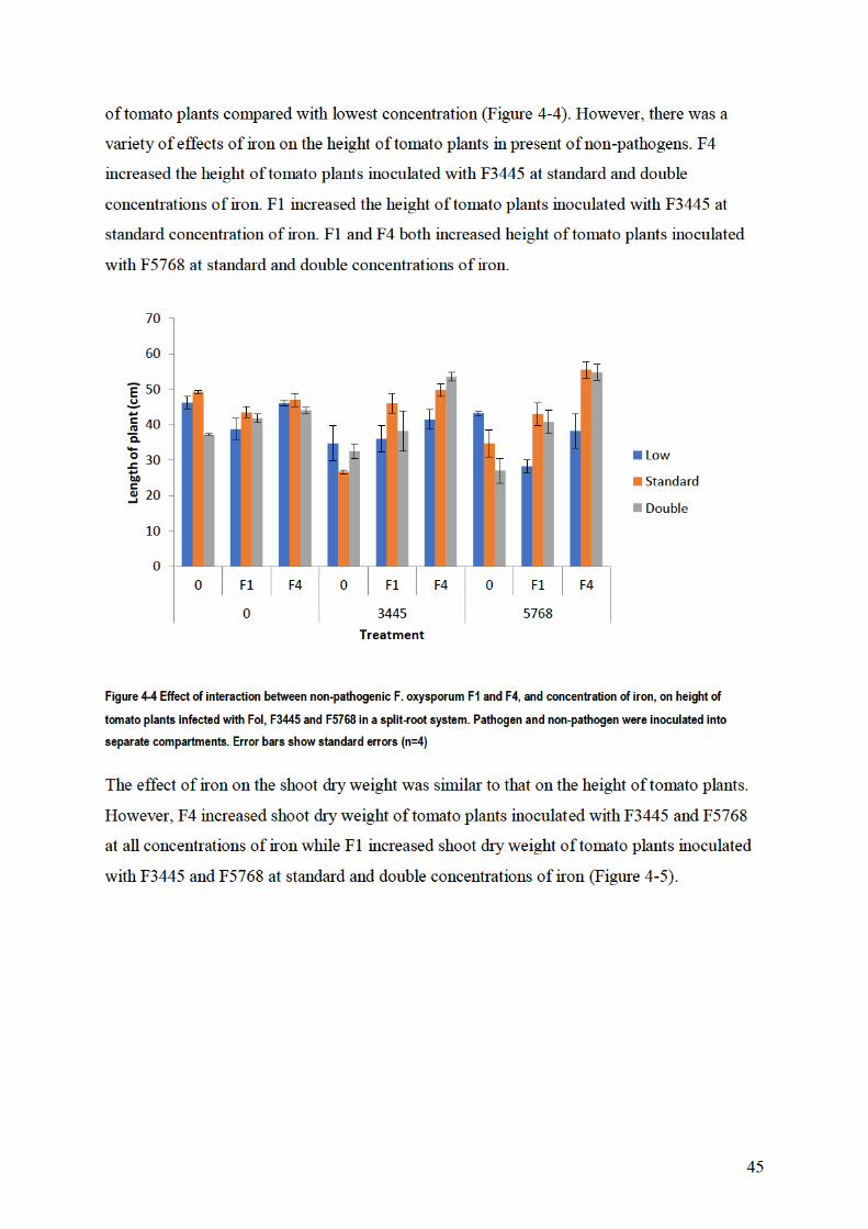


47
4.3.3. Effect of iron on the interaction between pathogen and
non-pathogen F4 together
There was a significant effect of iron on the interaction between non-pathogen F4 and
pathogen F3445 when they were inoculated into the same nutrient solution. F4 increased the
height of tomato plants inoculated with F3445 at all concentrations of iron. However, the
proportional increase in height due to F4 was greatest at the lowest level of iron compared
with highest level of iron (Figure 4-7).
Figure 4-7 Effect of nonpathogenic F. oxysporum F4 with different levels of iron on Fusarium wilt of tomato (height) caused by
Fol (F3445). Error bars show standard errors. 0 = Low Fe, 1 = Standard Fe, 2 = High Fe
There was a variety of effects of iron on the shoot dry weight of tomato plants inoculated
with F3445 in presence of non-pathogen. F4 increased the shoot dry weight of tomato plants
inoculated with F3445 at all concentrations of iron. However, the proportional increase in
shoot dry weight due to F4 was greatest at the lowest level of iron compared with highest
level of iron (Figure 4-8).
0
5
10
15
20
25
30
35
40
45
50
0 F4 0 F4 0 F4 0 F4 0 F4 0 F4
0 3445 0 3445 0 3445
0 1 2
Len
gth
of
pla
nt
(cm
)
Treatment
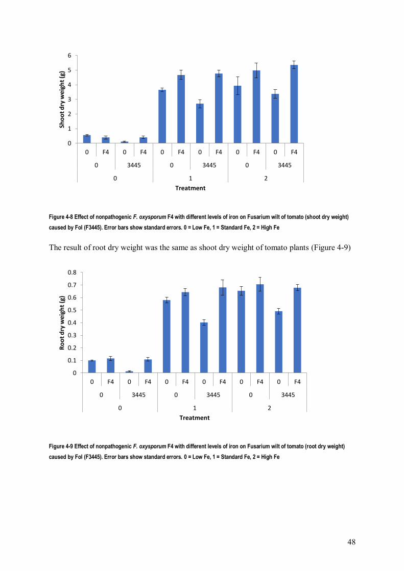
48
Figure 4-8 Effect of nonpathogenic F. oxysporum F4 with different levels of iron on Fusarium wilt of tomato (shoot dry weight)
caused by Fol (F3445). Error bars show standard errors. 0 = Low Fe, 1 = Standard Fe, 2 = High Fe
The result of root dry weight was the same as shoot dry weight of tomato plants (Figure 4-9)
Figure 4-9 Effect of nonpathogenic F. oxysporum F4 with different levels of iron on Fusarium wilt of tomato (root dry weight)
caused by Fol (F3445). Error bars show standard errors. 0 = Low Fe, 1 = Standard Fe, 2 = High Fe
0
1
2
3
4
5
6
0 F4 0 F4 0 F4 0 F4 0 F4 0 F4
0 3445 0 3445 0 3445
0 1 2
Sho
ot
dry
wei
ght
(g)
Treatment
0
0.1
0.2
0.3
0.4
0.5
0.6
0.7
0.8
0 F4 0 F4 0 F4 0 F4 0 F4 0 F4
0 3445 0 3445 0 3445
0 1 2
Ro
ot
dry
wei
ght
(g)
Treatment

49
4.5. Discussion
In this study, two biocontrol isolates were used to evaluate the ability to decrease disease
severity after inoculation of the strains in split root systems and non-split root systems. As
well, the role of Fe in enhancing induced resistance of tomato plants in the presence of non-
pathogens and pathogens was studied. Splitting the root systems in two parts to prevent any
direct contact between the pathogen and non-pathogen led to a significant reduction of the
disease severity by non-pathogens F1 and F4.
These results are in agreement with previous studies. The plant growth promoting
rhizobacteria (PGPR) strains 89B-27(Pseudomonas putida) and 90-166 (Serratia
marcescens) were inoculated on separate halves of roots of cucumber seedlings and planted
in separate pots. Both PGPR strains induced systemic resistance against F. oxysporum f. sp.
cucumerium by delaying disease symptom development (Liu et al., 1995). This result
strongly supports Hibar et al. (2007) that T. harzianum and T. viride were able to reduce
disease severity of Fol in split root system, when they were applied one week before
inoculation with the pathogen.
However, the efficacy of induced resistance varied according to the non-pathogen strain.
These results are consistent with those obtained by Olivain et al. ( 1995) who found that the
efficacy of the fungal biocontrol strain was different, with strain Fo47 shown to induce
resistance less efficiently than another non-pathogen F. oxysporum strain.
So, in the current work, non-pathogens F1 and F4 protected tomato plants from Fusarium
wilt, although there was no direct contact with the pathogen. This result indicated that
protection did not result from antagonism or competition between F1, F4, and pathogens and
shows that non-pathogen F1 and F4 induced resistance to Fusarium wilt in tomato. This could
consequently improve the health of the plant or make chemical or physical changes in the
plant that could reduce disease incidence. This result supported Tamietti et al. (1993), who
found that tomato plants grown in a soil made suppressive by the addition of non-pathogen
Fusarium strains showed physiological changes in the stems and leaves which are generally
related to systemic induced resistance. Also, non-pathogens could enhance activity of
chitinase which is confirmed by (Grenier and Asselin, 1990; Irving and Kuc, 1990; Tamietti
et al., 1993; Tuzun et al., 1989) or activity of β-1,3-glucanase or β-1,3-glucosidase (Tamietti
et al., 1993).

50
The induction of systemic resistance at high and standard iron availability by F1 and F4
agrees with observations that strains WCS374 and WCS417 of P. fluorescences and their
pseudobactin-minus mutant induced systemic resistance against Fusarium wilt of radish at
high concentration of iron (Leeman et al., 1996). Also, this result indicated that F1 and F4
reduced the toxicity of iron, by possibly consuming it. This result strongly supports Johnson
(2008), who found that siderophores are required for protection against iron-induced toxicity
in some fungal organisms. The significant effect of non-pathogen against pathogens at high
and standard concentration of iron, could be because the pathogen did not grow well or did
not have a big effect on the plant at low concentration of iron, so the pathogenicity was less
compared with high concentration of iron. This is supported by Lemanceau et al. (1993), who
found that increasing the concentration of Fe enhanced the mycelial growth of Fusarium
oxysporum f. sp. dianthi WCS816, and pathogen WCS816 was more dependent on iron
availability for growth than non-pathogen Fo47b10.
The higher overall antagonism at low iron availability by F4 agreed with observations that all
the isolates of Pseudomonas spp. induced systemic resistance against Fusarium wilt of
chickpea when iron availability in the nutrient solution was low (Saikia et al., 2005). Also,
the competition between non-pathogens and pathogen for iron led to induced suppressiveness
to Fusarium wilt pathogens. This result agrees with the finding by Scher and Baker (1982)
that the organization of Fe availability in the infection court, through Fe competition, can
induce suppressiveness to Fusarium wilt pathogens. Also, the competition between non-
pathogens and pathogen leads to decreased availability of iron in the nutrient solution for
pathogens, so that could cause decrease in spore germination of the pathogen. This
suggestion is strongly supported by Simeoni et al. (1987) who found that when availability of
iron dropped, Fe competition between the two organisms was enhanced and germination of
F.o. f.sp. cucumerinum was significantly decreased. So non-pathogens F1 and F4 stimulated
tomato plants to induced resistance against Fusarium wilt caused by pathogens F3445 and
F5768, although, there was no direct contact between them. Also at standard and high
concentrations of iron, non-pathogens F1 and F4 reduced disease severity of Fusarium wilt
caused by pathogen F3445 compared with low concentrations.

51
Chapter 5. Effect of root exudate on antagonism
5.1. Introduction
Many studies have investigated the role of plant root exudates in the spore germination,
growth and antagonism action of fungi. According to Marschner (1995) root exudates are
divided into two kinds of compounds. The first kind is low-molecular weight compounds
(amino acids, organic acids, sugar, phenolics, and other secondary metabolites). The second
kind is high-molecular weight exudates (polysaccharides and proteins). Organic compounds
released by the plant roots activate the microbial activity in the rhizosphere (Bacilio-Jim´enez
et al., 2003). Root exudates and their multiple components (e.g. sugars, organic acids, amino
acids, and phenolic compounds) play an important role in plant–microbe interactions in the
rhizosphere, especially in the initial phase of these interactions (Bais et al., 2006; Bertin et al.,
2003; Nelson, 1991). Plant root exudates can supply carbon compounds to stimulate specific
microbial populations in the rhizosphere as well as enhance biocontrol efficiency (Morgan et
al., 2005). Whipps (2001b) reported that plant pathogens can develop when plant root
exudate is applied, resulting in disease. In addition, root exudate had positive influences on
the hyphal diameter, length and degree of branching of Thielaviopsis basicola (Hood and
Shew, 1997).
According to Steinkellner et al. (2008), root exudate of tomato plants plays an important role
to increase the microconidial germination rate of biocontrol strain Fo 47 challenged with
tomato wilt pathogen Fol 007. Steinkellner et al. (2005) reported that root exudates at
different plant ages also varied in their effect on the spore germination of the tomato
pathogen F. oxysporum. When root exudate of tomato plants was applied, the germination of
soil-borne fungus Fol was inhibited (Scheffknecht et al., 2006). Also, in the vicinity of
tomato roots where exudates would occur, there was a big diversity in mycelial development
between strains of F. oxysporum (Steinberg et al., 1999).
Tomato root exudates supply the great energy source in the root zone for microbial activity
such as organic acid and sugar (Kravchenko et al., 2003). It was demonstrated previously that
citric acid was the biggest organic acid in root exudates. However, malic and succinic acid
were very important, but the levels of both of them were very dependent on the plant age
(Kamilova et al., 2006). As well as, in the exudate of tomato root, the major sugar

52
compounds were glucose, fructose and xylose (Kamilova et al., 2006). So, the main
objectives of this chapter were to assess the effect of root exudate and components such as
sugar, organic acid, and amino acid of tomato plants on the spore germination, growth and
antagonism action of fungi in laboratory and glasshouse experiments.
5.2. Materials and Methods
5.2.1. Root exudate production
Root exudates from tomato plants were produced following the method described by Shang
et al. (2000). Seeds of tomato cultivar Grosse Lisse were rinsed for 3 min with 1% sodium
hypochlorite for surface disinfestation. The seeds were washed three times with sterilized
distilled water. Then 10 tomato seeds were placed in plastic tubes (150 ml), containing 20 mL
of sterile Hoagland nutrient solution. The tubes were kept in a growth chamber at 25 C with
16 h light and 8 h dark. The nutrient solution containing root exudates was collected after 21
days. For checking microbial contamination, one millilitre was taken from the root exudate
and cultured on PDA medium and incubated at 25 C for 5 days. The solution was stored
frozen until used in experiments.
For the first series of experiments, four liquid media were used. These were root exudate in
Hoagland nutrient solution, Hoagland nutrient solution, Hoagland nutrient solution plus
sucrose at concentration 1.667 g/l or water (Tahat et al., 2010). The concentration of sucrose
was chosen because it was typical of the concentration of soluble C found in the rhizosphere
of plants (Cheng et al., 1993)
5.2.2. Effect of root exudate on the germination of fungal spores
The germination assay was achieved in sterile 96 well microplates. Aliquots of 200 μl of the
four liquid media were mixed with 1 μl of approximately 106 conidia/ml spore suspension of
F1, F2, F3, F4, F5, F6, F7, and F5769 separately and incubated at 25°C in the dark. Relative
microconidia germination and growth was determined after 48 h by measuring absorbance in
a microplate reader at 600 nm.

53
5.2.3. Effect of root exudate on antagonism in vitro
All seven antagonistic F. oxysporum isolates were used to test the effect of root exudates on
antagonism. 20 ml of the four liquid media were placed in Petri dishes. Molten water agar
was added to each Petri dish and mixed to give a final concentration of 20 g/l agar. Both the
antagonist and the pathogen F5769 were simultaneously inoculated at the opposite ends 5 cm
apart from each other with mycelial plugs of 5 mm diam. from the margin of 6 days old
cultures. In controls the pathogen was alone plated on one side of the plate at the periphery.
Inoculated plates were incubated at 25 °C for 7 to 10 days. The experiment was replicated
three times for each treatment. Results were expressed as means of radial fungal growth and
% inhibition of fungal growth of the pathogen (Ramezani, 2010; Subhani et al., 2013;
Whipps, 1987).
5.2.4. Effect of root exudate on fungal growth
Four liquid medium were used as above in 150 ml plastic tubes. Each tube had 20 ml filter-
sterilized liquid medium and 1 ml of approximately 106 conidia/ml spore suspension for non-
pathogens F1, F2, F3, F4, F5, F6, F7 and pathogen F5769 separately. After inoculation, tubes
were incubated for 6 days at 25 °C in a rotary shaker at 120 rpm. There were three replicated
for each treatment. Mycelial growth of each of the test isolates was harvested in Miracloth,
oven dried at 60 °C and weighed.
5.2.5. Effect of organic acid and sugar on antagonism in vitro
The effect of organic acids (malic, succinic and citric acid) and sugars (lactose, fructose,
glucose and xylose) on the antagonism between non-pathogens (F4, F1) and pathogen F5769,
was studied in dual culture on Czapek Dox agar medium. The organic acids and sugars were
used in place of the sucrose in Czapek Dox agar medium with five concentrations (5, 10, 20,
25, 30 g/l). The dual culture plates and controls were inoculated as described previously.
Inoculated Petri dishes were incubated at 25 °C for 7 to 10 days. There were three replicated
for each treatment. Results were expressed as means of radial of fungal growth and %
inhibition of fungal growth of the pathogen.
The effects of low concentrations of organic acids and sugars on antagonism between non-
pathogen F4 and pathogen F5769 was studied in a modification of the above experiment. The
organic acids and sugars were used in place of the sucrose in Czapek Dox agar medium with

54
two low concentrations (0.5 and 2 g/l). The dual culture plates and controls were inoculated
and incubated as described previously.
5.2.6. Effect of organic acids and sugars on antibiotic
production of non-pathogen
Non-pathogens F1 and F4 were grown in liquid Czapek Dox medium containing sugars
(lactose, fructose, xylose and glucose) or organic acids (citric, succinic and malic acid) at
concentrations of 5, 10, 20, 25, 30 g/l in place of sucrose. Five 5-mm discs of 6 day old non-
pathogen culture was transferred to each of 500 ml flasks contain 250 ml of medium and
incubated at 25 C on a rotary shaker at 120 rpm for 21 days. The mycelial biomasses were
filtered through Miracloth and filter paper (Whatman grade 41) and then culture filtrate was
filtered through a 0.45 µm membrane. 5 ml of culture filtrate was placed in the sterilized
Petri-dishes (9 cm3) and then 15 ml ¼ PDA was added and mixed carefully. Five mm disc
from 6 days old culture of pathogen was used for inoculation. The inoculated Petri dishes
were incubated at 25 °C. Mycelial growth of the pathogen was measured on each Petri dish.
The control was pathogen growth in Petri dishes containing ¼ PDA plus culture filtrate from
the pathogen. There were three replicated for each treatment. Radial growth of the pathogen
was measured and compared to control growth (Chen et al., 2012; Schalchli et al., 2012;
Whipps, 1987).
5.2.7. Effect of root exudate on antagonism in planta
Tomato seeds of the cultivar Grosse Lisse were planted in plug trays (plug size 3.4 by 3.4 by
5 cm, 6 plugs/tray) containing non sterile soilless potting mix. After 2 weeks tomato plants
were transplanted into 17 cm diameter pots containing non sterile soilless potting mix. Fungi
(F1, F4 and F5768) were grown in potato dextrose broth for 7 days on a rotary shaker at 120
rpm at 25 °C, blended, and propagule counts determined on a haemocytometer. After one
week conidial suspensions of non-pathogenic (F1 and F4) F. oxysporum (200 ml of
approximately 106 conidia/ml) were added to each pot containing 2 kg of potting mix and
then after two days the conidial suspensions of pathogenic F5768 (200 ml of approximately
106 conidia/ml) was added to each pot. Sugar (lactose and fructose) and organic acid (malic
and succinic) solutions were prepared. The concentrations of lactose, fructose, malic and
succinic were 0.171, 0.180, 0.134 and 0.118 g/l respectively. This was equivalent to 1 mM of
each compound except for the disaccharide lactose, which was 0.5 mM. 300ml from each

55
solution was added to the pots weekly. Each treatment consisted of four replicate pots of 1
plant /pot. Disease was observed by showing different growth between treatments and control
(pathogen only). After 6 weeks, height, shoot and root dry weights were determined. Shoot
height was taken from the base of the stem to apex. The shoots and roots were dried in an
oven at 70 °C until constant weight, then weighed individually (Larkin and Fravel, 2002;
Scher and Baker, 1982)
5.3. Results
5.3.1. Influence of root exudate on spore germination
There were significant effects of fungus and of medium on spore germination, but the
interaction between these was not significant. F5 and F6 had the highest absorbance, while F4
and F7 had the lowest (Figure 5-1). Germination was higher in Hoaglands solution and
Hoaglands plus exudate than in water, but did not differ between these two media (Figure 5-
2). Addition of sucrose to Hoaglands solution increased germination.
Figure 5-1 Spore germination of pathogen and non-pathogen Fusarium spp. Data are mean absorbance of germling
suspensions in four media. Error bars show standard errors (n = 12).
0
0.05
0.1
0.15
0.2
0.25
0.3
F1 F2 F3 F4 F5 F6 F7 Pathogen
Ab
sorb
ance
Isolate

56
Figure 5-2 Effect of root exudate, Hoagland nutrient solution, Hoagland nutrient solution plus sugar and water on germination
of spores of Fusarium spp. Error bars show standard errors (n = 12).
5.3.2. Effect of root exudates on antagonism in vitro
There was a significant effect of all treatments on the percentage growth inhibition for the
pathogen (Table 5-1). Root exudates increased the inhibition of growth of the pathogen for all
antagonists, while the Hoagland nutrient solution plus sugar decreased the inhibition of
growth of the pathogen relative to Hoagland solution by itself. Treatments F4 and F1 with
root exudates showed higher inhibition of growth of the pathogen compared with other fungi.
Table 5-1 Percentage inhibition of radial growth of pathogen F5769 in dual culture with antagonist on Hoagland solution plus
agar. The least significant difference for comparing data within a row is 3.44.
Fungi Root exudate plus
Hoagland Nutrient solution
Hoagland Nutrient
solution
Sugar plus Hoagland
Nutrient solution
F1 37.16 26.67 18.85
F2 33.63 26.67 17.91
F3 32.75 27.5 18.85
F4 43.37 25.82 16.98
F5 33.63 29.17 18.85
F6 31.85 27.5 21.68
F7 32.04 23.32 15.08
5.3.3. Effect of root exudates on fungal growth
Root exudates increased the weight of fungi compared to Hoagland nutrient solution while
the highest weights were in Hoagland solution with sucrose (Table 5-2).
0
0.05
0.1
0.15
0.2
0.25
0.3
Exudate H plus sugar Hoaglands Water
Ab
sorb
ance
Medium Growth

57
Table 5-2 Effect of three liquid medium (root exudate plus Hoagland nutrient solation, Hoagland nutrient solation and Hoagland
nutrient solation plus sugar on the dry weight (mg) of Fusarium spp. The least significant difference for comparing data within
a row is 2.04
Fungi Root exudate plus Hoagland
Nutrient solution
Hoagland Nutrient
solution
Sugar plus Hoagland
Nutrient solution
F1 12.33 8 18.33
F2 12 6.67 24.67
F3 10.67 7 17.67
F4 8 7.33 23
F5 15 13.67 23.33
F6 16 13 20.67
F7 16.33 12.33 23.67
5769 14.67 10 21.67
5.3.4. Effect of organic acid on antagonism in vitro
The inhibition of growth of pathogen F5769 by F1 decreased with increasing concentration of
malic acid from 33.5% at the lowest concentration to 16.2% at the highest concentration
However, the inhibition of growth of the pathogen by F4 increased with increasing
concentration of malic acid with F4 (Figure 5-3). Also, F4 had a larger effect on the mycelial
growth of pathogen F5769 compared to F1, in the presence of malic acid in dual culture
Figure 5-3 Inhibition of growth of Fol isolate F5679 by antagonists F1 and F4 on Czapek-Dox agar with a range of
concentrations of malic acid as sole organic source. Error bar shows standard error for comparing any two data points (n=3).
Succinic acid had a significant effect on the inhibitory action of F1 and F4 against pathogen
5769. The inhibition of growth of the pathogen by F1 and F4 decreased with increasing
0
10
20
30
40
50
0 5 10 15 20 25 30
Gro
wth
inh
ibit
ion
(%)
Concentration of malic acid (g/L)
F1
F4


60
Figure 5-8 Inhibition of growth of Fol isolate F5679 by antagonists F1 and F4 on Czapek-Dox agar with a range of
concentrations of glucose as sole carbon source. Error bar shows standard error for comparing any two data points (n=3).
Xylose had a significant effect on the inhibitory action of F1 and F4 against pathogen F5769.
As the concentration of xylose increased, the inhibition of growth of pathogen by F4
increased. Inhibition by F1 decreased slightly to a minimum at 20 g/l, as the concentration of
xylose increased (Figure 5-9). F4 had a larger effect on the mycelial growth of pathogen 5769
compared to F1, in the presence of xylose in dual culture.
Figure 5-9 Inhibition of growth of Fol isolate F5679 by antagonists F1 and F4 on Czapek-Dox agar with a range of
concentrations of xylose as sole carbon source. Error bar shows standard error for comparing any two data points (n=3).
In low concentrations of sugars (0.5 and 2.0g/L), there was no effect on antagonism between
antagonist and pathogen
5.3.6. Effect of organic acids and sugars on antibiotic
production of non-pathogen
Growth of the pathogen was inhibited by filtrate of F4 from all concentrations of fructose
except 20 g/l (Figure 5-10). Inhibition was greatest at the highest and lowest concentrations.
0
10
20
30
40
50
60
0 5 10 15 20 25 30
Gro
wth
inh
ibit
ion
(%)
Concentration of glucose (g/L)
F1
F4
0
10
20
30
40
50
0 5 10 15 20 25 30
Gro
wth
inh
ibit
ion
(%)
Concentration of xylose (g/L)
F1
F4

61
Growth of the pathogen was promoted by filtrate of F1 from the lowest concentration of
fructose, and showed only slight inhibition or promotion at other concentrations.
Figure 5-10 Inhibition of growth of Fol isolate F5679 by filtrate of F1 and F4 on ¼ PDA with a range of concentrations of frutose
as sole carbon source. Standard error is too small to show (n=3).
Concentration of lactose had a significant effect on action of filtrate of F1 and F4. Inhibition
of growth of pathogen by filtrate of F4 was greatest at 20 and 25 g/l, whereas inhibition of
growth of pathogen by filtrate of F1 was greatest at 20 g/l (Figure 5-11). As concentration of
lactose decreased or increased from 20 g/l, the inhibition of growth of pathogen was
decreased. Filtrate of F4 was slightly more inhibitory than filtrate of F1 on lactose.
Figure 5-11 Inhibition of growth of Fol isolate F5679 by filtrate of F1 and F4 on ¼ PDA with a range of concentrations of lactose
as sole carbon source. Standard error is too small to show (n=3).
Growth of the pathogen was promoted by filtrate of F4 from the lowest concentration of
glucose 5 and 10 g/l (Figure 5-12). However, the inhibition of growth of pathogen by filtrate
of F4 was greatest at 20 g/l of glucose. Filtrate of F1 promoted growth of pathogen from all
concentration of glucose except 30 g/l. Filtrate of F4 was slightly more inhibitory than filtrate
of F1 on glucose.
-10
-5
0
5
10
15
5 10 20 25 30
Gro
wth
inh
ibit
ion
(%)
Concentration of fructose (g/l)
F4
F1
-2
0
2
4
6
8
10
5 10 20 25 30
Gro
wth
inh
ibit
ion
(%)
Concentration of lactose (g/l)
F4
F1
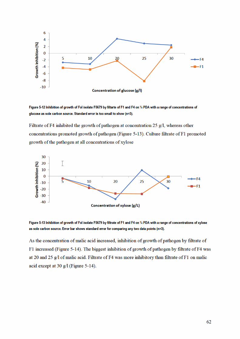
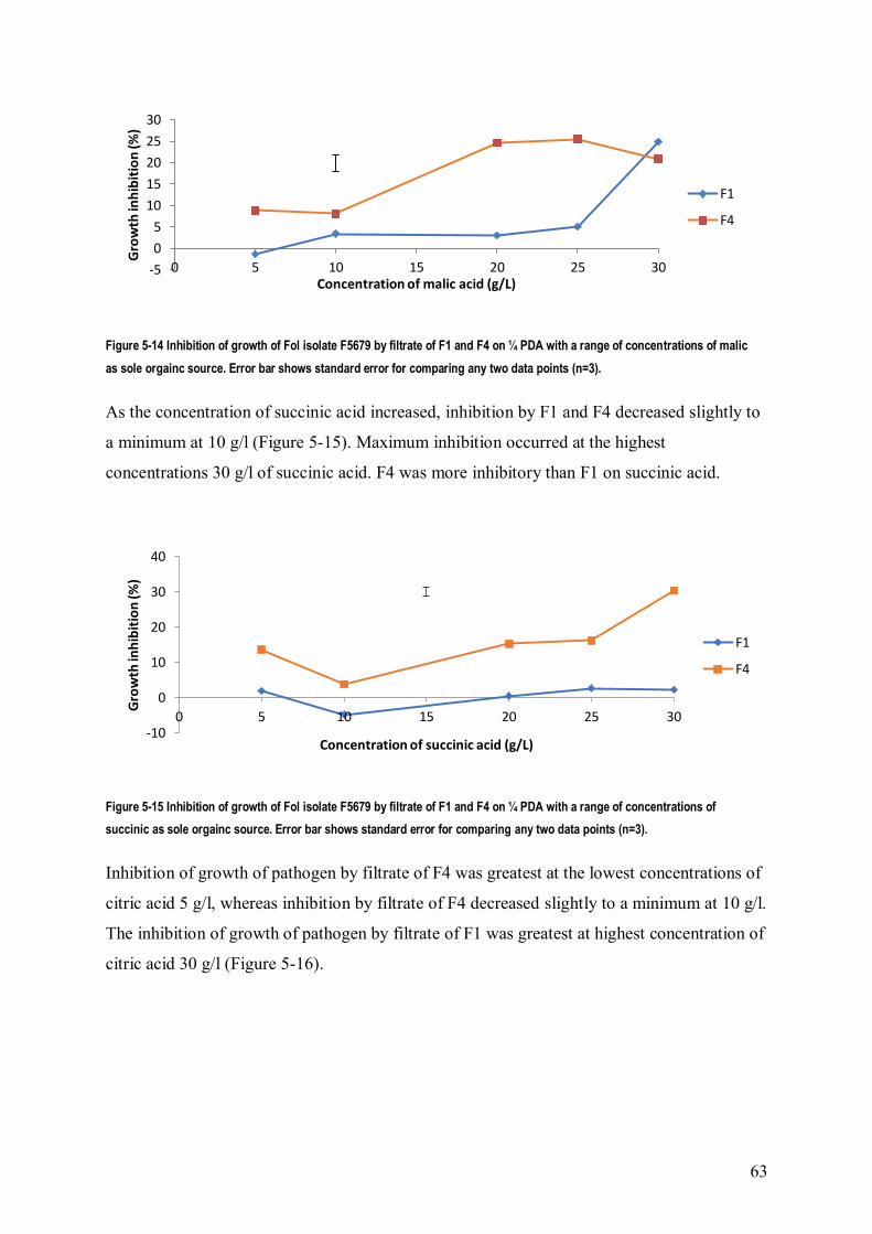
63
Figure 5-14 Inhibition of growth of Fol isolate F5679 by filtrate of F1 and F4 on ¼ PDA with a range of concentrations of malic
as sole orgainc source. Error bar shows standard error for comparing any two data points (n=3).
As the concentration of succinic acid increased, inhibition by F1 and F4 decreased slightly to
a minimum at 10 g/l (Figure 5-15). Maximum inhibition occurred at the highest
concentrations 30 g/l of succinic acid. F4 was more inhibitory than F1 on succinic acid.
Figure 5-15 Inhibition of growth of Fol isolate F5679 by filtrate of F1 and F4 on ¼ PDA with a range of concentrations of
succinic as sole orgainc source. Error bar shows standard error for comparing any two data points (n=3).
Inhibition of growth of pathogen by filtrate of F4 was greatest at the lowest concentrations of
citric acid 5 g/l, whereas inhibition by filtrate of F4 decreased slightly to a minimum at 10 g/l.
The inhibition of growth of pathogen by filtrate of F1 was greatest at highest concentration of
citric acid 30 g/l (Figure 5-16).
-5
0
5
10
15
20
25
30
0 5 10 15 20 25 30
Gro
wth
inh
ibit
ion
(%)
Concentration of malic acid (g/L)
F1
F4
-10
0
10
20
30
40
0 5 10 15 20 25 30
Gro
wth
inh
ibit
ion
(%)
Concentration of succinic acid (g/L)
F1
F4

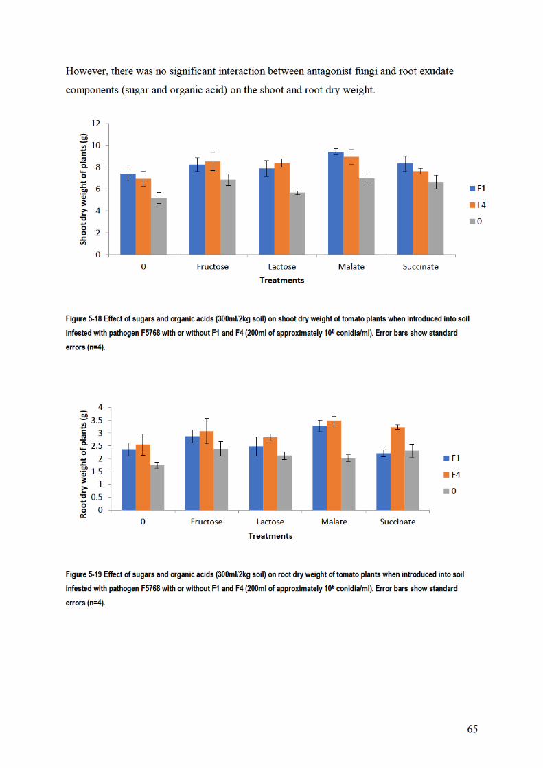

66
5.4. Discussion
The stimulation of microconidial germination of tomato pathogens and non-pathogens by
root exudate was found, confirming pervious finding showing that tomato root exudate
stimulated the microconidia of two tomato pathogens Fol 007 and FORL101587 and other F.
oxysporum strains (Steinkellner et al., 2005; Steinkellner et al., 2008). In the present study,
the germination of pathogen and non-pathogen was increased in the presence of root exudate,
compared to water only. This may be because root exudate contains sugars, amino acids and
organic acids which are known as general germination stimuli of spores (Nelson, 1991).
Root exudates played an important role in inhibition of growth of pathogen F5769 by all
antagonists. These results could be because root exudate has compounds such as sugar,
organic acid, amino acid and others that have effects on interactions between the pathogen
and non- pathogens. These results are consistent with those obtained by Bais et al. (2006);
Bertin et al. (2003); Nelson (1991), who observed that root exudates and their multiple
components (e.g. sugars, organic acids, amino acids, and phenolic compounds) play an
important role in plant–microbe interactions in the rhizosphere, especially in the initial phase
of these interactions. These compounds of root exudates also could stimulate antagonists to
produce antibiotics which inhibit growth of pathogens. Buxton (1960) found that pea root
exudate increased the ability of ten prevalent rhizosphere fungi to inhibit growth of the
pathogen F. oxysporum f.sp. pisi.
Also, root exudate enhanced growth of fungi and this result was most likely due to the sugars
in root exudate because the highest weights were found in Hoagland solution with sucrose.
This result confirmed what Kravchenko et al. (2003) found that rhizobacteria showed good
growth on a mixture of organic acids and sugars which is typical of the proportion of these
substances in tomato root exudate. Amino acids could also play an important role to increase
growth of fungi. This result agrees with those obtained by Simons et al. (1997), who observed
that amino acids in tomato root exudate assisted bacteria to colonize the root tip.
There were various effects of organic acids on the antagonistic action of F1 and F4 against
mycelial growth of pathogen 5769. This result could be due to the organic acids acting as
carbon sources which enhanced growth of fungi or could increase competition between fungi
for nutrients. This result strongly supports the theory of Kamilova et al. (2006); Kamilova et
al. (2005) that the major exudate carbon source, citric acid enhanced growth of bacteria in the

67
rhizoshere and competition for nutrient. Also, Chin-A-Woeng et al. (2000); Kamilova et al.
(2005) reported that organic acid usage by rhizobacteria is decisive for competitive tomato
root tip colonization, a process which is predominating essential for biocontrol.
Also, sugar had various effects on the antagonistic action of F1 and F4 against mycelial
growth of pathogen 5769. Sugars were used as sole carbon source. So some sugar compounds
assisted the non-pathogen to compete with the pathogen for growth. In the presence of sugar
compounds, the competition between non-pathogens and pathogen for the carbon source
increased. These results confirmed those of Kravchenko et al. (2003), who found sugar plays
an important role in antifungal activity of rhizobacteria in rhizosphere of plant.
So, organic acids and sugar separately had some influence on the antagonism between non-
pathogen F1, F4 and pathogen. These results could be due to sugar or organic acid when used
separately not being enough to enhance antagonist of non-pathogen against pathogen. These
results are consistent with those obtained by Lugtenberg et al. (1999). They observed that P.
fluorescens strain WCS365 and OE28 are very good users of sugars but poor colonizers of
tomato roots, therefore the strain must use another major carbon source for colonization such
as organic acids.
There was a different effect of sugar and organic acid on the antibiotic production of non-
pathogen F1 and F4 which it was using against the pathogen. Some of the sugar (fructose,
lactose and glucose) and organic (malic, succinic and citric acids) compounds stimulated the
non-pathogens to produce antibiotic against the pathogen. These results are consistent with
those obtained by Kravchenko et al. (2003). They believed that the antifungal activity of
rhizobacteria introduced into the plant rhizosphere depends on the sugar and organic acid
composition of the root exudates of these tomato plants. Also this result is confirmed by
Duchesne et al. (1988) who observed that Pinus resinosa root exudate from seedlings
inoculated with P. involutus led to increase in fungitoxic activity against Fusarium root. This
result was due to plant factors that control the genetic regulation of biosynthetic pathways in
P. involutus leading to antibiotic biosynthesis.
Plant root exudate can supply carbon compounds to activate specific microbial populations in
the rhizosphere and improved biocontrol efficiency (Morgan et al., 2005). However, plant
root exudate could also cause the development of plant pathogens, resulting in disease
(Whipps, 2001b). In the present study, adding sugar or organic acids to soil did not have an

68
effect on the interaction between non-pathogen and pathogen. This result could be due to
sugar or organic acid separately improving spore germination for both non-pathogens and
pathogen. This result strongly supports the finding of Steinkellner et al. (2005) that the root
exudate stimulate microcondia germination of Fol. As well, sugars are recognized to be
stimulatory to germination of Fusarium spp. (Nelson, 1991). Therefore the non-pathogen was
less suppressive towards the pathogen in pot trials. So most of the components of root
exudates such as sugar, organic and amino acids showed the ability to the improved
antagonist of non-pathogens against the pathogen in vitro. However, there was a little effect
of these components on the antagonist in pots trial

69
Chapter 6. Effect of nutrients on antagonism
6.1. Introduction
Previous studies have referred to the role of nitrogen and iron in biological control of soil
borne plant pathogens by competition for nutrients (Benson and Baker, 1970; Cook and
Schroth, 1965). The pathogen reacted to a wide range of nutritional sources with large
variation in its utilization
Nitrogen is considered an important component required for protein synthesis and other vital
functions (Khilare and Ahmed 2011). The availability of nitrogen, but not carbon, restricted
the activities of antagonists of Fusarium pseudograminearum in straw (Singh Lakhesar et al.,
2010). In addition, the form of nitrogen can impact the rate of displacement and mortality of
the pathogen in host residues (Singh Lakhesar et al., 2010). In Petri dishes, displacement of
Fusarium pseudograminearum from straw by Fusarium equiseti and Fusarium nygamai was
higher when urea and nitrate were used as nitrogen sources than when using ammonium as a
source of nitrogen. The population of Fusarium graminearum on buried wheat straw was
increased with ammonium nitrate, while nitrolime (calcium cyanamide) decreased the
populations (Yi et al., 2002). Also the conidial production of F. graminearum was
significantly decreased in the field by spraying urea onto pieces of maize stems (Khonga and
Sutton, 1991). The antagonistic activity of T. harzianum was developed against the mycelial
growth and the viability of sclerotia of S. rolfii in present of three nitrogen sources urea,
ammonium sulfate and potassium nitrate (Khattabi et al., 2004). When nitrate was used as
nitrogen source, the suppression of S. rolfsii was greater in soil by T. harzianum compared
with urea and ammonium (Khattabi et al., 2004). Fusarium species used both nitrate and
ammonium as a source of N, while Trichoderma species favorably used ammonium as a
source of N (Celar, 2003).
Jabnoun-Khiareddine et al. (2016) found that potassium salts decreased tomato wilt severity
incited by Fol and V. dahliae, and the potassium salts also enhanced growth of the tomato
plant. Organic and inorganic salts had various effects on the suppression of microbial strains
and the inhibition of growth of fungi in amended PDA medium was dependent on the
concentration used (Fagundes et al., 2013; Olivier et al., 1998). The inhibition of growth of
Botrytis cinerea was induced by adding calcium to the medium (Volpin and Elad, 1991).

70
Calcium decreased fungal infection of Botrytis cinerea and Penicillium expansum through
direct inhibition of growth and germination (Wisniewski et al., 1995). The growth of
Leucostoma persoonii was reduced 85% by using calcium propionate, 76% by using calcium
hydroxide and 73% by using calcium silicate (Biggs and El-Kooli, 1994). Golden Delicious
apples treated with calcium chloride solutions of 2% or 4% alone reduced decay by 40% and
60% respectively (Conway et al., 1994).
Previous studies suggested that the controlling of Fe availability in the infection court,
causing Fe competition, can induce suppressiveness to Fusarium wilt pathogens (Scher and
Baker, 1982). Selected isolates of P. fluorescens (Pf4-92 and PfRsC5) and P. aeruginosa
(PaRsG18 and PaRsG27) produced more salicylic acid at low iron (10mM EDDHA) than
high iron availability (10 mM Fe EDDHA), and all isolates produced more pseudobactin at
low iron than high iron availability except PaRsG27 (Saikia et al., 2005).
The aim of this study was to determine the effect of nutrients on the antagonistic action of
non-pathogenic F. oxysporum against the pathogen, as well as the effect of nutrients on the
antibiotic production of non-pathogen on the growth of the pathogen.
6.2. Materials and Methods
6.2.1. Effect of nutrients on antagonism in dual culture
To study the effect of different nutritional elements on the antagonist action of F1 and F4
against the mycelial growth of pathogen F5769, all nutrients were tested on Czapek Dox agar
medium. The recipe for the medium was varied to give different levels of the nutrients. For
nitrate levels, the quantity of NaNO3 in the medium was varied (0.4, 1.2, 2, 2.8, 3.6 g/l). For
ammonium, NH4Cl was substituted for the NaNO3 and five concentrations of NH4Cl were
used (0.24, 0.72, 1.24, 1.76, 2.24g/l). These gave the same range of concentrations of N in the
medium as for NaNO3. For calcium, Ca (NO3)2 was substituted for some of the NaNO3 and
five concentrations were used (0, 0.96, 1.92, 2.89, 3.85g/l). The total concentration of N was
kept constant. For potassium, the quantity of KCl was varied (0.32, 0.4, 0.5, 0.56, 0.64 g/l).
Mycelial plugs of 5 mm diam from the margin of 6 day old cultures of pathogen and non-
pathogens were used. Both the antagonist and the pathogen were simultaneously inoculated
at the opposite ends 5 cm apart from each other in the Petri dishes. In controls the pathogen
was plated alone on one side of the plate at the periphery. There were 3 replicate Petri plates

71
for each concentration. Inoculated plates were incubated at 25 °C for 7 to 10 days. Results
were expressed as means of radial fungal growth and percent inhibition of growth of the
pathogen. Percent inhibition was calculated as described in chapter 3. This experiment was
repeated for both NaNO3 and KCl, using the highest concentration (0.56, 0.64 g/l) and lowest
concentration (0.4 and 0.32 g/l) respectively (Khilare and Ahmed 2011; Ramezani, 2010;
Subhani et al., 2013; Whipps, 1987).
To study the effect of interactions between N and K on the antagonist action of F1 and F4
against the mycelial growth of the pathogen, both of them were tested on Czapek Dox agar
medium. The quantity of NaNO3 and KCl in the medium was used as source of N and of K
respectively. High level of N and low level of K (3.6 and 0.32 g/l) were used together and
lowest level of N with highest level of K (0.4 and 0.64 g/l) were used together. Plates were
inoculated, incubated, and inhibition calculated as above.
6.2.2. Effect of nutrients on antibiotic production
To study the effect of different nutritional component on the antibiotic action of F1 and F4
against the mycelial growth of pathogen F5679, all fungi were grown on modified Czapek
Dox liquid medium. Concentrations of NaNO3, NH4Cl, CaNO3 and KCl were varied in
Czapek Dox liquid medium as described above. Mycelial plugs of 5 mm diam from the
margins of 6 day old cultures of F1, F4 and F5769 were transferred to each of 500 ml flasks
contain 250 ml of medium and incubated at 25oC on a rotary shaker at 120 rpm for 21 days.
The mycelial biomasses were filtered through Miracloth and filter paper (Whitman grade 41)
and then the culture filtrate was filtered through a 0.45 µm membrane. 5 ml of culture filtrate
from each concentration was added to sterile Petri dishes (9 cm) and then 15 ml ¼ PDA was
added and mixed carefully. A 5 mm disc from a 6 day old culture of the pathogen was used
for inoculation. The inoculated Petri dishes were incubated at 25oC. Each treatment had three
replicates. Radial growth of the pathogen was measured. Growth on media amended with
culture filtrate of the pathogen was used as a control. The inhibition due to antibiotic effect
was calculated by dividing the difference in growth between the non-pathogen extract and the
pathogen extract by the growth on pathogen extract (Chen et al., 2012; Khilare and Ahmed
2011; Schalchli et al., 2012; Whipps, 1987).
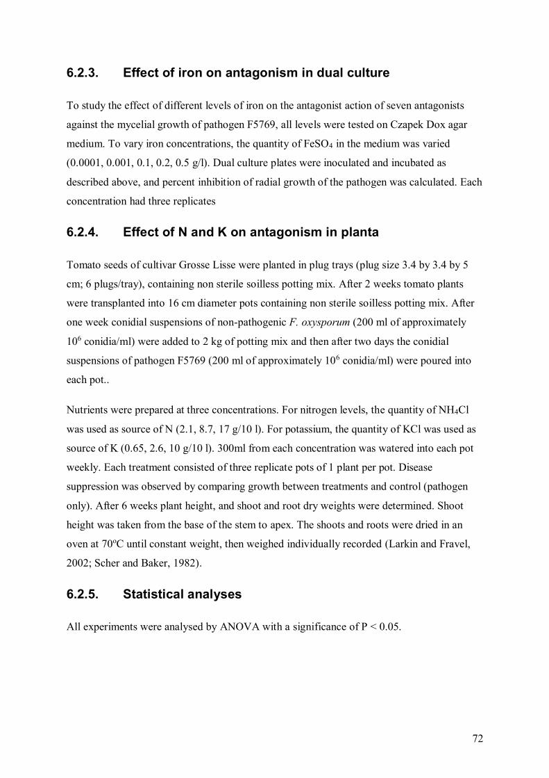
72
6.2.3. Effect of iron on antagonism in dual culture
To study the effect of different levels of iron on the antagonist action of seven antagonists
against the mycelial growth of pathogen F5769, all levels were tested on Czapek Dox agar
medium. To vary iron concentrations, the quantity of FeSO4 in the medium was varied
(0.0001, 0.001, 0.1, 0.2, 0.5 g/l). Dual culture plates were inoculated and incubated as
described above, and percent inhibition of radial growth of the pathogen was calculated. Each
concentration had three replicates
6.2.4. Effect of N and K on antagonism in planta
Tomato seeds of cultivar Grosse Lisse were planted in plug trays (plug size 3.4 by 3.4 by 5
cm; 6 plugs/tray), containing non sterile soilless potting mix. After 2 weeks tomato plants
were transplanted into 16 cm diameter pots containing non sterile soilless potting mix. After
one week conidial suspensions of non-pathogenic F. oxysporum (200 ml of approximately
106 conidia/ml) were added to 2 kg of potting mix and then after two days the conidial
suspensions of pathogen F5769 (200 ml of approximately 106 conidia/ml) were poured into
each pot..
Nutrients were prepared at three concentrations. For nitrogen levels, the quantity of NH4Cl
was used as source of N (2.1, 8.7, 17 g/10 l). For potassium, the quantity of KCl was used as
source of K (0.65, 2.6, 10 g/10 l). 300ml from each concentration was watered into each pot
weekly. Each treatment consisted of three replicate pots of 1 plant per pot. Disease
suppression was observed by comparing growth between treatments and control (pathogen
only). After 6 weeks plant height, and shoot and root dry weights were determined. Shoot
height was taken from the base of the stem to apex. The shoots and roots were dried in an
oven at 70oC until constant weight, then weighed individually recorded (Larkin and Fravel,
2002; Scher and Baker, 1982).
6.2.5. Statistical analyses
All experiments were analysed by ANOVA with a significance of P < 0.05.

73
6.3. Results
6.3.1. Effect of nutrients on antagonism in dual culture
In dual cultures of F1 or F4 with F5769 on the Czapek Dox agar medium containing different
concentrations of NaNO3 as source of N, F4 showed significantly stronger inhibition than F1
on mycelial growth of F5769 (Figure 6-1). The antagonistic action of F4 and F1 against
pathogen F5769 increased significantly with decreasing concentration of N. As well, the
effect of changes in N concentration on the inhibition was significantly stronger for F1 than it
was for F4.
In the repeat experiment, the result was the same as the first experiment. There were
significant differences between F1 and F4 in inhibition of mycelial growth of 5769 in the
presence of NaNO3 as source of N (Figure 6-1). F4 showed stronger inhibition than F1 on
mycelial growth of 5769. In addition, the level of inhibition of growth of 5769 increased as N
concentration decreased.
0
20
40
60
0 0.5 1 1.5 2 2.5 3 3.5 4
Gro
wth
inh
ibit
ion
(%)
Concentration of N (g/L)
a
F1
F4


75
In the repeat experiment, the result was similar to the first experiment. There were significant
differences between F1 and F4 on the inhibition of mycelial growth of 5769 (Figure 6-3). F4
showed stronger inhibition than F1 on mycelial growth of 5769. Moreover, the level of
inhibition of growth of 5769 increased as K concentration decreased.
Figure 6-3 Inhibition of growth of Fol isolate F5679 by antagonists F1 and F4 on Czapek-Dox agar with a range of
concentrations of KCl in two experiments (a and b). Error bar shows standard error for comparing any two data points (n=3).
On Czapek Dox agar medium containing different concentrations of Ca, the inhibitory effect
of F4 on the mycelial growth of the pathogen F5769 was significantly greater than F1. Also,
the level of inhibition of growth of F5769 increased as Ca concentration decreased (Figure 6-
4). The effect of Ca on the inhibition was stronger for F4 than it was for F1
0
20
40
60
0.3 0.35 0.4 0.45 0.5 0.55 0.6 0.65 0.7
Gro
wth
inh
ibit
ion
(%)
Concentration of K (K/L)
a
F1
F4
0
20
40
60
0.2 0.3 0.4 0.5 0.6 0.7
Gro
wth
inh
ibit
ion
(%)
Concentration of K (g/L)
b
F1
F4


77
F5769. F3 and F5 showed stronger inhibition than the other non-pathogens on mycelial
growth of F5769, while F4 was recorded as having least effect on the inhibition of growth of
pathogen 5769 (Figure 6-6). In addition, the level of inhibition of growth of F5769 increased
as iron concentration decreased (Figure 6-7). The interaction between non-pathogen isolate
and iron concentration was not significant.
Figure 6-6 Inhibition of growth of Fol isolate F5679 by antagonists on Czapek-Dox agar with a range of concentrations of
FeSO4. There was no significant interaction between isolate and concentration of FeSO4. Error bars show standard errors
(n=3).
Figure 6-7 Inhibition of growth of Fol isolate F5679 on Czapek-Dox agar with a range of concentrations of FeSO4. Error bars
show standard errors (n=3).
6.3.3. Effect of nutrients on antibiotic production
In general, adding culture filtrates of non-pathogens produced on different levels of NaNO3 to
¼ PDA medium showed that F4 had significantly stronger inhibition than F1 on mycelial
growth of F5769 (Figure 6-8). The level of inhibition of growth of F5769 increased as N
0
10
20
30
40
50
F1 F2 F3 F4 F5 F6 F7
Gro
wth
inh
ibit
ion
(%)
Fungi
0
5
10
15
20
25
30
35
40
45
50
0.0001 0.001 0.01 0.02 0.05
Gro
wth
inh
ibit
ion
(%
)
Concentration of Fe (g/L)

78
concentration increased and the effect of N on the inhibition was stronger for F4 than it was
for F1.
Figure 6-8 Inhibition of growth of Fol isolate F5679 by filtrate of F1 and F4 on ¼ PDA with a range of concentrations of NaNO3
as sole nitrogen source. Error bar shows standard error for comparing any two data points (n=3).
Culture filtrate of F4 from media containing NH4Cl as source of N showed stronger inhibition
than F1 on mycelial growth of F5769. For both non-pathogens, inhibition was greatest at the
high and low concentrations of NH4Cl and decreased at intermediate levels (Figure 6-9).
Figure 6-9 Inhibition of growth of Fol isolate F5679 by filtrate of F1 and F4 on ¼ PDA with a range of concentrations of NH4Cl as
sole nitrogen source. Error bar shows standard error for comparing any two data points(n=3).
In general, adding filtrate of non-pathogen produced from growth on different levels of KCl
to ¼ PDA medium showed significantly stronger inhibition by F4 than F1 on mycelial
growth of F5769 (Figure 6-10). The level of inhibition growth of F5769 increased as KCl
concentration increased and the effect of KCl on the inhibition was stronger for F4 than it
was for F1.
-20
0
20
40
60
0 0.5 1 1.5 2 2.5 3 3.5 4
Gro
wth
inh
ibit
ion
(%)
Concentration of N (g/L)
F1
F4
-10
-5
0
5
10
15
0 0.5 1 1.5 2 2.5
Gro
wth
inh
ibit
ion
(%)
Concentration of N (g/L)
F1
F4

79
Figure 6-10 Inhibition of growth of Fol isolate F5679 by filtrate of F1 and F4 on ¼ PDA with a range of concentrations of KCl.
Error bar shows standard error for comparing any two data points(n=3).
Adding the filtrate produced from growth of non-pathogens on different levels of Ca to ¼
PDA showed stronger inhibition by F4 than F1 on mycelial growth of F5769 (Figure 6-11).
The inhibition of growth of pathogen F5769 by F4 was increased as Ca decreased, while the
level of inhibition of growth of F5769 by F1 increased as Ca concentration increased. The
effect of Ca on the inhibition was stronger for F4 than it was for F1.
Figure 6-11 Inhibition of growth of Fol isolate F5679 by filtrate of F1 and F4 on ¼ PDA with a range of concentrations of CaNO3.
Error bar shows standard error for comparing any two data points (n=3).
Growth of pathogen F5769 on culture medium containing filtrate of F1 and F4 grown on
different concentrations of iron was inhibited more by F4 than by F1 (Figure 6-12). The level
of inhibition of growth of F5769 increased as iron concentration decreased. The effect of iron
on the inhibition was stronger for F4 than it was for F1.
-20
0
20
40
60
80
0 0.1 0.2 0.3 0.4 0.5 0.6 0.7Gro
wth
inh
ibit
ion
(%)
Concentration of K (g/L)
F1
F4
-20
-10
0
10
20
30
40
50
0 0.5 1 1.5 2 2.5 3 3.5 4 4.5
Gro
wth
inh
ibit
ion
(%)
Concentration of Ca (g/L)
F1
F4

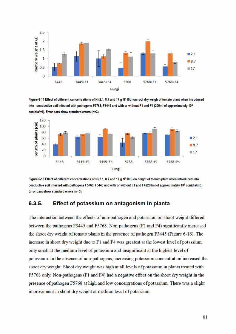


84
of F5769. This result agreed with El-Katatny et al. (2000) who found that anti-fungal enzyme
production by T. harzianum was significantly affected by the nitrogen source combined into
the medium. The level of inhibition growth of F5769 by culture filtrate increased as N
concentration increased. These results could be because N stimulated the non-pathogens to
produce secondary metabolites and enzymes that could have an effect on the growth of
pathogen. This is supported by Donzelli and Harman (2001); El-Katatny et al. (2000) who
found that nitrogen sources such as ammonium sulfate, urea and potassium nitrate, affected
the specific enzyme production by Trichoderma.
When NH4Cl was used as source of N the inhibition of growth of pathogen F5769 in dual
culture, and antibiotic production, was greatest at the high and low concentrations of NH4Cl
for both non-pathogens. This could be due to metabolite production of the non-pathogens
being enhanced at high concentrations of NH4Cl as sources of N. This is supported by
Donzelli and Harman (2001) who found that the activities of N-acetylhexosaminidase and
endochitinase were noticeably higher at 48 and 72h in a biocontrol strain of Trichoderma, in
the presence of high ammonium and low glucose levels.
The mechanisms of antagonism by non-pathogens are complex and diversified such as
competition for nutrients and induced resistance. This indicates that the effect of nitrogen
form on antagonism is more complex than just an effect on growth. So, there were direct and
indirect effects of nitrogen sources on the mechanism of activity of non-pathogens.
The dual culture trial of pathogen F5769 and non-pathogen F1 and F4 on the Czapek dox
agar medium containing potassium chloride result showed that K had a significant effect on
antagonism. The inhibition of growth of the pathogen by the non-pathogens was highest at
low level of K. Low concentrations of K created competition between non-pathogen and
pathogen for the nutrients, therefore the non-pathogens could grow faster than the pathogen
to get nutrient. Elad and Chet (1987) and Karimi and Zamani (2013) found that when fungi
grow faster, the level of chitosan production is higher which have an effect on the growth of
pathogen. This result was confirmed by Safaei et al. (2016) who found that potassium can
enhance chitosan production by fungi.
High concentrations of potassium chloride increased the production of antibiotics by F4. This
was due to the effect of concentration of K on the secondary metabolite production by the
non-pathogen. This could be due higher growth rate of F4 at higher concentrations of K.

85
High levels of calcium had a negative effect on antagonism by the non-pathogens against the
pathogen. This could be due to the growth of both non-pathogen and pathogen being reduced
by using high concentration of calcium. This result agreed with Zielinska et al. (2000) who
found that Fusarium culmorum growth on the medium containing calcium led to inhibition of
growth and development of this pathogen.
Calcium had different effects on the secondary metabolite production by non-pathogesn F1
and F4. The inhibition growth of pathogen F5769 by culture filtrate of F4 was increased at
low concentration of Ca. This could be because the Ca may reduce fungal growth of non-
pathogen at high concentration of Ca. This is supported by Wisniewski et al. (1995) who
found that the processes of growth and spore germination of Botrytis were affected by using
Ca They also found that isolate 247 of Candida oleophila did not inhibit the pathogen when
CaCl2 was used. However, the inhibition of growth of pathogen F5769 by culture filtrate of
F1 was increased at high concentration of Ca. This result could be because F1 may not be
affected by toxic levels of Ca while the pathogen was affected. This result agreed with
Wisniewski et al. (1995) who found that the pathogens Botrytis cinerea and Penicillium
expansum were inhibited by increasing concentration of calcium, whereas the growth of the
yeast isolate 182 of Candida oleophila was not affected.
Dual cultures of non-pathogens and F5769 on Czapeck-Dox agar medium containing five
levels of Iron, showed the level of inhibition of growth of F5769 increased as iron
concentration decreased. This result could be because at high level of iron there was enough
Fe available for both fungal pathogen and non-pathogen, whereas with low level of iron there
was not enough available iron for both fungi, so that could create competition between the
pathogen and non-pathogen which led to reduce growth of the pathogen. This result is in
agreement with Simeoni et al. (1987) who found that when Fe concentration dropped to
between 10-22 and 10-27 M, Fe competition of Pseudomonas putida was enhanced and
germination of Fusarium oxysporum f.sp. cucumerinum was significantly decreased.
Iron had different effects on the secondary metabolite production of F1 and F4. Culture
filtrate of F4 showed stronger inhibition than F1 on mycelial growth of F5769. This result
could be because F1 was more sensitive to the iron level than F4. Concentration of iron had a
big influence on the growth of F1 compared with F4, so the biomass of F4 was bigger than
F1 which led to greater production of secondary metabolites that had influence on the growth
of pathogen. Also the inhibition of growth of the pathogen at low concentration of iron was

86
greater compared with high concentration of iron. This could due to a high concentration of
iron causing stress to the non-pathogen F1 and F4. So the growth of both non-pathogen were
less compared with low concentration. Therefore the amount of secondary metabolites which
affected the growth of the pathogen was highest at low concentration compared with high
concentration of iron. This result agrees with that of Leeman et al. (1996) who found that at
low iron availability in vitro, P. fluorescens WCS374 and WCS417 produced 47 and 8 mg of
salicylic acid per millilitre, respectively, whereas the SA production by the both strains was
decreased with increasing iron availability.
In glasshouse experiments, the growth of shoot, root and height of tomato plant at high
concentration of nitrogen, was greater than at low concentration of nitrogen. This is because
nitrogen plays an important role in growth of tomato plants. The effect of the non-pathogens
in antagonism of the disease was greatest at low levels of nitrogen fertilization. The inhibition
of virulence of pathogen at low concentration of nitrogen could be due to first, nitrogen had
negative aeffect on the growth of pathogen at low concentration. This result agrees with
Sharma et al. (2013) who found that low levels of nitrogen in soil beneath Azadirachta indica
inhibited growth of F. oxysporum. Second, at low concentration of N there was competition
between non-pathogen and pathogen for the nutrient. So the non-pathogen could establish on
the root surface and prevent the pathogen from infecting the plant. This agrees with Elad and
Chet (1987) who found that the limitation in the general population of bacteria along the
roots caused by the biocontrol agents is probably due to competition on the nutrient. Also, the
form of nitrogen which is added to soil could have a different effect on the growth of fungi.
So, adding ammonium to the soil as a source of nitrogen could increase growth of the
pathogen. This agrees with Lakhesar et al. (2010) who found that adding ammonium to the
medium increased the radial growth of Fusarium pseudograminearum whereas, radial
growth of F. pseudograminearum was decreased by adding nitrate. Also F1 and F4 had less
effect at high concentration of N is because the plant is more tolerant to Fol when it has more
N. This results agreed with Sarhan et al. (1982) found that disease incidence of Fol decreased
with high levels of N.
In glasshouse experiments, at low concentration of K, F1 and F4 increased shoot dry weight
and that could be because potassium had a negative influence on the growth of non-pathogen
at high concentration of K. So the role of F1 and F4 to improve health of tomato plant found
in previous experiments become less. This result agreed with Jabnoun-Khiareddine et al.

87
(2016) who found that potassium salts had different effects on the inhibition of fungal growth
and reduced disease severity of tomato plant by Fol. Also the result showed increasing
potassium concentration to soil improved growth of plant in absence of non-pathogen. This
could due to the role of potassium to develop growth of tomato plant. Jabnoun-Khiareddine et
al. (2016) found that potassium salts enhanced growth of height, and root and shoot fresh
weight of tomato plant.
So, nitrogen and potassium at high and low concentration have effects on growth of the fungi
in dual culture, however nitrogen and potassium did not show effects on growth and
antagonism between non-pathogens F1 and F4 and pathogen F5769 in glasshouse.

88
Chapter 7. General Discussion
The aim of the work described in this thesis was to improve the biocontrol of Fusarium wilt
in tomato by understanding the environmental and host factors which effect the antagonism
of the pathogen Fusarium oxysporum f. sp. lycopersici by non-pathogenic strains of F.
oxysporum. This was done by first obtaining biocontrol agent strains of F. oxysporum that
could be used against Fusarium wilt caused by Fol. The ability of non-pathogens to induce
systemic resistance against Fusarium wilt was tested and role of iron in induced resistance
was examined. Also, the effects of mineral nutrients and root exudate on antagonism between
non-pathogens and pathogen were examined.
The pathogenicity of Fol was tested in two different ways, root dipping and adding conidial
suspension to soil. It was found that in root dipping, the pathogens were more aggressive
against tomato plants compared with adding conidial suspension to the soil. This could be
because in root dipping, spores were more likely to contact the root surface and reach the
elongation zone and develop disease than by adding conidial suspension (Olivain et al.,
2006). So the method of adding conidial suspension to the soil was used in the next
experiments because this method gave less severe disease, allowing the detection of the
effects of antagonists. Also adding spores to soil is more similar to the way that infection
occurs in the field. In addition, measuring the effect of disease on growth had advantages
over scoring incidence or severity of wilting. The symptoms were more consistent between
replicates and gave a quantitative measurement that was better suited to statistical analysis.
In dual culture and pot trials it was found that all non-pathogens reduced growth and disease
severity of pathogen Fol. However, the highest growth inhibition of the pathogen was
recorded by isolate F4. Isolates F1 and F4 protected all the plants from infection by the
pathogen when rated by showing no brown colour in the stem. This may be because F1 and
F4 have the greatest competition for nutrients or antibiotic production compared with the
other non-pathogens (Fravel et al., 2003; Kumar and Garampalli, 2013; Postma and
Luttikholt, 1996). Also, F1 and F4 improved the growth of tomato plants infected with Fol.
Therefore, F1 and F4 were chosen for further experiments.
Isolates F1 and F4 reduced the growth of tomato plants when applied in the absence of Fol
inoculation. A previous study found that nonpathogenic biocontrol F. oxysporum strain
70T01 had the ability to colonize the cortex of tomato roots and the densities in roots were

89
highest at inoculation sites compared with away from the inoculation sites (Bao and
Lazarovits, 2001). However, there appear to be no reports on the effects of root colonization
by biocontrol strains on tomato growth. The antagonists are called ‘non-pathogens’ because
they do not cause wilting or vascular browning, but they could still be parasites that reduce
the plant growth. So non-pathogens should not generally be used in the absence of disease.
F1 and F4 reduced the percentage of germination of tomato seeds, so the methods of using
non-pathogen F1 and F4 for the next experiments were to apply them after germination of the
seeds. It could be that antibiotic production by F1 and F4 had an effect on the seed
germination (Ibraheem et al., 1987; Owolade et al., 2005). So non-pathogens should be added
to seedlings before transplanting to the field.
Non-pathogens protected plants from Fusarium wilt even when there was no direct contact
with the pathogen. So F1 and F4 showed ability to induce resistance in tomato plants and this
could be due to either improved health of the plant or inducing specific defence mechanisms
in the plant (Tamietti et al., 1993). This is consistent with earlier work on non-pathogenic F.
oxysporum on tomatoes. Fuchs et al. (1997) found that non-pathogen Fo47 increased the
chitinase, β-1, 3-glucanase, and β-1, 4-glucosidase activity in tomato plants. Also a previous
study reported that adding non-pathogen Fusarium strains to soil led to stimulation of
systemic resistance by changes in the stems and leaves of tomato plants (Tamietti et al.,
1993).
Root exudates played an important role to stimulate germination of pathogen and non-
pathogen spores. This could be because components of root exudates such as sugars, organic
and amino acids stimulated spore germination (Nelson, 1991). However, in the presence of
root exudates, all antagonists inhibited the growth of pathogen F5769 in vitro. This result is
consistent with that of Buxton (1960) who found that root exudate of pea increased the ability
of rhizosphere fungi to inhibit growth of the pathogen F. oxysporum f.sp. pisi.
When organic acids and sugar were used as source of carbon, both of them enhanced
antifungal activity of the non-pathogens against the pathogen. Also, sugar and organic acids
stimulated the non-pathogens to produce antibiotics which acted against the pathogen
(Kravchenko et al., 2003).
In dual culture, the antagonism between non-pathogens F1 and F4 and pathogen F5769 was
affected by different sources of N. With increasing concentration of NaNO3 as source of N,

90
the inhibition of growth of pathogen F5769 by non-pathogens F1 and F4 was decreased.
However, at high and low concentration of NH4Cl as source of N, the inhibition of growth of
pathogen F5769 by non-pathogens F1 and F4 was increased. This difference in result could
be due to the form of N which played an important role to stimulate or limit the mechanisms
of non-pathogens which have an effect on their antagonistic activity (Khattabi et al., 2004).
At high concentration of NaNO3 as sole N source, the antibiotic production of F1 and F4 was
increased, whereas in dual culture the antagonism was decreased. This could be because
competition for nutrient between non-pathogen and pathogen at low concentration of NaNO3
as source of N (Elad and Chet, 1987) was more important than antibiotic production as a
mechanism of antagonism. With different levels of NaNO3 as source of N, the antibiotic
production of F4 gave stronger inhibition than F1 on growth of pathogen F5769. However the
levels of NH4Cl did not have different effects on antibiotic production of non-pathogens F1
and F4. This could be because F4 was more sensitive than F1 to NaNO3 as a source of N (El-
Katatny et al., 2000).
Using NH4Cl as source of N in dual culture and antibiotic production at high and low
concentration led to improved inhibition of growth of pathogen F5769 by non-pathogens F1
and F4. This could be due to the antibiotic production being improved at high levels of
NH4Cl as source of N (Donzelli and Harman, 2001).
In the glasshouse, N played an important role to develop healthy plants at high levels of
application. However, at low levels of N, the aggressiveness of the pathogen was reduced
compared with high N. This could be because at low level of N the pathogen did not grow
very well Sharma et al. (2013), or when N level was low that created competition for
nutrients between non-pathogens and pathogen. Therefore it could be that the non-pathogens
had the ability to establish on the root surface of plant which influenced the virulence of the
pathogen (Elad and Chet, 1987). This result was consistent with what was found in the dual
culture and antibiotic production experiments except with high level of N. That could be due
to tomato plants with high level of N becoming more tolerant to the pathogen Fol than at low
level of N. Sarhan et al. (1982) found that at high level of N the disease severity of Fusarium
wilt was decreased. So the non-pathogens will be less effective when soil N levels are high.
Meanwhile the non-pathogens will be more effective in soil that is poor in N.

91
In dual culture, the antagonistic activity of non-pathogens F1 and F4 against pathogen F5769
was enhanced at low level of K. This could be because the low level of K made the non-
pathogens grow faster than the pathogen to get nutrient (Elad and Chet, 1987; Karimi and
Zamani, 2013).
The antibiotic production of F4 was increased at high level of K. This could be because the
growth of non-pathogen was greater in media with high level of K which could affect the
secondary metabolite production.
In the glasshouse, non-pathogens improved shoot dry weight of diseased tomato plants at low
level of K more than at high levels. This could be because K at high level had a negative
effect on the growth of non-pathogens. However, at high level of K, the health of infected
tomato plants was improved even in the absence of non-pathogens. This could be due to the
role of K at high level to develop healthy plants (Jabnoun-Khiareddine et al., 2016). This
result supported what was found in dual culture, where low level of K improved the
antagonistic activity of non-pathogens F1 and F4 which may reduce the aggressiveness of the
pathogen. So the non-pathogens will be less effective in soil rich with K. Meanwhile, the
non-pathogens will have their greatest effect in soil poor in K.
The antagonistic activity of non-pathogens F1 and F4 against pathogen F5769 was negatively
affected by high levels of Ca. This could be because high level of Ca affected the growth of
non-pathogens (Zielinska et al., 2000), or reduced competition between the pathogen and
non-pathogens for Ca.
At low level of Ca, the antibiotic production of F4 inhibited growth of the pathogen F5769.
This could be due to the growth of non-pathogen being decreased at high level of Ca
(Wisniewski et al., 1995). However, at high level of Ca, the antibiotic production of F1
inhibited growth of pathogen F5769. This result could be due to toxicity of Ca to the
pathogen F5769 and F1 was not affected by high level of Ca (Wisniewski et al., 1995).
Iron also had various effects on the antibiotic production by non-pathogens F1 and F4. The
inhibition of growth of pathogen F5769 by culture filtrate of F4 was greater than culture
filtrate of F1. This could be because F1 was more influenced by iron levels than F4, so the
biomass of F4 was much bigger than biomass of F1. As a result, F4 produced a larger amount
of antibiotic production compared with F1 which had an effect on growth of the pathogen.
Also, at low level of iron, the inhibition of growth of pathogen F5769 by antibiotic

92
production of non-pathogens F1 and F4 was greater compared with high level. This could be
due to non-pathogens at high level of iron being exposed to stress which led to less growth.
So non-pathogens F1 and F4 produced a greater amount of antibiotic at low iron level
compared with high level which had influenced the growth of pathogen F5769 (Leeman et
al., 1996).
The induction of resistance in tomato by non-pathogens F1 and F4was greatest when iron was
at standard and high availability, this could be because at low iron concentration the pathogen
could not grow very well, or the pathogen had less effect on the plant at low iron
concentration. So the pathogenicity could be less (Leeman et al., 1996). On the other hand,
non-pathogens may produce antibiotics with high concentrations of iron which induced
systemic resistance of the tomato plant. This result contrasted with Saikia, et al. (2005) who
found that at low level of iron, the Pseudomonas spp. caused production of more salicylic
acid (SA) in chickpea than at high level of iron, which played an important role to induce
resistance.
When the pathogen and non-pathogen were applied with high concentrations of iron together
in solution culture, the greatest relative effect of F4 on disease occurred at low iron
concentration. This could be due to competition between the non-pathogen and pathogen for
iron availability before infection by the pathogen. This result is consistent with that of Scher
and Baker (1982) who found that reducing the iron availability in soil can induce
suppressiveness to Fusarium wilt by competition for iron in the infection site. Segarra et al.
(2010) also showed that competition for iron was a key mode of action for the antagonism of
Fusarium wilt in tomato by Trichoderma asperellum. At high levels of iron, the inhibition of
growth of pathogen F5769 by non-pathogens F1 and F4 was decreased. This could be due to
the availability of iron being enough for both pathogen and non-pathogen. However, at low
levels of iron there was not enough iron, so that could create competition between the
pathogen and non-pathogen which led to reduced growth of the pathogen (Simeoni et al.,
1987).
7.1. Suggestions to improve antagonism by non-
pathogens
Non-pathogens should be added to soil after seed germination to prevent decreased
germination. The non-pathogen should not be applied to soil in the absence of the pathogen

93
as non-pathogens had a negative effect on the growth of plants when used alone. Also, adding
non-pathogens to soil at least two days before transplanting seedlings to the field would allow
the non-pathogens to establish on the root surface of plants and stimulate induced resistance.
Seeds should be grown on sterilized soil and non-pathogen added after seed germination
before transplant to the field because root exudate played an important role in stimulating
spore germination for both non-pathogen and pathogen. So that gives more time for the non-
pathogen to establish on the root surface.
Applying non-pathogen to soil when iron level is high should be avoided, or we should add
low concentrations of iron to soil as fertilizer. Non-pathogens should not be applied to soil
when N levels are high. However, we should add non-pathogens to soil poor in N. Using low
concentration of N as fertilizer could improve the antagonistic activity of non-pathogens.
Also care should be taken about the form of N which is added to soil as fertilizer as each
form used as a source of N has a different effect on the plant and pathogen. Adding low
concentration of K as fertilizer could improve the antagonistic activity of non-pathogens.
Adding low concentration of Ca as fertilizer could improve the antagonistic activity of non-
pathogens.
7.1.1. Suggestions for future work
It would be a good idea to study why non-pathogens had a negative effect on plant growth in
the absence of the pathogen. Future work could determine if this is this because of the
reaction of the host plant toward non-pathogens or because the density of non-pathogen on
the root surface or root tissue can be high, so that non-pathogens could consume nutrients
from the roots. An analysis of the antibiotic production of non-pathogens could be done to
see if application to the plant has the ability to induce resistance of plants. That could give a
good opportunity to avoid use of non-pathogens directly which sometimes when applied
alone had an effect on the growth of plants. It is also important to examine the interaction
between plants and non-pathogenic F. oxysporum because there has not been much study
about whether the non-pathogens could become pathogenic or not in the future. It may also
be possible to select plants for resistance to the non-pathogens so that their growth is not
affected by the biocontrol agents in the absence of the pathogen.
It would be useful to find a way to apply non-pathogens with seeds without affecting seed
germination, like coating the seed with some chemical to inhibit the non-pathogen from

94
reaching the seed until germination. Or non-pathogens could be applied with some distance
from seeds, so it gives time for the seed to germinate before the fungus comes into contact
with it. This is very important as it allows the non-pathogen to establish around the seed area
to prevent the pathogen reaching the seed and infecting the plant. Also, it may be possible to
find some materials to apply with the non-pathogen that have ability to attract the non-
pathogen for a short time, so it gives time for germination of seeds. Also, more studies could
be done to find the optimum concentration of non-pathogen inoculation that has no effect on
the seed germination but still protects against Fusarium wilt.
It may be possible to apply sugar and organic acid to the field soil which has non-pathogens
and pathogen before transplanting the seedling to the field. This may stimulate germination
and interactions between pathogens and non-pathogens before sowing the plant. The non-
pathogens reduced growth of the pathogen in the presence of sugar and organic acid, so the
density of pathogen will decrease, and the possibility to infect the plant will be less.
Experiments should test whether stimulating the plant to increase production of sugar and
organic acids enhances biocontrol, because these components played an important role to
develop antifungal activities of non-pathogens
High levels of iron reduced the effect of the non- pathogens, so we should find a way that can
reduce iron concentration such as using siderophore-producing bacteria. That could help the
use of non-pathogens in soil rich in iron without affecting the antagonism of non-pathogens
against the pathogen. Also more studies should be done about the effect of iron levels on the
non-pathogens by using genetic tools to develop the ability of non-pathogens to tolerate the
high level of iron and retain the same ability to inhibit growth of the pathogen.

95
References
Alabouvette, C. (1986). Fusarium wilt suppressive soils from the châteaurenard region:
review of a 10-year study. Agronomie, 6(3), 273-284.
Alabouvette, C. and Couteaudier, Y. (1992). Biological Control of Fusarium Wilts with
Nonpathogenic Fusaria In P. In: Tjamos E.C., G.C., Cook, R.J. (Ed.), Biological
Control of Plant Diseases (pp. 415-426). Boston: Springer.
Alabouvette, C. , Olivain, C. , Migheli, Q. and Steinberg, C. (2009). Microbiological control
of soil-borne phytopathogenic fungi with special emphasis on wilt inducing Fusarium
oxysporum. New Phytologist, 184(3), 529-544.
Aldahadha, M. A. , Warwick, M. W. N. and Backhouse, D. (2012). Effects of Pythium
irregulare and root pruning on water-use efficiency of hydroponically grown wheat
under PEG-induced drought. Phytopathology, 160, 397–403. doi: 10.1111/j.1439-
0434.2012.01917.x
Altomare, C. , Norvell, W. A. , Bjorkman, T. and Harman, G. E. (1999). Solubilization of
phosphates and micronutrients by the plant-growth-promoting and biocontrol fungus
Trichoderma harzianum rifai 1295-22. Applied and Environmental Microbiology,
65(7), 2926-2933.
Alwathnani, A. H. and Perveen, K. (2012). Biological control of Fusarium wilt of tomato by
antagonist fungi and cyanobacteria. African Journal of Biotechnology, 11(5), 1100-
1105. doi: 10.5897/AJB11.3361
Alwathnani, A. H. , Perveen, K. , Tahmaz, R. and Alhaqbani, S. (2012). Evaluation of
biological control potential of locally isolated antagonist fungi against Fusarium
oxysporum under in vitro and pot conditions. African Journal of Microbiology, 6(2),
312-319. doi: 10.5897/AJMR11.1367
Amini, J. (2009). Induced resistance in tomato plants against Fusarium Wilt invoked by
nonpathogenic Fusarium, chitosan and bion. Plant Pathology Journal, 25(3), 256-262.
doi: 10.5423/ppj.2009.25.3.256
Armstrong, G. M. and Armstrong, J. K. (1981). Formae speciales and races of Fusarium
oxysporum causing wilt diseases. In T. T. A. Nelson P.E., Cook, R.J. (Ed.), Fusarium:
Diseases, Biology and Taxonomy (pp. 391–399). University Park: Pennsylvania State
University Pres.
Bacilio-Jim´enez, M. , Aguilar-Flores, S. , Ventura-Zapata, E. , P´erez- , Campos, E. ,
Bouquelet, S. and Zenteno, E. (2003). Chemical characterization of root exudates
from rice (Oryza sativa) and their effects on the chemotactic response of endophytic
bacteria. Plant and Soil, 249, 271–277
Baharvand, A. , Shahbazi, S. , Afsharmanesh, H. and Ebrahimi, M. A. (2014). Investigation
of gamma irradiation on morphological characteristics and antagonist potential of
Trichoderma viride against M. phaseolina. International Journal of Farming and
Allied Sciences, 3(11), 1157-1164.

96
Bais, P. H. , Weir, L. T. , Perry, G. L. , Gilroy, S. and Vivanco, M. V. (2006). The role of root
exudates in rhizosphere interactions with plants and other organisms. The Annual
Review of Plant Biology, 57, 233-266.
Bao, R. J. , Fravel, R. D. , O'Neill, R. N. , Lazarovits, G. and Berkum, P. v. (2002). Genetic
analysis of pathogenic and nonpathogenic Fusarium oxysporum from tomato plants.
Canadian Journal of Botany, 80(3), 271-279.
Bao, R. J. and Lazarovits, G. (2001). Differential colonization of tomato roots by
nonpathogenic and pathogenic Fusarium oxysporum strains may influence Fusarium
wilt control. Phytopathology, 91(5), 449-456.
Barakat, R. M. and Masri, M. I. (2009). Trichoderma harzianum in combination with sheep
manure amendment enhances soil suppressiveness of Fusarium wilt of tomato.
Phytopathology, 48, 385-395.
Belgrove, A. , Steinberg, C. and Viljoen, A. (2011). Evaluation of nonpathogenic Fusarium
oxysporum and Pseudomonas fluorescens for panama disease control. Plant Disease,
95(8), 951-959.
Benson, M. D. and Baker, R. (1970). Rhizosphere competition in model soil systems.
Phytopathology, 60(7), 1058-1061.
Bertin, C. , Yang, X. and Weston, A. L. (2003). The role of root exudates and allelochemicals
in the rhizosphere. Plant and Soil, 256(1), 67-83.
Biggs, R. A. and El-Kooli, M. M. (1994). Effect of calcium salt on growth, pectic enzyme
activity, and colonization of peach twigs by Leucostoma persoonii. Plant Disease, 78,
886-890.
Biles, L. C. and Martyn, D. R. (1989). Local and systemic resistance induced in watermelons
by formae speciales of Fusarium oxysporum. Phytopathology, 79, 856-860.
Bolwerk, A. , Lagopodi, A. L. , Lugtenberg, B. J. J. and Bloemberg, G. V. (2005).
Visualization of interactions between a pathogenic and a beneficial Fusarium strain
during biocontrol of tomato foot and root rot Molecular Plant Microbe Interactions,
18, 710-721.
Bost, S. (2011). Plant diseases: Tomato wilt problems. Report on Plant Diseases, from
https://ag.tennessee.edu/EPP/Extension%20Publications/Tomato%20Wilt%20Proble
ms.pdf
Brencic, A. and Winans, S. C. (2005). Detection of and response to signals involved in host-
microbe interactions by plant-associated bacteria. Microbiology and Molecular
Biology Reviews 69(1), 155-194.
Burgess, L. W. (1981). General ecology of the fusaria. In T. T. A. Nelson P.E., Cook, R.J.
(Ed.), Fusarium: Diseases, Biology, and Taxonomy (pp. 225-235). University Park:
Pennsylvania State University Pres.
Buxton, E. W. (1960). Effects of pea root exudate on the antagonism of some rhizosphere
micro-organisms towards Fusarium oxysporum f.sp. pisi Microbiology, 22, 678-689.

97
Cal, D. (1999). Effects of timing and method of application of Penicillium oxalicum on
efficacy and duration of control of Fusarium wilt of tomato. Plant Pathology, 48(2),
260-266.
Celar, F. (2003). Competition for ammonium and nitrate forms of nitrogen between some
phytopathogenic and antagonistic soil fungi. Biological Control, 28, 19-24.
Chen, L. H. , Cui, Y. Q. , Yang, X. M. , Zhao, D. K. and Shen, Q. R. (2012). An antifungal
compound from Trichoderma harzianum SQR-T037 effectively controls Fusarium
wilt of cucumber in continuously cropped soil. Australasian Plant Pathology, 41(3),
239-245. doi: 10.1007/s13313-012-0119-5
Cheng, W. , Coleman, C. D. , Carroll, C. R. and Hoffman, C. A. (1993). In situ measurement
of root respiration and soluble C concentrations in the rhizosphere. Soil Biology &
Biochemistry, 25(9), 1189-1196.
Cherif, M. and Benhamou, N. (1990). Cytochemical aspects of chitin breakdown during the
parasitic action of a Trichoderma sp. on Fusarium oxysporum f. sp. radicis-
lycopersici. Phytopathology, 80(12), 1406-1414.
Chin-A-Woeng, T. F. C. , Bloemberg, G. V. , Mulders, I. H. M. , Dekkers, L. C. and
Lugtenberg, B. J. J. (2000). Root colonization by phenazine-1-carboxamide producing
bacterium Pseudomonas chlororaphis PCL1391 is essential for biocontrol of tomato
foot and root rot. Molecular Plant- Microbe Interactions, 13, 1340-1345.
Clayton, E. E. (1923). The relations of temperature to Fusarium wilt of tomato. American
Journal of Botany, 10, 71-88.
Compant, S. , Duffy, B. , Nowak, J. , Clément, C. and Barka, E. (2005). Use of plant growth-
promoting bacteria for biocontrol of plant diseases: principles, mechanisms of action,
and future prospects. Appled and Environmental Microbiology, 71(9), 4951-4959.
Conrath, U. , Thulke, O. , Katz, V. , Schwindling, S. and Kohler, A. (2001). Priming as a
mechanism in induced systemic resistance of plants. European Journal of Plant
Pathology, 107, 113–119.
Conway, S. W. , Sams, E. C. , Wang, Y. C. and Abbott, A. J. (1994). Additive effects of
postharvest calcium and heat treatment on reducing decay and maintaining quality in
apples. American Society for Horticultural Science, 119(1), 49-53.
Cook, R. J. and Schroth, M. N. (1965). Carbon and nitrogen compounds and germination of
chlamydospores of Fusarium solani f. sp. phaseoli. Phytopathology, 55, 254-256.
Cotxarrera, L. , Trillas-Gay, M. I. , Steinberg, C. and Alabouvette, C. (2002). Use of sewage
sludge compost and Trichoderma asperellum isolates to suppress Fusarium wilt of
tomato. Soil Biology and Biochemistry, 34(4), 467-476.
Couteaudier, Y. and Alabouvette, C. (1990a). Quantitative comparison of Fusarium
oxysporum competitiveness in relation to carbon utilization. Microbial Ecology,
74(4), 261-267. doi: 10.1016/0378-1097(90)90678-J

98
Couteaudier, Y. and Alabouvette, C. (1990b). Survival and inoculum potential of conidia and
chlamydospores of Fusarium oxysporum f. sp. lini in soil. Canadian Journal of
Microbiology, 36(8), 551-556.
da Silva, C. J. and Bettiol, W. (2005). Potential of non-pathogenic Fusarium oxysporum
isolates for control of Fusarium wilt of tomato. Fitopatologia Brasileira, 30, 409-412.
Dakora, D. F. and Phillips, A. D. (2002). Root exudates as mediators of mineral acquisition in
low-nutrient environments. Plant and Soil, 245, 35-47.
Donzelli, B. G. G. and Harman, G. E. (2001). Interaction of ammonium, glucose, and chitin
regulates the expression of cell wall-degradation enzymes in Trichoderma atroviride
strain P1. Applied and Environmental Microbiology, 67(12), 5643–5647.
Dowling, N. D. and O'Gara, F. (1994). Metabolites of Pseudomonas involved in the
biocontrol of plant disease. Trends in Biotechnology, 12, 133-141.
Droby, S. , Chalutz, E. , Wilson, C. L. and Wisniewski, M. (1989). Characterization of the
biocontrol activity of Debaryomyces hansenii in the control of Penicillium digitatum
on grapefruit. Canadian Jornal of Microbiology 35, 794-800.
Droby, S. , Wisniewski, M. E. , Cohen, L. , Weiss, B. , Touitou, D. , Eilam, Y. and Chalutz,
E. (1997). Influence of CaCl2 on Penicillium digitatum, grapefruit peel tissue, and
biocontrol activity of Pichia guilliermondii. Phytopathology, 87, 310-315.
Dubey, C. S. , Suresh, M. and Singh, B. (2007). Evaluation of Trichoderma species against
Fusarium oxysporum f. sp. ciceris for integrated management of chickpea wilt.
Biological Control, 40, 118-127.
Duchesne, L. , Peterson, R. L. and Ellis, B. (1988). Pine root exudate stimulates the synthesis
of antifungal compounds by the ectomycorrhizal fungus Paxillus involutus. New
Phytologist, 108, 471-476.
Duffy, K. B. and Défago, G. (1997). Zinc improves biocontrol of Fusarium crown and root
rot of tomato by Pseudomonas fluorescens and represses the production of pathogen
metabolites inhibitory to bacterial antibiotic biosynthesis. Phytopathology, 87, 1250-
1257.
Duijff, B. J. , Pouhair, D. , Olivain, C. , Alabouvette, C. and Lemanceau, P. (1998).
Implication of systemic induced resistance in the suppression of Fusarium wilt of
tomato by Pseudomonas fluorescens WCS417r and by nonpathogenic Fusarium
oxysporum Fo47. Plant Pathology, 104, 903–910.
Edel-Hermann, V. , Brenot, S. , Gautheron, N. , Aim´ e, S. , Alabouvette, C. and Steinberg,
C. (2009). Ecological fitness of the biocontrol agent Fusarium oxysporum Fo47 in soil
and its impacton the soil microbial communities. FEMS Microbiology Ecology 68,
37-45.
El- Khallal, S. M. (2007). Induction and modulation of resistance in tomato plants against
Fusarium wilt disease by bioagent fungi (Arbuscular Mycorrhiza) and/or hormonal
elicitors (jasmonic acid&salicylic acid): 1- changes in growth, some metabolic
activities and endogenous hormones related to defence mechanism. Australian

99
Journal of Basic and Applied Sciences and Environmental Management, 1(4), 691-
705.
El-Katatny, M. H. and Emam, A., S. (2012). Control of postharvest tomato rot by spore
suspension and antifungal metabolites of Trichoderma harzianum. Microbiology,
Biotechnology and Food Sciences, 1, 1505-1528.
El-Katatny, M. H. , Somitsch, W. , Robra, K. H. , El-Katatny, M. S. and Gübitz, G. M.
(2000). Production of chitinase and β-1,3-glucanase by Trichoderma harzianum for
control of the phytopathogenic fungus Sclerotium rolfsii. Food Technology and
Biotechnology, 38(3), 173-180.
Elad, Y. and Chet, I. (1987). Possible role of competition for nutrients in biocontrol of
Pythium damping-off by bacteria. Phytopathology, 77, 190-195.
Elad, Y. and Henis, Y. (1982). Degradation of plant pathogenic fungi by Trichoderma
harzanum. Microbiology, 28, 719-725.
Endo, S. , Shinohara, M. , Ogawa, K. and Shibata, Y. (1975). The effect of soil moisture on
the occurrence of Fusarium wilt, Fusarium oxysorum f sp. lycopersici of tomato plant.
Bulletin of the College of Agriculture and Veterinary Medicine, 32, 79-86.
Fagundes, C. , Pérez-Gago, B. M. , Monteiro, R. A. and Palou, L. (2013). Antifungal activity
of food additives in vitro and as ingredients of hydroxypropyl methylcellulose-lipid
edible coatings against Botrytis cinerea and Alternaria alternata on cherry tomato
fruit. Food Microbiology, 166, 391-398.
Frank, Z. R. and Bakker, J. C. (1975). Soil aeration affecting resistance of tomato plants to
Fusarium oxysporum f sp. lycopresici. Phytoparasitica, 3, 70-71.
Fravel, D. R. , Olivain, C. and Alabouvette, C. (2003). Fusarium oxysporum and its
biocontrol. New Phytologist, 157(3), 493-502.
Friebe, A. , Vilich, V. , Hennig, L. , Kluge, M. and Sicker, D. (1998). Detoxification of
benzoxazolinone allelochemicals from wheat by Gaeumannomyces graminis var.
tritici, G. graminis var. graminis, G. graminis var. avenae, and Fusarium culmorum.
Applied and Environmental Microbilogy, 64, 2386-2391.
Fuchs, J.-G. , Moënne-Loccoz, Y. and Défago, G. (1997). Nonpathogenic Fusarium
oxysporum strain Fo47 induces resistance to Fusarium wilt in tomato. Plant Disease,
81(5), 492-496.
Gamliel, A. , Austerweil, M. and Kritzman, G. (2000). Non-chemical approach to soilborne
pest management–organic amendments. Crop Protection, 19(8), 847-853.
Garuba, T. , Abdulrahaman, A. A. , Olahan, G. S. , Abdulkareem, K. A. and Amadi, J. E.
(2014). Effects of fungal filtrates on seed germination and leaf anatomy of maize
seedlings (Zea mays L., Poaceae). Applied Sciences and Environmental Management,
18(4), 662-667.
Geiser, M. D. , Jime´nez-Gasco, M. M. , Kang, S. , Makalowska, I. , Veeraraghavan, N. ,
Ward, J. T. , Zhang, N. , Kuldau, A. G. and O’Donnell, K. (2004). Fusarium-ID v.

100
1.0: A DNA Sequence Database for Identifying Fusarium. Plant Pathology, 110 473–
479.
Gilreath, P. J. , Jones, P. J. and Overman, J. A. (1994). Soil-borne pest control in mulched
tomato with alternatives to methyl bromide. Proceedings of the Florida State
Horticultural Society, 107, 156-159.
Grenier, J. and Asselin, A. (1990). Some pathogenesis-related proteins are chitosamases with
lytic activity against fungal spores. Molecular Plant-Microbe Interactions, 3, 401-
407.
Hadar, Y. , Harman, G. E. and Taylor, A. G. (1984). Evaluation of Trichoderma koningii and
T. harzianum from New York soils for biological control of seed rot caused by
Pythium spp. Phytopathology, 74, 106-110.
He, C. , Hsiang, T. and Wolyn, D. J. (2001). Activation of defense responses to Fusarium
infection in Asparagus densiflorus. European Journal of Plant Pathology, 107, 473–
483.
He, Y. C. , Hsiang, T. and Wolyn, D. J. (2002). Induction of systemic disease resistance and
pathogendefence responses in Asparagus officinalis inoculated with nonpathogenic
strains of Fusarium oxysporum. Plant Pathology, 51, 225-230.
Hervás, A. , Trapero-Casas, J. L. and Jiménez-Díaz, R. M. (1995). Induced resistance against
Fusarium wilt of chickpea by nonpathogenic races of Fusarium oxysporum f. sp.
ciceris and nonpathogenic isolates of F. oxysporum. Plant Disease, 79, 1110-1116.
Hibar, K. , M., D.-R. and El Mahjoub, M. (2007). Induction of resistance in tomato plants
against Fusarium oxysporum f. sp. radicis-lycopersici by Trichoderma spp. Plant
Protection Journal, 2, 47-58.
Hoitink, H. A. J. , Inbar, Y. and Boehm, M. J. (1991). Status of compost-amended potting
mixes naturally suppressive to soilborne diseases of floricultural crops Plant Disease,
75, 869–873.
Hood, M. E. and Shew, H. D. (1997). The influence of nutrients on development, resting
hyphae and aleuriospore induction of Thielaviopsis basicola. Mycologia, 89(5), 793-
800.
Horinouchi, H. , Katsuyama, N. , Taguchi, Y. and Hyakumachi, M. (2008). Control of
fusarium crown and root rot of tomato in a soil system by combination of a plant
growth-promoting fungus, Fusarium equiseti, and biodegradable pots. Crop
Protection, 27, 859–864.
Horinouchi, H. , Muslim, A. , Suzuki, T. and Hyakumachi, M. (2007). Fusarium equiseti
GF191 as an effective biocontrol agent against Fusarium crown and root rot of tomato
in rock wool systems. Crop Protection, 26, 1514–1523.
Horinouchi, H. , Watanabe, H. , Taguchi, Y. , Muslim, A. and Hyakumachi, M. (2011).
Biological control of Fusarium wilt of tomato with Fusarium equiseti GF191 in both
rock wool and soil systems. Biocontrol, 56(6), 915-923. doi: 10.1007/s10526-011-
9369-3

101
Houssien, A. A. , Ahmed , S. M. and Ismail, A. A. (2010). Activation of tomato plant defense
response against Fusarium wilt disease using Trichoderma Harzianum and salicylic
acid under greenhouse conditions. Agriculture and Biological Sciences, 6, 328-338.
Hoynes, C. D. , Lewis, J. A. , Lumsden, R. D. and Bean, G. A. (1999). Biological control
agents in combination with fertilization or fumigation to reduce sclerotial viability of
Sclerotium rolfsii and disease of snap beans in the greenhouse. Phytopathology, 147,
175-182.
Hyakumachi, M. (1994). Plant growth-promoting fungi from turfgrass rhizosphere with
potential for disease suppression. Soil Microbiology, 44, 53-68.
Ibraheem, S. A. , Okesha, A. M. and Mhathem, K. T. (1987). Interrelationship between
protein and oil content of soybean seed with some associated fungi. Agriculture and
Water Resources Research, Plant Production 6, 53-66.
Ignjatov, M. , Milošević, D. , Nikolić, Z. , Gvozdanović-Varga, J. , Jovičić, D. and Zdjelar,
G. (2012). Fusarium oxysporum as causal agent of tomato wilt and fruit rot.
Pesticides and Phytomedicine (Belgrade), 27(1), 25–31.
Irving, H. R. and Kuc, J. (1990). Local and systemic induction of peroxidase, chitinase and
resistance in cucumber plants by K2HPO4. Physiological and Molecular Plant
Pathology, 37, 355-366.
Ishiba, C. , Tani, T. and Murata, M. (1981). Protection of cucumber against anthracnose by a
hypovirulent strain of Fusarium oxysporum f. sp. cucumerinum. Annals of the
Phytopathological Society of Japan, 47, 352-359.
Jabnoun-Khiareddine, H. , Abdallah, R. , El-Mohamedy, R. , Abdel-Kareem, F. , Gueddes-
Chahed, M. , Hajlaoui, A. and Daami-Remadi, M. (2016). Comparative efficacy of
potassium salts against soil-borne and air-borne fungi and their ability to suppress
tomato wilt and fruit rots. Microbial & Biochemical Technology, 8(2), 45-55.
Jasnic, M. S. , Vidic, B. M. , Bagi, F. F. and Dordevic, B. V. (2005). Pathogencity of
Fusarium Species in Soybean. The first scientific meeting Mycology, Mycotoxicology
and Mycoses, 109, 113-121.
Jenkins, S. F. and Averre, C. W. (1986). Problems and progress in integrated control of
southern blight of vegetables. Phytopathological Society, 70, 614-619.
Johnson, C. R. , Menge, J. A. , Schwab, S. and Ting, I. P. (1982). Interaction of photoperiod
and vesicular Arbuscular mycorrhizae on growth and metabolism of sweet orange.
New Phytologist, 90, 665-669.
Johnson, L. (2008). Iron and siderophores in fungal–host interactions. Mycological Research,
112, 170-183. doi: 10.1016/j.mycres.2007.11.012
Kamilova, F. , Kravchenko, L. , Shaposhnikov, A. , Azarova, T. , Makarova, N. and
Lugtenberg, B. (2006). Organic acids, sugars, and L-tryptophane in exudates of
vegetables growing on stonewool and their effects on activities of rhizosphere
bacteria. Molecular Plant-Microbe Interactions, 19, 250–256. doi: 10.1094/MPMI -
19-0250

102
Kamilova, F. , Validov, S. , Azarova, T. , Mulders, I. and Lugtenberg, B. (2005). Enrichment
for enhanced competitive plant root tip colonizers selects for a new class of biocontrol
bacteria. Environmental Microbiology, 7, 1809-1817. doi: 10.1111/j.1462-
2920.2005.00889.x
Karimi, K. and Zamani, A. (2013). Mucor indicus: biology and industrial application
perspectives: a review. Biotechnology Advances, 31, 466-481.
Katan, T. , Zamir, D. , Sarfatti, M. and Katan, J. (1991). Vegetative compatibility groups and
subgroups in Fusarium oxysporum f.sp. radicis-lycopersici. Phytopathology, 81, 255-
261.
Khattabi, N. , Ezzahiri, B. , Louali, L. and Oihabi, A. (2004). Effect of nitrogen fertilizers and
Trichoderma harzianum on Sclerotium rolfsii. Agronomie, 24(5), 281-288.
Khilare, V. C. and Ahmed , R. (2011). Effect of nutritional sources on the growth of
Fusarium oxysporum F. Sp. ciceri causing chickpea Wilt. Society of Science and
Nature 2(3), 524 - 528.
Khonga, B. E. and Sutton, C. J. (1991). Effects of acetic acid, propionic acid and urea on
inoculum production by Gibberella zeae in maize and wheat residues. Mycobiology
Research, 95(4), 409-412.
Kistler, H. C. , Momol, E. A. and Benny, U. (1991). Repetitive genomic sequences for
determining relatedness among strains of Fusarium oxysporum. Phytopathology,
81(33), 1-336.
Kollmorgen, J. F. (1974). Some effects of urea and Streptomyces griseus (2–A24) on
Fusarium avenaceum (Fr.) Sacc. Australian Journal of Agricultural Research, 25,
893-898.
Korobeinikova, M. A. V. (1960). The effect of nutrition, temperature, humidity, and pH on
the development of some species of the genus Fusarium. Trudy Instituta Biology
Ural’skii Filial Akademiy Nauk SSSR, 15, 71-81.
Kourtev, P. S. , Ehrenfeld, J. G. and Häggblom, M. (2002). Exotic plant species alter the
microbial community structure and function in the soil. Ecology, 83(11), 3152-3166.
Kravchenko, L. V. , Azarova, T. S. , Leonova-Erko, E. I. , Shaposhnikov, A. I. , Makarova,
N. M. and Tikhonovich, I. A. (2003). Root exudates of tomato plants and their effect
on the growth and antifungal activity of Pseudomonas strains. Microbiology, 72, 37-
41.
Kroon, B. A. M. , Scheffer, R. J. and Elgersma, D. M. (1991). Induced resistance in tomato
plants against Fusarium wilt invoked by Fusarium oxysporum f.sp. dianthi. Plant
Pathology, 97(6), 401-408.
Kuc, J. (1982). Induced immunity to plant disease. BioScience, 32, 854-860.
Kumar, A. and Garampalli, R. H. (2013). Screening of indigenous potential antagonistic
Trichoderma Species from tomato rhizospheric soil against Fusarium oxysporum f.
sp. lycoperscisi. Agriculture and Veterinary Science, 4, 42-47.

103
Lakhesar, D. P. S. , Backhouse, D. and Kristiansen, P. (2010). Nutritional constraints on
displacement of Fusarium pseudograminearum from cereal straw by antagonists.
Biological Control, 55(3), 241-247.
Larkin, R. P. and Fravel, D. R. (1998). Efficacy of various fungal and bacterial biocontrol
organisms for control of Fusarium wilt of tomato. Plant Disease, 82(9), 1022-1028.
doi: 10.1094/pdis.1998.82.9.1022
Larkin, R. P. and Fravel, D. R. (1999). Mechanisms of action and dose-response relationships
governing biological control of Fusarium wilt of tomato by nonpathogenic Fusarium
spp. Phytopathology, 89(12), 1152-1161. doi: 10.1094/phyto.1999.89.12.1152
Larkin, R. P. and Fravel, D. R. (2002). Effects of varying environmental conditions on
biological control of Fusarium wilt of tomato by nonpathogenic Fusarium spp.
Phytopathology 92, 1160_1166.
Leeman, M. , Den Ouden, F. M. , Van Pelt, J. A. , Dirkx, F. P. M. , Steijl, H. , Bakker, P. A.
H. M. and Schippers, B. (1996). Iron availability affects induction of systemic
resistance to Fusarium wilt of radish by Pseudomonas fluorescens. Phytopathology,
86, 149-155.
Leisinger, T. and Margraff, R. (1979). Secondary metabolites of the Fluorescent
Pseudomonads. Microbiological Review 43(3), 422-442.
Lemanceau, P. , Bakker, P. A. H. M. , De Kogel, W. J. , Alabouvette, C. and Schippers, B.
(1993). Antagonistic effect of nonpathogenic Fusarium oxysporum Fo47 and
Pseudobactin 358 upon pathogenic Fusarium oxysporum f. sp. dianthi. Applied and
Environmental Microbiology, 59(1), 74-82.
Liu, L. , Kloepper, J. W. and Tuzun, S. (1995). Induction of systemic resistance in cucumber
against Fusarium wilt by plant growth-promoting rhizobacteria. Phytopathology,
85(6), 695-698.
Lugtenberg, B. J. J. , Kravchenko, L. V. and Simons, M. (1999). Tomato seed and root
exudate sugars: composition, utilization by Pseudomonas Biocontrol strains and role
in rhizosphere colonization. Environmental Microbiology, 1, 439-446.
Maia, C. L. and Yano-Melo, M. A. (2001). Germination and germ tube growth of the
Arbuscular mycorrhizal fungi Gigaspora albida in different substrates. Brazilian
Journal of Microbiology 32, 281-285.
Mandeel, Q. and Baker, R. (1991). Mechanisms involved in biological control of Fusrium
wilt of cucumber with strains of nonpathogenic Fusrium oxysporum Phytopathology
81, 462-469.
Mark, G. L. , Dow, J. M. , Kiely, P. D. , Higgins, H. , Haynes, J. , Baysse, C. , Abbas, A. ,
Foley, T. , Franks, A. and Morrissey, J. (2005). Transcriptome Profiling of Bacterial
Responses to Root Exudates Identifies Genes Involved in Microbe-Plant Interactions.
Proceeding of the National Academy of Science, USA 102, 17454–17459.
Marschner, H. (1995). Marschner's mineral nutrition of higher plants (2nd ed.). New York:
Academic Press.

104
Martyniuk, S. , Stochmal, A. , Macías, F. A. , Marín, D. and Oleszek, W. (2006). Effects of
some benzoxazinoids on in vitro growth of Cephalosporium gramineum and other
fungi pathogenic to cereals and on Cephalosporium stripe of winter wheat.
Agricultural and Food Chemistry 54, 1036−1039.
Matti, D. and Sen, C. (1985). Integrated biocontrol of Sclerotium rolfsii with nitrogen
fertilizers and Trichoderma harzianum. Agricultural Science, 55, 464-468.
Mclauchlin, R. J. , Wilson, C. L. , Droby, S. , Ben-Arie, R. and Chalutz, E. (1992). Biological
control of postharvest diseases of grape, peach, and apple with the yeast Kloeckera
apiculata and Candida guilliermondii. Plant Disease, 76, 470-473.
Mohamed, H. A. A. and Haggag, W. M. (2005). Biocontrol potential of salinity tolerant
mutants of Trichoderma harzianum against Fusarium oxysporum causing tomato wilt
disease. Biotechnology, 8, 35-48.
Morgan, J. A. W. , Bending, G. D. and White, P. J. (2005). Biological costs and benefits to
plant–microbe interactions in the rhizosphere. Journal of Experimental Botany,
56(417), 1729-1739.
Mwangi, M. W. , Monda, E. O. , Okoth, S. A. and Jefwa, J. M. (2011). Inoculation of tomato
seedlings with Trichoderma harzianum and Arbuscular mycorrhizal fungi and their
effect on growth and control of wilt in tomato seedlings. Brazilian Journal of
Microbiology, 42(2), 508-513.
Nahalkova, J. , Fatehi, J. , Olivain, C. and Alabouvette, C. (2008). Tomato root colonization
by fuorescent-tagged pathogenic and protective strains of Fusarium oxysporum in
hydroponic culture differs from root colonization in soil. Federation of European
Microbiological Societies, 286, 152-157.
Naher, U. A. , Radziah, O. , Halimi, M. S. , Shamsuddin, Z. H. and Mohd Razi, I. (2009).
Influence of root exudate carbon compounds of three rice genotypes on rhizosphere
and endophytic diazotrophs. Pertanika Journal of Tropical Agricultural Science,
32(2), 209-223.
Naika, S. , Jeude, J. L. , Goffau, M. , Hilmi, M. and Dam, B. (2005). Cultivation of Tomato.
Wageningen: Agromisa Foundation and CTA
Nelson, E. B. (1991). Exudate molecules initiating fungal responses to seeds and roots. Plant
and Soil, 129, 61-73.
Norman, R. J. and Hooker, E. J. (2000). Sporulation of Phytophthora fragariae shows greater
stimulation by exudates of non-mycorrhizal than by mycorrhizal strawberry roots.
Mycobiology Research, 104(9), 1069–1073.
O’Donnell, K. , Kistler, H. C. , Cigelnik, E. and Ploetz, C. R. (1998). Multiple evolutionary
origins of the fungus causing panama disease of banana: concordant evidence from
nuclear and mitochondrial gene genealogies. Proceedings of the National Academy of
Sciences, 95(5), 2044-2049.
Olivain, C. and Alabouvette, C. (1997). Colonization of tomato root by a non-pathogenic
strain of Fusarium oxysporum. New Phytologist, 137, 14.

105
Olivain, C. , Humbert, C. , Nahalkova, J. , Fatehi, J. , L'Haridon, F. and Alabouvette, C.
(2006). Colonization of tomato root by pathogenic and nonpathogenic Fusarium
oxysporum strains inoculated together and separately into the soil. Applied and
Environmental Microbiology, 72(2), 1523-1531. doi: 10.1128/aem.72.2.1523-
1531.2006
Olivain, C. , Trouvelot, S. , Binet, M. , Cordier, C. , Pugin, A. and Alabouvette, C. (2003).
Colonization of flax roots and early physiological responses of flax cells inoculated
with pathogenic and nonpathogenic strains of Fusarium oxysporum. Applied and
Environmental Microbiology, 69, 5453-5462.
Olivier, C. , Halseth, E. D. , Mizubuti, G. S. E. and Loria, R. (1998). Postharvest application
of organic and inorganic salts for suppression of silver scurf on potato tubers. Plant
Disease, 82, 213-217.
Ordentlich, A. , Migheli, Q. and Chet, I. (1991). Biological control activity of three
Trichoderma isolates against Fusarium wilts of cotton and muskmelon and lack of
correlation with their lytic enzymes. Journal of Phytopathology, 133(3), 177-186. doi:
10.1111/j.1439-0434.1991.tb00152.x
Oritsejafor, J. J. (1986). Carbon and nitrogen nutrition in relation to growth and sporulation
of Fusarium oxysporum f.sp. elaeidis. Transactions of the British Mycological
Society, 87, 519-524.
Owolade, O. F. , Alabi, B. S. , Enikuomehin, O. A. and Atungwu, J. J. (2005). Effect of
harvest stage and drying methods on germination and seed-borne fungi of maize (Zea
mays L.) in south west nigeria. Biotechnology, 4(12), 1384-1389.
Pal, K. K. and Gardener, B. M. (2006). Biological control of plant pathogens. The Plant
Health Instructor, 1-25.
Papavizas, G. C. (1985). Trichoderma and Gliocladium: biology, ecology, and potential for
biocontrol. Phytopathology, 23, 23-54.
Pereyra, A. S. and Dill-Macky, R. (2004). Survival and inoculum production of Gibberella
zeae in wheat residue. Plant Disease, 88, 724-730.
Pocasangre, L. , Sikora, R. , Vilich, V. and Schuster, R. (2000). Survey of banana endophytic
fungi from central America and screening for biological control of the burrowing
nematode (Radopholus similis). InfoMusa, 9(1), 3-5.
Postma, J. and Luttikholt, A. J. G. (1996). Colonization of carnation stems by a
nonpathogenic isolate of Fusarium oxysporum and its effect on Fusarium oxysporum
f. sp. dianthi. Canadian Journal of Botany, 74(11), 1841-1851.
Ramamoorthy, V. , Viswanathan, R. , Raguchander, T. , Prakasam, V. and Samiyappan, R.
(2001). Induction of systemic resistance by plant growth promoting rhizobacteria in
crop plants against pests and diseases. Crop Protection, 20, 1-11.
Ramezani, H. (2010). Antagonistic effects of Trichoderma spp. against Fusarium oxysporum
f.sp. lycopersici causal agent of tomato wilt. Plant Protection Journal 2, 167-173.

106
Rudrappa, T. , Czymmek, K. J. , Paré, P. W. and Bais, H. P. (2008). Root-secreted malic acid
recruits beneficial soil bacteria. Plant Physiology 148, 1547–1556.
Safaei, Z. , Karimi, K. and Zamani, A. (2016). Impact of phosphate, potassium, yeast extract,
and trace metals on chitosan and metabolite production by Mucor indicus. Molecular
Sciences, 17, E1429. doi: 10.3390
Saikia, R. , Srivastava, K. A. , Singh, K. , Arora, D. K. and Lee, M. W. (2005). Effect of iron
availability on induction of systemic resistance to Fusarium wilt of chickpea by
Pseudomonas spp. Mycobiology, 33(1), 35-40.
Sarhan, A. R. T. , Barna, B. and Kiraly, Z. (1982). Effect of nitrogen nutrition on Fusarium
wilt of tomato plants. Annals of Applied Biology, 101(2), 245-250. doi:
10.1111/j.1744-7348.1982.tb00819.x
Sawane, A. and Sawane, M. (2014). Mycotoxigenicity of Aspergillus, Penicillium and
Fusarium spp. isolated from stored rice. International Journal of Current
Microbiology and Applied Sciences, 3(11), 116-121.
Schalchli, H. , Pardo, F. , Hormazábal, E. , Palma, R. , Guerrero, J. and Bensch, E. (2012).
Antifungal activity of wheat root exudate extracts on Gaeumannomyces graminis var.
tritici growth. Journal of Soil Science and Plant Nutrition, 12(2), 329-337.
Scheffknecht, S. , Mammerler, R. and Steinkellner, S. (2006). Root exudates of mycorrhizal
tomato plants exhibit a different effect on microconidia germination of Fusarium
oxysporum f. sp. lycopersici than root exudates from non-mycorrhizal tomato plants.
Mycorrhiza, 16, 365-370. doi: 10.1007/s00572-006-0048-7
Scher, M. F. and Baker, R. (1982). Effect of Pseudomonas putida and a synthetic iron
chelator on induction of soil suppressivenss to Fusarium wilt pathogen.
Phytopathology, 72(12), 1567-1573.
Segarra, G. , Casanova, E. , Aviles, M. and Trillas, I. (2010). Trichoderma asperellum strain
T34 controls Fusarium wilt disease in tomato plants in soilless culture through
competition for iron. Microbial Ecology, 59(1), 141-149. doi: 10.1007/s00248-009-
9545-5
Shang, H. , Grau, C. R. and Peters, R. D. (2000). Oospore germination of Aphanomyces
euteiches in root exudates and on the rhizoplanes of crop plants. Plant Disease, 84,
994-998.
Sharma, J. , Yadav, M. , Kumar, G. , Gupta, M. , Jayanand, D. and Rai, V. (2013). The study
of nitrogen concentration in soil of Azadirachta indica and its effect on growth of
Fusarium oxysporum International Journal of Current Research, 5(10), 2759-2761.
Shaw, K. R. , Shaik, M. , Mir, Z. A. , Prasad, M. S. L. , Prasad, D. R. and Senthilvel, S.
(2016). Establishing a high throughput screening method for large scale phenotyping
of castor genotypes for resistance to Fusarium wilt disease. Phytoparasitica, 44, 539–
548. doi: 10.1007/s12600-016-0535-0
Simeoni, A. L. , Lindsay, L. W. and Baker, R. (1987). Critical iron level associated with
biological control of Fusarium wilt. Phytopathology, 77, 1057-1061.

107
Simons, M. , Permentier, P. H. , de Weger, A. L. , Wijffelman, A. C. and Lugtenberg, J. B.
(1997). Amino acid synthesis is necessary for tomato root colonization by
Pseudomonas fluorescens strain WCS365. The American Phytopathological Society,
10, 102-106.
Singh Lakhesar, P. D. , Backhouse, D. and Kristiansen, P. (2010). Nutritional constraints on
displacement of Fusarium pseudograminearum from cereal straw by antagonists.
Biological Control 55 241–247.
Singh, P. U. , Sarma, K. B. and Singh , P. D. (2003). Effect of plant growth-promoting
rhizobacteria and culture filtrate of Sclerotium rolfsii on phenolic and salicylic acid
contents in chickpea (Cicer arietinum). Current Microbiology, 46, 131-140.
Sivan, A. and Chet, I. (1989). The possible role of competition between Trichoderma
harzianum and Fusarium oxysporum on rhizosphere colonization Phytopathology, 79,
198-203.
Smith, N. S. and Snyder, C., W. (1971a). Relationship of inoculum density and soil types to
severity of Fusarium wilt of sweet potato. Phytopathology, 61, 1049-1051.
Smith, S. E. , Jon, B. J. S. , Smith, F. A. and Bromley, L. J. (1986). Effects of mycorrhizal
infection on plant growth, nitrogen and phosphorus nutrition in glasshouse-grown
Allium cepa L. New Phytologist, 103, 359-373.
Smith, S. N. and Snyder, C. W. (1971b). Relationsbip of Inoculum Density and Soil Types to
Severity of Fusarium wilt of Sweet Potato. Phytopathology, 61, 1049-1051.
Spadaro, D. and Gullino, M. L. (2005). Improving the efficacy of biocontrol agents against
soilborne pathogens. Crop Protection, 24, 601-613.
Srinon, W. , Chuncheen, K. , Jirattiwarutkul, K. , Soytong, K. and Kanokmedhakul, S.
(2006). Efficacies of antagonistic fungi against Fusarium wilt disease of cucumber
and tomato and the assay of its enzyme activity. Agricultural Technology, 2(2), 191-
201.
Steinberg, C. , Whipps, J. M. , Wood, D. , Fenlon, J. and Alabouvette, C. (1999). Mycelial
development of Fusarium oxysporum in the vicinity of tomato roots. Mycological
Research, 103(06), 769-778.
Steinberg, R. A. (1950). Growth on synthetic nutrient solutions of some Fungi pathogenic to
tobacco. American Journal of Botany, 37, 711-714.
Steinkellner, S. , Mammerler, R. and Vierheilig, H. (2005). Microconidia germination of the
tomato pathogen Fusarium oxysporum in the presence of root exudates. Journal of
Plant Interactions, 1(1), 23-30.
Steinkellner, S. , Mammerler, R. and Vierheilig, H. (2008). Germination of Fusarium
oxysporum in root exudates from tomato plants challenged with different Fusarium
oxysporum strains. European Journal of Plant Pathology, 122(3), 395-401.
Sticher, L. , Mauch-Mani, B. and Metraux, J. P. (1997). Systemic acquired resistance.
Phytopathology, 35, 235–270.

108
Subhani, M. N. , Sahi, S. T. , Ali, L. , Hussain, S. , Iqbal, J. and Hussain, N. (2013).
Management of chickpea wilt caused by Fusarium oxysporum f. sp. ciceris through
antagonistic microorganisms. Plant Protection Journal, 1, 1-6.
Tahat, M. M. , Sijam, K. and Othman, R. (2010). The role of tomato and corn root exudate on
Glomus mosseae spores germination and Ralstonia solanacearum growth in vitro.
International Journal of Plant Pathology 1(1), 1-12.
Tamietti, G. and Alabouvette, C. (1986). Resistance des sols aux maladies. XIII. role des
Fusarium oxysporum non pathogenes dans les mecanismes De resistance d"un sol De
noirmoutier aux Fusarioses vasculaires. Agronomie, 6(6), 541-548.
Tamietti, G. , Ferraris, L. , Matta, A. and Abbattista, G. (1993). Physiological responses of
tomato plants grown in Fusarium suppressive soil. Phytopathology, 138, 66-76.
Tamietti, G. and Pramotton, R. (1990). La réceptivité Des sols Aux fusarioses vasculaires :
rapports entre résistance et microflore autochtone Avec référence particulière Aux
Fusarium non pathogènes. Agronomie, 10, 69-76.
Tanwar, A. , Aggarwal, A. and Panwar, V. (2013). Arbuscular mycorrhizal fungi and
Trichoderma viride mediated Fusarium wilt control in tomato. Biocontrol Science and
Technology, 23, 485-498.
Toussoun, T. A. (1975). Fusarium-suppressive soils. In: Bruehl GW, ed. Biology and control
of soil-borne plant pathogens. . In (pp. 145-151). St Paul, MN, USA: The American
Phytopathological Society.
Tuzun, S. , Nageswara Rao, M. , Vogeli, U. , Schardl, C. L. and Kuc, J. (1989). Induced
systemic resistance to blue mold: early induction and accumulation of B-1,3-
glucanases, chitinases, and other pathogenesis-related proteins (b- proteins) in
immunized tobacco. Phytopathology, 79, 979-983.
University of Illinois Extension. (1990). Fusarium wilt or "Yellows" of tomato (RPD 929).
Report on Plant Disease, from http://web.aces.uiuc.edu/vista/pdf_pubs/929.PDF
Validov, S. , Kamilova, F. , Qi, S. , Stephan, D. , Wang, J. J. , Makarova, N. M. and
Lugtenberg, B. (2007). Selection of bacteria able to control Fusarium oxysporum f.
sp. radicis-lycopersici in stonewool substrate. Journal of Applied Microbiology,
102(2), 461-471.
Van Peer, R. , Niemann, G. J. and Schippers, B. (1991). Induced resistance and phytoalexin
accumulation in biological control of Fusarium wilt of carnation by Pseudomonas sp.
strain WCS417r. Phytopathology, 81, 728-734.
Veloso, J. and Diaz, J. (2012). Fusarium oxysporum Fo47 confers protection to pepper plants
against Verticillium dahliae and Phytophthora capsici, and induces the expression of
defence genes. Plant Pathology, 61(2), 281-288. doi: 10.1111/j.1365-
3059.2011.02516.x
Volpin, H. and Elad, Y. (1991). Influence of calcium nutrition on susceptibility of rose
flowers. Phytopathology, 81, 1390-1394.
109
Walters, D. , Walsh, D. , Newton, A. and Lyon, G. (2005). Induced resistance for plant
disease control: maximizing the efficacy of resistance elicitors Phytopathology, 95,
1368-1373.
Whipps, J. M. (2001a). Microbial interactions and biocontrol in the rhizosphere.
Experimental Botany, 52, 487–511.
Whipps, M. J. (1987). Effect of media on growth and interactions between a range of soil-
borne glasshouse pathogens and antagonistic fungi. The New Phytologist, 107, 127-
142.
Whipps, M. J. (2001b). Microbial interactions and biocontrol in the rhizosphere. Journal of
Experimental Botany, 52(suppl 1), 487-511.
Whitelaw, A. M. , Harden, J. T. and Bender, L. G. (1997). Plant growth promotion of wheat
inoculated with Penicillium radicum sp. nov. Australian Journal of Soil Research, 35,
291-300.
Wilkes, M. , Marshall, D. and Copeland, L. (1999). Hydroxamic acids in cereal roots inhibit
the growth of take-all. Soil Biology and Biochemistry, 31, 1831-1836.
Wisniewski, M. , Droby, S. , Chalutz, E. and Eilam, Y. (1995). Effects of Ca2+ and Mg2+ on
Botrytis cinerea and Penicillium expansum in vitro and on the biocontrol activity of
Candida oleophila. Plant Pathology, 44, 1016-1024.
Wong, M., Y. (2003). Fusarium oxysporum f. sp. lycopersici (Sacc.) W.C. snyder and H.N.
hans. , 728.
Wu, K. , Yuan, S. , Xun, G. , Shi, W. , Pan, B. , Guan, H. , Shen, B. and Shen, Q. (2015).
Root exudates from two tobacco cultivars affect colonization of Ralstonia
solanacearum and the disease index. European Journal of Plant Pathology, 141(4),
667-677.
Yao, H. and Wu, F. (2010). Soil microbial community structure in cucumber rhizosphere of
different resistance cultivars to Fusarium wilt. FEMS Microbiology 72, 456–463.
Yi, C. , Kaul, H.-P. , Kübler, E. and Aufhammer, W. (2002). Populations of Fusarium
graminearum on crop residues as affected by incorporation depth, nitrogen and
fungicide application. Journal of Plant Diseases and Protection, 109, 252-263.
Zhang, N. , Wang, D. , Liu, Y. , Li, S. , Shen, Q. and Zhang, R. (2014). Effects of different
plant root exudates and their organic acid components on chemotaxis, biofilm
formation and colonization by beneficial rhizosphere-associated bacterial strains.
Plant Soil, 374, 689–700.
Zielinska, M. , Michniewicz, M. and Rozej, B. (2000). The effect of passaging of Fusarium
culmorum (W.G.Sm.) Sacc.on media containing calcium on the growth and
development of this fungus and on disease development in wheat seedlings. Acta
Agrobotanica, 53(2), 57-65.