Effect of early glucocorticoid treatment on MR and GR in late gestation ovine kidney
9
Kidney International, Vol. 61 (2002), pp. 405–413 Effect of early glucocorticoid treatment on MR and GR in late gestation ovine kidney VICKY HANTZIS,ANTHONY ALBISTON,DUANA MATSACOS, E. MARELYN WINTOUR, ARIANNE PEERS,IRENE KOUKOULAS,KATHY MYLES,KAREN MORITZ, and MIODRAG DODIC Howard Florey Institute of Experimental Physiology and Medicine, The University of Melbourne, and Baker Medical Research Institute, Melbourne, Victoria, Australia Effect of early glucocorticoid treatment on MR and GR in late homology in the ligand-binding domain. Within this sub- gestation ovine kidney. family MR and GR have the highest sequence identity, Background. The ontogeny of the renal mineralocorticoid sharing 94% amino acid identity in the DNA binding (MR) and glucocorticoid (GR) receptors in the ovine fetus, domain and approximately 57% identity in the ligand- and the effects of early exposure to synthetic or natural gluco- binding domain [2]. MR mediate aldosterone-induced corticoids on the expression of these genes in late gestation were examined. regulation of sodium ion transport in classical epithelial Methods. A partial cDNA sequence for the ovine MR was target tissues such as distal tubules of the nephron, distal cloned and used to generate primers and probes to measure colon, salivary and sweat glands [3]. In addition, MR MR mRNA expression by real-time polymerase chain reaction also play a role in non-epithelial tissues such as the brain (PCR). GR mRNA was also measured. Kidneys were collected and heart [4]. from ovine fetuses at various stages of gestation (days 60 to 140), twin ovine fetuses at 130 days, from ewes treated at days Mineralocorticoid receptors have been reported to 26 to 28 with either saline, dexamethasone or cortisol, and have two distinct physiological ligands depending on the adult sheep. Ligand binding was used to determine both GR type of cell and tissue in which they are expressed [1–4]. and MR protein levels in all 130-day-old fetuses and adults. Furthermore, MR have comparably high affinity for both Results. No significant changes in the expression of either renal MR or GR were detected throughout gestation. Cytosolic aldosterone and physiological glucocorticoids such as cor- protein levels were higher in the fetal kidneys than in the adult. tisol in humans and corticosterone in rats [1–4]. Circulat- There was a significant increase in both fetal MR and GR ing concentrations of glucocorticoids are approximately mRNA expression, but not protein levels in kidneys from ewes 100 to 1000 times higher than those of aldosterone. The pretreated with dexamethasone. principal factor conferring aldosterone selectivity on MR Conclusions. MR and GR mRNA are expressed throughout development in ovine fetal kidneys. Dexamethasone treatment in epithelial tissues is the enzyme 11-hydroxysteroid resulted in increased expression of MR and GR mRNA but dehydrogenase 2 (11-HSD2), which converts cortisol/ not protein levels. The dissociation between fetal mRNA and corticosterone into the inactive compounds cortisone protein levels, relative to adult kidneys, suggests that it may and 11-dehydrocorticosterone, respectively [5]. Addi- be confounding to draw conclusions based on mRNA levels tional mechanisms for aldosterone selectivity may in- alone. clude a longer half-life of MR–aldosterone complexes than MR–cortisol complexes, possible differences in ef- ficiency of transactivation of the MR-ligand complex, Mineralocorticoid (MR) and glucocorticoid (GR) re- and the reported differential propensity to form receptor ceptors are members of the steroid/thyroid/retinoic acid receptor (STR) family [1]. Together with the androgen hetero-/homodimers [6]. (AR), estrogen (ER), and progesterone receptors (PR) Whether renal MR need to be present throughout they form part of a subfamily sharing over 90% amino development is an open question. The fetus is supplied acid homology in the DNA binding domain and 50% with water and electrolytes via the placenta [7], the major organ regulating fluid and electrolyte balance during in- trauterine life. In mice in which the MR gene is inacti- Key words: cortisol, dexamethasone, glucocorticoid receptor, mineral- ocorticoid receptor, ontogeny, sheep, development. vated by homologous recombination (MR knockout) the fetus is born and develops relatively normally with symp- Received for publication May 17, 2001 toms of pseudohypoaldosteronism developing only dur- and in revised form September 4, 2001 Accepted for publication September 19, 2001 ing the first week of life [8]. Studies in which fetal sheep were bilaterally adrenalectomized in the last third of 2002 by the International Society of Nephrology 405
Transcript of Effect of early glucocorticoid treatment on MR and GR in late gestation ovine kidney

Kidney International, Vol. 61 (2002), pp. 405–413
Effect of early glucocorticoid treatment on MR and GR in lategestation ovine kidney
VICKY HANTZIS, ANTHONY ALBISTON, DUANA MATSACOS, E. MARELYN WINTOUR,ARIANNE PEERS, IRENE KOUKOULAS, KATHY MYLES, KAREN MORITZ, and MIODRAG DODIC
Howard Florey Institute of Experimental Physiology and Medicine, The University of Melbourne, and Baker Medical ResearchInstitute, Melbourne, Victoria, Australia
Effect of early glucocorticoid treatment on MR and GR in late homology in the ligand-binding domain. Within this sub-gestation ovine kidney. family MR and GR have the highest sequence identity,
Background. The ontogeny of the renal mineralocorticoid sharing 94% amino acid identity in the DNA binding(MR) and glucocorticoid (GR) receptors in the ovine fetus,domain and approximately 57% identity in the ligand-and the effects of early exposure to synthetic or natural gluco-binding domain [2]. MR mediate aldosterone-inducedcorticoids on the expression of these genes in late gestation
were examined. regulation of sodium ion transport in classical epithelialMethods. A partial cDNA sequence for the ovine MR was target tissues such as distal tubules of the nephron, distal
cloned and used to generate primers and probes to measure colon, salivary and sweat glands [3]. In addition, MRMR mRNA expression by real-time polymerase chain reactionalso play a role in non-epithelial tissues such as the brain(PCR). GR mRNA was also measured. Kidneys were collectedand heart [4].from ovine fetuses at various stages of gestation (days 60 to
140), twin ovine fetuses at 130 days, from ewes treated at days Mineralocorticoid receptors have been reported to26 to 28 with either saline, dexamethasone or cortisol, and have two distinct physiological ligands depending on theadult sheep. Ligand binding was used to determine both GR
type of cell and tissue in which they are expressed [1–4].and MR protein levels in all 130-day-old fetuses and adults.Furthermore, MR have comparably high affinity for bothResults. No significant changes in the expression of either
renal MR or GR were detected throughout gestation. Cytosolic aldosterone and physiological glucocorticoids such as cor-protein levels were higher in the fetal kidneys than in the adult. tisol in humans and corticosterone in rats [1–4]. Circulat-There was a significant increase in both fetal MR and GR ing concentrations of glucocorticoids are approximatelymRNA expression, but not protein levels in kidneys from ewes
100 to 1000 times higher than those of aldosterone. Thepretreated with dexamethasone.principal factor conferring aldosterone selectivity on MRConclusions. MR and GR mRNA are expressed throughout
development in ovine fetal kidneys. Dexamethasone treatment in epithelial tissues is the enzyme 11�-hydroxysteroidresulted in increased expression of MR and GR mRNA but dehydrogenase 2 (11�-HSD2), which converts cortisol/not protein levels. The dissociation between fetal mRNA and corticosterone into the inactive compounds cortisoneprotein levels, relative to adult kidneys, suggests that it may
and 11-dehydrocorticosterone, respectively [5]. Addi-be confounding to draw conclusions based on mRNA levelstional mechanisms for aldosterone selectivity may in-alone.clude a longer half-life of MR–aldosterone complexesthan MR–cortisol complexes, possible differences in ef-ficiency of transactivation of the MR-ligand complex,Mineralocorticoid (MR) and glucocorticoid (GR) re-and the reported differential propensity to form receptorceptors are members of the steroid/thyroid/retinoic acid
receptor (STR) family [1]. Together with the androgen hetero-/homodimers [6].(AR), estrogen (ER), and progesterone receptors (PR) Whether renal MR need to be present throughoutthey form part of a subfamily sharing over 90% amino development is an open question. The fetus is suppliedacid homology in the DNA binding domain and �50% with water and electrolytes via the placenta [7], the major
organ regulating fluid and electrolyte balance during in-trauterine life. In mice in which the MR gene is inacti-Key words: cortisol, dexamethasone, glucocorticoid receptor, mineral-
ocorticoid receptor, ontogeny, sheep, development. vated by homologous recombination (MR knockout) thefetus is born and develops relatively normally with symp-Received for publication May 17, 2001toms of pseudohypoaldosteronism developing only dur-and in revised form September 4, 2001
Accepted for publication September 19, 2001 ing the first week of life [8]. Studies in which fetal sheepwere bilaterally adrenalectomized in the last third of 2002 by the International Society of Nephrology
405

Hantzis et al: Programming kidney MR and GR406
gestation showed no significant effect on urinary sodium/ sheep kidney RNA using the following primer pair: for-ward primer: GAG AGT CTG GCC GTA ACT TCCpotassium excretion until close to term [9]. Aldosterone,
however, does cross the placenta to some extent [10], and TG, reverse primer: AGT CCA CTG GAG GGG ACAACA TAC ATA CAT CGC TCC. The resulting 574 bpthis may have physiological effects on the fetal kidney. A
number of studies have shown that the ovine fetal kidney PCR product was amplified, subcloned into the KSpBluescript vector and sequenced in an Applied Biosys-can respond to exogenous aldosterone [11–14]. Similarly,
when the MR antagonist spironolactone was infused, tem oligonucleotide Synthesizer (Perkin-Elmer, Nor-walk, CT, USA).alterations in fetal renal electrolyte excretion were seen,
demonstrating that endogenous aldosterone was ex-Ontogeny of mineralocorticoid and glucocorticoiderting a physiological effect on the ovine fetal kidneyreceptors in ovine kidney[13]. In addition, there are at least two reports of the
occurrence of oligohydramnios in human pregnancies in Pregnant ewes were killed by an overdose of pentobar-bitone sodium (100 mg/kg body weight; Lethabarb; Ar-which the fetuses were later found to have pseudohypo-
aldosteronism [15, 16]. Thus, in long gestation species nolds, Reading, UK). Tissue samples of kidneys werecollected from non-treated fetuses from each group atthere seems to be a functional MR acting over some part
of gestation. 60 to 65 days (N � 7), 100 to 115 days (N � 10), and140 days of gestation (N � 4) (term �145 to 150 days).There is substantial epidemiological evidence linking
an unfavorable intrauterine environment, resulting in Saline infused 130-day-old fetuses (N � 10) were usedfor the ontogeny study. The same group of animals alsolow birth weight for gestational age, with an increased
risk of developing cardiovascular disease (hypertension, was used as the control group in the study of the effectof glucocorticoid treatment on the MR and GR expres-coronary heart disease) and metabolic disease (type II
diabetes, dyslipidemia) in adult life [17, 18]. In the sheep sion in late gestation ovine kidney. Fresh tissue was fro-zen in liquid nitrogen to be used for total RNA extrac-model, exposure to excess glucocorticoid for a short pe-
riod (2 days) very early in gestation (26 to 28 days; term � tion. In addition kidneys were available from four adultnon-pregnant ewes.145 to 150 days) results in normal birth weight lambs
that develop hypertension due to increased cardiac out-Effects of early glucocorticoid treatment on the MRput and left ventricular hypertrophy as adults (unpub-and GR in late gestation ovine kidneylished data) [19]. Similarly, in rat models of intrauterine
programming the kidney has been implicated [20]; in Pregnant Merino ewes carrying twins at 26 to 28 daysof gestation were infused for 48 hours with saline (N �preliminary studies GR protein and mRNA were in-
creased in kidneys from fetal and neonatal rats fed a low 10; 5 sets of twins), 0.19 mL/h; cortisol (N � 10; 5 setsof twins, 5 mg/h) or dexamethasone (N � 8; 4 sets ofprotein diet, which is known to cause increased blood
pressure in offspring [21]. In the same study no change twins, 0.48 mg/h). Animals were killed at �130 days ofgestation and samples were collected as above.in renal MR gene expression was seen during this period.
Our aim was then twofold: (1) to determine whetherRNA extractionsMR and GR are present and expressed in the ovine fetal
kidney at various stages of gestation; and (2) to study Total RNA was isolated from 0.7 to 1.0 g frozen kidneytissue as previously described [23]. The kidney samplesthe effect of short-term (48 hours) steroid exposure, at
an early stage of gestation (26 to 28 days of gestation; obtained had both cortex and medulla tissues. Kidneysections were cut to ensure that each section had equalterm �150 days) on the renal expression of MR and GR
later in gestation. distribution of both cortical and medullary tissue. AfterDNase treatment, 0.5 �g of total RNA was reverse tran-scribed with appropriate controls to assess for genomic
METHODSDNA contamination, as previously described [24].
Cloning of the partial ovine MR cDNAReal-time PCRA sheep genomic library in EMBL3 (Clontech Labo-
ratories Inc., Palo Alto, CA, USA) was screened by Real-time PCR (using an ABI PRISM 7700 SequenceDetector; Applied Biosystems, Foster City, CA, USA)standard procedures with a 32P-labeled rat MR cDNA.
A clone encoding exon 9 of the sheep MR gene was was used to quantitate gene expression of the MR/GR.A multiplex comparative CT method was utilized in thisisolated and sequenced. The sequence encoding the ste-
roid binding domain of the receptor was isolated by RT- study, in which the target gene of interest (MR/GR) wasamplified with the endogenous reference 18S, in the samePCR with the 3� primer based on the sequence of exon
9 and the 5� primer designed based on homology between tube. The cycle number at which cDNA amplificationwas first detected above background in each well wasthe rat and human MR cDNAs. Reverse transcription-
polymerase chain reaction (RT-PCR) was performed on defined as the CT value. The primers for 18S were limited

Hantzis et al: Programming kidney MR and GR 407
Table 1. Mineralocorticoid receptor (MR) and glucocorticoid receptor (GR) primer and probe sequences,including final concentrations used in real-time PCR
Final concentrationSequence 5�–3� nmol/L
Forward primerMR TCCAAAGGATGGCCTCAAAA (465-484) 900GR ACTGCCCCAAGTGAAAACAGA (402-421) 900
Reverse primerMR ATCTTTCTCAGCTCCTTGATGTAATTT (513-539) 900GR AATAAAATGTCTGCCATTTCTGTTCAT (527-552) 900
TaqMan probeMR TCCTCATTTCTTCAAACGCAGCCTGG (486-511) 100GR AAAGAAGATTTTATCGAACTCTGCACCCCTG (425-455) 75
For each TaqMan probe, FAM (6-carboxyfluroscein) was attached at the 5� end and TAMRA (6-carboxy-tetraflurescein) at the 3� end. Nucleotide positions areshown in parenthesis. Accession numbers (GenBank/EMBL) are as follows; mineralococrticoid receptor, AF349768, and glucocorticoid receptor, S44554.
and a validation experiment was performed to ensure 2 mmol/L monothioglycerol, pH 7.4, and homogenizedimmediately. The homogenates were centrifuged atthat both test genes (MR/GR) as well as the 18S gene
were amplified at relatively equal efficiencies. 150,000 � g for 60 minutes at 4�C to yield a cytosol.Cytosol protein levels were determined by BradfordBoth MR and GR primers and probes were designed
using Primer Express� Version 1.0 (Applied Biosys- assay [25] using a Beckman DU�530 Life Science UV/Vis Spectrophotometer (Palo Alto, CA, USA).tems). For GR, a 79 bp fragment was amplified as pre-
viously described [22], and for MR, a 71 bp fragment was For MR binding studies, cytosols (100 �L) were incu-bated in glass tubes for approximately 16 to 18 hours atamplified, using the primers shown in Table 1. Optimal4�C with 100 �L of 1 �mol/L RU38486 plus doublingconcentrations of the primers and probes are given indilutions of [3H]-aldosterone (5 to 0.15 nmol/L) and assayTable 1. The TaqMan� probe and primers for 18S werebuffer (100 �L), giving a total incubation volume of 300supplied by Applied Biosystems in a control reagent kit.�L. Non-specific binding was determined at each dilutionThe coefficient of variation (CV) for one sample of adultof [3H] labeled aldosterone by the addition of 1 �mol/Lkidney, repeated five times in the one assay, was 24%(100 �L) of the unlabeled aldosterone in place of thefor GR and 33% for MR.assay buffer.Calculations:
For GR binding studies, cytosols (100 �L) were incu-�CT � CT (MR/GR) CT(18S) bated in glass tubes for 16 to 18 hours at 4�C with 100
�L of 1 �mol/L RU28318 plus double dilutions of [3H]��CT � �CT (MR/GR) �CT(calibrator)
dexamethasone (10 to 0.31 nmol/L) and assay buffer (100Amount of MR/GR relative to the calibrator 2-��CT: �L), giving a total incubation volume of 300 �L. Non-
The calibrator was either the mean of 130-day saline specific binding was determined at each dilution of [3H]kidneys or four adult kidneys. labeled dexamethasone by the addition of 1 �mol/L (100
�L) of the unlabeled dexamethasone in place of theLigand binding assayassay buffer.
[3H]-Aldosterone (60 to 80 Ci/mmol) was obtained from Bound and free steroids were separated by the addi-New England Corp. (Boston, MA, USA). [3H]-Dexa- tion of 300 �L of an ice-cold suspension of hydroxylapa-methasone (84 Ci/mmol) was obtained from Amersham tite (HAP; 15%, wt/vol) in 50 mmol/L Tris (hydroxy-(Buckinghamshire, UK); 17� hydroxy-11�-4-dimethyla- methyl) aminomethane and 10 mmol/L KH2PO4, pH 7.2.mino-phenyl-17-1-propynl-estra-4,9-dien-3-one), a highly After incubation for 20 minutes at 4�C with intermittentspecific synthetic glucocorticoid and progesterone antag- mixing, the tubes were centrifuged (1000 � g for 5 min),onist, and RU28318, a mineralocorticoid antagonist (the and the supernatant aspirated. The pellets were washedpotassium salt of 7-propyl spirolactone) were a gift three times with 1 mL of ice-cold washing buffer (8.5from Roussel-UCLAF (Paris, France); other unlabeled mmol/L Na2HPO4 · 12H2O, 1.5 mmol/L KH2PO4, 10 mol/Lsteroids were supplied from Sigma Chemical Co. (St. Na2MoO4 · 2H2O, pH 7.2). Washed HAP pellets wereLouis, MO, USA). resuspended in 2 mL of ethanol at room temperature
Kidney tissues were removed from 80�C freezer, cut for 15 minutes with intermittent mixing, and centrifugedinto small pieces on a glass petri dish on ice. Samples (1000 � g for 5min). The supernatant was added to 4 mLwere then added to an ice-cold assay buffer consisting to Insta-Gel Plus (Packard Bioscience B.V., Amsterdam,of 8.5 mmol/L Na2HPO4 · 12H2O, 1.5 mmol/L KH2HPO4, the Netherlands) and counted for five minutes on a Pack-
ard Tri-Carb Liquid Scintilation Analyzer 1900CA.10 mmol/L Na2MoO4 · 2H2O, 20% (vol/vol) glycerol,
Hantzis et al: Programming kidney MR and GR408
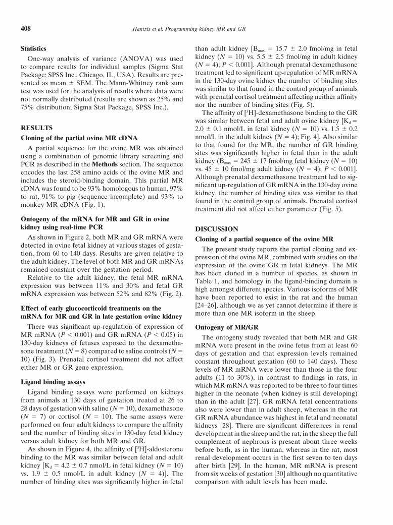
Statistics than adult kidney [Bmax � 15.7 � 2.0 fmol/mg in fetalkidney (N � 10) vs. 5.5 � 2.5 fmol/mg in adult kidneyOne-way analysis of variance (ANOVA) was used(N � 4); P � 0.001]. Although prenatal dexamethasoneto compare results for individual samples (Sigma Stattreatment led to significant up-regulation of MR mRNAPackage; SPSS Inc., Chicago, IL, USA). Results are pre-in the 130-day ovine kidney the number of binding sitessented as mean � SEM. The Mann-Whitney rank sumwas similar to that found in the control group of animalstest was used for the analysis of results where data werewith prenatal cortisol treatment affecting neither affinitynot normally distributed (results are shown as 25% andnor the number of binding sites (Fig. 5).75% distribution; Sigma Stat Package, SPSS Inc.).
The affinity of [3H]-dexamethasone binding to the GRwas similar between fetal and adult ovine kidney [Kd �
RESULTS 2.0 � 0.1 nmol/L in fetal kidney (N � 10) vs. 1.5 � 0.2nmol/L in the adult kidney (N � 4); Fig. 4]. Also similarCloning of the partial ovine MR cDNAto that found for the MR, the number of GR bindingA partial sequence for the ovine MR was obtainedsites was significantly higher in fetal than in the adultusing a combination of genomic library screening andkidney (Bmax � 245 � 17 fmol/mg fetal kidney (N � 10)PCR as described in the Methods section. The sequencevs. 45 � 10 fmol/mg adult kidney (N � 4); P � 0.001].encodes the last 258 amino acids of the ovine MR andAlthough prenatal dexamethasone treatment led to sig-includes the steroid-binding domain. This partial MRnificant up-regulation of GR mRNA in the 130-day ovinecDNA was found to be 93% homologous to human, 97%kidney, the number of binding sites was similar to thatto rat, 91% to pig (sequence incomplete) and 93% tofound in the control group of animals. Prenatal cortisolmonkey MR cDNA (Fig. 1).treatment did not affect either parameter (Fig. 5).
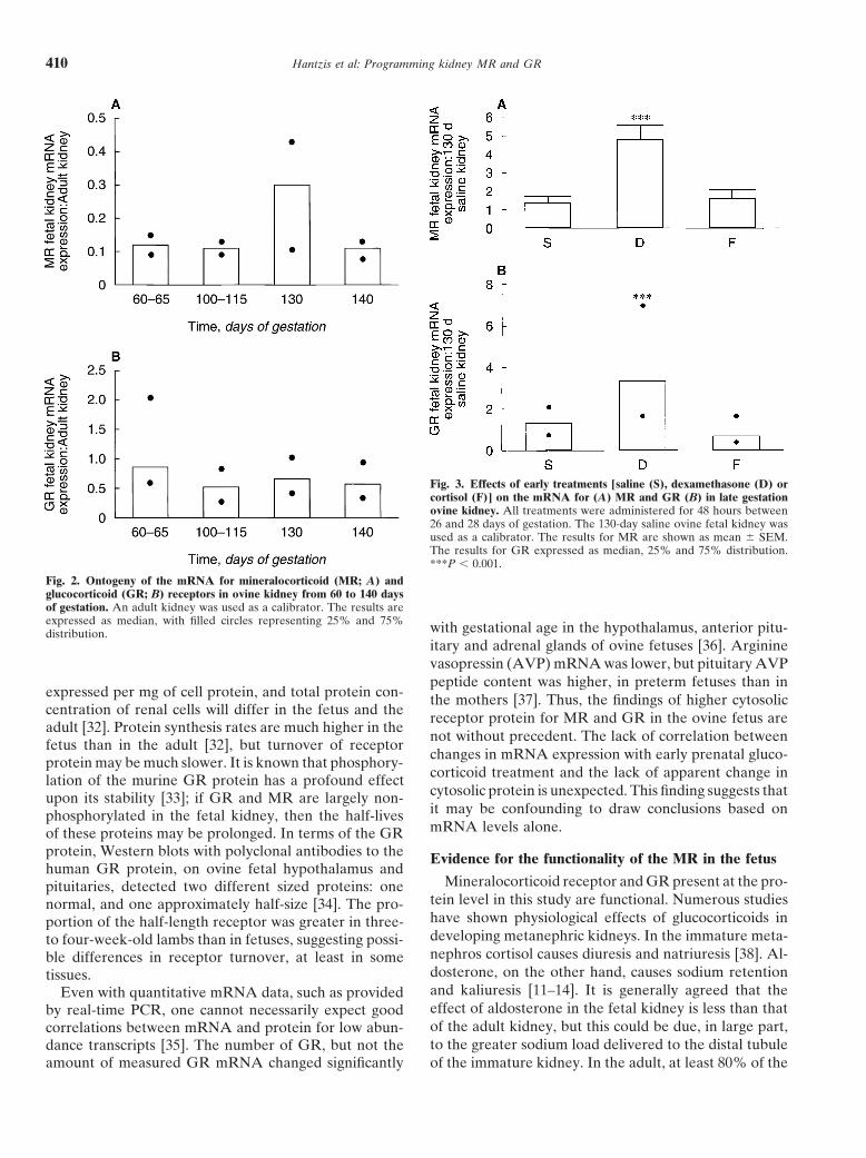
Ontogeny of the mRNA for MR and GR in ovinekidney using real-time PCR DISCUSSION
As shown in Figure 2, both MR and GR mRNA were Cloning of a partial sequence of the ovine MRdetected in ovine fetal kidney at various stages of gesta- The present study reports the partial cloning and ex-tion, from 60 to 140 days. Results are given relative to pression of the ovine MR, combined with studies on thethe adult kidney. The level of both MR and GR mRNAs expression of the ovine GR in fetal kidneys. The MRremained constant over the gestation period. has been cloned in a number of species, as shown in
Relative to the adult kidney, the fetal MR mRNA Table 1, and homology in the ligand-binding domain isexpression was between 11% and 30% and fetal GR high amongst different species. Various isoforms of MRmRNA expression was between 52% and 82% (Fig. 2). have been reported to exist in the rat and the human
[24–26], although we as yet cannot determine if there isEffect of early glucocorticoid treatments on themore than one MR isoform in the sheep.mRNA for MR and GR in late gestation ovine kidney
There was significant up-regulation of expression of Ontogeny of MR/GRMR mRNA (P � 0.001) and GR mRNA (P � 0.05) in The ontogeny study revealed that both MR and GR130-day kidneys of fetuses exposed to the dexametha- mRNA were present in the ovine fetus from at least 60sone treatment (N � 8) compared to saline controls (N � days of gestation and that expression levels remained10) (Fig. 3). Prenatal cortisol treatment did not affect constant throughout gestation (60 to 140 days). Theseeither MR or GR gene expression. levels of MR mRNA were lower than those in the four
adults (11 to 30%), in contrast to findings in rats, inLigand binding assays which MR mRNA was reported to be three to four times
Ligand binding assays were performed on kidneys higher in the neonate (when kidney is still developing)from animals at 130 days of gestation treated at 26 to than in the adult [27]. GR mRNA fetal concentrations28 days of gestation with saline (N � 10), dexamethasone also were lower than in adult sheep, whereas in the rat(N � 7) or cortisol (N � 10). The same assays were GR mRNA abundance was highest in fetal and neonatalperformed on four adult kidneys to compare the affinity kidneys [28]. There are significant differences in renaland the number of binding sites in 130-day fetal kidney development in the sheep and the rat; in the sheep the fullversus adult kidney for both MR and GR. complement of nephrons is present about three weeks
As shown in Figure 4, the affinity of [3H]-aldosterone before birth, as in the human, whereas in the rat, mostbinding to the MR was similar between fetal and adult renal development occurs in the first seven to ten dayskidney [Kd � 4.2 � 0.7 nmol/L in fetal kidney (N � 10) after birth [29]. In the human, MR mRNA is presentvs. 1.9 � 0.5 nmol/L in adult kidney (N � 4)]. The from six weeks of gestation [30] although no quantitative
comparison with adult levels has been made.number of binding sites was significantly higher in fetal

Hantzis et al: Programming kidney MR and GR 409
Fig. 1. Predicted amino acid sequence of the C-terminal end of the ovine mineralocorticoid receptor and multiple sequence alignment with thededuced amino acid sequences from monkey (T. belangery) (AAF63382), rat (P22199), human (P08235), shrew (Q29131), frog (Q91573) and pig(P79404; sequence incomplete). Identical and similar amino acids are boxed in black and gray, respectively.
In contrast to the mRNA studies, MR and GR protein, and GR proteins in the adult sheep kidney were similarin both studies.as determined by ligand binding, gave different results.
The concentration of GR protein (Bmax) was approxi-Discrepancy between mRNA and protein resultsmately five times greater in the late gestation fetal kidney
as in the adult, while that for MR was threefold higher Thus, there is an apparent discrepancy between themRNA results and the ligand binding studies for bothin the fetus. The Kd of the fetal and adult GR and MR
were comparable, and of the same order as that deter- receptors. There are several possible explanations forthis study result. The receptor protein concentration ismined by other workers [31]. The concentrations of MR
Hantzis et al: Programming kidney MR and GR410
Fig. 3. Effects of early treatments [saline (S), dexamethasone (D) orcortisol (F)] on the mRNA for (A) MR and GR (B) in late gestationovine kidney. All treatments were administered for 48 hours between26 and 28 days of gestation. The 130-day saline ovine fetal kidney wasused as a calibrator. The results for MR are shown as mean � SEM.The results for GR expressed as median, 25% and 75% distribution.***P � 0.001.
Fig. 2. Ontogeny of the mRNA for mineralocorticoid (MR; A) andglucocorticoid (GR; B) receptors in ovine kidney from 60 to 140 daysof gestation. An adult kidney was used as a calibrator. The results areexpressed as median, with filled circles representing 25% and 75% with gestational age in the hypothalamus, anterior pitu-distribution.
itary and adrenal glands of ovine fetuses [36]. Argininevasopressin (AVP) mRNA was lower, but pituitary AVPpeptide content was higher, in preterm fetuses than in
expressed per mg of cell protein, and total protein con- the mothers [37]. Thus, the findings of higher cytosoliccentration of renal cells will differ in the fetus and the receptor protein for MR and GR in the ovine fetus areadult [32]. Protein synthesis rates are much higher in the
not without precedent. The lack of correlation betweenfetus than in the adult [32], but turnover of receptor
changes in mRNA expression with early prenatal gluco-protein may be much slower. It is known that phosphory-corticoid treatment and the lack of apparent change inlation of the murine GR protein has a profound effectcytosolic protein is unexpected. This finding suggests thatupon its stability [33]; if GR and MR are largely non-it may be confounding to draw conclusions based onphosphorylated in the fetal kidney, then the half-livesmRNA levels alone.of these proteins may be prolonged. In terms of the GR
protein, Western blots with polyclonal antibodies to the Evidence for the functionality of the MR in the fetushuman GR protein, on ovine fetal hypothalamus and
Mineralocorticoid receptor and GR present at the pro-pituitaries, detected two different sized proteins: onetein level in this study are functional. Numerous studiesnormal, and one approximately half-size [34]. The pro-have shown physiological effects of glucocorticoids inportion of the half-length receptor was greater in three-developing metanephric kidneys. In the immature meta-to four-week-old lambs than in fetuses, suggesting possi-nephros cortisol causes diuresis and natriuresis [38]. Al-ble differences in receptor turnover, at least in somedosterone, on the other hand, causes sodium retentiontissues.and kaliuresis [11–14]. It is generally agreed that theEven with quantitative mRNA data, such as providedeffect of aldosterone in the fetal kidney is less than thatby real-time PCR, one cannot necessarily expect goodof the adult kidney, but this could be due, in large part,correlations between mRNA and protein for low abun-to the greater sodium load delivered to the distal tubuledance transcripts [35]. The number of GR, but not the
amount of measured GR mRNA changed significantly of the immature kidney. In the adult, at least 80% of the

Hantzis et al: Programming kidney MR and GR 411
Fig. 4. Affinity (Kd; A) and the concentration (Bmax; B) of MR and GRin late gestation ovine kidney compared to the adult kidney. The results Fig. 5. Effects of early treatments [saline (S), dexamethasone (D) orare shown as mean � SEM. ***P � 0.001. cortisol (F)] on the affinity (Kd; A) and the concentration (Bmax; B)
of MR and GR in late gestation ovine kidney. All treatments wereadministered for 48 hours between 26 and 28 days of gestation. Theresults are shown as mean � SEM.
filtered sodium is reabsorbed in the proximal convolutedtubule and pars recta, with remainder largely reabsorbeddistal to this point. In the fetal kidney the total fractional Programming effects of dexamethasone versus cortisolreabsorption of sodium is lower, at 96.5% (rather than Exposure of the ovine fetus to dexamethasone, at a99%) and only �65% is reabsorbed in the proximal time at which the only functioning kidney is a mesoneph-segment [38, 39]. Therefore, even under the influence ros [22], caused a significant increase in mRNAs for bothof very high concentrations of plasma aldosterone the MR and GR, whereas the exposure to maximal cortisolfractional excretion of sodium is greater in the fetal kid- levels did not. This may have been due to dexamethasoneney (1.9%) than in the normal adult kidney (�1.0%) accessing the fetal compartment more efficiently than[14]. If some of fetal kidney GR mRNA encodes a GR� cortisol, or the dexamethasone exerting this ‘program-receptor (as yet unknown in sheep), and this dimerizes ming’ effect via a different receptor, such as the pregnanewith the MR, this also may reduce the physiological X receptor (PXR), which ‘sees’ dexamethasone but not
cortisol [41]. No studies have shown the presence ofeffectiveness of aldosterone [40].

Hantzis et al: Programming kidney MR and GR412
25), in Principles of Perinatal-Neonatal Metabolism (2nd ed), editedsuch a receptor in the tissues of the ovine fetus early inby Cowett RM, Springer, 1998, pp 511–536
gestation. However, it is quite remarkable that treatment 8. Berger S, Bleich M, Schmid W, et al: Mineralocorticoid receptorknockout mice: Pathophysiology of Na metabolism. Proc Natlof a fetus, at a stage when the permanent metanephricAcad Sci USA 5:9424–9429, 1998kidney is represented by only some metanephrogenic
9. Benson CA, Wintour EM: The effect of bilateral fetal adrenalec-mesenchyme and the first branching of the ureteric bud, tomy on fluid balance in the ovine fetus. J Physiol 489:235–241,
1995can affect gene expression in the developed kidney some10. Wintour EM, Coghlan JP, Hardy KJ, et al: Placental transfer ofone hundred days later.
aldosterone in the sheep. J Endocrinol 86:305–310, 198011. Lingwood B, Hardy KJ, Coghlan JP, et al: Effect of aldosterone
on urine composition in the chronically cannulated ovine fetus.CONCLUSION J Endocrinol 76:553–554, 1978
12. Robillard JE, Nakamura KT, Lawton WJ, et al: Effects of aldo-In cloning the partial sequence of the ovine MR, to sterone on urinary kallikrein and sodium excretion during foetalour knowledge for the first time we were able to monitor life. Pediatr Res 19:1048–1052, 1985
13. Kairaitis K, Lumbers ER: The influence of endogenous mineralo-changes in MR mRNA and protein in the ovine fetus.corticoids on the composition of foetal of fetal urine. J Dev Physiol
The level of both MR and GR mRNA remains relatively 13:347–351, 199014. Mann SE, Nijland MJM, Ross MG: Fetal absorption of intra-constant throughout gestation. The synthetic glucocorti-
amniotic aldosterone: Effects on urine composition. J Soc Gynecolcoid, dexamethasone, increased levels of both MR andInvestig 6:252–257, 1999
GR mRNA administered short term in early gestation, 15. Greenberg D, Abramson O, Phillip M: Fetal pseudohypoaldoste-ronism: Another cause of hydramnios. Acta Paediatr 84:582–584,whereas the natural glucocorticoid cortisol had no effect1995on MR or GR gene expression. Thus, up-regulation of
16. Narchi H, Santos M, Kulaylat N: Polyhydramnios as a sign ofgene expression for both MR and GR is evidence for a fetal pseudohypoaldosteronism. Int J Gynaecol Obstet 69:53–54,
2000programming effect occurring in utero.17. Osmond C, Barker DJP: Fetal, infant, and childhood growth areThe dissociation between fetal mRNA and protein predictors of coronary heart disease, diabetes and hypertension in
levels, relative to adult kidneys, suggests that it may be adult men and women. Environ Health Perspect 108:545–553, 200018. Huxley RR, Sheill AW, Law CM: The role of size at birth andconfounding to draw conclusions based on mRNA levels
postnatal catch-up growth in determining systolic blood pressure:alone. A review of the literature. J Hypertens 18:815–831, 200019. Dodic M, May CN, Wintour EM, et al: An early prenatal exposure
to excess glucocorticoid leads to hypertensive offspring in sheep.ACKNOWLEDGMENTSClin Sci 94:149–155, 1998
20. Woods LL: Fetal origins of adult hypertension: A renal mecha-The studies were supported by Block Grants to the Howard Floreynism? Curr Opin Nephrol Hypertens 9:419–425, 2000Institute (983001) and the Baker Institute (993001) from the National
21. Bertram C, Trowern AR, Whorwood CB, et al: The maternalHealth and Medical Research Council of Australia, and grants-in-aiddiet during pregnancy programs altered expression of the glucocor-from the Jack Brockhoff Foundation and BHP, Australia. The Appliedticoid receptor and type 2 11 beta-hydroxysteroid dehydrogenase:Biosystems PRISM Sequence Detector System was purchased withPotential molecular mechanisms underlying the programming offunds donated by the Philip Bushell Foundation, the Harold and Corahypertension in utero. Endocrinology 142:2841–2853, 2001Brennen Benevolent Trust, the Viertel and Ramaciotti Foundations.
22. Peers A, Hantzis V, Dodic M, et al: Functional glucocorticoidThe authors wish to thank Professor John W. Funder for critical readingreceptors in the mesonephros of the ovine foetus. Kidney Intof the manuscript.59:425–433, 2001
23. Myles K, Funder JW: Type I (mineralocorticoid) receptors in theReprint requests to Dr. Miodrag Dodic, Howard Florey Institute,guinea pig. Am J Physiol 267:E-268–E272, 1994The University of Melbourne, Victoria 3010, Australia.
24. Kwak SP, Patel PD, Thompson RC, et al: 5�-Heterogeneity of theE-mail: [email protected] receptor messenger ribonucleic acid: Differen-tial expression and regulation of splice variants with the rat hippo-
REFERENCES campus. Endocrinology 133:2344–2350, 199325. Bradford MM: A rapid and sensitive method for quantitation of
1. Funder JW, Krozowski Z, Myles K, et al: Mineralocorticoid re- microgram quantities of protein utilizing the principle of proteinceptors, salt and hypertension. Recent Prog Horm Res 52:247–263, dye binding. Anal Biochem 72:248–254, 19761997 26. Zennaro MC, Farman N, Bonvalet JP, et al: Tissue-specific ex-
2. Arriza LJ, Weinberger C, Cerelli G, et al: Cloning of human pression of alpha and beta messenger ribonucleic acid isoforms ofmineralocorticoid receptor complementary DNA: Structural and the human mineralocorticoid receptor in normal and pathologicalfunctional kinship with the glucocorticoid receptor. Science 237: states. J Clin Endocrinol Metab 82:1345–1352, 1997268–275, 1987 27. Kalinyak JE, Bradshaw JG, Perlman AJ: The role of develop-
3. Marver D, Stewart J, Funder JW, et al: Renal aldosterone recep- ment and adrenal steroids in the regulation of the mineralocorticoidtors: Studies with [3H] aldosterone and the anti-mineralocorticoid receptor messenger RNA. Horm Metab Res 24:106–109, 1992[3H] spirolactone. Proc Natl Acad Sci USA 71:1431–1435, 1975 28. Kalinyak JE, Griffin CA, Hamilton RW, et al: Developmental
4. Reul JM, Gesing A, Droste S, et al: The brain mineralocorticoid and hormonal regulation of glucocorticoid receptor messengerreceptor: Greedy for ligand, mysterious in function. Eur J Pharma- RNA in the rat. J Clin Invest 84:1843–1848, 1989col 405:235–249, 2000 29. Moritz KM, Wintour EM: Functional development of the meso-
5. Albiston AL, Obeyesekere VR, Smith RE, et al: Cloning and and metanephros. Pediatr Nephrol 13:171–178, 1999tissue distribution of the human 11�-hydroxysteroid dehydroge- 30. Condon J, Gosden C, Gardener D, et al: Expression of type 2 11nase type 2 enzyme. Mol Cell Endocrinol 105:R11–R17, 1994 beta-hydroxysteroid dehydrogenase and corticosteroid hormone
6. Farman N, Rafestin-Oblin ME: Multiple aspects of mineralocorti- receptors in early life. J Clin Endocrinol Metab 83:4490–4497, 1998coid selectivity. Am J Physiol (Renal Physiol) 280:F181–F192, 2001 31. Roesch DM, Keller-Wood M: Differential effects of pregnancy
on mineralocorticoid and glucocorticoid receptor availability and7. Wintour EM: Water metabolism in the fetal-placental unit (chapt

Hantzis et al: Programming kidney MR and GR 413
immunoreactivity in cortisol feedback sites. Neuroendocrinology gestational age and after adrenocorticotropin treatment. Endocri-nology 126:11–17, 199070:55–62, 1999
32. Liechty EA, Boyle DW: Protein metabolism in the fetal-placental 37. Zhao X, Nijland MJ, Ervin MG, et al: Regulation of hypothalamicarginine vasopressin messenger ribonucleic acid and pituitary argi-unit, in Principles of Perinatal-Neonatal Metabolism, edited by
Cowett RM, Springer, 1998, pp 369–387 nine vasopressin content in fetal sheep: Effects of acute tonicityalterations and fetal maturation. Am J Obstet Gynecol 179:899–905,33. Webster JC, Jewell CM, Bodwell JE, et al: Mouse glucocorticoid
receptor phosphorylation status influences multiple functions of 199838. Towstoless MK, McDougall JG, Wintour EM: Gestationalthe receptor protein. J Biol Chem 272:9287–9293, 1997
34. Saoud CJ, Wood CE: Developmental changes and molecular changes in renal responsiveness to cortisol in the ovine fetus. Pedi-atr Res 26:6–10, 1989weight of immunoreactive glucocorticoid receptor protein in the
ovine fetal hypothalamus and pituitary. Biochem Biophys Res 39. Lumbers ER, Hill KJ, Bennett VJ: Proximal and distal tubularactivity in chronically catheterized fetal sheep compared with theComm 229:916–921, 1996
35. Gygi SP, Rochon Y, Franza BR, et al: Correlation between pro- adult. Can J Physiol Pharmacol 66:697–702, 198840. Vottero A, Chrousos GP: Glucocorticoid receptor beta: View 1.tein and mRNA abundance in yeast. Mol Cell Biol 19:1720–1730,
1999 Trends Endocrinol Metab 10:333–338, 199941. Moore JT, Kliewer SA: Use of the nuclear receptor PXR to36. Yang K, Jones SA, Challis JRG: Changes in glucocorticoid recep-
tor number in the hypothalamus and pituitary of the sheep with predict drug interactions. Toxicology 153:1–10, 2000