Effect of dipyrone and thalidomide alone and in combination on STZ-induced diabetic neuropathic pain
12
ORIGINAL ARTICLE Effect of dipyrone and thalidomide alone and in combination on STZ-induced diabetic neuropathic pain Neha Chauhan & Rajeev Taliyan & Pyare Lal Sharma Received: 6 August 2011 /Accepted: 28 December 2011 / Published online: 17 January 2012 # Springer-Verlag 2012 Abstract Diabetic neuropathy is recognized as one of the most common complications of chronic diabetes, but its path- ophysiological mechanism is complex and yet to be completely explored. Monotherapy with conventional analgesics fails to provide adequate pain relief in peripheral diabetic neuropathy. There are a number of evidence suggesting that tumor necrosis factor (TNF-α) plays an important role in the pathogenesis of peripheral diabetic neuropathy. TNF-α up-regulation activates nuclear factor κB, which further up-regulates cyclooxygenase (COX)-2 leading to altered prostaglandin profile. Inhibition of TNF-α and COX-2 provides beneficial effect on diabetic neuropathy by decreasing the oxidative stress level and by preventing neuronal hypersensitivity due to an increased prostaglandin level. The present study was designed to assess the effect of dipyrone and thalidomide on streptozo- tocin (STZ)-induced neuropathic pain behavior in rats. STZ 50 mg/kg, i.p. was administered to induce experimental dia- betes in the rats. Three weeks following STZ, dipyrone (300 and 600 mg/kg, i.p.) and thalidomide (25 and 50 mg/kg, i.p.) alone and subeffective dose combination of dipyrone and thalidomide (300 and 25 mg/kg -1 , i.p.) administered daily for 2 weeks significantly attenuated thermal hyperalgesia, mechanical allodynia, and formalin-induced phase-2 flinching response. Moreover, the subeffective dose combination of dipyrone and thalidomide and preemptive treatment with tha- lidomide (50 mg/kg) reduces oxidative stress in diabetic rats. In conclusion, the combination of subeffective dose of dipyr- one and thalidomide prevented the development and mainte- nance of experimental diabetic neuropathy. The combination of thalidomide (TNF-α inhibitor) and dipyrone (COX inhib- itor) may be used as a potential therapeutic agent for the treatment of diabetic neuropathy. Keywords Diabetic neuropathy . Hyperalgesia . Allodynia . Tumor necrosis factor . Cyclooxygenase Introduction One of the most common chronic complications of diabetes mellitus is diabetic neuropathy, but it remains probably the least understood complications (Greene et al. 1997), which is mainly characterized by spontaneous pain, abnormal sen- sations such as paresthesia, allodynia (pain responses to innocuous stimuli), and hyperalgesia (exaggerated pain responses to noxious stimuli). The contribution of hypergly- cemia in pathogenesis of diabetic neuropathy is beyond controversy, which eventually leads to accumulation of ad- vanced glycation end-products (Brownlee 2005), protein kinase C isoform activation, mitochondrial dysfunction (Vinik et al. 2003), and activation of nuclear factor-κB (NF-κB) (Wang et al. 2006). All these pathways converge in the production of oxidative stress. Reactive oxygen/nitrogen species (Pop-Busui et al. 2006a, b) and inflammatory cytokine tumor necrosis factor α (TNF-α) (Taliyan et al. 2010) play a key role in diabetic neuropathy, starting from the development of the initial stages of diabetes to the progression of the later stages of N. Chauhan : R. Taliyan : P. L. Sharma (*) Department of Pharmacology, I.S.F College of Pharmacy, Moga 142001 Punjab, India e-mail: [email protected] Naunyn-Schmiedeberg's Arch Pharmacol (2012) 385:527–538 DOI 10.1007/s00210-011-0724-9
-
Upload
neha-chauhan -
Category
Documents
-
view
219 -
download
7
Transcript of Effect of dipyrone and thalidomide alone and in combination on STZ-induced diabetic neuropathic pain

ORIGINAL ARTICLE
Effect of dipyrone and thalidomide alone and in combinationon STZ-induced diabetic neuropathic pain
Neha Chauhan & Rajeev Taliyan & Pyare Lal Sharma
Received: 6 August 2011 /Accepted: 28 December 2011 /Published online: 17 January 2012# Springer-Verlag 2012
Abstract Diabetic neuropathy is recognized as one of themost common complications of chronic diabetes, but its path-ophysiological mechanism is complex and yet to be completelyexplored. Monotherapy with conventional analgesics fails toprovide adequate pain relief in peripheral diabetic neuropathy.There are a number of evidence suggesting that tumor necrosisfactor (TNF-α) plays an important role in the pathogenesis ofperipheral diabetic neuropathy. TNF-α up-regulation activatesnuclear factor κB, which further up-regulates cyclooxygenase(COX)-2 leading to altered prostaglandin profile. Inhibition ofTNF-α and COX-2 provides beneficial effect on diabeticneuropathy by decreasing the oxidative stress level and bypreventing neuronal hypersensitivity due to an increasedprostaglandin level. The present study was designed toassess the effect of dipyrone and thalidomide on streptozo-tocin (STZ)-induced neuropathic pain behavior in rats. STZ50 mg/kg, i.p. was administered to induce experimental dia-betes in the rats. Three weeks following STZ, dipyrone (300and 600 mg/kg, i.p.) and thalidomide (25 and 50 mg/kg, i.p.)alone and subeffective dose combination of dipyrone andthalidomide (300 and 25 mg/kg−1, i.p.) administered dailyfor 2 weeks significantly attenuated thermal hyperalgesia,mechanical allodynia, and formalin-induced phase-2 flinchingresponse. Moreover, the subeffective dose combination ofdipyrone and thalidomide and preemptive treatment with tha-lidomide (50 mg/kg) reduces oxidative stress in diabetic rats.
In conclusion, the combination of subeffective dose of dipyr-one and thalidomide prevented the development and mainte-nance of experimental diabetic neuropathy. The combinationof thalidomide (TNF-α inhibitor) and dipyrone (COX inhib-itor) may be used as a potential therapeutic agent for thetreatment of diabetic neuropathy.
Keywords Diabetic neuropathy . Hyperalgesia . Allodynia .
Tumor necrosis factor . Cyclooxygenase
Introduction
One of the most common chronic complications of diabetesmellitus is diabetic neuropathy, but it remains probably theleast understood complications (Greene et al. 1997), whichis mainly characterized by spontaneous pain, abnormal sen-sations such as paresthesia, allodynia (pain responses toinnocuous stimuli), and hyperalgesia (exaggerated painresponses to noxious stimuli). The contribution of hypergly-cemia in pathogenesis of diabetic neuropathy is beyondcontroversy, which eventually leads to accumulation of ad-vanced glycation end-products (Brownlee 2005), proteinkinase C isoform activation, mitochondrial dysfunction(Vinik et al. 2003), and activation of nuclear factor-κB(NF-κB) (Wang et al. 2006). All these pathways convergein the production of oxidative stress.
Reactive oxygen/nitrogen species (Pop-Busui et al.2006a, b) and inflammatory cytokine tumor necrosis factorα (TNF-α) (Taliyan et al. 2010) play a key role in diabeticneuropathy, starting from the development of the initialstages of diabetes to the progression of the later stages of
N. Chauhan :R. Taliyan : P. L. Sharma (*)Department of Pharmacology, I.S.F College of Pharmacy,Moga 142001 Punjab, Indiae-mail: [email protected]
Naunyn-Schmiedeberg's Arch Pharmacol (2012) 385:527–538DOI 10.1007/s00210-011-0724-9

neuropathic pain (Taliyan et al. 2011). TNF-α or interleukin(IL) is released by macrophages, Schwann cells, and lym-phocytes in diabetic nerves in humans and animals (Conti etal. 2002; Yagihashi et al. 2007). These studies suggest a roleof TNF-α in the regulation of development of hyperalgesiaand allodynia and apoptosis in diabetic animals, inflammatoryor immunological disease. This leads to much effort recentlyin finding ways to down-regulate its production or inhibit itseffects. A number of chimeric TNF-α antibodies such asAdlimumab, Etanercept, and CDP571 have been developedto treat conditions associated with elevated TNF-α, but theseantibodies have certain limitations including their high costand potential adverse effect (Scheinfeld 2004). Thalidomide,a derivative of glutamic acid, inhibits TNF-α synthesis bydecreasing the half-life of TNF-αmRNA and was reported topossess various beneficial pharmacological properties includ-ing antiinflammatory, immunomodulatory, and antiangio-genic effects (Ribeiro et al. 2000; Ye et al. 2006) and wasreintroduced, despite its powerful teratogenic nature, as treat-ment for diverse chronic immunological/inflammatory dis-eases, and it is suggested as a promising treatment forneurodegenerative diseases (Sampaio et al. 1991). Thalido-mide’s immunomodulatory effects and inhibition of the syn-thesis and release of proinflammatory cytokines as well asincreases the release of anti-inflammatory cytokines are basedon its capacity to modify T-helper cell phenotype from aproinflammatory Th1 to an anti-inflammatory Th2 pattern,on the basis of the type of cytokines produced (Corrala andKaplan 1999; Ribeiro et al. 2000; Sommer et al. 2001).
TNF-α activation further regulates the production ofadditional cytokines and nerve growth factor, macrophagerecruitment, myelin removal, regeneration, and neuropathicpain by different mechanisms. In addition, there is simulta-neous activation of cyclooxygenase (COX)-2 in the periph-eral nerves of STZ diabetic rats (Pop-Busui et al. 2002),contributing to diabetes-induced neuropathic pain. COX-2 up-regulation results in altered prostaglandin profile in whichthere is an increased production of vasoconstricting prosta-glandin H2 (PGH2), thromboxane A2, and prostaglandin F2-alpha (PGF2-α) and reduction in vasodilatory prostacyclin(PGI2). In addition, COX-2 up-regulation increases reactiveoxygen species (ROS) generation, which further exacerbatesoxidative stress.
Current treatment of peripheral diabetic neuropathy(PDN) involves the use of tricyclic antidepressant, selectiveserotonin reuptake inhibitors (Mckeage 2007), anticonvul-sants, opioids and antioxidant protein kinase C inhibitors,COX-2 inhibitors (Kellog et al. 2008), and nonsteroidalanti-inflammatory drugs as mild analgesics and so on Treat-ment with these drugs is often limited because of partial
effectiveness and side effects associated with these drugs(Chong and Hester 2007; O’Connor 2009). Thus, there is aneed of new therapeutic interventions targeting primarymechanisms resulting in nerve damage in PDN.
Dipyrone (COX inhibitor) and thalidomide (TNF-α inhib-itor) have been evaluated for efficacy in STZ-induced neuro-pathic pain in rats. Dipyrone acts as an effective analgesic andantipyretic agent (Ceraso. 1994), exerting its antinociceptiveeffect by inhibition of prostaglandin synthesis in the periph-eral and the central nervous system (Abbate et al. 1990;Shimada et al. 1994), although its precisemechanism of actionremains unclear.
Initially, analgesia by dipyrone was explained by an inhib-itory action on PG synthesis. However, Nikolova et al. (1980)suggested that the profile of the pharmacological effects ofdipyrone is certainly different from that of other nonsteroidalanti-inflammatory drugs. Lorenzetti and Ferreira (1996) haveindicated that the involvement of arginine–nitric oxide (NO)pathway in primary sensory neurons contributes to dipyrone-induced spinal and peripheral analgesia. Moreover, it has beenreported that the peripheral analgesic effect of dipyrone mayresult from direct blockade of hyperalgesia rather than fromprevention of the release of prostaglandins in inflamed tissues(Lorenzetti and Ferreira 1985). In addition to this, severalstudies indicated that dipyrone induces an antinociceptiveeffect both by peripheral and central mechanisms (Akman etal. 1996). Both these drugs have shown efficacy in variousinflammatory models. However, there is no study reported onthe use of these drugs alone and in combination on STZ-induced neuropathic pain model in rats, which is addressedin the present study.
Experimental animals
Wistar rats weighing 200–280 g were used for behavioralparadigm of PDN. The experimental protocol was approvedby the Institutional Animal Ethics Committee.
Induction and assessment of diabetes in rats
Experimental diabetes was induced by a single intraperito-neal (i.p.) injection of STZ (50 mg kg−1) freshly dissolved incitrate buffer pH. Serum glucose level was assessed byusing enzymatic glucose oxidase peroxidase commerciallyavailable kit method, 72 h after STZ induction. Only ratswith blood glucose concentration more than 240 mg/dl wereconsidered diabetic and used for the study. Body weight andserum glucose were measured before and at the end of theexperiment to see the effect of pharmacological interven-tions on these parameters.
528 Naunyn-Schmiedeberg's Arch Pharmacol (2012) 385:527–538
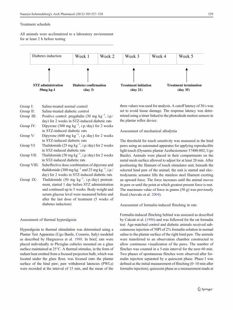
Treatment schedule
All animals were acclimatized to a laboratory environmentfor at least 2 h before testing
Diabetes induction Week 1 Week 2 Week 3 Week 4 Week 5
Treatment termination STZ administration50mg kg-1 (day 35)(day 21)(day 3)
Treatment initiationDiabetes confirmation
Group I: Saline-treated normal controlGroup II: Saline-treated diabetic controlGroup III: Positive control: pregabalin (30 mg kg−1, i.p./
day) for 2 weeks in STZ-induced diabetic ratsGroup IV: Dipyrone (300 mg kg−1, i.p./day) for 2 weeks
in STZ-induced diabetic ratsGroup V: Dipyrone (600 mg kg−1, i.p./day) for 2 weeks
in STZ-induced diabetic ratsGroup VI: Thalidomide (25 mg kg−1, i.p./day) for 2 weeks
in STZ-induced diabetic ratsGroup VII: Thalidomide (50 mg kg−1, i.p./day) for 2 weeks
in STZ-induced diabetic ratsGroup VIII: Subeffective dose combination of dipyrone and
thalidomide (300 mg kg−1 and 25 mg kg−1, i.p./day) for 2 weeks in STZ-induced diabetic rats
Group IX: Thalidomide (50 mg kg−1, i.p./day) pretreat-ment, started 1 day before STZ administrationand continued up to 5 weeks. Body weight andserum glucose level were measured before andafter the last dose of treatment (5 weeks ofdiabetes induction)
Assessment of thermal hyperalgesia
Hyperalgesia to thermal stimulation was determined using aPlantar Test Apparatus (Ugo Basile, Comerio, Italy) modeledas described by Hargreaves et al. 1988. In brief, rats wereplaced individually in Plexiglas cubicles mounted on a glasssurface maintained at 25°C. A thermal stimulus, in the form ofradiant heat emitted from a focused projection bulb, which waslocated under the glass floor, was focused onto the plantarsurface of the hind paw; paw withdrawal latencies (PWLs)were recorded at the interval of 15 min, and the mean of the
three values was used for analysis. A cutoff latency of 30 s wasset to avoid tissue damage. The response latency was deter-mined using a timer linked to the photodiodemotion sensors inthe plantar reflex device.
Assessment of mechanical allodynia
The threshold for touch sensitivity was measured in the hindpaws using an automated apparatus for applying reproduciblelight touch (Dynamic plantar Aesthesiometer 37400-002; UgoBasile). Animals were placed in their compartments on themetal mesh surface allowed to adjust for at least 20 min. Afterpositioning the filament of touch stimulator unit, beneath theselected hind paw of the animal, the unit is started and elec-trodynamic actuator lifts the stainless steel filament exertingan upward force. The force increases until the animal movesits paw or until the point at which greatest present force is met.The maximum value of force in grams (50 g) was previouslyfixed (Arevalo et al. 2004).
Assessment of formalin-induced flinching in rats
Formalin-induced flinching behind was assessed as describedby Calcutt et al. (1996) and was followed for the rat formalintest. Age-matched control and diabetic animals received sub-cutaneous injection of 50Pl of 2% formalin solution in normalsaline to the plantar surface of the right hind paw. The animalswere transferred to an observation chamber constructed toallow continuous visualization of the paws. The number offlinches was counted in a 5-min interval for the next 60 min.Two phases of spontaneous flinches were observed after for-malin injection separated by a quiescent phase. Phase I wasdefined as the initial measurement of flinching (0–10min afterformalin injection), quiescent phase as a measurement made at
Naunyn-Schmiedeberg's Arch Pharmacol (2012) 385:527–538 529

10–20 min, and phase II as all the subsequent measurementsafter formalin injection. The results are expressed as the sumof flinching responses in phases 1 and 2 of the formalin test.
Collection of blood and tissue samples in rats
In this study, at the end of treatment schedule on day 35, bloodwas collected for serum glucose estimation, and the animalswere euthanized by cervical dislocation immediately afterbehavioral assays, followed by collection of sciatic nerve forestimation of markers of oxidative stress, and sciatic nerveswere rapidly removed, washed with sterile normal saline, andweighed. A 10% (wt/vol) tissue homogenate was prepared in0.1 M phosphate buffer (pH 7.4) and centrifuged for 15 min at2,000×g to obtain the clear supernatant for the estimation ofoxidative stress markers.
Biochemical assessment
Estimation of lipid peroxidation
Lipid peroxidation in the sciatic nerve was estimated colori-metrically by measuring thiobarbituric acid reactive substancesby the method of Niehius and Samuelsson (1968). Supernatant(0.1 ml) of sciatic nerve homogenate was treated with 2 ml of(1:1:1 ratio) thiobarbituric acid (0.37%)–trichloroacetic acid(15%)–hydrochloric acid (0.25 N) reagent and placed in hotwater bath for 15 min, cooled, and centrifuged, and then clearsupernatant was measured at 532 nm (UV-1700 Spectropho-tometer; Shimadzu, Japan) against a blank solution. Finally, thevalues are expressed as nanomoles per gram of tissue.
Estimation of reduced glutathione
The concentration of endogenous antioxidant-reduced glu-tathione (GSH) level in the sciatic nerve was estimatedfollowing the method described by Lou et al. (1988). In thismethod, 0.2 ml of supernatant was mixed with 1.78 ml of1.0 M Tris buffer (pH 8.2) with 0.02 M ethylenediaminete-trachloroacetic acid. Then, 20PL of 0.1 M 5,5′-dithio-bis-2-nitrobenzoic acid (Ellman’s reagent) was added to the mix-ture, and absorbance was noted at 412 nm (UV-1700 Spectro-photometer, Shimadzu); the values are expressed as picomolesper gram of tissue.
Measurement of nitrite
The nitrite concentration in the serumwas measured by Griessreaction (Sastry et al. 2002). In this method, 0.1 ml of super-natant of the nerve homogenate wasmixed with 0.25ml of 1%sulfanilamide (prepared in 3 N HCL) and 0.25 ml of 0.1% N-(1-naphthyl) ethylenediaminedihydrochloride with shaking.After 10 min, absorbance was measured at 545 nm (UV-
1700 Spectrophotometer; Shimadzu), and the values of nitriteconcentration were obtained from sodium nitrite standardcurve and are expressed in nanomoles per gram of tissue.
Drugs and chemicals
Thalidomide, dipyrone, pregabalin, streptozotocin (STZ;Sigma Aldrich Corporation, Bangalore, India) and formalin(37% formaldehyde) (SD Fine Chemicals, Mumbai, India)were used in this study. Glucose oxidase peroxidase estima-tion kit was purchased from Erba, Transasia Bio-Medicals,Mumbai, India. Unless stated, all other chemicals and bio-chemical reagent of highest analytical grade quality wereused. Dipyrone for i.p. administration was freshly preparedby solubilizing in sterile normal saline. Thalidomide for i.p.administration was dissolved in 10% dimethylsulfoxide. For-malin was diluted with sterile normal saline. Dose of dipyrone(Hernandez-Delgadillo and Cruz 2004), and others were se-lected on the basis of a previous report that was replicate by apilot study (n03).
Statistical analysis
The results are expressed as mean±SD. The behavioral datawere analyzed using two-way analysis of variance followed bybetween-group differences by Bonferroni post hoc test formultiple comparison. p<0.05 was considered statisticallysignificant.
Results
Effect of dipyrone and thalidomide alone and in combinationon body weight and on serum glucose level
Rats injected with STZ (50 mg/kg, i.p.) showed a significantrise in serum glucose and a significant decline in body weight(Table 1), as compared to age-matched normal control rats(vehicle treated). Monotherapy with dipyrone and thalidomideand subeffective dose combination of (dipyrone and thalido-mide) in STZ diabetic rats did not alter the 5-week diabetichyperglycemia and reduced body weight, as compared tovehicle-treated diabetic rat. Furthermore, pretreatment with tha-lidomide (50 mg kg−1, i.p.) also did not affect hyperglycemia indiabetic rats and their body weight.
Behavioral assessment
Effect of dipyrone and thalidomide alone or in combinationon thermal hyperalgesia
The nociceptive threshold was significantly reduced in dia-betic control rats compared to normal control rats (Fig. 1). In
530 Naunyn-Schmiedeberg's Arch Pharmacol (2012) 385:527–538

this study, monotherapy with subeffective dose of dipyrone(300 mg kg−1, i.p.) and thalidomide (25 mg kg−1, i.p.) for3 weeks had no effect on PWLs in age-matched diabetic rats,whereas monotherapy with a high dose of dipyrone
(600 mg kg−1, i.p.) and thalidomide (50 mg kg−1, i.p.) signif-icantly attenuated the development of thermal hyperalgesia indiabetic rats compared to untreated diabetic rats. Moreover,treatment with subeffective dose combination of dipyrone
Table 1 Effect of dipyrone and thalidomide alone or in combination on body weight and blood glucose level in rats
Treatment (mg kg−1) Body weight (g) Blood glucose (mg dl−1)
Initial Final Initial Final
NC 227.33±14.03 256.5±17.98 108.86±4.18 108.38±10.19
DC 260.66±29.64 186.83±23.41* 110.34±3.92 487±18.70*
D + P 30 256.83±16.43 191.83±17.67 110.16±4.708 477±19.01
D + DPN 300 204±26.31 164.8±16.05 15.96±4.48 461±29.81
D + DPN 600 266.8±10.03 185.2±28.46 110.6±4.03 446.2±24.65
D + TH 25 235±9.71 142.66±18.83 109.5±4.50 412.16±18.33
D + TH 50 32.5±12.78 153±26.19 111.33±2.8 426.16±16.82
D + DPN 300 + TH 25 236.2±10.51 131.8±11.10 107.8±3.86 473.44±17.33
D + TH 50 (pre) 234.66±9.77 131.16±9.96 110.16±2.63 419±18.27
Values are mean±SD
NC normal control; DC diabetic control; D + P 30 pregabalin-treated diabetic rats; DPN + D 300 and D + DPN 600 dipyrone-treated diabetic rats;D + TH 25 and D + TH 50 thalidomide-treated diabetic rats; D + DPN 300 + T H 25 dipyrone and thalidomide combination-treated diabetic rats; D+ TH 50 (pre) pretreatment with thalidomide in diabetic rats
*p<0.05 vs. normal control
Fig. 1 Effect of dipyrone and thalidomide alone or in combination onthermal hyperalgesia, in control and STZ-injected diabetic rats. Valuesare expressed as mean±SD. n06. *p<0.05 vs. normal control; #p<0.05vs. diabetic control; $p<0.05 vs. dipyrone (300 mg kg−1); +p<0.05 vs.thalidomide (25 mg kg−1). NC normal control; DC diabetic control; D +P30 pregabalin (30mg kg−1)-treated diabetic group;D+D300 andD+D
600 dipyrone (300 and 600 mg kg−1)-treated diabetic groups; D + T 25andD+T 50 thalidomide (25 and 50mg kg−1)-treated diabetic group;D+D 300 + T 25 dipyrone (300 mg kg−1) and thalidomide (25 mg kg−1)combination-treated diabetic group. Arrow indicates day of initiation oftreatment
Naunyn-Schmiedeberg's Arch Pharmacol (2012) 385:527–538 531
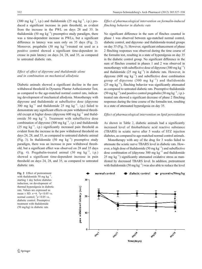
(300 mg kg−1, i.p.) and thalidomide (25 mg kg−1, i.p.) pro-duced a significant increase in pain threshold, as evidentfrom the increase in the PWL on days 28 and 35. Inthalidomide (50 mg kg−1) preemptive study paradigm, therewas a time-dependent increase in PWLs, but a significantdifference in latency was seen only on 35 days (Fig. 2).Moreover, pregabalin (30 mg kg−1)-treated rat used as apositive control showed a significant time-dependent in-crease in pain latency on days 24, 28, and 35, as comparedto untreated diabetic rats.
Effect of effect of dipyrone and thalidomide aloneand in combination on mechanical allodynia
Diabetic animals showed a significant decline in the pawwithdrawal threshold in Dynamic Plantar Asthesiometer Test,as compared to the age-matched normal control rats, indicat-ing development of mechanical allodynia. Monotherapy withdipyrone and thalidomide at subeffective dose (dipyrone300 mg kg−1 and thalidomide 25 mg kg−1, i.p,) failed todemonstrate any significant effects on paw withdrawal thresh-old except at higher doses (dipyrone 600 mg kg−1 and thalid-omide 50 mg kg−1). Treatment with subeffective dosecombination of dipyrone (300 mg kg−1, i.p.) and thalidomide(25 mg kg−1, i.p.) significantly increased pain threshold asevident from the increase in the paw withdrawal threshold ondays 24, 28, and 35, as compared to untreated diabetic animal(Fig. 3). In thalidomide (50 mg kg−1) preemptive studyparadigm, there was an increase in paw withdrawal thresh-old, but a significant effect was observed on 28 and 35 days(Fig. 4). Pregabalin-treated animal (30 mg kg−1, i.p.)showed a significant time-dependent increase in painthreshold on days 24, 28, and 35, as compared to untreateddiabetic rats.
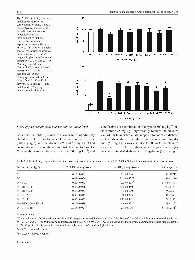
Effect of pharmacological intervention on formalin-inducedflinching behavior in diabetic rats
No significant difference in the sum of flinches counted inphase 1 was observed between age-matched normal control,diabetic control, and dipyrone- and thalidomide-treated groupson day 35 (Fig. 5). However, significant enhancement of phase2 flinching responses was observed during the time course ofthe formalin test, resulting in a state of hyperalgesia on day 35in the diabetic control group. No significant difference in thesum of flinches counted in phases 1 and 2 was observed inmonotherapywith subeffective dose of dipyrone (300mg kg−1)and thalidomide (25 mg kg−1) in diabetic rats. However, indipyrone (600 mg kg−1) and subeffective dose combinationgroup of dipyrone (300 mg kg−1) and thalidomide(25 mg kg−1), flinching behavior was significantly attenuatedas compared to untreated diabetic rats. Preemptive thalidomide(50mg kg−1) and positive control pregabalin (30mg kg−1, i.p.)-treated rats showed a significant decrease of phase 2 flinchingresponses during the time course of the formalin test, resultingin a state of attenuated hyperalgesia on day 35.
Effect of pharmacological intervention on lipid peroxidation
As shown in Table 2, diabetic animals had a significantlyincreased level of thiobarbituric acid reactive substance(TBARS) in sciatic nerve after 5 weeks of STZ injectiondiabetes, as compared to age-matched normal control animals.
Monotherapy with any of the drug for 3 weeks failed toattenuate the sciatic nerve TBARS level in diabetic rats. How-ever, a high dose of thalidomide (50 mg kg−1) and subeffectivedose combination of (dipyrone 300 mg kg−1 and thalidomide25 mg kg−1) significantly attenuated oxidative stress as man-ifested by decreased TBARS level. In addition, pretreatmentwith thalidomide (50mg kg−1) was also able to reduce the level
Fig. 2 Effect of pretreatmentwith thalidomide 50 mg kg−1,starting 1 day before diabetesinduction, on development ofthermal hyperalgesia in diabeticrats. Values are expressed asmean ± SD. n06. *p<0.05 vs.normal control; #p<0.05 vs.diabetic control. Preemptivetreatment with thalidomide(50 mg/kg) in diabetic rats
532 Naunyn-Schmiedeberg's Arch Pharmacol (2012) 385:527–538

of TBARS significantly. Three-week treatment with pregaba-lin (30 mg kg−1) in diabetic animals produced a significantreduction in TBARS levels in sciatic nerve.
Effect of pharmacological intervention on reducedglutathione
As shown in Table 2, 5-week treatment of diabetic animalsshowed a significantly decreased level of GSH in sciatic nerve,as compared to age-matched control animals. Pregabalin
(30 mg kg−1)-treated rats in the positive control group showedimproved GSH level of sciatic nerve of diabetic rats on day 35,as compared to diabetic untreated rats, whereas monotherapywith low and high doses of dipyrone (300 and 600 mg kg−1)and thalidomide (25 and 50 mg kg−1) and pretreatment withthalidomide (50 mg kg−1) did not improve the reduced GSHlevel in diabetic rats. On the other hand, subeffective dose lowcombination of dipyrone 300 mg kg−1 and thalidomide25 mg kg−1 showed significant improvement in GSH level insciatic nerve of diabetic rats compared to untreated diabetic rats.
Fig. 3 Effect of dipyrone and thalidomide alone or in combination onmechanical allodynia, in control and STZ injected diabetic rats. Valuesare expressed as mean±SD. n06. *p<0.05 vs. normal control; #p<0.05vs. diabetic control; $p<0.05 vs. dipyrone (300 mg/kg); +p<0.05 vs.thalidomide (25 mg/kg). NC normal control; DC diabetic control; D +
P 30 pregabalin (30 mg kg−1)-treated group; D + D 300 and D + D 600dipyrone (300 and 600 mg kg−1)-treated group; D + T 25 and D + T 50thalidomide (25 and 50 mg kg−1)-treated group; D + D300 + T 25dipyrone (300 mg kg−1)-and thalidomide (25 mg kg−1)-treated combi-nation group. Arrow indicates day of initiation of treatment
Fig. 4 Effect of pretreatmentwith thalidomide 50 mg kg−1,starting 1 day before diabetesinduction, on the developmentof mechanical allodynia indiabetic rats. Values areexpressed as mean±SD. n06.*p<0.05 vs. normal control;#p<0.05 vs. diabetic control.Pretreatment with thalidomidein diabetic rats (50 mg/kg)
Naunyn-Schmiedeberg's Arch Pharmacol (2012) 385:527–538 533
Effect of pharmacological intervention on nitrite level
As shown in Table 2, serum NO levels were significantlyelevated in the diabetic rats. Treatment with dipyrone(300 mg kg−1) and thalidomide (25 and 50 mg kg−1) hadno significant effect on the serum nitrite level up to 2 weeks.Conversely, administration of dipyrone (600 mg kg−1) and
subeffective dose combination of dipyrone 300 mg kg−1 andthalidomide 25 mg kg−1 significantly reduced the elevatedlevel of nitrite in diabetic rats compared to untreated diabeticcontrol rats on day 35. Similarly, pretreatment with thalido-mide (50 mg kg−1) was also able to attenuate the elevatedserum nitrite level in diabetic rats compared with age-matched untreated diabetic rats. Pregabalin (30 mg kg−1)
Fig. 5 Effect of dipyrone andthalidomide alone or incombination on phase 1 and 2nociceptive responses in theformalin test indicative ofhyperalgesia on thedevelopment of diabeticneuropathy. Values areexpressed as mean±SD. n06.*p<0.05; #p<0.05 vs. diabeticcontrol. NC normal control; DCdiabetic control; D + P 30pregabalin (30 mg kg−1)-treatedgroup; D + D 300 and D + D600 dipyrone (300 and600 mg kg−1)-treated diabeticgroup; D + T 25 and D + T 50thalidomide (25 and50 mg kg−1)-treated diabeticgroup; D + D 300 + T 25dipyrone (300 mg kg−1)- andthalidomide (25 mg kg−1)-treated combination group
Table 2 Effect of dipyrone and thalidomide alone or in combination on (sciatic nerve) TBARS, GSH level, and (serum) nitrite level in rats
Treatment (mg kg−1) TBARS (μmol/g tissue) GSH (μmol/g tissue) Nitrite (μmol/l)
NC 0.15±0.051 7.1±0.586 33±8.717
DC 0.48±0.056* 3.45±0.427* 98±2.280*
D + P 30 0.22±0.042 # 4.73±0.225# 54.33±4.501#
D + DPN 300 0.40±0.046 3.85±0.450 90±5.76
D + DPN 600 0.36±0.053# 4.3±0.670 75±2.607#
D + TH 25 0.39±0.045 3.68±0.213 84±2.68
D + TH 50 0.36±0.053 4.13±0.382 79±2.96
D + DPN 300 + TH 25 0.29±0.039# 4.6±0.328# 71±1.897#
D + TH 50 (pre) 0.388±0.027# 4.08±0.196 81.16±2.71#
Values are mean±SD
NC normal control; DC diabetic control; D + P 30 pregabalin-treated diabetic rats; D + DPN 300 and D + DPN 600 dipyrone-treated diabetic rats;D + TH 25 and D + TH 50 thalidomide-treated diabetic rats; D + DPN 300 + TH 25 dipyrone and thalidomide combination-treated diabetic rats; D+ TH 50 (pre) pretreatment with thalidomide in diabetic rats; GSH reduced glutathione
*p<0.05 vs. normal control# p<0.0.5 vs. diabetic control
534 Naunyn-Schmiedeberg's Arch Pharmacol (2012) 385:527–538

treatment for 2 weeks significantly attenuated an increase inNO levels.
Discussion
This study demonstrated the effect of dipyrone, a central andperipheral COX inhibitor, and thalidomide, a TNF-α inhib-itor, on the development and maintenance of STZ-inducedpain behavior in rats.
Studies in the experimental animal models such as theSTZ-induced diabetic model had helped to define the patho-physiology of diabetic neuropathic pain. It is a well-establishedfact that diabetic rats display exaggerated hyperalgesic behav-ior in response to noxious stimuli that may mimic the aspectsof painful diabetic neuropathy in humans (Freshwater et al.2002), and for this reason, STZ-diabetic rats have been in-creasingly used as a model of painful diabetic neuropathy.
It has been reported earlier that STZ-induced diabetic neu-ropathic pain is characterized by hyperalgesia and allodynia(Meeus and Nijs 2007; Velazques et al. 2007) and was alsofound in the present study after the third week following STZinjection. This is in line with various other observations(Kuhad et al. 2008; Ohsawa and Kamei 1999). However,some studies suggest that STZ-induced hypernociception isnot only associated with hyperglycemia (Romanovsky et al.2004); there is a possibility of STZ sensitizing the peripheralafferent nociceptors and central nociceptive neurons (Cunha etal. 2009). Furthermore, diabetic rats showed an increasedfrequency of flinching following paw formalin injection thatis indicative of hyperalgesia in this model (Courteix et al.1993). In this study, diabetic rats displayed exaggeratedflinching behavior only in the second phase of the formalintest in diabetic animals, which is in agreement with the otherreport (Tourandokht et al. 2005).
The pathogenesis of NP is complex and yet to be explored.It is well documented that oxidative stress in diabetes plays akey role in modulating diabetes-induced thermal hyperalgesiaand mechanical allodynia, thereby altering the pain perception(Shukla and Tang Wang 2006). Hyperglycemia is reported toinduce oxidative stress through multiple pathways such asredox imbalances secondary to enhanced aldose reductaseactivity (Yagihashi et al. 2001); increased advanced glycationend-products (Brownlee et al. 1988); altered protein kinase Cactivity, especially b-isoforms (Cameron et al. 1999); prosta-noid imbalances (Pop-Busui et al. 2002); and mitochondrialoverproduction of superoxide (Brownlee 2003). All thesepathways converge in the production of oxidative stress.Key mediators of oxidative stress in the progression anddevelopment of diabetic neuropathy are marked increase inROS, higher concentration of nitrite (an index of amount ofNO released, which is a source of peroxynitrite), and thedecreased antioxidant defenses in the tissue of diabetic
animals (Schmeichel et al. 2003). Oxidative stress has beendocumented in peripheral nerve (Cameron et al. 1999;Obrosova et al. 1998; Song et al. 2003), dorsal root andsympathetic ganglia (Low et al. 1997), and the vasculatureof the peripheral nervous system (Coppey et al. 2001) andcontributes to nerve blood flow and conduction deficits, im-paired neurotrophic support, changes in signal transductionand metabolism, and morphological abnormalities character-istic of PDN (Pop-Busui et al. 2006a, b). In the present study,there was a significant increase in the various markers ofoxidative stress such as TBARS, nitrite, and reduction inendogenous antioxidant enzymes activity, that is, reducedglutathione in STZ-treated rats compared with vehicle-treated control rats. The STZ-injected rats had significantlyhigher blood glucose level and decreased body weight thatwas observed throughout study.
Furthermore, increased oxidative stress triggers NF-κB(Faux and Howden 1997), which consequently leads toTNF-α activation (Ignatowski et al. 1999; Kuhad et al.2008), COX-2 mRNA induction (Kiritoshi et al. 2003), andCOX-2 gene expression (Pop-Busui et al. 2006a, b). COX-2up-regulation increases the rate of prostaglandin G2 (PGG2)to PGH2 conversion and ROS generation, further exacerbat-ing oxidative stress. COX inhibitors have been reported toameliorate pain behavior in rats. COX inhibitors such asindomethacin and piroxicam have been reported to preventthe neuropathic pain behavior in experimental model. How-ever, dipyrone, COX inhibitor, at a low dose that was used inthis study, on the basis of pilot study in rats (n04), failed toprovide a beneficial effect, but a higher dose significantlyattenuated the STZ-induced hyperalgesia and allodynia.
Another inflammatory enzyme regulated by NF-κB is in-ducible NO synthase (iNOS) (Kim et al. 2008). Like COX-2,iNOS both induces and is induced by NF-κB, leading to avicious cycle of inflammation (Kim et al. 2008). The NOgenerated by iNOS directly modulates the blood supply tothe nerves and participates in microvascular changes follow-ing injury (Levy and Zochodne 2004). Excessive local levelsof NO during inflammation may damage axons and growthcones. NO avidly combines with superoxide to form perox-ynitrite, which rapidly causes protein nitration or nitrosyla-tion, lipid peroxidation, DNA damage, and cell death, and hasdirect toxic effects on the nerve tissue leading to neuropathicpain (Kim et al. 2008). Although the level of peroxynitrite wasnot measured, NO, an indicator of nitrosative stress, wasmeasured and found to be increased in the STZ-diabetic rats.
Under chronic hyperglycemia, oxidative stress acceleratesendogenous TNF-α production in microvascular and neuraltissues, which undergo an increased microvascular permeabil-ity, hypercoagulability, and nerve damage, thus initiating andpromoting the development of characteristic lesions of diabeticmicroangiopathy and polyneuropathy (Satoh et al. 2003). Fur-thermore, TNF-α up-regulates COX-2 enzyme, resulting to an
Naunyn-Schmiedeberg's Arch Pharmacol (2012) 385:527–538 535

enhanced level of PGs (Campbell and Meyer 2006; Yi et al.2007). In one study, administration of TNF-α significantlydecreased motor nerve conduction velocity (MNCV) in dia-betic rats, although it did not influence the MNCV in nondia-betic rats (Satoh et al. 1998). This finding implies that TNF-αcontributes to diabetic nerve dysfunction and indicates thatsuppression of enhanced TNF-α production in a diabetic statemight attenuate the progression of diabetic polyneuropathy.Moreover, TNF-α has been reported to initiate the release ofother inflammatory cytokines including IL-1β and IL-2 thatare responsible for causing neuropathic pain (Watkins andMaier 2003; Wang et al. 2006). In addition, proinflammatorycytokines release leads to accumulation of free radicals (Leiteet al. 2007) and activates enzymes like COX-2 and iNOS,further releasing PGs and NO, well-known mediators that areinvolved in spinal hypersensitization (Thacker et al. 2007).Therefore, it seems that TNF-α production is involved in theincipient stage of diabetic peripheral neuropathy.
In the present study, monotherapy with a high dose ofdipyrone (600 mg kg−1, i.p.) and thalidomide (50 mg kg−1)partially corrected the altered thermal hyperalgesia and me-chanical allodynia in diabetic animals. However, subeffectivedose combination of dipyrone (300 mg kg−1, i.p.) and thalido-mide (25 mg kg−1, i.p.) reversed STZ-induced thermal hyper-algesia and allodynia. Moreover, preemptive treatment withthalidomide prevented the development of STZ-induced ther-mal hyperalgesia and mechanical allodynia. Our results are infull agreement with Zanella et al. (2008) and Dogrul et al.(2011), who reported on improvement in thermal hyperalgesiawith etanercept (TNF-α-antibodies). In contrast, recently, it hasbeen reported that TNF-α elevates neurite outgrowth throughan NF-κB-dependent pathway in cultured adult sensory neu-rons, and the diminished expression of TNF-α in diabetes maycontribute to sensory neuropathy (Saleh et al. 2011).
In addition, formalin-induced flinching response in phase 2was inhibited by dipyrone (600 mg kg−1, i.p.)-treated diabeticgroup and the subeffective dose combination of dipyrone(300 mg kg−1, i.p.) and thalidomide (25 mg kg−1, i.p.). Also,in thalidomide (50 mg kg−1, i.p.) preemptive treatment group,the exaggerated flinching behavior was blunted. Thus, it isclear from the behavioral studies that preemptive thalidomideand subeffective dose combination of dipyrone and thalido-mide attenuated the development of dipyrone. In the presentstudy, we have targeted oxidative stress and antioxidant de-fence factors in diabetic neuropathy by the inhibition of TNF-α (proinflammatory cytokine) and COX inhibition. Mono-therapy with high-dose dipyrone (600 mg kg−1, i.p.), subef-fective dose combination of dipyrone (300 mg kg−1, i.p.) andthalidomide (25 mg kg−1, i.p.), and preemptive thalidomide(50 mg kg−1, i.p.) resulted in the reduction of oxidative stress,particularly TBARS in sciatic nerve of diabetic rats; whereasonly the combination of dipyrone- and thalidomide-treatedgroup restores the endogenous antioxidant GSH in sciatic
nerve of diabetic rats. Moreover, the combination therapyand preemptive thalidomide treatment reduced the serum ni-trite level in diabetic rats, thereby reducing nitrosative stress.
Hence, it may be concluded that subeffective dose com-bination of thalidomide and dipyrone significantly inhibitedSTZ-induced neuropathic pain behaviors.
Conclusion
The results of the present study demonstrate that the combina-tion of subeffective dose of dipyrone and thalidomide preventsthe development and maintenance of experimental diabeticneuropathy and that their antihyperalgesic and antiallodyniceffects are mediated by inhibition of TNF-α and COX activa-tion and by modulating oxidative and nitrosative stress insciatic nerve.
References
Abbate R, Gori M, Pinto S, Attanasio M, Paniccia R, Coppo M,Castellani S, Giusti B, Boddi M, Neri SGG (1990) Cyclooxyge-nase and lipoxygenase metabolite synthesis by polymorphonucle-ar neutrophils: in vitro effect of dipyrone. Prostaglandins LeukotEssent Fatty Acids 41:89–93
Akman H, Aksu F, Gultekin I, Ozbek H, Oral U, Doran F, Baysal F(1996) A possible central antinociceptive effect of dipyrone inmice. Pharmacology 53:71–78
Arevalo MI, Escribano E, Calpena A, Domenech J, Queralt J (2004)Thermal hyperalgesia and light touch allodynia after intradermalmycobacterium butyricum administration in rat. Inflammation27:293–299
Brownlee M (2005) The pathobiology of diabetic complications: aunifying mechanism. Diabetes 54:1615–1625
Brownlee M (2003) A radical explanation for glucose-induced beta celldysfunction. J Clin Invest 112:1788–1790
Brownlee M, Cerami A, Vlassara H (1988) Advanced products ofnonenzymatic glycosylation and the pathogenesis of diabetic vas-cular disease. Diabetes Metab Rev 4:437–451
Calcutt N, Jorge M, Yaksh T, Chaplan S (1996) Tactile allodynia andformalin hyperalgesia in streptozotocin-diabetes rats: effect of insu-lin, aldose reductase inhibition and lidocaine. Pain 68:293–299
Cameron NE, Cotter MA, Jack AM, Basso MD, Hohman TC (1999)Protein kinase C effects on nerve function, perfusion, Na(t)K(t)-ATPase activity and glutathione content in diabetic rats. Diabetologia42:1120–1130
Campbell JN, Meyer RA (2006) Mechanism of neuropathic pain.Neuron 52:77–92
Ceraso OL (1994) Los Analgesicos antitermicos, Lopez Libreros Editores,Buenos Aires, pp 31-155
Chong MS, Hester J (2007) Diabetic painful neuropathy: current andfuture treatment options. Drugs 67:569–585
Conti G, Scarpini E, Baron P, Livraghi S, Tiriticco M, Bianchi R,Vedeler C, Scarlato G (2002) Macrophage infiltration and deathin the nerve during the early phases of experimental diabeticneuropathy: a process concomitant with endoneurial inductionof IL-1beta and p75NTR. J Neurol Sci 195:35–40
Coppey LJ, Gellett JS, Davidson EP, Dunlap JA, Lund DD, Yorek MA(2001) Effect of antioxidant treatment of streptozotocin induceddiabetic rats on endoneurial blood flow, motor nerve conduction
536 Naunyn-Schmiedeberg's Arch Pharmacol (2012) 385:527–538

velocity, and vascular reactivity of epineurial arterioles of thesciatic nerve. Diabetes 50:1927–1937
Corrala LG, Kaplan G (1999) Immunomodulation by thalidomide andthalidomide analogues. Ann Rheum Dis 58:I107–I113
Courteix C, Eschalier A, Lavarenne J (1993) Streptozotocin-induceddiabetic rats: behavioral evidence for a model of chronic pain. Pain53:81–88
Cunha JM, Funez MI, Cunha FQ, Parada CA, Ferreira SH (2009)Streptozotocin-induced hypernociception is not dependent on hy-perglycemia. Braz J Med Biol Res 42:197–206
Dogrul A, Gul H, Yesilyurt O, Ulas UH, Yildiz O (2011) Systemic andspinal administration of etanercept, a tumor necrosis factor alphainhibitor, blocks tactile allodynia in diabetic mice. Acta Diabetol48(2):135–142
Faux SP, Howden PJ (1997) Possible role of lipid peroxidation in theinduction of NF-kappa B and AP-1 in RFL-6 cells by crocidoliteasbestos: evidence following protection by vitamin E. EnvironHealth Perspect 105:1127–1130
Freshwater JD, Svensson CI, Malmberg AB, Calcutt NA (2002) Ele-vated spinal cyclooxygenase and prostaglandin release duringhyperalgesia in diabetic rats. Diabetes 51:2249–2255
Greene DA, Sima AAF, Feldman EL, Stevens MJ (1997) Ellenbergand Rifkin diabetic neuropathy. In: Rifkin H, Porte D, Sherwin R(eds) Diabetes mellitus. Appleton and Lange, Stanford, pp 1009–1076
Hargreaves K, Dubner R, Brown F, Flores C, Joris J (1988) A new andsensitive method for measuring thermal nociception in cutaneoushyperalgesia. Pain 32(1):77–88
Hernandez-Delgadillo GP, Cruz SL (2004) Dipyrone potentiatesmorphine-induced antinociception in dipyrone-treated andmorphine-tolerant rats. Eur J Pharmacol 502(1-2):67–73
Ignatowski TA, Covey WC, Knight PR, Severin CM, Nickola TJ,Spengler RN (1999) Brain-derived TNFalpha mediates neuro-pathic pain. Brain Res 841:70–77
Kellog AP, Cheng HT, Pop-Busui R (2008) Cycloxygenase-2 pathwayas a potential therapeutic target in diabetic peripheral neuropathy.Curr Drug Targets 9:68–76
Kim YW, Zhao RJ, Park SJ, Lee JR, Cho IJ, Yang CH (2008) Antiin-flammatory effects of liquiritigenin as a consequence of the inhi-bition of NFkappaB dependent iNOS and proinflammatorycytokines production. Br J Pharmacol 154:165–173
Kiritoshi S, Nishikawa T, Sonoda K, Kukidome D, Senokuchi T,Matsuo T, Matsumura T, Tokunaga H, Brownlee M, Araki E(2003) Reactive oxygen species from mitochondria inducecyclooxygenase-2 gene expression in human mesangial cells:potential role in diabetic nephropathy. Diabetes 52:2570–2577
Kuhad A, Sharma S, Chopra K (2008) Lycopene attenuates thermalhyperalgesia in a diabetic mouse model of neuropathic pain. Eur JPain 12:624–632
Leite D, Lima J, Ferreira S, Calixto J, Rumjanek V (2007) ABCCtransporter inhibition reduces zymosan-induced peritonitis. J LeukocBiol 82:630–637
Levy D, Zochodne DW (2004) NO pain: potential roles of nitric oxidein neuropathic pain. Pain Pract 4(1):11–18
Lorenzetti BB, Ferreira SH (1985) Mode of analgesic action of dipyrone:direct antagonism of inflammatory hyperalgesia. Eur J Pharmacol114:375–381
Lorenzetti BB, Ferreira SH (1996) Activation of the arginine–nitric oxidepathway in primary sensory neurons contributes to dipyrone-induced spinal and peripheral analgesia. Inflamm Res 45:308–311
Lou M, Dickerson J, Garadi R, York B (1988) Glutathione depletion inthe lens of galactosemic and diabetic rats. Exp Eye Res 46:517–530
Low PA, Nickander KK, Tritschler HJ (1997) The roles of oxidativestress and antioxidant treatment in experimental diabetic neurop-athy. Diabetes 46(suppl 2):38–42
McKeage K (2007) Treatment options for the management of diabeticpainful neuropathy: best current evidence. Curr Opin Neurol20:553–557
Meeus M, Nijs J (2007) Central sensitization: a biophysiologicalexplanation for chronic widespread pain in patients withfibromyalgia and chronic fatigue syndrome. Clin Rheumatol26:465–473
Niehius WG, Samuelsson D (1968) Formation of malondialdehydefrom phospholipids arachidonate during microsomal lipid perox-idation. Eur J Biochem 6:126–130
Nikolova MD, Stefanova R, Nikolov R, Daleva L (1980) Comparativestudy of dipyrone (analgin) and acetylsalicylic acid: analgesiceffects. In: Ovtcharov R, Pola W (eds) Proceedings of DipyroneSymposium. Stuttgart, Germany, p 83
Obrosova IG, Sone H, Masterson JA (1998) Evaluation of α1-adrenoceptor antagonist and antioxidant therapy on diabetes in-duced changes in retinal NAD(P)-redox status: evidence against“pseudohypoxia?” Diabetes 47(suppl. 1): 40.-228
O’Connor AB (2009) Neuropathic pain: quality-of-life impact, costsand cost effectiveness of therapy. PharmacoEconomics 27:95–112
Ohsawa M, Kamei J (1999) Possible involvement of spinal proteinkinase C in thermal allodynia and hyperalgesia in diabetic rat. EurJ Pharmacol 372:221
Pop-Busui R, Marinescu V, Van Huysen C, Li F, Sullivan K, GreeneDA, Larkin D, Stevens MJ (2002) Dissection of metabolic, vas-cular, and nerve conduction interrelationships in experimentaldiabetic neuropathy by cyclooxygenase inhibition and acetyl-L-carnitine administration. Diabetes 51:2619–2628
Pop-Busui R, Sima A, Stevens M (2006a) Diabetic neuropathy andoxidative stress. Diabetes Metab Res Rev 22:257–273
Pop-Busui R, Sima A, Stevens M (2006b) Diabetic neuropathy andoxidative stress. Diabetes Metab Res Rev 22:257–273
Ribeiro RA, Vale ML, Ferreira SH, Cunha FQ (2000) Analgesic effectof thalidomide on inflammatory pain. Eur J Pharmacol 391:97–103
Romanovsky D, Hastings SL, Stimers JR, Dobretsov M (2004) Rele-vance of hyperglycemia to early mechanical hyperalgesia instreptozotocin-induced diabetes. J Pheripher Nerv Syst 9:62–69
Saleh A, Smith DR, Balakrishnan S, Dunn L, Martens C, Tweed CW,Fernyhough P (2011) Tumor necrosis factor-α elevates neuriteoutgrowth through an NF-κB-dependent pathway in culturedadult sensory neurons: diminished expression in diabetes maycontribute to sensory neuropathy. Brain Res 1423:87–95
Sampaio EP, Sarno EN, Galilly R, Cohn ZA, Kaplan G (1991) Tha-lidomide selectively inhibits tumor necrosis factor-α productionby stimulated human monocytes. J Exp Med 173:699–703
Sastry K, Moudgal R, Mohan J, Tyagi J (2002) Spectrophotometricdetermination of serum nitrite and nitrate by Cu-Cd alloy. AnalBiochem 306:79–82
Satoh J, Qiang X, Sagara M, Toyota T (1998) Treatment of diabeticneuropathy with antioxidants/TNF-α suppressants. Gendaiiryo30:55–62
Satoh J, Yagihashi S, Toyota T (2003) The possible role of tumornecrosis factor-alpha in diabetic polyneuropathy. Exp DiabesityRes 4:65–71
Scheinfeld N (2004) A comprehensive review and evaluation of theside effects of the tumor necrosis factor alpha blockers etanercept,infliximab and adalimumab. J Dermatolog Treat 15:280–294
Schmeichel A, Schmelzer J, Low P (2003) Oxidative injury andapoptosis of dorsal root ganglia neurons in the chronic experi-mental diabetic neuropathy. Diabetes 52:165–171
Shimada SG, Otterness IG, Stitt JT (1994) A study of the mechanism ofaction of the mild analgesic dipyrone. Agents Actions 41:188–192
Shukla PK, Tang Wang ZJ (2006) Phosphorylation of neurogranin,protein kinase C, and Ca2+/calmodulin dependent protein kinase
Naunyn-Schmiedeberg's Arch Pharmacol (2012) 385:527–538 537

II in opioid tolerance and dependence. Neurosci Lett 404(3):266–269
Sommer C, Lindenlaub T, Teuteberg P, Schafers M, Hartung T, ToykaKV (2001) Anti-TNF antibodies reduce pain-related behavior intwo different mouse models of painful mononeuropathy. BrainRes 913:86–89
Song Z, Fu DT, Chan YS, Leung S, Chung SS, Chung SK (2003)Transgenic mice over expressing aldose reductase in Schwanncells show more severe nerve conduction velocity deficit andoxidative stress under hyperglycemic stress. Mol Cell Neurosc23:638–664
Taliyan R, Sharma PL (2011) Possible mechanism of protective effectof thalidomide in STZ-induced-neuropathic pain behavior in rats.Inflammopharmacol. doi:10.1007/s10787-011-0106-4
Taliyan R, Singh M, Sharma PL (2010) Beneficial effect of cyclosporinein experimental diabetes induced neuropathic pain in rats. Int JPharmacol 6(4):355–361
Thacker MA, Clark AK, Marchand F, McMahon SB (2007) Patho-physiology of peripheral neuropathic pain: immune cells andmolecules. Anesth Analg 105:838–847
Tourandokht B, Mehrdad R, Farshad RG (2005) Antinociceptive effectof Teucrium polium leaf extract in the diabetic rat formalin test. JEthnopharmacol 97:207–210
Velazques KT, Mohammad H, Swetzer SM (2007) Protein kinase inpain: involvement of multiple isoforms. Pharmacol Res 55:578–589
Vinik AI, Maser RE, Mitchell BD, Freeman R (2003) Diabetic auto-nomic neuropathy. Diabetes Care 26:1553–1579
Wang Y, Schmeichel AM, Iida H (2006) Enhanced inflammatoryresponse via activation of NFkappaB in acute experimental diabeticneuropathy subjected to ischemia reperfusion injury. J Neurol Sci247:47–52
Watkins LR, Maier SF (2003) Glia: a novel drug discovery target forclinical pain. Nat Rev 2:973–984
Yagihashi S, Yamagishi SI, Wada R, Baba M, Hohman TC, Yabe-Nishimura C, Kokai Y (2001) Neuropathy in diabetic rat over-expressing human aldose reductase and effects of aldose reductaseinhibitor. Brain 124:2448–2458
Yagihashi S, Yamagishi S, Wada R (2007) Pathology and pathogeneticmechanisms of diabetic neuropathy: correlation with clinical signsand symptoms. Diabetes Res Clin Prac 77(Suppl 1):184–189
Ye Q, Chen B, Tong Z, Nakamura S, Sarria R, Costabel U, Guzman J(2006) Thalidomide reduces IL-18, IL-8 and TNF-α release fromalveolar macrophages in interstitial lung disease. Eur Respir J28:824–831
Yi J, Park S, Kapadia R, Vemuganti R (2007) Role of transcriptionfactors in mediating post-ischemic cerebral inflammation andbrain damage. Neurochem Int 50:1014–1027
Zanella JM, Burright EN, Hildebrand K, Hobot C, Cox M, ChristofersonL, McKay WF (2008) Effect of etanercept, a tumor necrosis factor-alpha inhibitor, on neuropathic pain in the rat chronic constrictioninjury model. Spine 33(3):227–234
538 Naunyn-Schmiedeberg's Arch Pharmacol (2012) 385:527–538