EFFECT OF COLCEMID ON THE CENTRIOLE CYCLE IN...
18
J. Cell Sd. 53, 15S-171 (1982) 155 Printed in Great Britain © Company of Biologists Limited 1982 EFFECT OF COLCEMID ON THE CENTRIOLE CYCLE IN CHINESE HAMSTER OVARY CELLS RYOKO KURIYAMA National Institute for Basic Biology, Okazaki 444, Japan SUMMARY The structural changes in the centrioles in Chinese hamster ovary cells were monitored by electron microscopy of whole mount preparations to investigate the effects of colcemid on the events in the centriole cycle. The population of mitotic cells increased with time of incubation with colcemid, but the arrest at mitosis by this drug was soon overcome, resulting in the forma- tion of nuclei and a change in the shape of the cells again spreading over the substrate. The maximal mitotic index was reached every 25 h in the presence of either o-io or 0-91 /ig/ml of colcemid. During this time, cells became multinucleated, increased greatly in size, and accumu- lated 8 to 10-nmfilamentousbundles in the cytoplasm instead of microtubules, almost all of which had been depolymerized after exposure to colcemid. In the cells that were continuously treated with colcemid, a pair of centrioles became dis- oriented and each subsequently produced a daughter centriole. However, these daughter centrioles elongated to only half their full length; many unusual figures in the centriolar pairs resulted from their proceeding normally to the phases for disorientation and nucleation for centrioles in the next cycle. Although the rate of centriole elongation and the frequency of formation of the daughter centrioles were decreased by increasing the concentration of colcemid, the disorientation of the centrioles was not disturbed by this drug. The inhibitory effect of colcemid on centriolar nucleation and elongation was found to be totally reveisible; the formation and elongation of new daughter centrioles occurred again just after removal of the drug. Prolonged treatment of cells with colcemid caused ultrastructural changes in the centrioles, such as the outgrowth of microtubules from the wall of centriolar triplets or the formation of unusual bundles of microtubules around the centrioles. INTRODUCTION In animal cells, the progression of centriolar events through the cell cycle is tightly coordinated with other cellular events. The number of centrioles in each cell is under very strict control, and neither too many nor too few centrioles are observed. Almost all observations concerning centrioles have had to rely on electron microscopy of thin sections, despite its time-consuming procedure. A recent advance in technique had allowed the visualization of centrioles in mam- malian cultured cells by electron microscopy of whole mount preparations (Kuriyama & Borisy, 1981), and has made it possible to re-examine the structural changes in centrioles as a function of the cell cycle (Kuriyama & Borisy, 1981). A method was developed to document the centriole cycle in a graphic form, in which centriolar profiles were placed in six categories according to their orientation and the ratio of length of daughter centrioles to that of parents. The morphological changes in the centriole cycle were characterized by the three distinct events of nucleation, elongation 6-2
Transcript of EFFECT OF COLCEMID ON THE CENTRIOLE CYCLE IN...

J. Cell Sd. 53, 15S-171 (1982) 155Printed in Great Britain © Company of Biologists Limited 1982
EFFECT OF COLCEMID ON THE CENTRIOLE
CYCLE IN CHINESE HAMSTER OVARY CELLS
RYOKO KURIYAMANational Institute for Basic Biology, Okazaki 444, Japan
SUMMARY
The structural changes in the centrioles in Chinese hamster ovary cells were monitored byelectron microscopy of whole mount preparations to investigate the effects of colcemid on theevents in the centriole cycle. The population of mitotic cells increased with time of incubationwith colcemid, but the arrest at mitosis by this drug was soon overcome, resulting in the forma-tion of nuclei and a change in the shape of the cells again spreading over the substrate. Themaximal mitotic index was reached every 25 h in the presence of either o-io or 0-91 /ig/ml ofcolcemid. During this time, cells became multinucleated, increased greatly in size, and accumu-lated 8 to 10-nm filamentous bundles in the cytoplasm instead of microtubules, almost all ofwhich had been depolymerized after exposure to colcemid.
In the cells that were continuously treated with colcemid, a pair of centrioles became dis-oriented and each subsequently produced a daughter centriole. However, these daughtercentrioles elongated to only half their full length; many unusual figures in the centriolar pairsresulted from their proceeding normally to the phases for disorientation and nucleation forcentrioles in the next cycle. Although the rate of centriole elongation and the frequency offormation of the daughter centrioles were decreased by increasing the concentration ofcolcemid, the disorientation of the centrioles was not disturbed by this drug. The inhibitoryeffect of colcemid on centriolar nucleation and elongation was found to be totally reveisible;the formation and elongation of new daughter centrioles occurred again just after removal ofthe drug.
Prolonged treatment of cells with colcemid caused ultrastructural changes in the centrioles,such as the outgrowth of microtubules from the wall of centriolar triplets or the formation ofunusual bundles of microtubules around the centrioles.
INTRODUCTION
In animal cells, the progression of centriolar events through the cell cycle is tightlycoordinated with other cellular events. The number of centrioles in each cell is undervery strict control, and neither too many nor too few centrioles are observed. Almostall observations concerning centrioles have had to rely on electron microscopy of thinsections, despite its time-consuming procedure.
A recent advance in technique had allowed the visualization of centrioles in mam-malian cultured cells by electron microscopy of whole mount preparations (Kuriyama& Borisy, 1981), and has made it possible to re-examine the structural changes incentrioles as a function of the cell cycle (Kuriyama & Borisy, 1981). A method wasdeveloped to document the centriole cycle in a graphic form, in which centriolarprofiles were placed in six categories according to their orientation and the ratio oflength of daughter centrioles to that of parents. The morphological changes in thecentriole cycle were characterized by the three distinct events of nucleation, elongation
6-2

156 R- Kuriyama
and disorientation. The proportion of centrioles in each category was then plotted asa frequency histogram in which the centriolar events were represented. The resultsobtained from the whole mount preparations of lysed Chinese hamster ovary (CHO)cells fully confirmed the pattern of events in the centriole cycle as determined byelectron microscopy of thin sections (Stubblefield, 1968; Robbins, Jentzsch & Micali,1968; Rattner & Phillips, 1973). This is summarized in Fig. 1. Further application ofthis whole mount method had already demonstrated the effects on the cycle of severaltreatments, such as inhibition of DNA synthesis or enucleation (Kuriyama & Borisy,1981).
IV
n. n=, \L nVI
Fig. 1. Diagram of the 6 categories of centrioles, which are arranged according totheir orientation and the length ratio of daughter to parent centrioles. Category Irepresents profiles of 2 centrioles with full-sized parent and full-sized or almost full-sized daughters, but not in orthogonal configuration. In categories II through VI,the parent and daughter centrioles are situated perpendicular to each other and theratio of length of daughter to parent centrioles increases: II, 00-0-2; III, 0-2-0-4;IV, 0-4-0-6; V, 0-6-0-8; VI, o-8-i-o. The progress in the centriolar profile from I toII represents the nucleation of the daughter centrioles; categories II through VIrepresent the elongation of the daughter centriole; and from VI to I representsdisorientation.
The lower diagram shows the centriole cycle, in which the morphological changesof centrioles are characterized by 4 distinct events. Daughter cells formed by celldivision receive a pair of orthogonally arranged, almost full-sized centrioles whichbecome disoriented in Gi-phase. During late G± or early S, a short daughter centrioleappears at the proximal end of each parent, oriented perpendicularly. The daughtercentrioles elongate slowly from 5 to G,-phase and attain almost full size at prophase,when the 2 pairs of centrioles separate and begin to migrate towards opposite endsof the nucleus. They are positioned at each spindle pole and are segregated to eachdaughter cell by mitosis (M), after which a new centriole cycle is begun.

Effect of colcemid on centrioles 157
Colchicine and its derivative, colcemid, are well known inhibitors of mitosis thatinterfere with the structure of the mitotic spindle; they have long been used forobtaining synchronized mitotic cell populations (Stubblefield & Klevecz, 1965). Themolecular mechanism of the inhibition is based upon their specific binding to themicrotubule protein, tubulin (Taylor, 1965; Borisy & Taylor, 1967). Therefore, thesealkaloids cause disruption of the cytoplasmic microtubules as well as prevention oftubulin assembly into microtubules. Recent work done in vivo showed us the detailedmechanism of colchicine inhibition on micotubule assembly. Margolis & Wilson (1977)suggested that colchicine binds first to soluble tubulin dimer, which then adds to thegrowing microtubule as a colchicine-dimer (CD) complex and effectively 'caps' themicrotubule and aborts further polymerization. Insensitivity of the centriole to thesedrugs is well documented, but few reports have been presented on the structuralevents in the centrioles after exposure to colchicine or colcemid. The research de-scribed in this paper is concerned with the effects of colcemid on the centriole cycle andits coordination with nuclear events in CHO cells.
MATERIALS AND METHODS
Cell culture and synchronization
All experiments were performed on Chinese hamster ovary cells grown as monolayer cultures,as described in a previous paper (Kuriyama & Borisy, 1981), with some modifications. CHOcells were maintained in Ham's F-12 medium (Nissui Seiyaku Co., Ltd, Tokyo, Japan) supple-mented with 10% foetal bovine serum (Flow Laboratories, Stanmore, N.S.W., Australia),antibiotics and 15 mM-iV-2-hydroxyethylpiperazine-iV'-2-ethane sulphonic acid (HEPES) atpH 7-2 in a humid atmosphere with 5 % CO, at 37 °C. The cells had a mean generation time of17 h as determined by periodic cell counts in several particular areas.
Synchronization of cells at S- and M-phase was performed as described in the previous paper(Kuriyama & Borisy, 1981). Cells at 5 stage were obtained by the addition to a non-confluentculture of thymidine to a final concentration of 2-10 HIM. After 10—15 h> t n e monocultures werethen washed free of thymidine and returned to fresh medium. In order to get mitotic cells, thecells were kept in culture in fresh medium for 4-5 h after removal of thymidine. Then colcemidwas added at a concentration of o-io /tg/ml and the cells cultured for an additional 6-7 h untilthe mitotic index was at a maximum value (40%: refer to curve A in Fig. 2). Cells arrested atmitosis by colcemid were collected by centrifugation at 500 g for 3-5 min, and then platedinto new dishes for further culturing with colcemid for each experiment.
Preparation of cells for determining and mitotic index and nuclear numberin colcemid-treated CHO cells
After culture of synchronized cells for the desired period, they were collected by directscraping off from the plastic dishes with a rubber policeman. The cells were then pelleted ina centrifuge and resuspended in a medium containing 10 mM-PIPES (1,4-piperazine-7V-iV'-bis-(2-ethane sulphonic acid)), 05 mM-MgCl2> and 1 mM-EGTA (ethylene glycol-bis(/?-amino-ethyl ether)-iV,iV,A''',iV'-tetraacetate) at pH 67. Under these hypotonic conditions, the cellsswelled and did not lyse, favouring clearer observations of chromosomes or nuclei within thecells by phase-contrast microscopy.
Preparation and visualization of centrosomes from colcemid-treated cells
Interphase or mitotic cells were lysed as described previously (Kuriyama & Borisy, 1981).Sedimented cells were resuspended for 1-2 min in 10 vol. distilled water, then lysed in a

158 R. Kuriyama
solution of 2% Triton X-100 and 1 mM-PIPES by the addition of an equal volume of 4%Triton X-100 in 2 mM-PIPES at pH 6-7. As reported (Kuriyama & Borisy, 1981), the nucleusbecomes increasingly swollen and pale over the time of incubation at room temperature, asobserved under phase-contrast microscopy; this process makes it possible to visualize centriolesattached to the nucleus, by electron microscopy of whole mount preparations. After 20—50 min,when the nuclei were judged to be sufficiently pale, 05 vol. of 3 % glutaraldehyde in distilledwater was added to stop the extraction of the nucleus. Colcemid-treated cells showed a smallincrease in the number of nuclei that retained no centrosomes. Free centriolar profiles werecounted for making distribution histograms. In the case of mitotic cells, free centrosomes wereproduced after lysis of cells because of the absence of nuclei; therefore, glutaraldehyde fixationwas done immediately afterwards.
Microscopy
Whole mount samples were prepared as described by Gould & Borisy (1977) with somemodifications (Kuriyama & Borisy, 1981). Several drops of fixed samples were sedimented ontoionized Formvar-coated 400-mesh grids, which had been heavily coated with carbon. Afterwashing with distilled water, grids were stained with 2 % phosphotungstic acid with its pHadjusted to 65 with NaOH, and examined in a JEM 100 CX or Hitachi H-500H electronmicroscope operated at 75-100 kV.
Frequency histogram profiles of centriolar configurations were constructed by photographingand measuring the arrangement and the length of the daughter and parent centrioles. Theywere then classified into one of 6 categories (Fig. 1) as described in detail in the previous paper(Kuriyama & Borisy, 1981). In order to construct a frequency histogram for one sample, 100-150photographs of centrioles or centrosomes were taken. The set of histograms in each figurerepresents experiments run in parallel in the same culture.
For preparation of thin sections, samples were fixed with 25 % glutaraldehyde, postfixedin 1 % OsO4, and stained with 05 % uranyl acetate for 2 h at room temperature. After dehydra-tion through an ethanol series, they were infiltrated and embedded in the Epon formulation ofSpun according to the standard procedure. Thin sections were cut on a Sorvall Porter-BlumII Ultramicrotome with glass knives, picked up on Formvar-coated 200- or 400-mesh gridsand stained with uranyl acetate and lead citrate.
RESULTS
Effect of prolonged culture ofCHO cells with colcemid
In order to obtain cells synchronously arrested at mitosis, o-io/tg/ml of colcemidwas added to the cell culture as described in Materials and Methods. The populationof the mitotic cells increased with time of incubation (Fig. 2, curved), but this blockageat mitosis was found not to be permanent. When the mitotic index reached a maximalvalue (40%) at 10 h after removal of thymidine, floating cells in the medium werecollected and placed into new dishes to continue further culturing of cells with colcemid.Cells eventually became nucleated and spread over the substrate. In Fig. 2, curves Band C, the number of cells with condensed chromosomes was plotted against the timeof incubation with o-io and 0-91/tg/ml of colcemid, respectively. At 25 h, nucleicould be detected in over 90% cells in the populations; thereafter they advanced tothe mitotic phase, with the second maximal mitotic index appearing about 13 h later.No difference in the effect of colcemid concentration between doses of o-io and0-91 /ig/ml was observed until around 50 h. The third mitotic cycle actually appearedless clearly in the presence of 0-91 /tg/ml of colcemid. Few cytoplasmic microtubules inthese cells were detected by electron microscopy, so it is suggested that microtubules

Effect of colcemid on centrioles 159
may not participate in the attachment and spreading of colcemid-treated cells over thesubstrate.
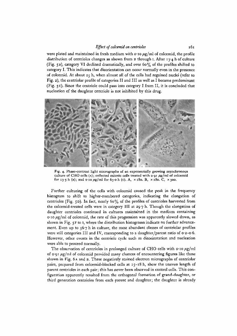
Fig. 3 presents the change in nuclear number within one cell upon incubating thecells in a medium containing o-io/tg/ml of colcemid. It is evident from these histo-grams that a single nucleus was found in over 70% of cells after the first mitotic stagein colcemid solution. Further culturing resulted in the formation of many multi-nucleated progeny, at which time not only cellular size but also the form of spreadingover the substrate were more or less changed. In the control, attached and spreadingcells on the plastic dishes had a rather long and slender asymmetric shape (Fig. 4A),while colcemid-treated cells seemed to lose their stretched appearance and to flatteninto a relatively uniform sheet (Fig. 4B). As shown in Fig. 4c, blocked cells also becamelarger as the nuclear number increased.
100 -
70 8020 30 40 50 60Time after removal of thymidine
Fig. 2. Change in the mitotic index according to the time of incubation with colcemidin CHO cell cultures. Curve A, at 4 h after removal of thymidine (arrow), 010 /*g/mlof colcemid was added to block the cells at mitosis. When a maximum mitotic indexabout 40 % was obtained at 10 h, mitotic cells were collected and placed into newculture dishes which contained o-io/fg/ml (curve B) or 091 /Jg/ml (curve C) ofcolcemid.
Changes in centriolar profile of CHO cells in the presence of colcemid
The centriole cycle in colcemid-blocked cells. As described in a previous paper(Kuriyama & Borisy, 1981), centrosomes in mammalian cultured cells attach firmly tothe nucleus. The profile of the centriolar pair on the nucleus can be easily visualizedby extracting the isolated nucleus-centrosome complex with Triton X-100 in a mediumof low ionic strength. This method was also applicable to the cells being kept in culturewith colcemid for a long time, since this complex was found to be quite stable evenin the presence of colcemid, in spite of the tendency for increased numbers of freecentrosomes in blocked cells.
Frequency histograms show many changes in the profiles of centrioles as the timeof incubation with colcemid increased. This is summarized in Fig. 5, which represents

i6o R. Kuriyama
100
50
50
50
a? 50
jenc
y
1 50LL
50
25
25
25
25
1LL-1
A
B
C
D
E
F
G
H
1
J
•
10-5
16-0
230
290
360
41 0
46-8
540
59-3
725
1 2 3 4 5 6 7 8 9 101112131415
Nuclear number in cell
Fig. 3. Change in the nuclear number in CHO cells during culture with colcemid.After treatment with thymidine for 12 h, cells were washed and transferred to thefresh medium at zero time. Colcemid (010 fig/wi) was added at 5 h, and the cultureof the collected mitotic cells was continued in the same medium. Incubation timesin hours are shown on the right.
the centriole cycle of CHO cells starting from Af-phase, through incubation witho-io/ig/ml of colcemid, until they were lysed. Fig. 5 A shows the centriole profileof rounded cells in mitosis obtained 7-5 h after washing out of thymidine, and 2-5 hafter addition of colcemid. The most abundant categories were VI and I, that is twoalmost full-sized centrioles either in an orthogonal configuration relative to each other(VI), or not (I). When the mitotic cells gathered at 10 h after removal of thymidine
Effect of colcemid on centrioles 161
were plated and maintained in fresh medium with o-io/ig/ml of colcemid, the profiledistribution of centrioles changes as shown from B through i. After 13-4 h of culture(Fig. 5B), category VI declined dramatically, and over 60% of the profiles shifted tocategory I. This indicates that disorientation can occur normally even in the presenceof colcemid. At about 25 h, when almost all of the cells had regained nuclei (refer toFig. 2), the centriolar profile of categories II and III as well as I became predominant(Fig. 5 c). Since the centriole could pass into category I from II, it is concluded thatnucleation of the daughter centriole is not inhibited by this drug.
Fig. 4. Phase-contrast light micrographs of an exponentially growing asynchronousculture of CHO cells (A); collected mitotic cells treated with 0-91 /tg/ml of colcemidfor I3'5 h (B); and 010/ig/ml for 63-0 h (c). A, x 180. B, x 180. C, x 300.
Further culturing of the cells with colcemid caused the peak in the frequencyhistogram to shift to higher-numbered categories, indicating the elongation ofcentrioles (Fig. 5D). In fact, nearly 60% of the profiles of centrioles harvested fromthe colcemid-treated cells were in category III at 297 h. Though the elongation ofdaughter centrioles continued in cultures maintained in the medium containingo-io/fg/ml of colcemid, the rate of this progression was apparently slowed down, asshown in Fig. 5 F to 1, where the distribution histograms indicate no further advance-ment. Even up to 567 h in culture, the most abundant classes of centriolar profileswere still categories III and IV, corresponding to a daughter/parent ratio of O-2-O-6.However, other events in the centriole cycle such as disorientation and nucleationwere able to proceed normally.
The observation of centrioles in prolonged culture of CHO cells with o-io/tg/mlof 0-91 /tg/ml of colcemid provided many chances of encountering figures like thoseshown in Fig. 6 A and B. These negatively stained electron micrographs of centriolarpairs, prepared from colcemid-blocked cells at 15-18 h, show the uneven length ofparent centrioles in each pair; this has never been observed in control cells. This con-figuration apparently resulted from the orthogonal formation of grand-daughter, orthird generation centrioles from each parent and daughter; the daughter is already

162 R, Kuriyama
not in perpendicular configuration to the parent, but is only partially elongated becauseof the inhibitory effect of colcemid on centriole elongation. It might be concludedthat complete elongation of the daughter centriole is not indispensable for the grand-daughter's nucleation. However, it seems worth mentioning here that the ratio oflengths of daughter to parent centrioles is always slightly larger when grand-daughters
30
30
30
30
jen
cy
p
JT 3 0L
30
30
30
30
A
^ _ B
-IC
D
- _ |
^ _ E
F
" ^ ^
C
- ^ ^
H
1
7-5(-2-5)
134(3-4)
24-6(14-6)
297(197)
34-6(24-6)
390(290)
450(350)
49-9(39-9)
56-7(46-7)
I II III IV V VI
Fig. 5. Frequency histograms of centriole profiles in CHO cells treated continuouslywith o-io/Jg/ml of colcemid. Ordinate: frequency in percentage; abscissa: categoryof centriole profile. After removal of thymidine at zero time, o-io fig/ml of colcemidwas added at 5 h to block the cells at M-phase. Isolated mitotic cells were placed intonew culture dishes with o-io/*g/ml of colcemid and kept in culture until they werelysed. Times of incubation shown on right, in hours. Times in parenthesis are afterisolation of mitotic cells.

Effect of colcemid on centrioles 163
are present than the ratio of lengths in category III. At the same time, abnormalpairs of centrioles become conspicuous, as illustrated in Fig. 6 c. The daughtercentriole (arrow) can be observed to nucleate from the longer parent at right angles,but not to nucleate from the shorter parent. It seems possible that nucleation in thecentriole cycle cannot occur from a parent centriole that is too short. Though nodirect evidence to verify this idea is now available, the result suggests that a criticallength of parent centriole is necessary for new centriole formation.
B
Fig. 6. Whole mount electron micrographs of centrioles obtained from CHO cellstreated with 0-06 /Jg/ml of colcemid for 15 h (A) or with o-io /*g/ml for 18 h (B) and25 h (c). Note the short centriole (arrow) in C. A, X 40500. B, x 41 goo. C, x 40500.
Inhibition of daughter centriole elongation by colcemid. That the primary effect ofcolcemid is to inhibit elongation of daughter centrioles was also confirmed by aseparate experiment, in which the investigation of the effect of colcemid on thecentriole cycle was begun from synchronized 5-phase cells. Results are shown in Fig. 7.After blockage of the cells with 5 mM-thymidine for 10 h (Fig. 7A), the centriolarprofiles were found to be clustered in categories II, III and IV, where the centriolepairs contain daughters less than 60% of full length. When the cells were washedfree of thymidine and cultured in fresh medium for 7 h, the peak in the histogrammoved towards longer daughter centrioles. Also centrioles in category I began toincrease as shown in Fig. JB. This histogram should be compared to the ones shownin c and D, in which the cell were cultured in media containing o-io and 0-91 /tg/mlof colcemid, respectively, after removal of thymidine. In spite of an advance in theelongation of daughter centrioles in colcemid-treated cells, the delay in the progressof the cycle is remarkable. The higher the concentration of colcemid, the more evidentthe delay in the elongation process.
Inhibition of centriole nucleation by colcemid. It was also found that higher concen-tration of the drug caused the frequency of daughter centriole nucleation, as well asthe rate of elongation, to decrease. Fig. 8 shows the change in profile distribution ofcentrioles harvested from cells cultured with various concentrations of colcemid. After

164 R. Kuriyama
exposure of collected mitotic cells to o-o6/4g/ml of colcemid for 17 h, the mostabundant class of centriolar profiles was category II (Fig. 8 A). With increasing con-centration, however, delay in the progression of the centriole cycle became obvious(Fig. 8B to D). Finally, formation of the daughter centriole is not detectable at all inthe cells treated with 0-91 fig/ml of colcemid. At the same time, elongation of thedaughter centriole itself appeared to stop immediately as shown in D, where theprofile distribution was quite similar to the mitotic cells employed here as starting
40
20
40
* 20
i-req
uenc
y i
S
20
40
20
A
B
"l —^l_ ^ _ C
- j_ D
•Jk.
60
40
20
40
20
40
20.
40
20
B
C
D
I II III I V V V I I II III I V V V IFig. 7 Fig. 8
Fig. 7. Change in the frequency histograms of centriole profiles in CHO cells uponstarting colcemid treatment from iS-phase. After treatment with 5 mM-thymidinefor 10 h, cells were washed and cultured for 7 h in fresh medium, which contained:B> °/*g/ml; C, o-io fig/wl; D, 0-91 fig/ml of colcemid. A. The centriole profiles ofcells just after removal of thymidine.Fig. 8. Change in the frequency histograms of centriole profiles in CHO cells in thepresence of various concentrations of colcemid. After isolation, the mitotic cellscontinued in culture for 17 h with: A, o-o6/ig/ml; B, o-io/tg/ml; c, o-i3/tg/ml;D, 0-91 fig/ml of colcemid.
material. This implies no more progress in the centriole cycle. In spite of these potentselective effects of colcemid on nucleation and elongation, the centriolar event ofdisorientation was not itself directly disrupted by this drug as far as we observed.Therefore, a centriolar pair appeared, composed of a parent and daughter each existingin a separate configuration. In fact, attempts were unsuccessful to detect any regularlyperpendicular arrangement between the centrioles after 23 h culture of cells with0-91 fig/ml of colcemid.

Effect of colcemid on centrioles 165
Due to the inhibitory effect of colcemid on nucleation and elongation, mitotic cellsobtained after prolonged treatment with colcemid came to have different centriolarprofiles than normal mitotic cells. However, when centrosomes containing thecentrioles with abnormal profiles were tested in an in vitro preparation of brain micro-tubules by the method developed by Gould & Borisy (1977), their nucleating activitywas as high as those in normal mitotic cells.
40
20
40
E 20
Fre
quen
cy
20
40
20
A
c
D
JjI II III I V V V I
Fig. 9. Change in the frequency histograms of centriole profiles in CHO cells afterremoval of colcemid. Isolated mitotic cells were cultured for 15 hwith 0-91 /tg/mlof colcemid. After removal of colcemid, they were cultured in fresh medium for:A, o h; B, 2-5 h; c, 6-5 h. In D, cells were cultured for 6-45 h in the medium containing0-91 ug/ml of colcemid.
Reversibility of the inhibitory effect of colcemid on centriole nucleation and elongation.Fig. 9 shows the reversible effect of colcemid on the centriolar events of nucleationand elongation. Synchronked M-phase cells were placed into culture dishes with0-91 /ig/ml of colcemid. Fifteen hours later the drug was washed out and the cellswere returned to fresh medium. Fig. 9A illustrates the frequency distribution ofcentrioles in the cells just prior to removal of colcemid. As before, the profiles wereclustered in categories V, VI and I, and also most of the pairs were disoriented regard-less of the length of daughter centrioles. After 2-5 h culture in fresh medium (Fig. 9B),the peak in the histogram shifted to category II, indicating the occurrence of centriolenucleation. As shown in Fig. 9 c, further culturing of the cells permits these daughtercentrioles to elongate. This distribution should be compared to the one shown in D,which presents the profiles of centrioles obtained from the cells cultured in a colcemid-containing medium for the same duration as those in c. This histogram was essentiallythe same as the control illustrated in A. Therefore, it may be concluded that the

R. Kuriyama
inhibitory effect of colcemid on the centriole cycle is completely reversible, at leastin terms of centriolar nucleation and elongation.
Ultrastructural changes in CHO cells by colcemid treatment
As described above, longer-term incubation of cells with colcemid caused severalstructural changes in CHO cells. The fine ultrastructural change in cells at 27-5 h afteraddition of o-io/tg/ml of colcemid was easily characterized from sectioned electronmicrographs. In treated cells almost all cytoplasmic microtubules depolymerized.Instead, a massive accumulation of filamentous bundles became prominent. Theyformed discrete large bundles of aligned 8 to 10-nm filaments in the cytoplasm of eachcell (Fig. 10A, B). Although this filamentous structure appeared most conspicuouslyin the cytoplasm of interphase cells, abundant filaments running through the cyto-plasm could also be observed within the cells at the mitotic stage (Fig. 10D).
Fig. 10. Thin-section electron micrographs of colcemid-treated CHO cells withnucleus (A, B, C) or chromosomes (D). Arrows in A show the bundle structure offilaments. B shows a high magnification of the area outlined in A. A filamentous bundlenear the centriole is illustrated in C. A, x 3400. B, X 20500. c, x 51 800. D, x 10800.
Distinct changes in centriolar ultrastructure were also produced by colcemid. Somerepresentative electron micrographs of whole mount preparations of treated cells arepresented in Fig. 11. Fig. 11A shows the outgrowth of a microtubule from the wallof centriolar triplet microtubules. In some cases, a triplet (B) or doublet microtubule(c) and plural microtubules from different triplets (c) were detected jutting out fromdifferent triplets. The outgrowing microtubules are stable, unlike cytoplasmic ones,since conditions employed here in whole mount preparations to visualize centrioles

Effect of colcemid on centrioles 167
A
Fig. 11. Whole mount electron micrographs of centrioles in abnormal configurations,obtained from CHO cells treated with 006-091 /ig/ml of colcemid for 4-44.5 h.A, X 39OOO. B, X 4 2 8 0 O . C, X 27OOO. D, X45OOO. E, X 383OO. F, X 27OOO. G, X 33 3 0 0 .H, X 280OO. I, X 2SOOO.
attached to the nuclei made cytoplasmic microtubules depolymerize completely.Stable microtubules were also observed near centrioles as bunching clusters (Fig. n D,E, F). We do not have any data to relate these short bundle of microtubules to theabnormal nucleating form of daughter centrioles formed under the influence ofcolcemid. However, we did have many chances to detect several kinds of abnormalcombinations of centrioies (Fig. 11G to i).

168 R. Kuriyama
DISCUSSION
The present results suggest that, even in the presence of colcemid, CHO cells retaintheir ability to go through their own specific nuclear cycle after overcoming thearrested metaphase caused by this alkaloid. The same endogenous recovery fromcolchicine or colcemid has already been demonstrated in a variety of organisms(Stubblefield, 1964; Kleinfeld & Sisken, 1966; Rosenbaum& Carlson, 1969; Fragata,1972; Wunderlich & Heumann, 1974). In their report on cilia regeneration in Tetra-hymena, Rosenbaum & Carlson (1969) observed many vacuoles with yellow crystalsin the cell coincident with the recovery from the colchicine-induced arrest; theyfinally postulated that the overcoming of the colchicine inhibition in Tetrahymena wasprobably a unique phenomenon related to the feeding and excretory mechanisms ofthis organism. Cultured mammalian cells (Stubblefield, 1964; Kleinfeld & Sisken,1966) including the CHO cells described in this report, however, appeared to retaina considerable amount of colcemid in the cytoplasm, sufficient to suppress any forma-tion of cytoplasmic microtubules. We do not have any data to explain reasonably thecellular control mechanism for recovery from colcemid blockage, but in vitro experi-ments suggest a possibility that colchicine inhibition on microtubule assembly is notabsolute (Sternlicht & Ringel, 1979; Farrell & Wilson, 1980). Some free tubulin dimeraddition occurs over the colchicine-tubulin dimer block to form copolymer, eventuallyresulting in recovery from the effect of the block. Further studies are awaited forelucidation.
Graphic representation of the centriole cycle provides us with a means for deter-mining the specific effects of colcemid on each centriolar event. It is elongation, ratherthan other events in the centriole cycle, that is preferentially inhibited by colcemid.This results in the perpendicular formation of a short daughter centriole from theparent that is not full-sized. Therefore, it might be concluded that the several centriolarevents can occur, to some extent, independently of each other. However, the selectiveeffects of colcemid on the centriolar structure were found to be concentration-dependent: culturing the cells with 0-91/tg/ml of colcemid produced interferencenot only with elongation, but also with nucleation itself. The potent ability of centriolesto replicate and mature in the presence of colcemid (Stubblefield, 1968) may be due,at least in part, to the drug concentration used in the experiments. In spite of theaforementioned effect of colcemid on the centriole cycle, there are a few reportsobserving the considerable number of centrioles produced in the pituitary (Hubert,Flament-Durand & Dustin, 1974; Flament-Durand, Hubert & Dustin, 1976) by theincubation of dissected tissues with microtubular poisons such as colchicine or vin-cristine: this treatment also led to ciliogenesis or formation of macrotubules andcrystalline structures within the cells. Since centriolar formation appears to be greatlyinfluenced by the conditions used in each experiment, involving such variables asdehydration (Hubert et al. 1974; Flament-Durand et al. 1976), Tris-buffer medium(Margulis, Banerjee & White, 1969) or drug concentration, it is conceivable thatthese apparently contradictory phenomena in pituitary and tissue culture cells resultedfrom different sensitivities to treatment in each specific cell.

Effect of colcemid on centrioles 169
Much has been observed concerning the morphological changes in cells afterexposure to microtubular poisons. At the cellular level, these drugs cause disruptionof cytoplasmic or spindle microtubules, resulting in an apparent increase in thenumber of 10-nm filaments similar to that reported in colchicine-treated cells (Ishikawa,Bischoff & Holtzer, 1968; Wisniewski, Shelanski & Terry, 1968; Krishan & Hsu,1969; Olmsted et al. 1970; Goldman, 1971). A recent paper by Pytela & Wiche (1980)suggested that high molecular weight polypeptides (HMWPs) prepared from thefraction of intermediate filaments might have a dual role in the cell, serving not onlyas regulators of microtubule assembly but also as linker components between micro-tubules and intermediate filaments. Therefore, it seems highly probable that afterdestruction of microtubules by colcemid, the remaining HMWPs caused the 10-nmfilaments to associate with one another, consequently forming discrete large filamentousbundles in the cytoplasm. The most prominent alteration in the centriole revealed byelectron microscopy of whole mount preparations was the growing out of microtubulesfrom the triplet microtubules on one side of the centriole. It is not clear whether thisoutgrowth results from colcemid disturbing the programming for centriole construction;however, the same morphological change has already been reported (Zuckerberg &Solari, 1973) in the seminiferous epithelium of the mouse after treatment with vinblastine.
It is well known that, irrespective of their morphological similarities, microtubulesobtained from various sources show a remarkable difference in their structural stability(Behnke & Forer, 1967). Generally, cytoplasmic microtubules are very labile, beingeasily disassembled by treatments such as low temperature or high pressure. On theother hand, microtubules from motile structures or from the centriole are relativelystable and resistant to those treatments. Colchicine or colcemid prevents tubulinassembly into cytoplasmic microtubules both in vivo and in vitro, but these drugs arenever able to depolymerize ciliary, flagellar or centriolar microtubules.
As for the effect of these drugs on the assembly of stable microtubules, however,several investigators have already reported that ciliary and flagellar regeneration couldnot proceed in the presence of the alkaloids in Chinese hamster fibroblasts (Stubblefield& Brinkley, 1966), Chlamydomonas reinhardii (Rosenbaum, Moulder & Ringo, 1969),Tetrahymena pyriformis (Rosenbaum & Carlson, 1969), Stentor coeruleus (Neviackas &Margulis, 1969) or in sea urchin embryos (Iwaikawa, 1977). They postulated that itwas the potent binding activity of the drugs to tubulin, the microtubule subunitprotein (Taylor, 1965; Borisy & Taylor, 1967), that interfered with regeneration ofcilia or flagella by preventing the assembly of tubulin into motile microtubules.Results obtained in this work also demonstrate an inhibitory effect of colcemid oncentriole formation in CHO cells. Therefore, it seems plausible that the stable ciliaryand flagellar as well as centriolar microtubules are generated from colcemid-sensitivetubulin subunits sharing an identical unit for labile cytoplasmic microtubules. Further-more, we have already obtained some evidence concerning the instability of recon-stituted microtubules from ciliary and flagellar outer-doublet tubulin (Kuriyama,1976). Differences in structural stability might not be due to the characteristics of thetubulin molecule itself. Rather, they could be due to the properties of the additionalsupporting substances surrounding the motile or centriole microtubules, or some

170 R. Kuriyama
conformational changes occurring in tubulin subunits just at the time of formation ofthe cilia, flagella or centrioles in vivo.
The author wishes to express her thanks to Professor H. Kanatani of the National Institutefor Basic Biology and Professor H. Sakai of the University of Tokyo for their kind criticism inpreparing the manuscript. This work was supported by a postdoctoral fellowship in specialareas from the Ministry of Education, Science and Culture in Japan.
REFERENCES
BEHNKB, O. & FORER, A. (1967). Evidence for four classes of microtubules in individual cells.J. Cell Set. 2, 169-192.
BORISY, G. G. & TAYLOR, E. W. (1967). The mechanism of action of colchicine. Binding ofcolchicine-3H to cellular protein. J. Cell Biol. 34, 525-533.
FARRELL, K. W. & WILSON, L. (1980). Proposed mechanism for colchicine poisoning ofmicrotubules ressaembled in vitro from Strongylocentrotus purpiiratus sperm tail outerdoublet tubulin. Biochemistry 19, 3048—3054.
FLAMENT-DURAND, J., HUBERT, J. P. & DUSTIN, P. (1976). Centriolo- and ciliogenesis in therat's pituicytes under the influence of microtubule poisons in vitro. Exptl Cell Res. 99, 435-438.
FRAGATA, M. (1972). Endogenous recovery from colchicine-induced inhibition of growth.Experientia 28, 506—507.
GOLDMAN, R. D. (1971). The role of three cytoplasmic fibers in BHK-21 cell motility. I.Microtubules and the effects of colchicine. J. Cell Biol. 51, 752-762.
GOULD, R. R. & BORISY, G. G. (1977). The pericentriolar material in Chinese hamster ovarycells nucleates microtubule formation. J. Cell Biol. 73, 601-615.
HUBERT, J.-P., FLAMENT-DURAND, J. & DUSTIN, P. (1974). Centrioles and cilia multiplicationin the pituitary of the rat after Furosemid and colchicine treatment. Cell Tiss. Res. 149,349-301-
ISHIKAWA, H., BISCHOFF, R. & HOLTZER, H. (1968). Mitosis and intermediate-sized filamentsin developing skeletal muscle. J. Cell Biol. 38, 538-555.
IWAIKAWA, Y. (1977). Effect of colcemid on the cilia development in the sea urchin embryo.Coll. gen. Educ, Nagoya Univ., Res. Bull, zi, 81-88.
KLEINFELD, R. G. & SISKEN, J. E. (1966). Morphological and kinetic aspects of mitotic arrestby and recovery from colcemid. J. Cell Biol. 31, 369-379.
KRISHAN, A. & Hsu, D. (1969). Observations on the association of helical polyribosomes andfilaments with vincristine-induced crystals in Earle's L-cell fibroblasts. jf. Cell Biol. 43,553-563.
KURIYAMA, R. (1976). In vitro polymerization of flagellar and ciliary outer fiber tubulin intomicrotubules. J. Biochem., Tokyo 80, 153-165.
KURIYAMA, R. & BORISY, G. G. (1981). The centriole cycle in Chinese hamster ovary cellsas determined by whole mount electron microscopy.^. Cell Biol. (In Press.)
MARGOLIS, R. L. & WILSON, L. (1977). Addition of colchicine-tubulin complex to micro-tubule ends: The mechanism of substoichiometric colchicine poisoning. Proc. natn. Acad.Set. U.S.A. 74, 3466-3470.
MARGULIS, L., BANERJEE, S. & WHITE, T. (1969). Colchicine-inhibited cilia regeneration:explanation for lack of effect in Tris buffer medium. Science, N. Y. 164, 1177-1178.
NEVIACKAS, J. A. & MARGULIS, L. (1969). The effect of colchicine on regenerating membranellarcilia in Stentor coeruleus.J. Protozool. 16, 165-171.
OLMSTED, J. B., CARLSON, K., KLEBE, R., RUDDLE, F. & ROSENBAUM, J. (1970). Isolation ofmicrotubule protein from cultured mouse neuroblastoma cells. Proc. natn. Acad. Set.U.S.A. 65, 129-136.
PYTELA, R. & WICHE, G. (1980). High molecular weight polypeptides (270000-340000) fromcultured cells are related to hog brain microtubule-associated proteins but copurify withintermediate filaments. Proc. natn. Acad. Sci. U.S.A. 77, 4808-4812.

Effect of colcemid on centrioles 171
RATTNBR, J. B. & PHILLIPS, S. G. (1973). Independence of centriole formation and DNAsynthesis. J. Cell Biol. 57, 359-372.
ROBBINS, E., JENTZSCH, G. & MICALI, A. (1968). The centriole cycle in synchronized HeLacells. J. Cell Biol. 36, 329~339-
ROSENBAUM, J. L. & CARLSON, K. (1969). Cilia regeneration in Tetrahymena and its inhibitionby colchicine. J. Cell Biol. 40, 415-425.
ROSENBAUM, J. L., MOULDER, J. E. & RINGO, D. L. (1969). Flagellar elongation and shorteningin Chlamydomonas. The use of cycloheximide and colchicine to study the synthesis andassembly of flagellar proteins. J. Cell Biol. 41, 600-619.
STEBNLICHT, H. & RINGEL, I. (1979). Colchicine inhibition of mictrotubule assembly viacopolymer formation. J. biol. Chetn. 254, 10540-10550.
STUBBLEFIELD, E. (1964). DNA synthesis and chromosomal morphology of Chinese hamstercells cultured in media containing iV-deacetyl-JV-methylcolchicine (Colcemid). In Cyto-genetics of Cells in Culture (ed. R. J. C. Harris), pp. 223-248. New York: Academic Press.
STUBBLEFIELD, E. (1968). Centriole replication in a mammalian cell. In The Proliferation andSpread of Neoplastic Cells, pp. 175-193. Baltimore, Md: Williams and Wilkins.
STUBBLEFIELD, E. & BRINKLEY, B. R. (1966). Cilia formation in Chinese hamster fibroblasts invitro as a response to Colcemid treatment. J. Cell Biol. 30, 645-652.
STUBBLEFIELD, E. & KLEVECZ, R. (1965). Synchronization of Chinese hamster cells by reversalof Colcemid inhibition. Exptl Cell Res. 40, 660-664.
TAYLOR, E. W. (1965). The mechanism of colchicine inhibition of mitosis. I. Kinetics of inhibi-tion and the binding of 3H-colchicine. J. Cell Biol. 25, 145-160.
WISNIEWSKI, H., SHELANSKI, M. L. & TERRY, R. D. (1968). Effects of mitotic spindle inhibitorson neurotubules and neurofilaments in anterior horn cells. J. Cell Biol. 38, 224-229.
WUNDERLICH, F. & HEUMANN, H.-G. (1974). In vivo reassembly of microtubules in the presenceof intracellular colchicine. Cytobiologie 10, 140-151.
ZUCKERBERG, C. & SOLARJ, A. J. (1973). Centriolar changes induced by vinblastine sulphate inthe seminiferous epithelium of the mouse. Expl Cell Res. 76, 470-475.
(Received 16 March 1981)