Effect of ciprofloxacin incorporation in PVA and PVA ...
40
HAL Id: hal-00915089 https://hal.archives-ouvertes.fr/hal-00915089 Submitted on 12 Dec 2013 HAL is a multi-disciplinary open access archive for the deposit and dissemination of sci- entific research documents, whether they are pub- lished or not. The documents may come from teaching and research institutions in France or abroad, or from public or private research centers. L’archive ouverte pluridisciplinaire HAL, est destinée au dépôt et à la diffusion de documents scientifiques de niveau recherche, publiés ou non, émanant des établissements d’enseignement et de recherche français ou étrangers, des laboratoires publics ou privés. Effect of ciprofloxacin incorporation in PVA and PVA bioactive glass composite scaffolds Mostafa Mabrouk, Amany Mostafa, Hassane Oudadesse, Azza A. Mahmoud, Mohamed I. El-Gohary To cite this version: Mostafa Mabrouk, Amany Mostafa, Hassane Oudadesse, Azza A. Mahmoud, Mohamed I. El-Gohary. Effect of ciprofloxacin incorporation in PVA and PVA bioactive glass composite scaffolds. Ceramics International, Elsevier, 2013, pp.In Press. 10.1016/j.ceramint.2013.09.033. hal-00915089
Transcript of Effect of ciprofloxacin incorporation in PVA and PVA ...
HAL Id: hal-00915089https://hal.archives-ouvertes.fr/hal-00915089
Submitted on 12 Dec 2013
HAL is a multi-disciplinary open accessarchive for the deposit and dissemination of sci-entific research documents, whether they are pub-lished or not. The documents may come fromteaching and research institutions in France orabroad, or from public or private research centers.
L’archive ouverte pluridisciplinaire HAL, estdestinée au dépôt et à la diffusion de documentsscientifiques de niveau recherche, publiés ou non,émanant des établissements d’enseignement et derecherche français ou étrangers, des laboratoirespublics ou privés.
Effect of ciprofloxacin incorporation in PVA and PVAbioactive glass composite scaffolds
Mostafa Mabrouk, Amany Mostafa, Hassane Oudadesse, Azza A. Mahmoud,Mohamed I. El-Gohary
To cite this version:Mostafa Mabrouk, Amany Mostafa, Hassane Oudadesse, Azza A. Mahmoud, Mohamed I. El-Gohary.Effect of ciprofloxacin incorporation in PVA and PVA bioactive glass composite scaffolds. CeramicsInternational, Elsevier, 2013, pp.In Press. �10.1016/j.ceramint.2013.09.033�. �hal-00915089�
1
Effect of ciprofloxacin incorporation in PVA and PVA bioactive
glass composite scaffolds
M. Mabrouka,b
, A. A. Mostafaa,b
, H. Oudadessea, A.A. Mahmoud
c, and M. I. El-Gohary
d
aUniversité de Rennes 1, UMR CNRS 6226, 263 av. du général leclerc, 35042 Rennes Cedex,
France. bBiomaterials Department ., National Research Centre NRC, Cairo, Egypt.
cPharmaceutical Technology Department., National Research Centre NRC, Cairo, Egypt. dPhysics Department., Faculty of Science, Al -Azhar University, Cairo, Egypt.
Abstract
Scaffolds are implants used to deliver cells, drugs, and genes into the body in a local
controlled release pattern which offers many advantages over systematic drug delivery.
Composite scaffolds of polyvinyl alcohol (PVA) and quaternary bioactive glass (46S6
system) with different ratios of glass contents were prepared by lyophilisation technique.
The broad spectrum antibiotic ciprofloxacin (Cip) was impregnated to the scaffold during
the fabrication in a concentration of 5, 10 and 20%. Biodegradation rate and in-vitro
mineralization of the prepared scaffolds were performed by soaking the scaffolds in
simulated body fluid (SBF). Phase identification, microstructure, porosity, bioactivity,
mechanical properties and drug release pattern in PBS were characterized by XRD, SEM
coupled with EDS, Hg-porosimeter, inductively coupled plasma-optical emission
spectroscopy (ICP-OES), universal testing machine, fourier transform infrared (FTIR)
and UV-spectrophotometer, respectively. A porous scaffold has been obtained with
porosity up to 85%. By increasing the glass contents in the prepared scaffold the porosity
and the degradation rate decrease however, the compressive strength was enhanced. A
sustained drug release pattern was observed with a quasi-Fickian diffusion mechanism.
The formulated ciprofloxacin loaded porous polyvinyl alcohol scaffold gave an
acceptable physicochemical properties and was able to deliver the drug in a prolonged
release pattern which offers a distinguish treatment for osteomylitis as well as local
antibacterial effect.
Keywords: Tissue engineering, scaffolds, ciprofloxacin, drug release, freeze drying,
polyvinyl alcohol.
2
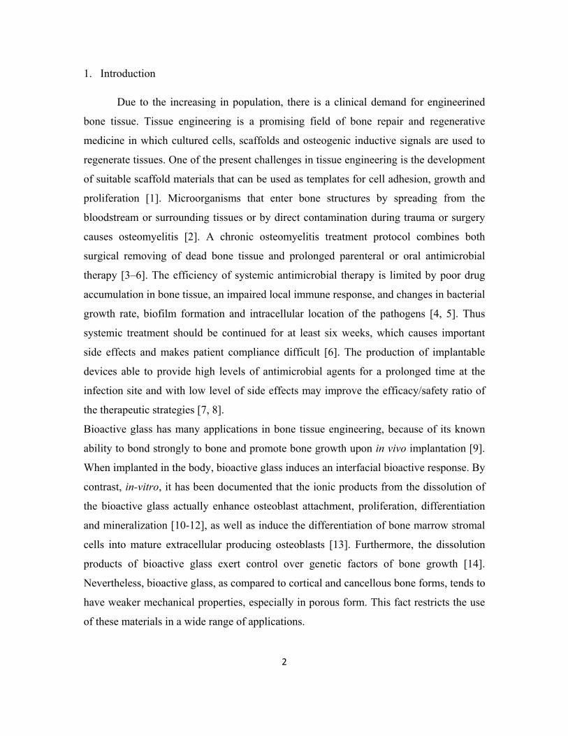
1. Introduction
Due to the increasing in population, there is a clinical demand for engineerined
bone tissue. Tissue engineering is a promising field of bone repair and regenerative
medicine in which cultured cells, scaffolds and osteogenic inductive signals are used to
regenerate tissues. One of the present challenges in tissue engineering is the development
of suitable scaffold materials that can be used as templates for cell adhesion, growth and
proliferation [1]. Microorganisms that enter bone structures by spreading from the
bloodstream or surrounding tissues or by direct contamination during trauma or surgery
causes osteomyelitis [2]. A chronic osteomyelitis treatment protocol combines both
surgical removing of dead bone tissue and prolonged parenteral or oral antimicrobial
therapy [3–6]. The efficiency of systemic antimicrobial therapy is limited by poor drug
accumulation in bone tissue, an impaired local immune response, and changes in bacterial
growth rate, biofilm formation and intracellular location of the pathogens [4, 5]. Thus
systemic treatment should be continued for at least six weeks, which causes important
side effects and makes patient compliance difficult [6]. The production of implantable
devices able to provide high levels of antimicrobial agents for a prolonged time at the
infection site and with low level of side effects may improve the efficacy/safety ratio of
the therapeutic strategies [7, 8].
Bioactive glass has many applications in bone tissue engineering, because of its known
ability to bond strongly to bone and promote bone growth upon in vivo implantation [9].
When implanted in the body, bioactive glass induces an interfacial bioactive response. By
contrast, in-vitro, it has been documented that the ionic products from the dissolution of
the bioactive glass actually enhance osteoblast attachment, proliferation, differentiation
and mineralization [10-12], as well as induce the differentiation of bone marrow stromal
cells into mature extracellular producing osteoblasts [13]. Furthermore, the dissolution
products of bioactive glass exert control over genetic factors of bone growth [14].
Nevertheless, bioactive glass, as compared to cortical and cancellous bone forms, tends to
have weaker mechanical properties, especially in porous form. This fact restricts the use
of these materials in a wide range of applications.
3
One approach to enhancing the mechanical properties of materials is the elaboration of
inorganic–organic composites, as they often show an excellent combination between
strength and toughness, as well as improved characteristics, when compared to their
individual components.
Bioactive scaffolds were able to provide a three dimensional (3-D) architecture for
enhancing osteogenesis and angiogenesis. Most of the scaffolds were built on complex
constructs of several composition and additives. Natural polymer such as chitosan,
alginate or gelatin [15-17] or synthetic polymer such as poly lactic acid (PLA), poly
glycolic acid(PGA) an poly lactic –co-glycolic acid (PLGA) are widely used for polymer
scaffold. However, some drawbacks still exist upon using polymers including: formation
of acidic species, this leads to a decrease in pH and tissue inflammation [18, 19]. Among
several choices of polymers, poly (vinyl alcohol) (PVA), a hydrophilic semicrystalline
polymer, has been frequently explored as an implant material in wide array of biomedical
applications such as drug delivery systems, wound dressings, membranes, surgical repairs
and artificial skin, mainly due to its excellent mechanical strength, biocompatibility, and
nontoxicity [15, 16].
Direct application of antibiotics or growth factors to the defect site and the control release
of the drug delivery systems have been developed [20, 21]. Antibiotics can be
incorporated into bone substitutes [22]. The sustained release of antibiotics from scaffold
structures has been widely demonstrated to control the kinetic of the release [23]. Drug
releasing polymer scaffolds should have certain criteria such as a homogeneity of the
drug distribution, good binding affinity of the drug to the scaffold, define amount of the
drug loading as well as release kinetic and stability [23-25]. The most widely acceptable
agents in local delivery system for chronic osteomylitis are amino glycosis and
quinolones. Ciprofloxacin (1-cyclopropyl-6-fluro-1, 4-dihydro-4-oxo-7- (1-pipera
Zinyl)-3- quinoline carboxylic acid) is a fluroquinolone derivative, widely used in
osteomyelitis because of its favorable penetration and bactericidal effect on all the
probable osteomyelitis pathogens [29]. The main purpose of the current study was to
develop and fabricate a construct of bioactive scaffold combining an antibiotic
(ciprofloxacin). Bioactive glass powders were used as powder fillers to reinforce poly
4
vinyl alcohol scaffold. Characterization of these scaffolds before and after addition of the
drug has been investigated by TEM, XRD, FTIR, mercury prosimeter, SEM coupled with
EDS, ICP-OES, universal testing machine and UV-spectrophotometer. Bioactivity of the
composite scaffolds and their use as drug carriers to effectively deliver therapeutic agent
in a sustained and controlled manner has been studied.
2. Materials and methods
2.1. Fabrication of PVA/SG-B –Cip scaffolds
46S6 bioactive glass powder was synthesized by sol-gel method SG-B as following.
A colloidal solution of 46S6 was prepared by sol-gel. Initially, 56 ml of tetraethoxysilane
(TEOS:Fluka, Mwt=208.33) was added to 350 ml of distilled water and 350 ml of
ethanol at room temperature . pH was adjusted at 2 by nitric acid with continuous stirring
for 1hr, addition of 24.5 g of calcium nitrate hydrate (Fluka, Mwt=236) to the above
solution continue stirring till dissolving, addition of 21.07 g of sodium hydroxide (Prolab,
Mwt= 40) to the above mixture (the previous mixture was named solution A), 10 g of
polyethylene glycol (PEG-Fluka Mwt=600) was dissolved in 400 ml of distillate water at
room temperature. 3.43 g of ammonium dihydrogen phosphate (MERK, Mwt=115.03)
was added to the PEG solution, this mixture was named solution B. Furthermore, solution
(B) was gradually added on solution (A) with continuous stirring over night. The resulted
solution was filtrated and washed with distillate water for 3 times and with ethanol for 1
time using centrifuge with 1650 rpm for 10 min drying of the washed gel at 70ºC for
overnight. The dried powder was calcinied at 600οC for 2hr.
2.1.1. Composite preparation
PVA/SG-B-Cip composite scaffolds were prepared by employing freeze drying technique
as demonstrated in figure (1.1). Firstly, PVA (ALDRICH, Mwt= 67.000) was dissolved
in distilled water at 80oC for 2hr using a polymer concentration of 15 wt%. Three
different concentrations of SG-B (nano particles see TEM image figure (1.2)) 33.5,50 and
5
66.5 wt%, were added to the PVA solution and continue stirred for overnight using a
magnetic stirrer in order to break the SG-B agglomerates and ensure a better
(homogenous) distribution of SG-B particles in the composite scaffolds. Three different
concentrations of ciprofloxacin 5, 10 and 20 wt% were added to the above mixture
continue stirred for 1hr (scaffolds with the same composition was prepared without drug
loading as a control). Scaffolds were casted in Teflon molds and kept at -18oC for
overnight, and freeze dried for 24 hr then the scaffolds were removed from the molds and
kept in the desecrator for further analysis as mentioned below.
2.2. Morphological and microstructural properties
The microarchitecture of these scaffolds was assessed qualitatively using scanning
electron microscopy (SEM) and quantitatively using mercury intrusion porosimetry
(MIP) and liquid displacement method.
2.2.1. SEM
SEM analyses were performed on a thin piece of scaffold sheared from the center using a
sharp razor blade after soaking in liquid nitrogen for 2 minutes. Scaffolds were observed
using (max. of 20 kV) SEM with gold palladium coating to avoid beam damage of the
polymer, which can be prominent on these scaffolds that have very fine microstructure.
2.2.2. MIP
MIP was performed using (PORESIZER 9320 V2.08) to determine median pore
diameter, and percent porosity.
2.2.3. Liquid displacement method
Scaffold samples were submersed in cyclohexan for 1 hr. The volume of a scaffold
immersed in the fluid is equal to the volume of the displaced fluid, and we can calculate
the porosity from the following equation:
P % = [(W1-W3)/ (W2-W3)] x100 (1)
6
Where W1: weight of the scaffold before immersion, W2: weight of the scaffold after
immersion and W3: weight after drying by this method we can just get the porosity
percentage (P %).
2.3. Mechanical properties of the prepared scaffolds
Bones are often submitted to compression stress in the body. It has been broadly accepted
by the research community to perform compression assays for evaluating biomaterials for
potential use as bone repair. For that reason, the mechanical behavior of the composites
was evaluated by compression tests. Specimens were evenly cut from the most
homogeneous region of the foam to form blocks measuring 10 × 10 × 10mm3. These
samples were positioned between parallel plates using equipment EMIC DL 3000 and
compressed with a crosshead speed of 0.5mm·min−1
and a 1.0 kN load cell. At least three
samples (n = 3) of each hybrid system were measured and the results averaged.
Compressive strength tests were carried out to determine the effect of bioactive glass and
the drug concentrations on the mechanical strength of scaffolds.
2.4. Bioactivity
2.4.1. Phase analysis by X-ray diffraction (XRD).
X-ray diffraction (XRD) technique (PhilipsX’Pert-MPD system with a CuKa wavelength
of 1.5418A°) was used to analyze the structure of the prepared SG-B and PVA/ SG-B
composite scaffolds. The diffract meter was operated at 40kV and 30mA at a 2ɵ range of
10–70°employing a step size of 0.058/s.
2.4.2. Infrared studies
Fourier transformed infrared analysis (FTIR; Nicolet Magna-IR 550 spectrometer,
Madison, Wisconsin) was performed to identify the nature of the chemical bonds
between atoms. The samples were small pellets, of 0.5 cm diameter, obtained by pressing
the scaffolds powder with KBr.
7
2.4.3. SEM coupled with EDS
The morphology of surfaces of scaffolds was studied by using scanning electron
microscopy (SEM) (Jeol JSM 6301). It is a technique of morphological analysis based on
the principle of electron-matter interactions. To allow surface conduction, the scaffolds
were metalized by gold-palladium layer (a few µm of thickness) before being introduced
into the analysis room. Semi quantitative chemical analysis on scaffolds surfaces after
immersion in SBF, covered by gold-palladium layer to allow surface conduction, was
performed by energy dispersive spectroscopy (EDS) in Jeol JSM 6400.
2.4.4. ICP-OES
The concentrations of (Ca, p and Si) elements after each soaking time in SBF were
measured by using Inductively Coupled Plasma-Optical - Emission Spectrometry (ICP-
OES). This method offers a high sensitivity, less than 1µg/g depending on the analyzed
matrix and offers a high accuracy. The principle is based on the determination of the
amount of each element present in solution by analyzing the intensity of the radiation
emitted at the specific elemental frequency after the nebulisation of atoms.
2.5. In vitro degradation studies
The degradation pattern of the composite scaffold was studied in PBS medium at 37 ◦C.
groups of scaffolds (3 scaffolds in each) were immersed in PBS and incubated for up to
30 days. After each period time one of the scaffolds was washed two times by distilled
water to remove ions adsorbed on the surface and was dried. Initial weight of the scaffold
was noted as Wo and dry weight as Wt. The degradation of scaffolds was calculated
using the following formula:
Degradation % = (Wo – Wt)/Wo × 100 (2)
8
2.6. Ciprofloxacin release behavior
Drug incorporation into the scaffolds was investigated by means of XRD, FTIR and SEM
coupled with EDS.
Phosphate buffer solution (PBS), pH 7.4 (10 ml), previously heated at 37⁰C, was added to
test tubes containing freshly prepared scaffolds. The tubes were kept at 37⁰C with shaking
(50 oscillations min-1
) and, at pre-established times, 1 ml samples of the release medium
were taken and the drug concentration was determined spectrophotometrically at 277 nm
(Jenway 6705 UV/Vis, UK). The samples were replaced with fresh buffer in order to
keep constant volume of medium. All experiments were carried out in triplicate.
Ciprofloxacin release was monitored for 360 hr.
2.7. Mechanism of ciprofloxacin release
Korsmeyer–Peppas model [31] was used to find out the mechanism of drug release from
the investigated scaffolds:
Mt/ M∞= Ktn (3)
Where, Mt / M∞ is fraction of drug released at time t, k is the rate constant and n is the
release exponent. In case of quasi-Fickian diffusion the value of n < 0.5, Fickian
diffusion n =0.5, non-Fickain or anomalus transport n =0.5-1.0 and Case II transport n =
1.0.
9
3. Results and discussion
3.1. Morphological and microstructural properties
The effect of the particle size of SG-B particles on the properties of PVA/SG-B scaffolds,
which are being developed for tissue engineering applications [32]. The morphology of
the prepared scaffolds is presented in (figure 2); in which we can observe that all the
prepared scaffolds have wide range of interconnected pores including macro, micro and
nanopores as it also confirmed by mercury porosimeter. PVA scaffold shows highly
interconnected pores with smooth pore walls. Incorporation of ciprofloxacin into the
PVA scaffold changes the arraying and shapes of pores and its thickness due to the
interaction between ciprofloxacin and PVA. As the glass content increases the porosity
decreases and the pore walls becomes thicker. Among several processing techniques, the
freeze drying method was chosen since it could provide easy control of the pore structure
[33]. The co-existence of macropores and micropores is not only favorable for the
ingrowth of cells and new tissue but also beneficial to the exchange of nutrients and
metabolic waste [34]. The porosity percentage for the prepared scaffolds was determined
by MIP and liquid displacement methods and there was no significant difference between
the two methods as it is demonstrated in table (1) [32].
3.2. Mechanical properties
The mechanical behavior of the prepared scaffolds was characterized by determining the
compressive strength. The PVA alone exhibit low compressive strength as shown in
figure (3). In the produced scaffolds, a marked change could be observed, as the amount
of glass and drug increased the compressive strength increase. The incorporation of SG-B
into PVA polymer enhances the compressive strength, due to their small particle size and
large surface area which results in great attachment of SG-B particles to the polymer
matrix as reported before [33-38].
3.3. Bioactivity
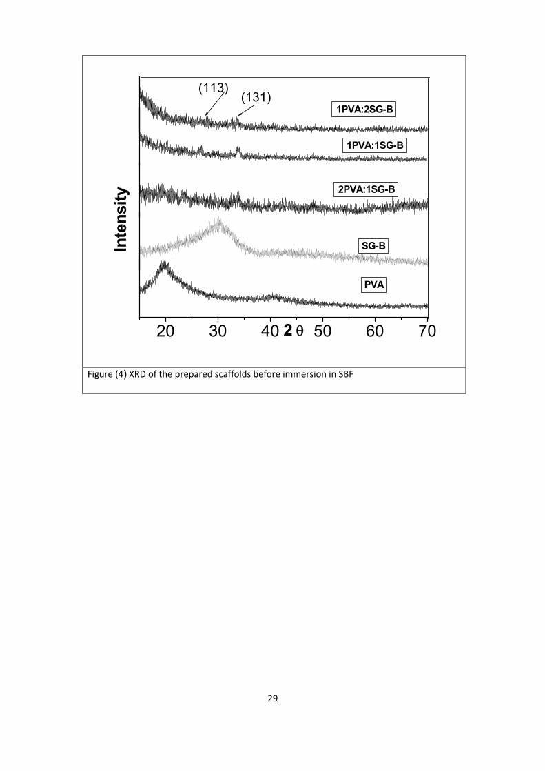
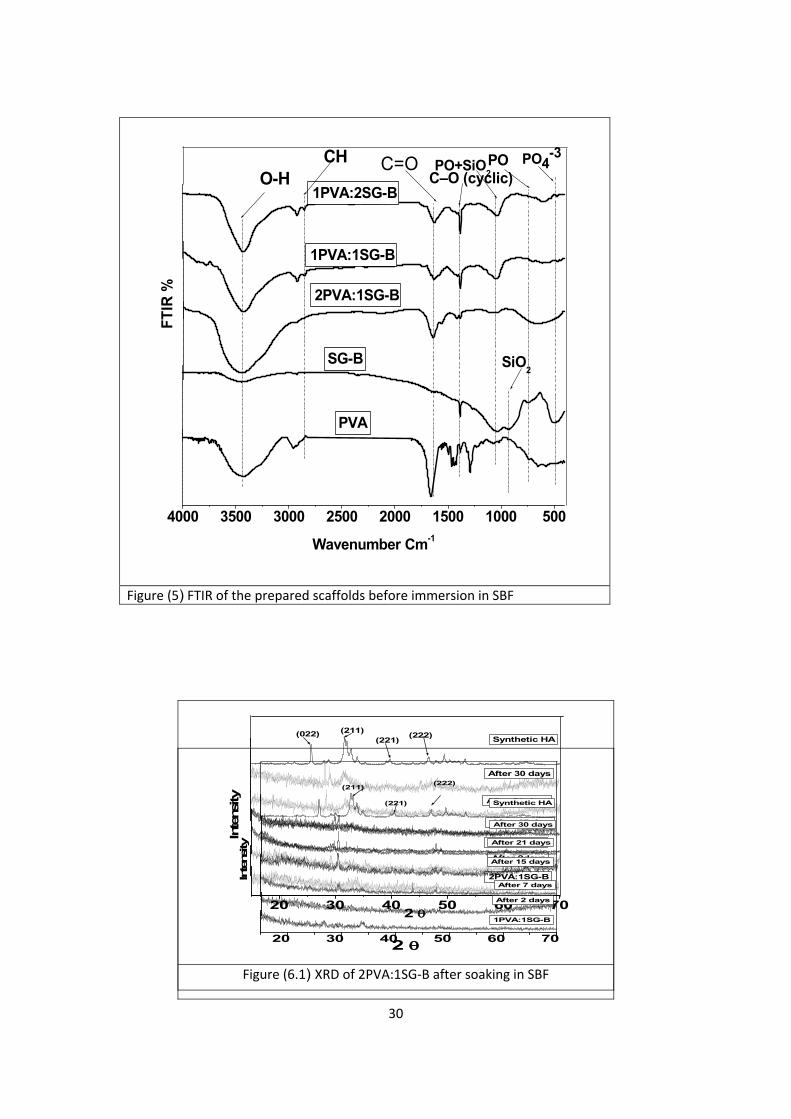
Figures (4) and figure (5) represent XRD and FTIR respectably of the prepared PVA/SG-
B composite scaffolds with SG-B and PVA as references before immersion in SBF.
10
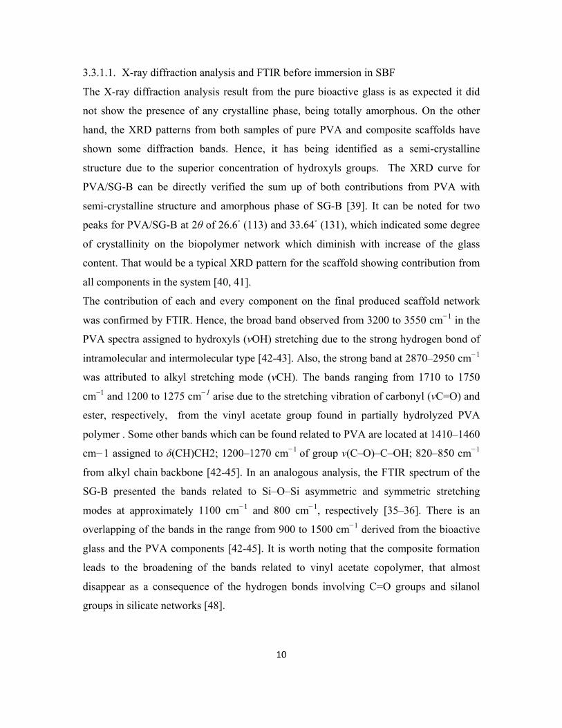
3.3.1.1. X-ray diffraction analysis and FTIR before immersion in SBF
The X-ray diffraction analysis result from the pure bioactive glass is as expected it did
not show the presence of any crystalline phase, being totally amorphous. On the other
hand, the XRD patterns from both samples of pure PVA and composite scaffolds have
shown some diffraction bands. Hence, it has being identified as a semi-crystalline
structure due to the superior concentration of hydroxyls groups. The XRD curve for
PVA/SG-B can be directly verified the sum up of both contributions from PVA with
semi-crystalline structure and amorphous phase of SG-B [39]. It can be noted for two
peaks for PVA/SG-B at 2θ of 26.6◦ (113) and 33.64
◦ (131), which indicated some degree
of crystallinity on the biopolymer network which diminish with increase of the glass
content. That would be a typical XRD pattern for the scaffold showing contribution from
all components in the system [40, 41].
The contribution of each and every component on the final produced scaffold network
was confirmed by FTIR. Hence, the broad band observed from 3200 to 3550 cm−1
in the
PVA spectra assigned to hydroxyls (νOH) stretching due to the strong hydrogen bond of
intramolecular and intermolecular type [42-43]. Also, the strong band at 2870–2950 cm−1
was attributed to alkyl stretching mode (νCH). The bands ranging from 1710 to 1750
cm−1
and 1200 to 1275 cm−1
arise due to the stretching vibration of carbonyl (νC=O) and
ester, respectively, from the vinyl acetate group found in partially hydrolyzed PVA
polymer . Some other bands which can be found related to PVA are located at 1410–1460
cm−1 assigned to δ(CH)CH2; 1200–1270 cm−1
of group ν(C–O)–C–OH; 820–850 cm−1
from alkyl chain backbone [42-45]. In an analogous analysis, the FTIR spectrum of the
SG-B presented the bands related to Si–O–Si asymmetric and symmetric stretching
modes at approximately 1100 cm−1
and 800 cm−1
, respectively [35–36]. There is an
overlapping of the bands in the range from 900 to 1500 cm−1
derived from the bioactive
glass and the PVA components [42-45]. It is worth noting that the composite formation
leads to the broadening of the bands related to vinyl acetate copolymer, that almost
disappear as a consequence of the hydrogen bonds involving C=O groups and silanol
groups in silicate networks [48].
11
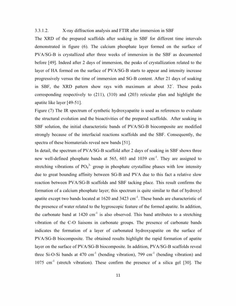
3.3.1.2. X-ray diffraction analysis and FTIR after immersion in SBF
The XRD of the prepared scaffolds after soaking in SBF for different time intervals
demonstrated in figure (6). The calcium phosphate layer formed on the surface of
PVA/SG-B is crystallized after three weeks of immersion in the SBF as documented
before [49]. Indeed after 2 days of immersion, the peaks of crystallization related to the
layer of HA formed on the surface of PVA/SG-B starts to appear and intensity increase
progressively versus the time of immersion and SG-B content. After 21 days of soaking
in SBF, the XRD pattern show rays with maximum at about 32◦. These peaks
corresponding respectively to (211), (310) and (203) reticular plan and highlight the
apatite like layer [49-51].
Figure (7) The IR spectrum of synthetic hydroxyapatite is used as references to evaluate
the structural evolution and the bioactivities of the prepared scaffolds. After soaking in
SBF solution, the initial characteristic bands of PVA/SG-B biocomposite are modified
strongly because of the interfacial reactions scaffolds and the SBF. Consequently, the
spectra of these biomaterials reveal new bands [51].
In detail, the spectrum of PVA/SG-B scaffold after 2 days of soaking in SBF shows three
new well-defined phosphate bands at 565, 603 and 1039 cm-1
. They are assigned to
stretching vibrations of PO43-
group in phosphate crystalline phases with low intensity
due to great bounding affinity between SG-B and PVA due to this fact a relative slow
reaction between PVA/SG-B scaffolds and SBF tacking place. This result confirms the
formation of a calcium phosphate layer; this spectrum is quite similar to that of hydroxyl
apatite except two bands located at 1620 and 3423 cm-1
. These bands are characteristic of
the presence of water related to the hygroscopic feature of the formed apatite. In addition,
the carbonate band at 1420 cm-1
is also observed. This band attributes to a stretching
vibration of the C-O liaisons in carbonate groups. The presence of carbonate bands
indicates the formation of a layer of carbonated hydroxyapatite on the surface of
PVA/SG-B biocomposite. The obtained results highlight the rapid formation of apatite
layer on the surface of PVA/SG-B biocomposite. In addition, PVA/SG-B scaffolds reveal
three Si-O-Si bands at 470 cm-1
(bending vibration), 799 cm-1
(bending vibration) and
1075 cm-1
(stretch vibration). These confirm the presence of a silica gel [30]. The
12
appearance of apatite mineral and a silica gel indicate the interactions between the
scaffolds and SBF as described by Hench et al. This mechanism could be explained
through the following steps:
(a) rapid exchange of protons H3O+ from the SBF with Ca
2+ , Na
+ ions in bioglass to form
the Si-OH groups, (b) loss of soluble silica as Si(OH)4 by breaking of Si-O-Si bridging
links and subsequent formation of surface silanol groups in the process, (c) condensation
and repolymerization of surface silanols to form SiO2-rich surface layer, (d) migration of
Ca2+
and PO43-
through the surface silica-rich layer and formation of a Ca-P rich layer on
the surface of biocomposite, (e) incorporation of OH-, CO3
2- from the solution and
subsequent crystallization of the Ca-P layer to form HCA [53-56]. The obtained results
confirm the bioactivity of PVA/BG biocomposite. Especially, they highlight the positive
effect of SG-B particle size and SG-B bounding strength with PVA controls the
formation rate of well crystallized apatite layer on its surface.
3.2.2. SEM with EDS after immersion in SBF
Two compositions of the prepared scaffolds have been under investigated by SEM
coupled with EDS , figure (8), to evaluate their surface changes after soaking in SBF for
21 days (PV and 1PVA:2SG-B). This scaffolds had exhibit excellent bioactivity and high
fracture toughness. The hydroxy apatite crystals formed with condensed manure on the
surface of the biocomposite scaffolds but the surface of PVA scaffold is not changed yet.
Incorporation of PVA with SG-B induces a great modification to PVA bioactivity. SEM
analysis suggested the excitants of strong molecular interaction between SG-B particles
and PVA network, causing SG-B to be dispersed uniformly in the composite scaffolds.
The presence of Ca, P, Na and Cl elements on the surface of the prepared composite
scaffolds were determined by EDS. The phosphocalcic ratio Ca/P after 21 days of
immersion in SBF is nearly equal to the stoichiometric apatite [33, 49 and 57-60].
3.3.3. Evaluation of elemental concentrations in SBF
The change of ions concentrations in SBF was demonstrated in figure (9). For P and Si
ions they take the same behavior for all the prepared scaffolds with little difference in
13
their amount in the SBF. Which is due to the limit of the integrate combination between
SG-B and PVA. This little difference is according to bounding and incorporation of SG-B
into PVA. The SG-B particle size is affecting on the amount of P and Si in the SBF as its
confirmed by XRD, FTIR and SEM with EDS. The ions concentration of Ca was found
to be completely different for each composition of scaffolds. This is much believed to be
according to the glass content in the scaffolds and the small particle size of SG-B as they
in turn changes the porosity and the degradation rate in the SBF [35, 49, 50, 54, 57, 58,
and 59].
3.4. Degradation
Biodegradation rate of the prepared scaffolds was investigated in SBF at different time
intervals with PVA alone as control as shown in figure (10). PVA scaffold exhibit higher
degradation rate (100% after 2 days) than those of PVA/SG-B scaffolds. Ideally, in tissue
engineering, a scaffold is usually intended to temporary fill a defect, while gradually
degrading as neo-tissue is formed. In due course, the scaffold is replaced by new bone
tissue [61]. After implantation, the scaffold interacts with the tissue fluids, uptaking them
at some extent, starting the degradation process [62]. A relative low degradation rate is
much favorable for cell attachment and differentiation. Furthermore, increases of the
glass amount in the scaffold decreases the degradation rate due to the fact that
incorporation of inorganic filler into polymer matrix decreases the porosity as confirmed
by MIP and liquid displacement methods and as documented before [63 and 64]. Porosity
decrease lead to decreases of the exposed surface area from the scaffold to the SBF. This
decreasing prolong the consumed time for biodegradation, giving more time for cells
attachment and proliferation [65].
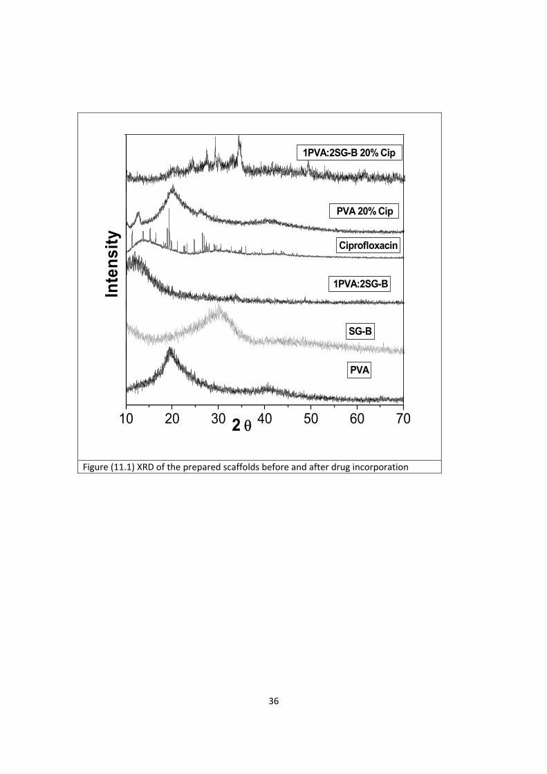
3.5. Ciprofloxacin incorporation
The success of incorporation of ciprofloxacin into PVA and PVA/SG-B scaffolds was
confirmed by XRD, FTIR and SEM coupled with EDS.
14
3.5.1. XRD
Figures (11.1) represent the XRD for PVA and PVA/SG-B scaffolds with and without the
drug. Ciprofloxacin has specific sharp crystal peaks while PVA, SG-B and PVA/SG-B
have broad peaks. When ciprofloxacin was entrapped into the scaffold matrix, its sharp
crystal peaks were overlapped with the noise of the surrounded polymer and disappeared
indicating that ciprofloxacin was successfully entrapped into the scaffold matrix system
and formation of a new solid phase for ciprofloxacin with low crystallinity[66-70].
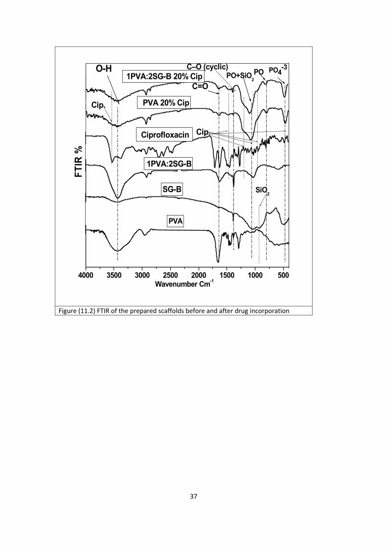
3.5.2. FTIR
The FTIR for ciprofloxacin loaded scaffolds are demonstrated in figures (11.2). The
FTIR spectrum of ciprofloxacin shows one prominent characteristic band between 3500
and 3450 cm-1
, which was assigned to stretching vibration of OH groups Another band at
3000- 2950 cm-1
represent alkene and aromatic C-H stretching, mainly υ=C-H was
demonstrated. The 1950 to 1450 cm-1
region exhibited FTIR absorption from a wide
variety of double-bonded functional groups. The band at 1750 to 1700 cm-1
represented
the carbonyl C=O stretching i.e., υC=O. The peak between 1650 and 1600 cm -1
was
assigned to quinolones. The band from 1450 to 1400 cm-1
represented υC-O and at 1300
to 1250 cm-1
suggested bending vibration of O-H group which proved the presence of
carboxylic acid. A strong absorption band between 1050 and 1000cm-1
was assigned to
C-F group. The FTIR for the PVA scaffolds loaded with ciprofloxacin indicate the
presence of new bands at 3522, 1744, and 1473.52 cm-1
when compared with those of
non-medicated scaffold due to the presence of ciprofloxacin. These bands were indicated
also for PVA/SG-B scaffolds loaded with ciprofloxacin beside another band at 1088 cm-1
with high intensity due to combination of drug with glass particles into the polymer
matrix. A shorter band appeared in the region of 1500–1200 cm-1
that could be ascribed
to the hydrated bonds with ciprofloxacin molecules [66-73].
The FTIR spectra indicate that, although a physical interaction between the drug and the
scaffold components occurs with both PVA/SG-B scaffolds. This is probably because
PVA/SG-B has a greater content of pendant hydroxyl groups that are more accessible for
establishing hydrogen bonds with the drug [69].
15
3.5.3. SEM coupled with EDS
The SEM image of the drug shows rod shape crystals and its EDS indicate the presence
of F and Cl elements which are the main components of the drug as demonstrated in
figure (11.3). SEM images for the cross-section of scaffold loaded with the ciprofloxacin
reveal the rod shape of ciprofloxacin crystal in the scaffold matrix system [70, 75]Also
the EDS confirm the presence of F and Cl elements in the scaffolds loaded with
ciprofloxacin . Therefore, XRD, FTIR and SEM coupled with EDS indicate and confirm
the success incorporation of ciprofloxacin into PVA and PVA/SG-B scaffolds.
3.6 Release behavior of ciprofloxacin
The release behavior of ciprofloxacin from the prepared scaffolds is presented in figure
(12). Considering the hydrophilic molecule, ciprofloxacin is expected to exhibit burst
release from the investigated system. Moreover, a sustained drug release profile was
observed form the investigated figures with quasi-Fickian diffusion mechanism (n-values
less than 0.5). This mechanism indicates that the polymer is hydrated, swell and then the
drug diffuses through the swollen matrix system, which ultimately slows down the
kinetic release.
The release profile for ciprofloxacin from scaffold prepared from PVA/SG-B loaded with
5, 10 and 20% ciprofloxacin was higher than that for scaffold loaded with PVA scaffolds
loaded with 5, 10 and 20% of ciprofloxacin. This could be due to the interaction between
PVA alone with ciprofloxacin resulting in great bounding affinity between the drug and
PVA polymer. On the other hand presence of SG-B particles in the polymer matrix leaves
no free space for the ciprofloxacin causing fast release for the drug in the PBS. [67, 71
and 75].
16
Conclusions
In this study, the PVA/SG-B biocomposite scaffolds loaded with ciprofloxacin with well
interconnected pore structure were fabricated via freeze drying technique. The
degradation rate and physicochemical properties of the prepared scaffold by freeze drying
for tissue engineering could be controlled by controlling glass content and drug
concentrations. The pore size achieved is suitable for cell activation and tissue
regeneration. Drug loaded scaffolds with ciprofloxacin exhibit a good drug delivery
system with sustained drug release. The biodegradation rate and structural morphology of
the prepared scaffolds could be controlled by adjusting ciprofloxacin percentage.
17
References
[1]. Kretlow, J. D. and Mikos, A. G.: from material to tissue biomaterial
development, scaffold fabrication, and tissue engineering. J AIChE 2008; 54:
3048–67.
[2]. Brook, I.: Microbiology and management of joint and bone infections due to
anaerobic bacteria. J Orthop Sci 2008; 13:160–9.
[3]. Makinen, TJ., Veiranto, M. and Lankinen, P.: In vitro and in vivo release of
ciprofloxacin from osteoconductive bone defect filler. J Antimicrob Chemother
2005; 56:1063–8.
[4]. Brady, RA., Leid, JG. and Calhoun, JH.: Osteomyelitis and the role of
biofilms in chronic infection. FEMS Immunol Med Microbiol 2008; 52:13–22.
[5]. Garcia-Lechuz, J. and Bouza, E.: Treatment recommendations and strategies
for the management of bone and joint infections. Expert Opin Pharmacol 2009;
10:35–55.
[6]. Lazzarini, L., Lipsky, BA. and Mader, JT.: Antibiotic treatment of
osteomyelitis: what have we learned from 30 years of clinical trials. Int J Infect
Dis 2005; 9:127–38.
[7]. Kanellakopoulou, K.: Local treatment of experimental seudomonas aeruginosa
osteomyelitis with a biodegradable dilactide polymer releasing ciprofloxacin.
Antimicrob Agents Chemother 2008; 52:2335–9.
[8]. Lepretre, S.: Prolonged local antibiotics delivery from hydroxyapatite
functionalised with cyclodextrin polymers. Biomate 2009; 30:6086–93.
[9]. Hench, L. L.: Genetic design of bioactive glass. J Eur Ceram Soc 2009; 7:
1257–65.
[10]. Kaufmann, E. E., Ducheyne, P. and Shapiro I.: Effect of varying physical
properties of porous, surface modified bioactive glass 45S5 on osteoblast
proliferation and maturation. J Biomed Mater Res 2000; 52: 783–96.
[11]. Jones, J. R., Tsigkou, O. and Coates, E. E. : Extracellular matrix formation
and mineralization on a phosphate-free porous bioactive glass scaffold using
primary human osteoblast (HOB) cells. Biomate 2007; 28: 1653–63.
18
[12]. Valerio, P., Pereira, M. M. and Goes, A. M.: The effect of ionic products
from bioactive glass dissolution on osteoblast proliferation and collagen
production. Biomate 2004; 25: 2941–8.
[13]. Radin, S., Reilly, G. and Bhargave, G.: Osteogenic effects of bioactive glass
on bone marrow stromal cells. J Biomed Mater Res A 2005; 73: 21–9.
[14]. Xynos, I. D., Buttery, L. D. and Hench L. L.: Ionic products of bioactive glass
dissolution increase proliferation of human osteoblasts and induce IGF II m RNA
expression and protein synthesis. Biochem Biophys Res Commun. 2000; 276:
461–5.
[15]. Yilgor, P., Tuzlakoglu, K., Reis, R.L., Hasirci, N. and Hasirci, V.:
Incorporation of a sequential BMP-2/BMP-7 delivery system into chitosan-based
scaffolds for bone tissue engineering, Biomate 2009;30: 3551–9.
[16]. Xu, J., Li J.B, Lian, X., Ayers, D.C. and Song, J.: Sustained and localized in
vitro release of -2/7, RANKL, and tetracycline from FlexBone, an elastomeric
osteoconductive bone substitute, J Orthop Res. 2009; 27:1306– 11.
[17]. Choi, D.H., Park, C.H., Kim, I.H., Chun, H.J., Park, K. and Han, D.K.,
Fabrication of core-shell microcapsules using PLGA and alginate for dual growth
factor delivery system, J. Control. Release 2010; 147:193–201.
[18]. Mikos AG, McIntire LV, Anderson JM and Babensee JE.: Host response to
tissue engineered devices. Adv Drug Deliv Rev 1998; 33:111–39.
[19]. Böstman O, Hirvensalo E, Vainionpää S, Mäkelä A, Vihtonen K and Törmälä
P, Ankle fractures treated using biodegradable internal fixation. Clin Orthop Relat
Res 1989; 238:195–203.
[20]. Chen, R.R. and Mooney, D.J.: Polymeric growth factor delivery strategies for
tissue engineering, Pharm. Res. 2003; 20: 1103–12.
[21]. Han, D., Liu, W., Ao, Q., Wang, and G.: Optimal delivery systems for bone
morphogenetic proteins in orthopedic applications should model initial tissue
repair structures by using a heparin-incorporated fibrin-fibronectin matrix, Med.
Hypotheses 2008;71: 374–8.
[22]. Silverman, L.D., Lukashova, L., Herman, O.T., Lane, J.M. and Boskey, A.L.,
Release of gentamicin from a tricalcium phosphate bone implant, J. Orthop. Res.
2007; 25: 23–29.
19
[23]. Sokolsky-Papkov, M, Agashi, K, Olaye, A, Shakesheff, K and Domb, AJ.
Polymer carriers for drug delivery in tissue engineering. Adv Drug Deliv Rev
2007; 59:187–206.
[24]. Griffith, LG and Naughton, G. Tissue engineering-current challenges and
expanding opportunities. Sci 2002; 295:1009–14.
[25]. Nair, LS and Laurencin, CT. Polymers as biomaterials for tissue engineering
and controlled drug delivery. Adv Biochem Eng Biotechnol 2006; 102:47–90.
[26]. Yamaoka, T., Tabata, Y. and Ikada, Y.: Comparison of body distribution of
poly (vinyl alcohol) with other water-soluble polymers after travenous
administration. J Pharma and Pharmaco 1995; 47 no. 6:479–86.
[27]. Chiellini, E., Corti, A., D’Antone, S. and Solaro, R.: Biodegradation of poly
(vinyl alcohol) based materials. Progr in Polym Sci, 2003; 28, no. 6: 963–1014.
[28]. Alta, V., Bitschnaua, A. and Osterlinga, J.: The effects of combined
gentamicin–hydroxyapatite coating for cementless joint prostheses on the
reduction of infection rates in a rabbit infection prophylaxis model. Biomate.
2006; 27: 4627-34.
[29]. Nayak, A. K. and Sen, K. K.: Hydroxyapatite-ciprofloxacin minipellets for
bone-implant delivery: Preparation, characterization, in-vitro drug adsorption and
dissolution studies. Int J Drug Dev Res 2009; 1: 47–59.
[30]. Dietrich, E., Oudadesse, H., Lucas-Girot, A. and Mami, M.: In vitro
bioactivity of melt-derived glass 46S6 doped with magnesium. J of Biomed Mater
Res. 2009; 88: 1087-96.
[31]. Korsmeyer, R. W., R. Gurny, et al. (1983). "Mechanisms of solute release
from porous hydrophilic polymers." Int J Pharm15: 25-35.
[32]. Misra, SK., Nazhat, SN. and Valappil, SP.: Fabrication and characterization
of biodegradable Poly (3-hydroxybutyrate) composite containing bioglass.
Biomacromolecules 2007; 8:2112-9.
[33]. Yazdanpanah, A., Reza, K. and Moztarzadeh, F.: Enhancement of the fracture
toughness in bioactive glass-based nanocomposites with nanocrystalline forsterite
as advanced biomaterials for bone tissue engineering applications. Ceram Inter
2012; 38:5007-14.
[34]. Wong Sh., Baji, A. and Alan, N.: Effect of specimen thickness on fracture
20
toughness and adhesive properties of hydroxyapatite-filled polycaprolactone.
Compos : Part A, 2008; 39: 579–87.
[35]. Julian, R. J.: New trends in bioactive scaffolds: The importance of
nanostructure. J Eur Cer Soci. 2009; 29: 1275–81.
[36]. Nallaa, R.K., Kinneyb, J.H., and Ritchie R.O.: Effect of orientation on the in
vitro fracture toughness of dentin: the role of toughening mechanisms. Biomate
2003; 24:3955–68.
[37]. Mahinda, D. K. and Ken, P. Ch.: Fracture toughness testing of brittle materials
using semi-circular bend (SCB) specimen. Engi Fract Mech , 2012; 91: 133–50.
[38]. Cook, R.B., Curwen, C., Tasker, T. and Zioupos, P.: Fracture toughness and
compressive properties of cancellous bone at the head of the femur and
relationships to non-invasive skeletal assessment measurements. Med Engi & Phy
, 2010; 32: 991–7.
[39]. María, C., Gutiérrez, Zaira Y., Carvajal, G. and Jobbágy M.: Poly (vinyl
alcohol) Scaffolds with Tailored Morphologies for Drug Delivery and Controlled
Release. Adv Funct Mater. 2007; 17: 3505–13.
[40]. Hutmacher, DW.: Scaffolds in tissue engineering bone and cartilage. Biomate
2000; 21:2529–43.
[41]. Tao, W., Mahir, T. and Gunasekaran, S.: Selected properties of pH-sensitive,
biodegradable chitosan–poly (vinyl alcohol) hydrogel, Polym. Int. 2004; 53: 911–
8.
[42]. Andrade, G., Barbosa-Stancioli, E. F., Mansur, A. A. Piscitelli, W
Vasconcelos, L. and Mansur H. S.: Design of novel hybrid organic-inorganic
nanostructured biomaterials for immunoassay applications,”. Biomed Mate , 2006;
1, no. 4: 221–34.
[43]. Mansur, H.S. and Costa, H. S.: Nanostructured poly(vinyl alcohol)/bioactive
glass and poly(vinyl alcohol)/chitosan/ bioactive glass hybrid scaffolds for
biomedical applications,”. Chem Engi J . 2008; 137, no. 1: 72–83.
[44]. Mansur, H. S., Or´efice, R. L. and Mansur, A. A.: Characterization of poly
(vinyl alcohol)/poly (ethylene glycol) hydrogels and PVA-derived hybrids by
small-angle X-ray scattering and FTIR spectroscopy,”. Poly, 2004; 45, no. 21:
7193–02.
21
[45]. Coates, J.: Encyclopedia of analytical chemistry: interpretation of infrared
spectra, a practical approach,” in Encyclopedia of Analytical Chemistry, R. A.
Meyers, Ed., 2000; JohnWiley & Sons, Chichester, UK, 10815–37.
[46]. Almeida, R. M. and Pantano, C. G.: Structural investigation of silica gel films
by infrared spectroscopy. J App Phy, 1990; 68, no. 8: article 4225, 8 pages.
[47]. Wang, T., Turhan, M. and Gunasekaran, S.: “Selected properties of pH-
sensitive, biodegradable chitosan-poly(vinyl alcohol) hydrogel,” Poly Int. , 2004;
53, no. 7: 911–8.
[48]. Shin, S.-H. and Kim, H.-I.: Contribution of hydrogen bond and coupling
reaction improvement in compatibility of organic polymer/silica
nanocomposites,”. Ind. & Engi Chem Res., 2001; 7: 147–52.
[49]. Mami, M., Oudadesse, H. and Doebez-Sridi, R.: Synthesis and in-vitro
characterization of melt derived 47S CaO–P2O5–SiO2–Na2O bioactive glass.
Ceram − Silik .2008; 52 (3):121-9.
[50]. Oudadesse, H., Mami, M. and Doebez-Sridi, R.: Study of the Bioactivity of
Various Mineral Compositions of Bioactive Glasses. Bioceram Develop and App.
2011; 1: Article ID D110151, 3 pages.
[51]. Superb, K. M., Dirk, M. and Brunner Tobias, J.: Comparison of nanoscale and
microscale bioactive glass on the properties of P(3HB)/Bioglass composites.
Biomat. 2008; 29: 1750-61.
[52]. Luo, P.: Methods of synthesizing hydroxyapatite powders and bulk materials.
United States Patent 1999.
[53]. Hench, L.L., Splinter, R.J., Allen, W.C. and Greenlee, T.K.: Bonding
mechanisms at the interface of ceramic prosthetic materials. J Biomed Mater Res
1971, 5(6), pp. 117-41.
[54]. Oudadesse, H., Mami, M., Doebez-Sridi, R., Pellen, P., Perez, F., Jeanne S.,
Chauvel-Lebret D., Mostafa A. and Cathelineau G., Study of various mineral
compositions and their bioactivity of bioactive glasses. Biocera 2009;22 : 379-82.
[55]. Hench, L. L., The story of bioglass. J Mater Sci: Mater Med 2006; 17: 967-78.
[56]. Hench, L.L. and West, J.K.: Biological applications of bioactive glasses, Life
Chem Rep 1996, Vol. 13, pp. 187-241.
22
[57]. Oudadesse, H., Bui, X. V. and Yann L.: Chitosan Effects on Bioactive Glass
for Application as Biocopmosite Biomaterial. Int J of biolo and biomed engi .
2011; 5: 49-56.
[58]. Oudadesse, H., Mostafa, A. and Bui X. V.: Physico-chemical assessment of
biomimetic nano-hydroxyapatite/polymer matrix for use in bony surgery. Int J of
biolo and Biomed Engi. 2011; 5: 103-10.
[59]. Mami, M., Lucas-Girot, A. and Oudadesse, H.: Investigation of the surface
reactivity of a sol–gel derived glass in the ternary system SiO2–CaO–P2O5 . App
Sur Sci 2008; 54:7386–93.
[60]. Bellucci, D., Cannillo, V. and Sola, A.: Macroporous Bioglass®-derived
scaffolds for bone tissue regeneration. Ceram Int 2011; 37: 1575–85.
[61]. Gaalen, S.M.V., Kruyt, M.C. and Meijer, G.J.: Tissue engineering of bone, in:
C.v. Blitterswijk, J. Sohier (Eds.), Tissue Engineering, Academic Press, Elsevier,
UK, 2008, pp. 555–606.
[62]. Gomes, M.E., Azevedo, H.S., Moreira, A.R., Ellä, V., Kellomäki, M. and
Reis, R.L.: Starchpoly(- caprolactone) and starch-poly(lactic acid) fibre-mesh
scaffolds for bone tissue engineering applications: structure, mechanical
properties and degradation behavior. J Tiss Engi Rege Medici. 2008; 2: 243–52.
[63]. Zhang, F., Chuanglong H. and Lijun Cao.: Fabrication of gelatin–hyaluronic
acid hybrid scaffolds with tunable porous structures for soft tissue engineering. Int
J Biolo Macromolec, 2011; 48: 474–81.
[64]. Wu, F., Liu, Ch. and O’Neill, B.: Fabrication and properties of porous scaffold
of magnesium phosphate/polycaprolactone biocomposite for bone tissue
engineering. App Surf Sci, 2012; 258:7589–95.
[65]. Peter, M., Binulal, N.S. and Nair, S.V.: Novel biodegradable chitosan–
gelatin/nano-bioactive glass ceramic composite scaffolds for alveolar bone tissue
engineering. Chem Engi J, 2010; 158:353–61.
[66]. Unnithan, A. R., Barakat, N. A.M. and Tirupathi Pichiah, P.B., Wound-
dressing materials with antibacterial activity from electrospun polyurethane–
dextran nanofiber mats containing ciprofloxacin HCl. Carbohyd Poly, 2012;90 :
1786–93.
23
[67]. Wang, Q., Zedong, Du Y. and Kennedy, J.: Controlled release of ciprofloxacin
hydrochloride from chitosan/polyethylene glycol blend films. Carbohyd.
Polym.2007; 69:336–43.
[68]. Sahoo, S., Charkaborti, Ch. K. and Behera, P. K.: Qualitativ analysis of a
ciprofloxacin / HPMC mucoadhesive suspension. Int J Pharma and Bio Sci , 2012;
3: 558-76.
[69]. Rodrı´guez-Tenreiro, C., Alvarez-Lorenzo, C., Concheiro, A. and Torres-
Labandeira, J.J.: Characterization of cyclodextrin-carbopol interactions by DSC
and FTIR. J Therm Anal Calorim. 2004; 77:403–11.
[70]. Nayak, A. K., Laha, B. and Sen, K. K.: Development of hydroxyapatite-
ciprofloxacin bone-implants using Quality by design. Acta Pharm. 2011; 61: 25–
36.
[71]. Sunitha , A. and Kumar, S.: Study on Effect of Solvents & Nonsolvents on
Microspheres of Ciprofloxacin: Coacervation Phase Separation. J Adv Sci Res,
2010; 1(2): 24-33.
[72]. Sunitha , A. and Kumar, S.: Study on the effect of polymers on the release
rate of drug from ciprofloxacin hydrochloride microspheres .J Pharmaceu
Cosmetolo 2010;1(1): 1-8.
[73]. Kesavan, S. and Alamelu Bai, S.: Effect of surfactant on the release of
ciprofloxacin from gelatin microspheres. J. ARS Pharmaceu Ars Pharm, 2010;51
n 1: 1-16.
[74]. Thakre, Y. M. and Choudhary, M. D.: Synthesis, characterization and
evaluation of derivative of Ciprofloxacin (1-cyclopropyl-6-fluoro-4-oxo-7-[4-
(phenyl carbonyl) piperazin-1-yl]-1, 4-dihydroquinoline-3-carboxylic acid) and
their complexes. J Chem Pharm Res, 2011; 3(5):390-8.
[75]. Puga, A. M., Rey-Rico, A. and Magariños B.: Hot melt poly-e-
caprolactone/poloxamine implantable matrices for sustained delivery of
ciprofloxacin. Acta Biomate, 2012; 8:1507–18.
Table
Table (1): porosity percentage and pore diameter measured by mercury Hg porosimeter and liquid displacement techniques.
Porosity % pore diameter range (4V/A)
Liquid displacement
With drug
20% 10% 5%
Without drug
MIP nm µm
Sample name
66 69 72 85.47 88.14 6.2 139 PVA
73 74 77 79.46 74.95 6.3 131 2PVA:1SG‐B
67 69 70 70.30 67.60 6.3 119 1PVA:1SG‐B
55 58 59 60.50 46.68 6.3 110 1PVA:2SG‐B
24
Figures
Freezing at ‐ 18оC for overnight
The resulted mixture was casted in Teflon molds.
Addition of BG on the dissolved PVA continues stirring for overnight.
Drug addition on the above mixture continues stirring for 1 hr.
PVA dissolving in distilled water at 80 оC with 15% W/V.
Lyophilization at ‐56оC for 24 hr.
Figure (1.1)
25
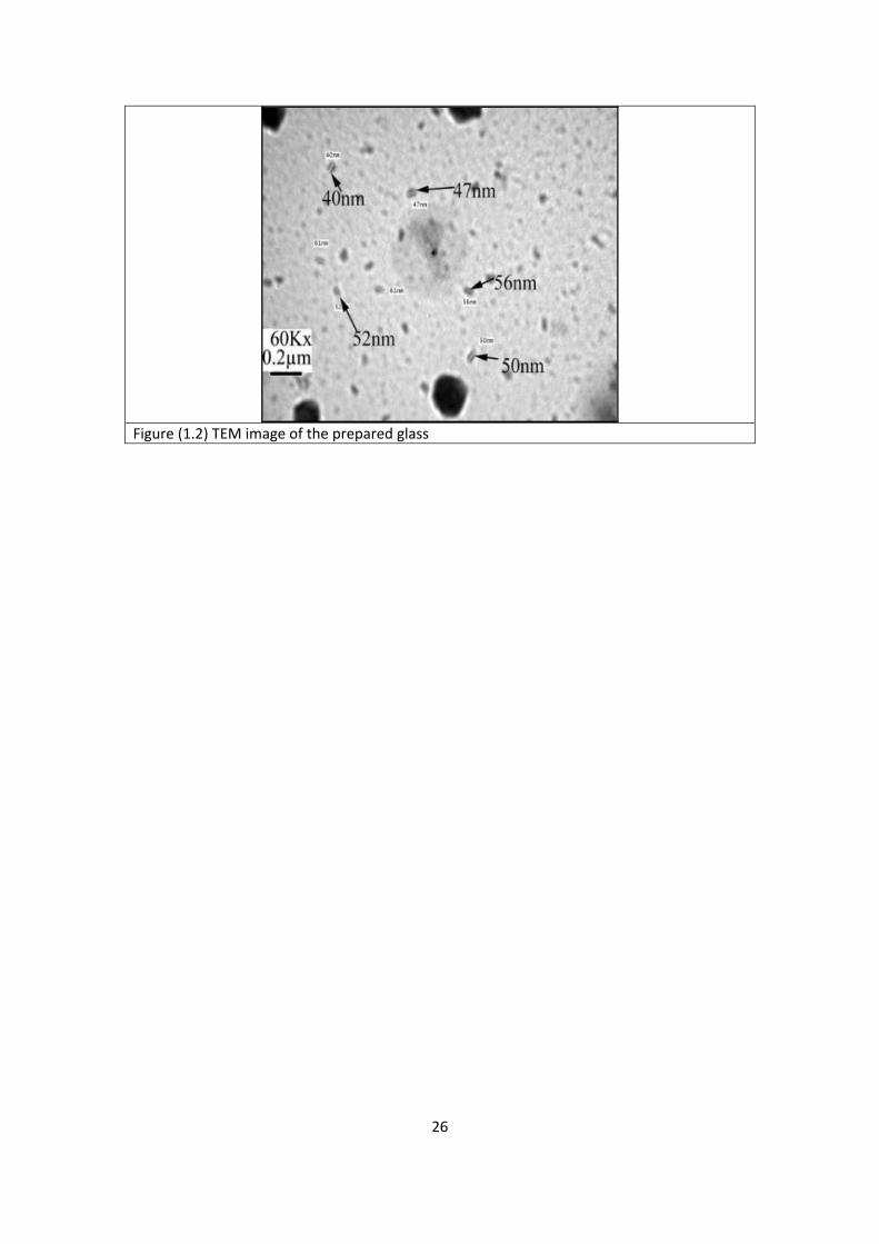
Figure (1.2) TEM image of the prepared glass
26
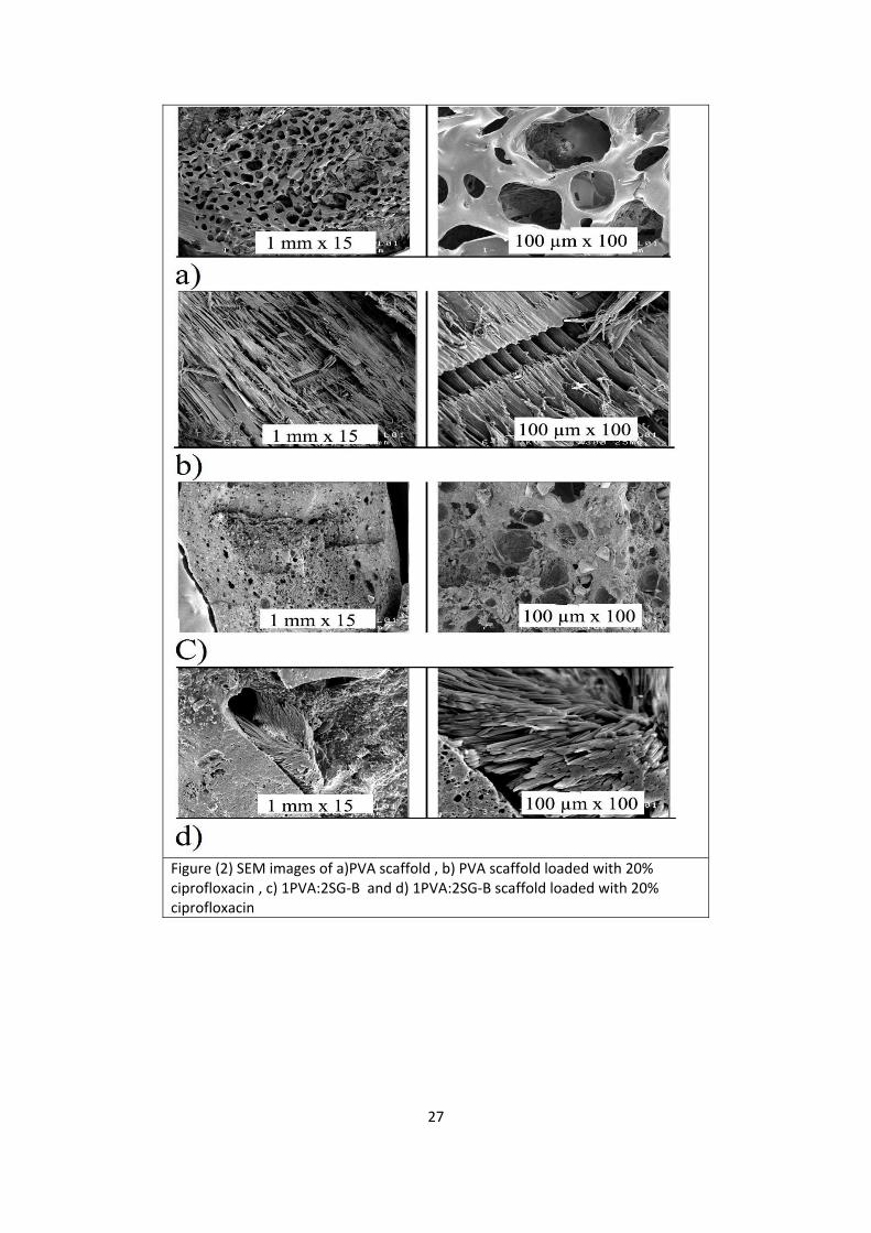
Figure (2) SEM images of a)PVA scaffold , b) PVA scaffold loaded with 20% ciprofloxacin , c) 1PVA:2SG‐B and d) 1PVA:2SG‐B scaffold loaded with 20% ciprofloxacin
27
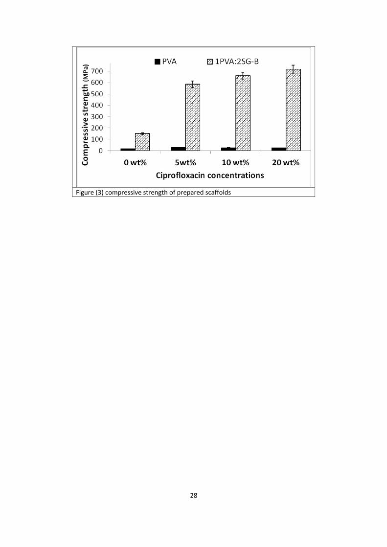
Figure (3) compressive strength of prepared scaffolds
28
20 30 40 50 60 70
Inte
nsit
y
2
PVA
SG-B
2PVA:1SG-B
1PVA:1SG-B 1PVA:2SG-B
(113)(131)
Figure (4) XRD of the prepared scaffolds before immersion in SBF
29
4000 3500 3000 2500 2000 1500 1000 500
FT
IR %
Wavenumber Cm-1
PVA
C=O
1PVA:2SG-B
1PVA:1SG-B
2PVA:1SG-B
SG-B
SiO2
CH
O-H C–O (cyclic)PO+SiO
2 PO PO4
-3
Figure (5) FTIR of the prepared scaffolds before immersion in SBF
20 30 40 50 60 70
Inte
nsity
2
2PVA:1SG-B
After 2days
After 15 days
After 7 days
Synthetic HA
After 21 days
After 30 days
(022)(221)
(222)(211)
Figure (6.1) XRD of 2PVA:1SG‐B after soaking in SBF
20 30 40 50 60 70
(222)
Intensity
2 1PVA:1SG-B
After 2 days
After 7 days
After 15 days
After 21 days
After 30 days
(211)
(221)
Synthetic HA
30
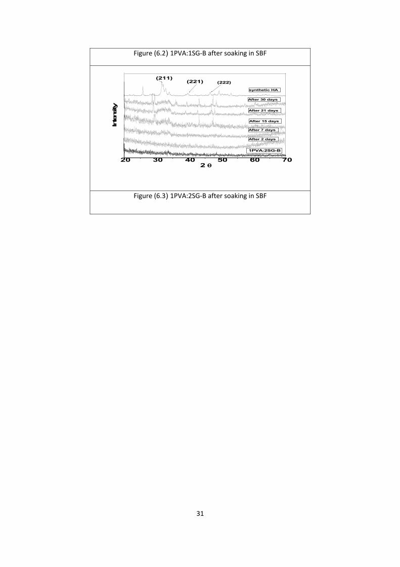
Figure (6.2) 1PVA:1SG‐B after soaking in SBF
20 30 40 50 60 70
Intensity
2 1PVA:2SG-B
After 2 days
After 7 days
After 15 days
After 21 days
(221)(211)
After 30 days
(222)
synthetic HA
Figure (6.3) 1PVA:2SG‐B after soaking in SBF
31
4000 3500 3000 2500 2000 1500 1000 500
O-H C=O C–O (cyclic)
PO+SiO2 PO
PO4-3
Synthetic HA
After 30 days
After 21 days
After 15 days
After 7 days
After 2days
FTIR
Wavenumber Cm-1
2PVA:1SG-B
Figure (7.1) 2PVA:1SG‐B after soaking in SBF
4000 3500 3000 2500 2000 1500 1000 500
O-H C=O
Synthetic HA
After 30 days
After 21 days
After 15 days
After 7 days
After 2days
FTIR
Wavenumber Cm-1
1PVA:1SG-B
C–O (cyclic)
PO+SiO2
PO4-3
Figure (7.2) 1PVA:1SG‐B after soaking in SBF
4000 3500 3000 2500 2000 1500 1000 500
PO PO4-3
Synthetic HA
After 30 days
After 21 days
After 15 days
After 7 days
After 2days
FTIR
Wavenumber Cm-1
1PVA:2SG-B
O-H
C=OC–O (cyclic)
PO+SiO2
Figure (7.3) 1PVA:2SG‐B after soaking in SBF
32
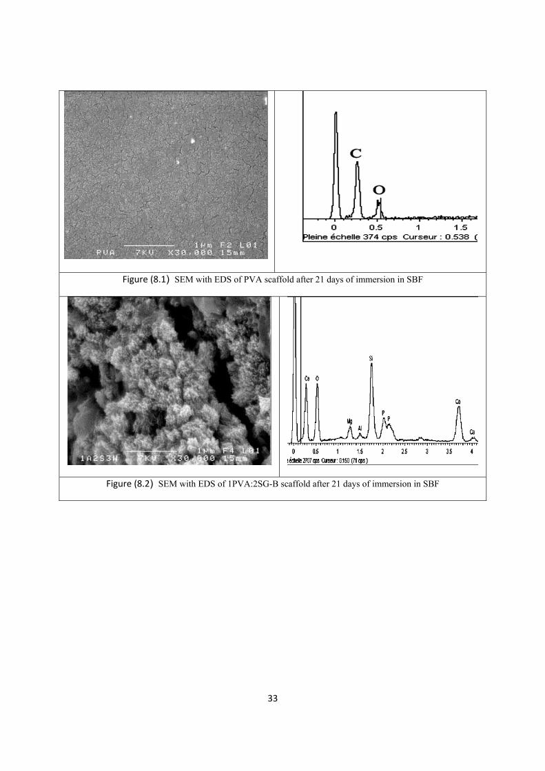
Figure (8.1) SEM with EDS of PVA scaffold after 21 days of immersion in SBF
Figure (8.2) SEM with EDS of 1PVA:2SG-B scaffold after 21 days of immersion in SBF
33
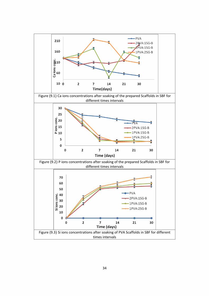
Figure (9.1) Ca ions concentrations after soaking of the prepared Scaffolds in SBF for
different times intervals
Figure (9.2) P ions concentrations after soaking of the prepared Scaffolds in SBF for
different times intervals
Figure (9.3) Si ions concentrations after soaking of PVA Scaffolds in SBF for different
times intervals
34
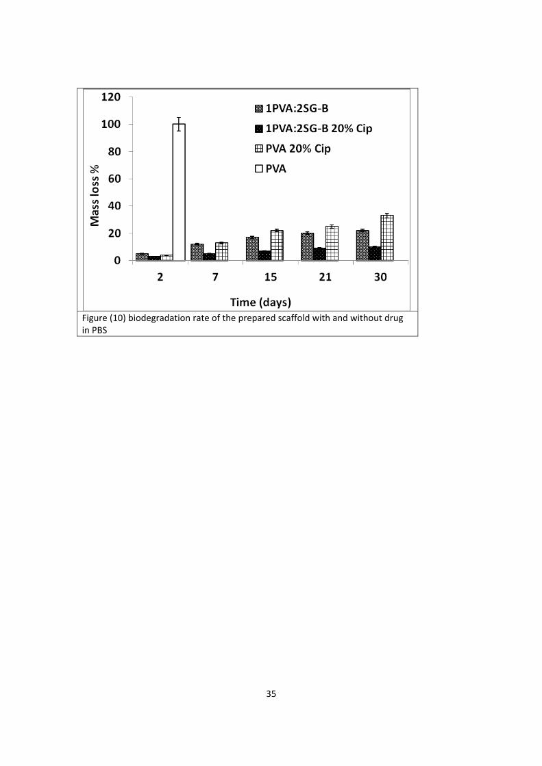
Figure (10) biodegradation rate of the prepared scaffold with and without drug in PBS
35
10 20 30 40 50 60 70
Inte
ns
ity
2
PVA
SG-B
1PVA:2SG-B
Ciprofloxacin
PVA 20% Cip
1PVA:2SG-B 20% Cip
Figure (11.1) XRD of the prepared scaffolds before and after drug incorporation
36
4000 3500 3000 2500 2000 1500 1000 500
FT
IR %
Wavenumber Cm-1
PVA
C=O
1PVA:2SG-B
SG-B
SiO2
O-H C–O (cyclic)PO+SiO
2 PO PO4
-3
Cip.
Cip.
1PVA:2SG-B 20% Cip
Ciprofloxacin
PVA 20% Cip
Figure (11.2) FTIR of the prepared scaffolds before and after drug incorporation
37
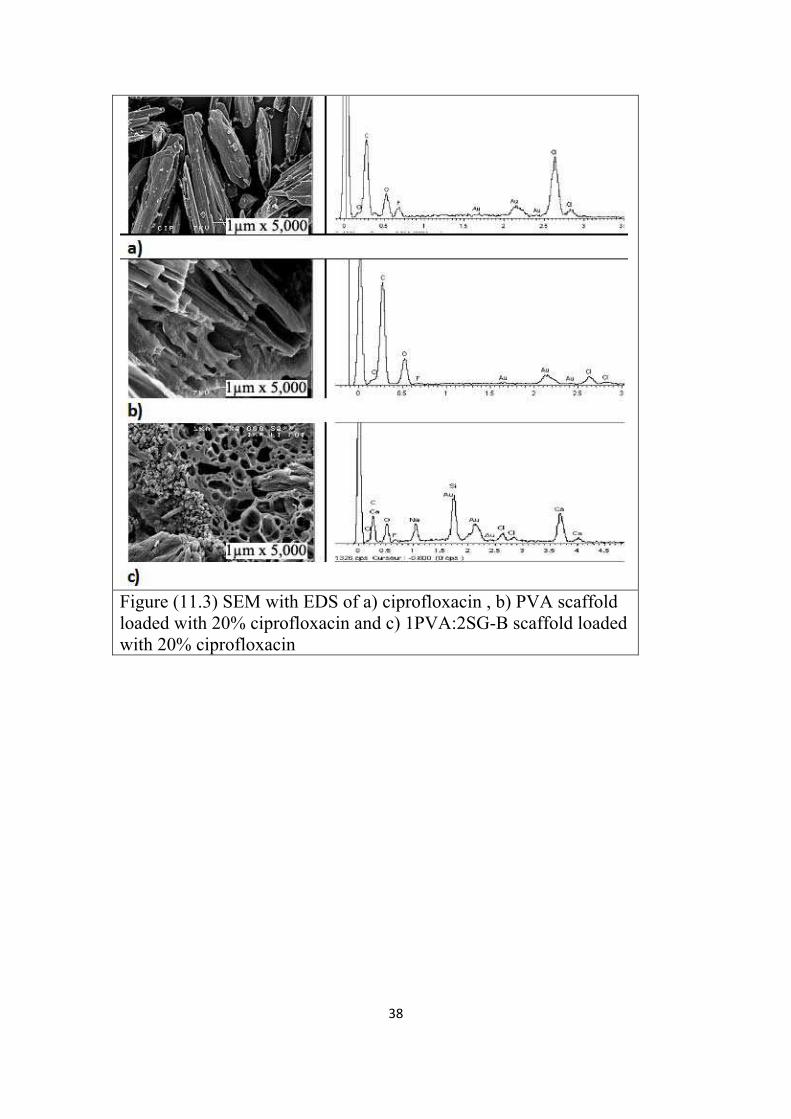
Figure (11.3) SEM with EDS of a) ciprofloxacin , b) PVA scaffold
loaded with 20% ciprofloxacin and c) 1PVA:2SG-B scaffold loaded
with 20% ciprofloxacin
38
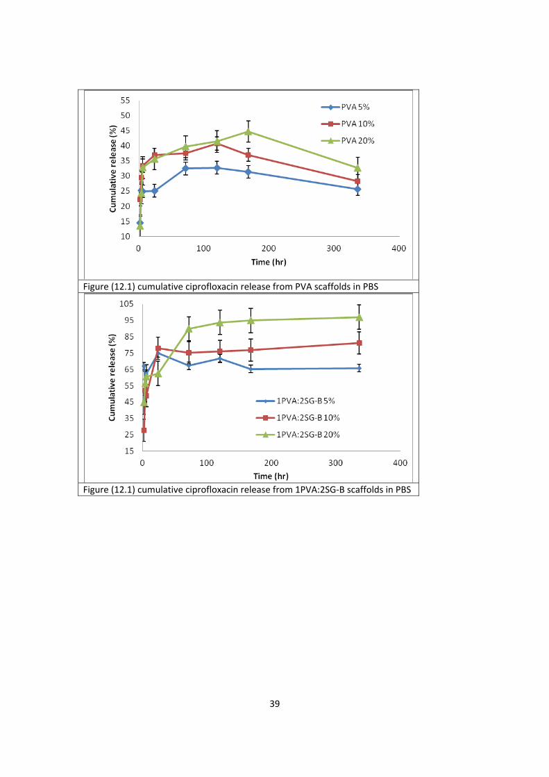
Figure (12.1) cumulative ciprofloxacin release from PVA scaffolds in PBS
Figure (12.1) cumulative ciprofloxacin release from 1PVA:2SG‐B scaffolds in PBS
39