AI Lecture 17 Planning Noémie Elhadad (substituting for Prof. McKeown)

Int.J.Curr.Microbiol.App.Sci (2018) 7(3): 3725-3736
3725
Original Research Article https://doi.org/10.20546/ijcmas.2018.703.431
Effect of Chlorophyllin on Biomphalaria alexandrina Snails and
Schistosoma mansoni Larvae
Heba A. Elhadad1, Bassem A. El-Habet
1, Rania M. Azab
2, Hanaa M. Abu El Einin
3,
Wael M. Lotfy4 and Hassan A. Atef
5*
1Department of Parasitology, Medical Research Institute, Alexandria University,
Alexandria, Egypt 2 National Institute of Laser Department, National Institute of Laser Enhanced Science
(NILES), Cairo University, Giza, Egypt 3Department of Environmental Researches and Medical Malacology, Theodor Bilharz
Research Institute, Imbaba, Giza, Egypt 4Department of Community Health Nursing, Faculty of Nursing, Alexandria University-
Matrouh Branch, Marsa Matrouh, Egypt 5Department of Mycology and Mycotoxins, Animal Health Research Institute, Agriculture
Research Centre, Dokki, Cairo, Egypt
*Corresponding author
A B S T R A C T
The present study was undertaken to investigate the lethal activity of
chlorophyllin against different developmental stages of Biomphalaria
alexandrina snails and Schistosoma mansoni aquatic larvae under
laboratory conditions. In all experiments, the studied organisms were
incubated in different chlorophyllin concentrations in the dark, and then
exposed to sunlight to stimulate the lethal photosensitizing action of
chlorophyllin. Snails lethal concentrations for six hours of sunlight
exposure were as follows: LC90 (131.86 ppm), LC50 (82.68 ppm), LC25
(56.93 ppm) and LC10 (33.76 ppm). The lethal action of chlorophyllin is
affected by several factors including: light source, duration of sunlight
exposure, the developmental stage of the snails and the presence of
infection. On the cellular level, histological sections revealed marked
destruction of certain tissues with loss of their landmarks. Chlorophyllin
also had a profound lethal effect on the larval stages of Schistosoma
mansoni. So, chlorophyllin is a promising substance of plant origin that
could be used in snail control programs.
K e y w o r d s
Schistosomiasis,
Snail, Control,
Chlorophyllin,
Photosensitization
Accepted:
28 February 2018
Available Online:
10 March 2018
Article Info
International Journal of Current Microbiology and Applied Sciences ISSN: 2319-7706 Volume 7 Number 03 (2018) Journal homepage: http://www.ijcmas.com

Int.J.Curr.Microbiol.App.Sci (2018) 7(3): 3725-3736
3726
Introduction
Schistosomiasis is one of the most important
snail borne parasitic infections with a
significant public health impact (King, 2009).
Hence, the introduction of novel drugs for
disease treatment and improvement in water
supply and sanitation facilities in endemic
areas, resulted in that the snail control is
perhaps employed less often as a means of
combating the disease. However, it remains an
important and effective measure, especially
where transmission occurs to a significant
extent to children throughout playing in
contaminated water with parasite. This type of
water contact is not likely to be changed
through health education and the provision of
safe water supplies (Useh, 2012). Several
studies evaluated the Snail control by using
chemical and plant molluscicides, biological
predators and ecological methods and
indicated that the chemical control was one of
the most important tool for the control of the
pulmonate snail intermediate hosts
(McCullough, 1992).
On the other hand, other studies reported that
the Photosensitization is the administration of
a photoactive compound that selectively
accumulates in the target cells which will be
killed following irradiation with visible light
(Luksiene, 2005; Redmond, 2008). It was
effective in inactivation of different
microorganisms such as bacteria, yeasts,
viruses and parasites. Photosensitization can
open new and interesting avenues for the
development of novel, effective and
ecologically friendly photopesticides and
antimicrobial agents.
Since several decades, the development of
photosensitizers came along through many
stages and many studies specifically designed
to investigate the insecticidal activity of
various dyes in the presence of visible light.
Where, Graham (1963) was one of the first
scientists who paid attention to the possibility
of using ‘‘photosensitizing agents’’ as
insecticides.
Recently, many studies were done using
different photosensitizers to investigate
photosensitic effect on parasites, snails and
mosquito larvae all in aquatic ecosystems.
(Salama et al., 2002; El-Tayeb, 2003; El-
Tarky, 2005; Wohllebe, 2010 and Ragheb,
2013)
As human Schistosomiasis mansoni is still one
of the major health problems in Egypt
(Barakat, 2013 and Lotfy, 2009). Using
chlorophyllin in as a photosensitizer for
control of the intermediate host, Biomphalaria
snails by (Mahmoud et al., 2013; Ragheb,
2013; Ragheb et al., 2013). However, the used
different preparations of chlorophyllingave
different inaccurate lethal concentration
(LC50). (Mahmoud et al., 2013; Ragheb, 2013
and Ragheb et al., 2013). Hence, it was
important to use a standard chlorophyllin
preparation and carry out the experiments
under standard conditions to obtain the
accurate LC50 values.
Therefore, the present study was undertaken to
investigate the lethal effect of chlorophyllin on
Biomphalaria alexandrina snails and free-
living stages of S. mansoni under standard
conditions which were studied for the first
time.
Materials and Methods
Chlorophyllin sodium copper salt
It was purchased from Sigma Aldrich
(Commercial code: c 6003). A stock solution
(1gm/L) was prepared in distilled water and
was kept in the dark. The required dilutions
were prepared by using an appropriate volume
of the stock solution to be completed to 100
ml with dechlorinated water.

Int.J.Curr.Microbiol.App.Sci (2018) 7(3): 3725-3736
3727
Biomphalaria alexandrina snails
Biomphalaria alexandrina snails were
obtained from Theodor Bilharz Research
Institute (TBRI), Imbaba, Giza, Egypt. They
were maintained in dechlorinated water at
(24±1oC) and were fed oven dried lettuce
leaves daily. Fish food TetraMin® and blue
green algae, mainly Nostoc muscorm, were
also used as an additional food source for
newly hatched and juvenile snails.
Schistosoma mansoni miracidia and
cercariae
Schistosoma mansoni eggs were obtained
from Schistosomiasis Biological Supply
Center TBRI, Imbaba, Giza, Egypt. Miracidia
were hatched in a small amount of
dechlorinated water at 25±1°C and used
directly. While, cercariae were obtained from
experimentally infected Biomphalaria
alexandrina snails by light-stimulated
shedding in a small amount of dechlorinated
water and were used directly.
Experimental Design (Mahmoud et al.,
2013)
Chlorophyllin, as a photosensitizing agent,
necessitates being darkly incubated with the
examined organisms for a period of time to
allow its accumulation within the organisms'
tissues followed by sunlight irradiation for
activation of its lethal potential. Accordingly,
in the different experiments, 5 B. alexandrina
snails (6-8mm) were incubated in
chlorophyllin in darkness overnight. After
that, snails were exposed to sunlight for
different periods then transferred to
dechlorinated water to recover in darkness for
24 hours. Later on, viability was assessed:
snails showing no vital signs (movements or
reflexes after tipping with a needle) were
considered dead. In each experiment, 3
replicates were used.
Light and dark controls
Light and dark controls were allowed to run
along with the test samples. In light control,
the tested organisms (snails, miracidia and
cercariae) were exposed to the same
experimental conditions without being
incubated with chlorophyllin. While the dark
control involved incubation of the tested
organisms with the highest concentration of
chlorophyllin applied in the experiment in the
dark under the same experimental conditions
without sunlight exposure.
Standardization of chlorophyllin sodium
copper salt application method
Effect of light source on molluscicidal
properties
Two series of B. alexandrina snails were
incubated in 100 ml of 150, 100 and 50 mg/l
of chlorophyllin solution overnight. The 1st
series was exposed to artificial light (desk
lamp, 100 w/15cm height) and the 2nd
series
was exposed to sunlight for 6 hours.
Thereafter, the snails were thoroughly washed
and transferred to clean dechlorinated water to
recover in the dark and their viability was
assessed the next day.
Effect of snail recovery from chlorophyllin
before exposure to sunlight
Two series of B. alexandrina snails were
incubated in 100 ml of the concentrations 150,
100 and 50 mg/l of chlorophyllin solution
overnight, and then were exposed to sunlight
for 6 hours. The1st series was recovered from
chlorophyllin into dechlorinated water before
sunlight exposure. The 2nd
series was exposed
without recovery from chlorophyllin.
Thereafter, they were thoroughly washed and
transferred to clean dechlorinated water to
recover in the dark. Viability was then
assessed.

Int.J.Curr.Microbiol.App.Sci (2018) 7(3): 3725-3736
3728
Determination of the lethal concentrations
of chlorophyllin sodium copper salt to
snails
Two groups of 10 B. alexandrina snails were
added to 200 ml of the concentrations 250,
200, 150, 125, 100, 50, 25 and10 mg/l of
chlorophyllin.
They were incubated in the dark overnight,
and then were transferred to clean
dechlorinated water. One group was exposed
to sunlight for 6 hours and the second was
exposed for 9 hours.
Thereafter, their viability was assessed and
lethal concentrations were calculated using
IBM SPSS statistics program with probit
analysis (Finney, 1970).
Evaluation the factors affecting the
molluscicidal potency of chlorophyllin
lethal concentrations (calculated for six
hours of sunlight exposure)
Effect of duration of exposure to sunlight
Three series of LC10, LC25, LC50 and LC90 of
chlorophyllin were prepared. Snails were
incubated in each concentration overnight,
transferred to clean dechlorinated water and
exposed to sunlight. The 1st series was
exposed to sunlight for 2 hours, the 2nd
series
for 4 hours and the 3rd
series for 6 hours.
Viability was then assessed.
Effect of chlorophyllin on different
developmental stages of snails (Gawish et
al., 2009)
LC25, LC50 and LC90 of chlorophyllin were
prepared. Subsequently, egg masses, juvenile
snails (2-4mm), adult snails (6-8mm) and (>8
mm) were incubated overnight. They were
exposed to sunlight, allowed to recover and
their viability was assessed.
Effect of chlorophyllin on infected snails
LC25, LC50 and LC90 of chlorophyllin were
prepared. Infected snails were incubated
overnight and exposed to sunlight for six
hours. Thereafter, snail mortality was
assessed.
Histopathological effects of chlorophyllin
on B. alexandrina snails
Snails were incubated in LC50 of chlorophyllin
and exposed to sunlight for six hours.
Thereafter, exposed snails together with light
control snails were fixed using Bouin’s
solution, embedded in paraffin wax, sectioned
(5-8 μm) and stained by H and E.
Effect of chlorophyllin on S. mansoni
aquatic larvae
Ten millilitres of dechlorinated water
containing approximately 500 freshly hatched
miracidia or 100 cercariae were mixed with 10
ml of LC25 (57 mg/l) to obtain a concentration
of 28 mg/l according to the method of Mostafa
and Gawish in 2009 with some modifications.
(Mostafa and Gawish, 2009) Then, aliquots
from the mixture, each containing about 30
miracidia, were incubated for different periods
(30, 60, 90 minutes). They were then exposed
to sunlight. Thereafter, microscopical
assessment of larval viability was done
alongside with the light and dark control
groups. Cessation of movement for more than
one minute was considered a sign of larval
death. Finally, the dead organisms were
counted and recorded.
Statistical analyses
Statistical analyses were run on IBM
compatible PC using SPSS for windows
statistical package (SPSS, 2006). Lethal
concentrations were calculated using probit
analysis. The mortality rates of experimental

Int.J.Curr.Microbiol.App.Sci (2018) 7(3): 3725-3736
3729
groups were compared using Pearson's chi-
squared test and if conditions of calculation
were not possible, Fisher's exact or Monte
Carlo exact tests were used. The value of p
below 0.05 was considered significant.
Results and Discussion
The molluscicidal activity of chlorophyllin
against B. alexandrina snails and S. mansoni
larvae using different concentrations was
investigated under several experimental
laboratory conditions.
The current results of the effect of light source
on chlorophyllin application revealed that no
mortality was noticed in the group exposed to
artificial light with chlorophyllin. While, the
exposed group to sunlight showed increased
mortality rate with increase in chlorophyllin
concentration (Table 1).
Whereas, no mortality was noticed in the dark
control group denoting efficiency of
chlorophyllin only after sunlight exposure.
Regarding the effect of recovering snails into
dechlorinated water before sunlight exposure,
it was observed that snails not recovered
showed consistently higher mortality than
those recovered, however the difference was
not statistically significant (Table 2).
On the other hand, the use of chlorophyllin
concentrations varying between 10 - 250 mg/l
under conditions of 6 or 9 hours of sunlight
exposure revealed that the exposure 9 hours to
sunlight had resulted in higher snails' mortality
even with very low chlorophyllin
concentration (Table 3). However, the values
of lethal concentrations were calculated for 6
hours of sunlight exposure after overnight
incubation in order to mimic average duration
of daylight in different seasons (LC90 (131.86
mg/l), LC50 (82.68 mg/l), LC25 (56.93 mg/l)
and LC10 (33.76 mg/l).
Currently, there were effects of some variables
on chlorophyllin molluscicidal potency as the
duration of sunlight exposure -after incubation
with different lethal concentrations- caused
profound effect on snail’s mortality. In
addition, it was observed that the exposure for
2 hours of sunlight resulted in no mortality.
While, the exposure for 4hours and 6 hours
resulted in significant snail mortality which
was proportionally related to the increase in
both the lethal concentration and the duration
of sunlight exposure (Table 4).
In the present study, the effect of
chlorophyllin on different developmental
stages of snails was evaluated. The results
revealed that the mortality rates were higher
among adult snails size (6-8mm).While, in
Juvenile snails and adult snails more than
8mm as well as egg masses showed lower
sensitivity to the effect of chlorophyllin.
Whereas, the difference in mortality rate was
not statistically significant between the
different groups except at LC90 (Table 5).
Significantly, the effect of chlorophyllin was
greatly enhanced against infected snails
compared to uninfected snails especially in the
low lethal concentrations (LC25 and LC50)
(Table 6).
In our study, the influence of chlorophyllin
treatment on B. alexandrina snail tissues was
investigated by histological sections of head
foot region, digestive and hermaphrodite
glands (Figure 1). Sections of exposed snails
to chlorophyllin treatment were friable with
marked necrosis and vacuolar degeneration.
The head foot region showed the appearance
of a large central space that led to the collapse
of the foot region as a result of marked
cellular destruction with appearance of many
vacuoles due to necrosis of unicellular glands
and muscle fibres (Figure 1-B). While, there
was evident complete destruction of salivary
glands.

Int.J.Curr.Microbiol.App.Sci (2018) 7(3): 3725-3736
3730
Table.1 The incidence of mortality rate of snails exposed to artificial light or sunlight after dark
chlorophyllin exposure
Chlorophyllin
concentration(mg/l)
Mortality rate (%) Test value p
Artificial light
(n=15)
Sunlight
(n=15)
50 0.0 26.7 - FEp= 0.100
100 0.0 46.7 - FEp= 0.006
150 0.0 86.7 =22.941 <0.001
Both light and dark controls showed zero mortality rate
Table.2 The incidence of mortality rate of snails recovered from chlorophyllin before sunlight
exposure compared to non-recovered snails
Chlorophyllin
concentration(mg/l)
Mortality rate (%) Test value p
Recovered before
sunlight exposure
(n=15)
Not recovered
before sunlight
exposure
(n=15)
50 26.7 46.7 =1.292 0.256
100 46.7 60.0 =0.536 0.464
150 86.7 100.0 - FEp=0.483
The light and dark control snail groups showed no mortality.
Table.3 The incidence of mortality rate of snails incubated in ascending concentrations of
chlorophyllin followed by sunlight exposure for six or nine hours
Chlorophyllin concentration
(mg/l)
Mortality rate (%) according to sunlight exposure period
Six hours (n=10) Nine hours (n=10)
10 0.0 80.0
25 0.0 80.0
50 40.0 100.0
100 60.0 100.0
125 80.0 100.0
150 100.0 100.0
200 100.0 100.0
250 100.0 100.0
Light and dark control groups showed no mortality
Table.4 The incidence of the mortality rate of snails incubated with different lethal
concentrations of chlorophyllin followed by sunlight exposure for two, four and six hours
Lethal concentration Mortality rate of snails (%) Test value p
Two hours
(n=15)
Four hours
(n=15)
Six hours
(n=15)
LC 10 0.0 0.0 0.0 - -
LC 25 0.0 6.6 20.0 - MCp= 0.306
LC50 0.0 20.0 46.6 - MCp=0.009
LC90 0.0 40.0 86.6 =23.138 <0.001
The light and dark control snail groups showed zero mortality rate

Int.J.Curr.Microbiol.App.Sci (2018) 7(3): 3725-3736
3731
Table.5 The incidence of mortality rate of snail developmental stages incubated with
chlorophyllin followed by sunlight exposure for six hours
Lethal
concentration
Mortality rate (%) Test
value
p
Snail
eggs
(n=40)
Juvenile
snails
(2-4 mm)
(n=15)
Adult snails
(6-8 mm)
(n=15)
Adult snails
(> 8 mm)
(n=15)
LC 25 10.0 0.0 20.0 6.6 - MCp=0.38
7
LC50 30.0 20.0 46.6 13.3 - MCp=
0.222
LC90 52.5 26.7 86.6 26.7 =14.6
90
0.002
The light and dark control for different developmental groups showed zero mortality rate
Table.6 The incidence of mortality rate of uninfected snails compared to infected snails exposed
to the same experimental conditions
Lethal concentration Mortality rate (%) Test value p
Uninfected snails
(n=15)
Infected snails
(n=15)
LC25 20 73.3 =8.571 0.003
LC50 46.7 93.3 - FEp=0.014
LC90 86.7 100 - FEp=0.100
The light and dark control groups of the infected snails showed zero mortality rate
Table.7 The incidence of mortality rate of chlorophyllin (28 mg/l) exposed miracidia at different
incubation and exposure times
Light exposure
time
Mortality rate according to incubation time in chlorophyllin
(%)
Test
value
p
30 minutes
(n=30)
60 minutes
(n=30)
90 minutes
(n=30)
15 minutes 10.0 30.0 50.0 11.429 0.003
30 minutes 46.7 70.0 100.0 21.378 <0.001
45 minutes 83.3 100.0 100.0 7.978 0.009
60 minutes 100.0 100.0 100.0 - -
The light and dark control groups of miracidia showed no mortality
Table.8 The incidence of mortality rate of chlorophyllin (28 mg/l) exposed cercariae at different
incubation and exposure times
Light exposure
time
Mortality rate according to dark incubation time in
chlorophyllin (%)
Test
value
p
30 minutes
(n=10)
60 minutes
(n=10)
90 minutes
(n=10)
15 minutes 20.0 60.0 100.0 - MCp=0.011
30 minutes 70.0 100.0 100.0 - MCp=0.086
45 minutes 100.0 100.0 100.0 - -
The light and dark control groups of cercariae showed no mortality.
Int.J.Curr.Microbiol.App.Sci (2018) 7(3): 3725-3736
3732
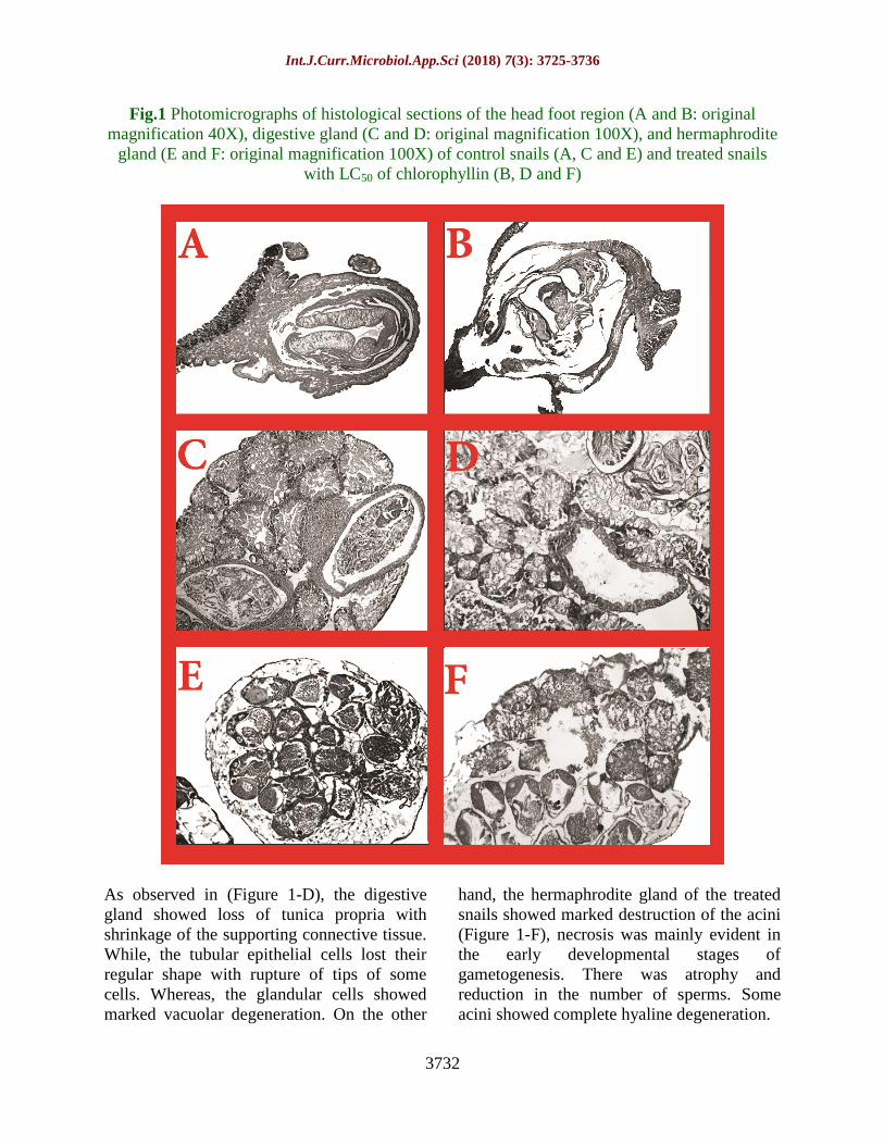
Fig.1 Photomicrographs of histological sections of the head foot region (A and B: original
magnification 40X), digestive gland (C and D: original magnification 100X), and hermaphrodite
gland (E and F: original magnification 100X) of control snails (A, C and E) and treated snails
with LC50 of chlorophyllin (B, D and F)
As observed in (Figure 1-D), the digestive
gland showed loss of tunica propria with
shrinkage of the supporting connective tissue.
While, the tubular epithelial cells lost their
regular shape with rupture of tips of some
cells. Whereas, the glandular cells showed
marked vacuolar degeneration. On the other
hand, the hermaphrodite gland of the treated
snails showed marked destruction of the acini
(Figure 1-F), necrosis was mainly evident in
the early developmental stages of
gametogenesis. There was atrophy and
reduction in the number of sperms. Some
acini showed complete hyaline degeneration.

Int.J.Curr.Microbiol.App.Sci (2018) 7(3): 3725-3736
3733
Similarly, in case of Schistosoma larval
stages, chlorophyllin treatment showed
miracidicidal (Table 7) and cercaricidal
(Table 8) effects, which were mainly
governed by the dynamics of both incubation
time in darkness and exposure time to
sunlight. Brief incubation of larvae with
chlorophyllin up to 90 minutes with LC25
resulted in 100% death of both miracidia and
cercariae after 60 and 45 minutes of sunlight
exposure.
The Schistosomiasis is a worldwide disease of
poverty that leads to chronic health hazards
(Bhattacharyya et al., 2014), during the past
decades, numerous efforts have been made to
control schistosomiasis throughout the world
(Costa et al., 2014).
One of the popular methods to control the
infection is to de-link the life cycle by killing
the snail intermediate hosts (Jaiswal and
Singh, 2008). Synthetic molluscicides have
been widely used for the effective control of
snails, but because of serious environmental
hazards more researches are now being
focused on molluscicides of plant origin
(Srivastava and Singh, 2005; Kumar and
Singh, 2006 and Jaiswal et al., 2008).
Various studies indicated that several factors
may affect the lethal concentration and
actions of chlorophyllin. Regarding the effect
of different light sources, it was found that
artificial light resulted in no mortality, while
exposure to sunlight resulted in significant
snail mortality. Under sunlight exposure, snail
mortality was directly proportional to the
increase in chlorophyllin concentration during
the dark incubation phase. Similar findings
were reported in earlier study on Chaoborus
crystallinus which revealed that a minimum
of 36 W/m2 of visible daylight was needed to
induce photodynamic destruction of the larvae
using chlorophyllin (Erzinger et al., 2011).
While, other study evaluated the effect of
light source with other photosensitizers like
carbamide perhydrate and it was reported that
photosensitizing effect was only associated
with sunlight exposure (Gawish et al., 2009).
Concerning the effect of recovery of B.
alexandrina snails from chlorophyllin before
sunlight exposure, it was noted that snails not
significantly recovered showed a relatively
higher mortality. This observation might be
attributed to elevation of temperature of
chlorophyllin solution, which may add
another factor (effect of heat) during sunlight
exposure. So, the recovery of snails from
chlorophyllin was recommended to assess
only the effect of absorbed chlorophyllin
during incubation period.
In the current study, evaluation the
molluscicidal properties of chlorophyllin
revealed that the snail mortality rate was
governed by the accumulation of
chlorophyllin within the snails' tissues during
dark incubation period together with the
duration of sunlight exposure. In addition, the
LC50 of chlorophyllin sodium copper salt for
B. alexandrina snails, after six hours of
sunlight exposure was (82.68 mg/l) and the
lethal concentrations vary with different
organisms. Several authors as (Wohllebe et
al., 2009) noted that LC50 value for Culex sp.
larvae was about 6.88 mg/L. While, (Erzinger
et al., 2011) showed that for Chaoborus sp.
larvae LC50 was approximately 24.18 mg/L.
Also, Wohllebe et al., (2012) found that
Ichthyophthiriu smulftifiliis was killed using
LC50 of about 0.67 mg/L. A higher LC50 was
reported by (Mahmoud et al., 2013) who,
noted that LC50 was about 30 mg/L for the
snails Lymnaeastagnalis, Biomphalaria spp.
And Physamarmorata..
Regarding the effect of light dose on snail
mortality, it was greatly affected and directly
proportional to the duration of sunlight
exposure. This may be attributed to the

Int.J.Curr.Microbiol.App.Sci (2018) 7(3): 3725-3736
3734
minimum time required for the initiation and
the promotion of the photodynamic action of
the photosensitizer. Where, chlorophyllin
needed a minimum of four hours of sunlight
exposure to exert its photodynamic action.
These results came in accord with a previous
study of (Mahmoud et al., 2013), who showed
that the increased duration of sunlight
exposure after chlorophyllin dark incubation
was associated with higher mortality among
Biomphalaria spp. snails.
In the present study, the effect of lethal
concentrations of chlorophyllin on different
snail developmental stages was observed that
the molluscicidal effect of chlorophyllin
varied with the size or age of the snails.
Where, chlorophyllin had a limited lethal
effect on immature snails, on adult snails
more than 8 mm and on egg masses. This
limited effect on adult snails (>8 mm) may be
attributed to the thick shell of snails which
may interfere with light penetration into the
snail's tissues needed to stimulate the
absorbed chlorophyllin to produce its
photosensitization effect. On the other hand,
the limited effect on immature snails may be
attributed to the great ability of juvenile
snail's tissue to regenerate. These finding
were in contrary with the findings of
Mahmoud et al., (2013) who reported that
chlorophyllin had resulted in 100% death of
immature snails after three hours of sunlight
exposure using concentrations up to 15µg/ml.
While, the lethal effect on egg masses
observed in the present study was also noticed
by (Mahmoud et al., 2013) who reported 70%
death of egg masses after three hours using
concentrations up to 15µg/ml compared to
100% death of snails exposed to the same
experimental conditions, denoting a lower
effect on egg masses.
On the other hand, the effect of chlorophyllin
on infected snails resulted in a greatly
enhanced lethal effect compared to uninfected
adult snails (6-8mm). This may be explained
by the added effect of photosensitization
induced cellular damage to the already
weakened snails.
Histopathological examination for assessment
of the photosensitization effect of
chlorophyllin on snail's tissues observed its
destructive effect on different cells. However,
studies assessing phototoxicity at sub-cellular
level are difficult because of the extreme
complexity of cells (Spikes, 1989).
While, during present study for the effect of
chlorophyllin on miracidia and cercariae, it
was found that it had a larvicidal effect which
was mainly dependant on both incubation and
exposure time to sunlight. This larvicidal
effect of chlorophyllin was different from
other photosensitizers like carbamide
perhydrate which had no biocidal activity
against S. mansoni miracidia and cercariae
even after their exposure to double the LC50
or LC90 for 20 minutes in sunlight (Gawish et
al., 2009)
From the foregoing results, It is concluded y
that chlorophyllin is a promising plant derived
product with potential molluscicidal and
larvicidal proprieties. Further evaluation and
conditions for optimization their effects were
urgently required for efficient field
application is recommended.
References
Barakat, R. M. R.2013. Epidemiology of
Schistosomiasis in Egypt: Travel
through Time: Review. J Adv Res, 4(5),
425-432.
Bhattacharyya, T., Ayandeh, A., Falconar, A.
K., Sundar, S., El-Safi, S., Gripenberg,
M. A., Miles, M. A. 2014. IgG1 as a
potential biomarker of post-
chemotherapeutic relapse in visceral
leishmaniasis, and adaptation to a rapid

Int.J.Curr.Microbiol.App.Sci (2018) 7(3): 3725-3736
3735
diagnostic test. PLoS Negl Trop Dis,
8(10), e3273. doi: 10.1371/journal.
pntd.0003273
Costa, L. E., Goulart, L. R., Pereira, N. C.,
Lima, M. I., Duarte, M. C., Martins, V.
T.,... Coelho, E. A. 2014. Mimotope-
based vaccines of Leishmania infantum
antigens and their protective efficacy
against visceral leishmaniasis. PLoS
One, 9(10), e110014. doi: 10.1371/
journal.pone.0110014
El-Tarky, A. E. 2005. Semi field studies to
control schistosomiasis free larval
vectors using selected sensitizers with
sun light and laser radiation. Ph.D.
thesis, National Institute of Laser
Enhanced Science, Cairo University,
Egypt.
El-Tayeb, T. 2003. Laser scanning
microscopy for determination of the
efficiency of hematoporphyrin in control
of Culex pipiens larvae and the snail
vector of Fasciola gigantica. Ph.D.
Thesis. National Institute of Laser
Enhanced Sciences, Cairo University,
Egypt.
Erzinger, G. S., Wohllebe, S., Vollrath, F.,
Souza, S. C., Richter, P., Lebert, M.,
and Häder, D.-P. 2011. Optimizing
conditions for the use of chlorophyll
derivatives for photodynamic control of
parasites in aquatic ecosystems.
Parasitology research, 109(3), 781-786.
Finney, D. J. 1970. Probit Analysis, 3rd ed.
London Cambridge University Press.
Gawish, F. A., El-Sherbini, S., and Aly, H.
F.2009. Effect of photosensitization
process of carbamide perhydrate on
Biomphalaria alexandrina snails and
their infection with Schistosoma
mansoni. J Appl Sci Res, 5, 46-56.
Graham, K. 1963. Concepts of forest
Entomology. New York, NY: Reinhold
Publ Corp/Chapman and Hall Ltd.
Jaiswal, P., and Singh, D.2008. Molluscicidal
activity of Carica papaya and Areca
catechu against the freshwater snail
Lymnaea acuminata. Veterinary
parasitology, 152(3), 264-270.
Jaiswal, P., Singh, V., and Singh, D.2008.
Enzyme inhibition by molluscicidal
component of Areca catechu and Carica
papaya in the nervous tissue of vector
snail Lymnaea acuminata. Pesticide
biochemistry and physiology, 92(3),
164-168.
King, C. H. (2009). Toward the elimination of
schistosomiasis. N Engl J Med, 360(2),
106-109.
Kumar, P., and Singh, D.2006. Molluscicidal
activity of Ferula asafoetida, Syzygium
aromaticum and Carum carvi and their
active components against the snail
Lymnaea acuminata. Chemosphere,
63(9), 1568-1574.
Lotfy, W. M. 2009. Human schistosomiasis in
Egypt: historical review, assessment of
the current picture and prediction of the
future trends. J Med Res Inst, 30, 1-7.
Luksiene, Z. 2005. New approach to
inactivation of harmful and pathogenic
microorganisms by photosensitization.
Food Technol Biotechnol, 43(4), 411-
418.
Mahmoud, M. S., Richter, P., Shalaby, H. A.,
Kandil, O. M., and Hader, D. P. 2013.
Molluscicidal activity of chlorophyll
extraction against the freshwater snails.
J Coast Life Med, 1(2), 85-88. doi:
10.12980/jclm.1.2013c824
McCullough, F. S.1992. The role of
mollusciciding in schistosomiasis
control. Geneva, Switzerland: WHO.
Mostafa, S. S., and Gawish, F. A. 2009.
Towards to control Biomphalaria
alexandrina snails and the free living
larval stages of Schistosoma mansoni
using the microalga Spirulina platensis.
Austr. J. Bas. Appl. Sci, 3(4), 4112-
4119.
Ragheb, M. 2013. Photothermal and
photosensitization, novel control

Int.J.Curr.Microbiol.App.Sci (2018) 7(3): 3725-3736
3736
modalities on certain biological and
histological parameters of
Biomphalaria alexandrina snails. Ph.D.
Thesis. Faculty of Science, Cairo
University, Egypt
Ragheb, M., El-Tayeb, T., Al Emam, M.,
Amer, M., and Bashtar, A. 2013.
Copper chlorophyllin and magnesium
chlorophyllin as molluscicidal agents
against Biomphalaria alexandrina
snails Asia Acad Res J Multidiscip,
1(16), 229-254.
Redmond, R. W. 2008. Photophysics and
Photochemistry in Photodynamic
Therapy. In M. R. H. and and P. Mroz
(Eds.), Advances in Photodynamic
Therapy: Basic, Translational, and
Clinical (pp. 41-58). Boston and
London: Artech House.
Salama, E. M., El-Sherbini, S., Abdel-Kader,
M. H., and Jori, G. 2002. Site of action
of hematoporphyrin (a photo-activated
insecticide) in Culex pipiens larvae.
Egypt J Biol, 4, 133-141.
Srivastava, P., and Singh, D. 2005. Control of
harmful snails: Tejpat (Cinnamomum
tamala) a potential molluscicide. J Appl
Biosci, 31(2), 128-132.
Spikes, J. 1989. Photosensitization. In S. C. K
(Ed.), The Science of photobiology
(second edition ed., pp. 79-110). New
york USA: plenum press.
SPSS, (2006). Statistical Package for Social
Science, SPSS for windows Release
16.0.0, and 12 June, 2006. Standard
Version, Copyright SPSS Inc., 1989-
2006, All Rights Reserved, Copyright ®
SPSS Inc.
Useh, M. F. 2012. Control of schistosomiasis.
In M. B. Rokni (Ed.), Schistosomiasis
(pp. 73-102): InTech.
Wohllebe, S. 2010.Combating parasites in
aquatic ecosystems by natural
photosensitizers. Ph.D. Thesis.
University of Erlangen, Erlangen,
Germany.
Wohllebe, S., Richter, P., and Häder, D.-P.
2012. Chlorophyllin for the control of
Ichthyophthirius multifiliis (Fouquet).
Parasitology research, 111(2), 729-733.
Wohllebe, S., Richter, R., Richter, P., and
Häder, D.-P. 2009. Photodynamic
control of human pathogenic parasites
in aquatic ecosystems using
chlorophyllin and pheophorbid as
photodynamic substances. Parasitology
research, 104(3), 593-600.
How to cite this article:
Heba A. Elhadad, Bassem A. El-Habet, Rania M. Azab, Hanaa M. Abu El Einin, Wael M.
Lotfy and Hassan A. Atef. 2018. Effect of Chlorophyllin on Biomphalaria alexandrina Snails
and Schistosoma mansoni Larvae. Int.J.Curr.Microbiol.App.Sci. 7(03): 3725-3736.
doi: https://doi.org/10.20546/ijcmas.2018.703.431