Effect of Bisphenol-A on Brain Tissue in Pregnant Rat
13
Int.J.Curr.Microbiol.App.Sci (2016) 5(8): 677-689 677 Original Research Article http://dx.doi.org/10.20546/ijcmas.2016.508.077 Effect of Bisphenol-A on Brain Tissue in Pregnant Rat T. Geetharathan* Department of Biotechnology, Sri Padmavati Mahila Visvavidyalayam, Tirupati-517502, Andhra Pradesh, India *Corresponding author ABSTRACT Introduction Among many stabilizers of plastics, bisphenol-A (BPA) is a popular stabilizer that mimics the actions of estrogen and affects the reproduction, endocrine glands in vivo and in vitro (Kuiper et al., 1998). Animal studies have shown that in utero exposure to BPA produces prenatal and postnatal adverse effects on multiple tissues, including the brain (Richter et al., 2007). Prenatal BPA exposure affects brain development, sexual differentiation, social and anxiety-like behavior, and learning (or) memory (Kundakovic et al., 2011). In humans, emerging evidence for BPA- associated disruption to neurodevelopment is consistent with the rodent data and has revealed sex-specific effects of gestational BPA levels on emotional regulation and aggression in children (Perera et al., (2012). Growing evidence suggests that BPA is associated with oxidative stress (Sui et al., 2012). Indeed, it has been revealed that BPA can disturb oxidative homeostasis through direct or indirect pathways, International Journal of Current Microbiology and Applied Sciences ISSN: 2319-7706 Volume 5 Number 8 (2016) pp. 677-689 Journal homepage: http://www.ijcmas.com For more than ten years, there has been a scientific and journalistic controversy whether BPA causes adverse effects on human. Humans are routinely exposed to BPA, an estrogenic chemical present in food and beverage containers, dental composites, and many products in the home and workplace. In order to study the effects of BPA on the organ of pregnant rat brain, the albino rats were administrated to low& high concentrations of BPA (50 mg/kg. d & 500 mg/kg. d). We aimed to study the toxic effect of BPA on the body & brain weight, developmental structure of cerebellum in albino pregnant rat. After 8 days (Doses were administrated gestational day 8 th to 15 th ), the antioxidant enzymes, superoxide dismutase (SOD), catalase (CAT), malondialdehyde (MDA) and glutathione (GSH) concentrations in the organ was measured. Results showed that the MDA contents were increased with the added concentration of the BPA. But the SOD, CAT, GSH content was decreased. BPA induction also caused histopathological changes in the rat brain and cerebellum. All BPA treatment also revealed an increase in DNA fragmentation as evidenced by an increase in concentration of dose. It is indicated BPA can induce the rat’s oxidative damage and the effect is much easier for the brain. Keywords BPA, Pregnant rat, Brain, Cerebellum, MDA, GSH. Accepted: 28 July 2016 Available Online: 10 August 2016 Article Info
Transcript of Effect of Bisphenol-A on Brain Tissue in Pregnant Rat

Int.J.Curr.Microbiol.App.Sci (2016) 5(8): 677-689
677
Original Research Article http://dx.doi.org/10.20546/ijcmas.2016.508.077
Effect of Bisphenol-A on Brain Tissue in Pregnant Rat
T. Geetharathan*
Department of Biotechnology, Sri Padmavati Mahila Visvavidyalayam,
Tirupati-517502, Andhra Pradesh, India *Corresponding author
A B S T R A C T
Introduction
Among many stabilizers of plastics,
bisphenol-A (BPA) is a popular stabilizer
that mimics the actions of estrogen and
affects the reproduction, endocrine glands in
vivo and in vitro (Kuiper et al., 1998).
Animal studies have shown that in utero
exposure to BPA produces prenatal and
postnatal adverse effects on multiple tissues,
including the brain (Richter et al., 2007).
Prenatal BPA exposure affects brain
development, sexual differentiation, social
and anxiety-like behavior, and learning (or)
memory (Kundakovic et al., 2011). In
humans, emerging evidence for BPA-
associated disruption to neurodevelopment
is consistent with the rodent data and has
revealed sex-specific effects of gestational
BPA levels on emotional regulation and
aggression in children (Perera et al., (2012).
Growing evidence suggests that BPA is
associated with oxidative stress (Sui et al.,
2012). Indeed, it has been revealed that BPA
can disturb oxidative homeostasis through
direct or indirect pathways,
International Journal of Current Microbiology and Applied Sciences ISSN: 2319-7706 Volume 5 Number 8 (2016) pp. 677-689
Journal homepage: http://www.ijcmas.com
For more than ten years, there has been a scientific and journalistic controversy
whether BPA causes adverse effects on human. Humans are routinely exposed to
BPA, an estrogenic chemical present in food and beverage containers, dental
composites, and many products in the home and workplace. In order to study the
effects of BPA on the organ of pregnant rat brain, the albino rats were
administrated to low& high concentrations of BPA (50 mg/kg. d & 500 mg/kg. d).
We aimed to study the toxic effect of BPA on the body & brain weight,
developmental structure of cerebellum in albino pregnant rat. After 8 days (Doses
were administrated gestational day 8th to 15
th), the antioxidant enzymes, superoxide
dismutase (SOD), catalase (CAT), malondialdehyde (MDA) and glutathione (GSH)
concentrations in the organ was measured. Results showed that the MDA contents
were increased with the added concentration of the BPA. But the SOD, CAT, GSH
content was decreased. BPA induction also caused histopathological changes in the
rat brain and cerebellum. All BPA treatment also revealed an increase in DNA
fragmentation as evidenced by an increase in concentration of dose. It is indicated
BPA can induce the rat’s oxidative damage and the effect is much easier for the
brain.
K e y w o r d s
BPA,
Pregnant rat,
Brain,
Cerebellum,
MDA, GSH.
Accepted:
28 July 2016
Available Online:
10 August 2016
Article Info

Int.J.Curr.Microbiol.App.Sci (2016) 5(8): 677-689
678
including mitochondrial function (Ooe et al.,
2005), modulation of antioxidant enzymes
and increase thiobarbituric acid-reactive
substances in the brain of mice exposed
throughout embryonic (or) fetal route
(Kabuto et al., 2004). Oxidative stress has
been implicated in aging and many
pathological disorders, such as ischemic
diseases, neurodegenerative diseases,
diabetes, and cancer, although the
underlying mechanisms are not always
completely understood (Lenaz et al., 2012).
The most meaningful studies on the effects
on brain development, neuroendocrine
signaling, and behavior concern the in utero
and perinatal exposure to BPA. The
exposure of pups or of pregnant rats or mice
to BPA results in an increased expression of
ERs (Ramos et al., 2003) and ER-α labeled
neurons in diverse brain areas and in a
reduced number of dopamine containing
neurons (Masuo et al., 2004). CD-1 mice
developmental exposure to BPA result in a
significant change in the locus coeruleus
(Kubo et al., 2001), while maternal exposure
disrupt normal neocortical development in
fetuses by accelerating neuronal
differentiation (or) migration (Nakamura et
al., 2006). Moreover, five studies have
demonstrated low dose effects of adult BPA
exposures on brain endpoints such as
synapse formation and remodeling (Leranth
et al., 2008). Some of these effects were
observed only in response to additional
hormone treatments (e.g., BPA attenuates
the effects of estrogens on synapse
formation (or) remodeling).
Epidemiological studies examining
neurobehavioral outcomes correlates pre-
natal exposure to BPA exposure and
neurobehavioral outcomes, such as
aggressivity and hyperactivity (Braun et al.,
2009) or anxious or depressed behavior with
poor emotional control (Braun et al., 2011).
These observations are in agreement with
results of behavioral tests conducted on
laboratory rodents exposed to low doses of
BPA such as anxious behaviour, increase of
aggressiveness, impaired learning, hyper-
reactivity to painful or fear-provoking
stimuli, altered adult play and other socio-
sexual behaviors (Ryan and Vandenbergh,
2006). The intrauterine foet al life
environment is critical for the normal
development. The rat cerebellum during the
first period after birth is considered to be
equivalent to the third trimester of human
pregnancy (Hamre and West, 1993; Thomas
et al., 1998). Chemicals effects and changes
in the levels of hormones can lead to
changes in brain function and consequently
in behavior (Jacobson and Jacobson, 1996;
Faroon et al., 2001). Factors that affect the
normal development of the cerebellum can
cause pathological effects, depending on the
developmental stage (Laure-Kamionowska
and Maliska, 2009). To date, there have
been few in vivo studies that have examined
the impact of BPA exposure during
gestation and lactation on brain development
(Facciolo et al., 2002).
In addition, BPA (25–50 mg/kg/day, i.p.
injection) has been shown to enhance
oxidative stress and lipid peroxidation
promoting the cellular death in several
organs (e.g. brain, liver, and kidney) of
exposed rodents (Aydogan et al., 2008;
Kabuto et al., 2003). This study was
designed to assess the effect of BPA on the
prenatal developmental structure of
cerebellum in brain of pregnant albino rat.
Therefore, the aims of this study were to
illustrate the effects of BPA on the acute
oxidative damage in targeted tissues and
speculate the possible mechanism, by
detecting the activity of the contents of
MDA, and the contents of SOD, CAT, and
GSH. The present study demonstrates DNA
fragmentation by agarose gel electrophoresis
in targeted tissues of rats exposed to low and
high-doses BPA.

Int.J.Curr.Microbiol.App.Sci (2016) 5(8): 677-689
679
Materials and Methods
Chemicals: Bisphenol-A and all chemicals
were purchased from Sd Fine Chemicals,
Bombay, BPA was dissolved in Sesame oil
(vehicle) as stock before administration.
Animals and Experimental design
Experimental animals
Pregnant wistar albino rats weighing 250-
300 g were used as experimental animals.
The animals were obtained from the M/S
Raghavendra Enterprises, Bangalore, India.
They were maintained on stock diet and kept
under fixed appropriate conditions of
housing and handling. Different
experimental groups of the animals were
caged separately and an average of 6
animals per cage was maintained. The
control as well as other treated groups of rats
was given free access to standard chow and
water ad libitum. All these animals were
housed in wooden cages and provided water
in glass bottles.
Experimental Design
The animals were divided into 3 groups.
Animals of group (1) served as control and
received a daily oral administration of
sesame oil, Animals of group (2) were
administered orally 50 mg/kg of BPA for 8
days and group (3) were administered orally
500mg/kg of BPA for 8 days respectively.
The doses of BPA were administered 8 days
(gestational day 8th
to 15th
). The lower dose
of BPA (50 mg/kg) in this study was chosen
on the base of previous studies (Bian et al.,
2006; Richter et al., 2007).The weight of
each animal was recorded. The rats were
anesthetized with diethyl ether and
sacrificing, the brain of each rat was
dissected and removed and its weight was
recorded. Cerebellums were dissected then
fixed for histopathological examinations.
Malondialdehyde Contents (MDA) Assay
0.5 mL sample dilution was removed to the
test tube, and 2 ml of 0.6% TBA solution
was added. TBA solu- tion was dissolved by
a small amount of 1 mol/L sodium hyroxide,
and then diluted to the concentration by 10%
trichloroacetic acid. The test tube was sealed
by plastic wrap with a little hole. After in the
water bath boiling for 15 minutes, the test
tube was cooled in the water, and then
centrifuged at 10,000 rpm for 10 minutes.
The absorbance value of the supernatant was
measured at 450 nm, 532 nm and 600 nm of
wavelength. The formula C = 6.45 × (D532
− D600) − 0.56 × D450 was used to
calculated the concentration of MDA.
Glutathione (GSH) Contents Assay
Low molecular weight thiols (predominantly
GSH) in cells were examined using
Multiskan Spectrum, which produces a
highly absorbance at 412 nm upon its
reaction with thiols groups. 200 μL of the
supernatant was took and mingled with 50
μL of 10% TCA, so that the protein was
precipitated, then centrifuged the liquid for
the sec- ond time so as to eliminate the
protein. The supernatant was diluted with
PBS-EDTA, regulated pH with sodium
hydroxide to 7.5, and then 50 μL sample
solution and 150 μL DTNB of 60 μg/mL
were joined into 96 wells micro plate, kept
in dark place for 5 minutes at room
temperature. Finally detected the OD values
at the wave- length of 412 nm, calculate the
concentration of GSH according to the
standard curve.
Light microscopy
All specimens for light microscope
examination were cut into small pieces and
fixed in a solution of 10% formaldehyde and
processed to get paraffin sections of 5 µm
thickness. Sections were stained with

Int.J.Curr.Microbiol.App.Sci (2016) 5(8): 677-689
680
Haematoxylin and Eosin (H&E) (Bancroft
and Stevens, 1996). Slides were mounted
using entellan and covered with cover slips
prior to viewing and photography by (Nikon
Eclipse E 200) light microscope.
DNA fragmentation studies
Tissue (brain) slices were placed in the
digestion buffer (10 mM Tris–HCl with pH
8.0; 0.1 M EDTA with pH 8.0; 1% SDS and
proteinase K with concentration of 1 mg/10
ml) and were incubated (14–18 h, 55◦C) in a
shaking water bath. The DNA contents were
extracted, precipitated, and stored as
described below for the analysis by agarose
gel electrophoresis. After digestion, samples
were extracted three times with 10%
saturated phenol/chloroform/isoamyl alcohol
(24:24:1) and were precipitated using
ethanol. The precipitates were rinsed two
times with 70% ethanol, air dried, and
resuspended in Tris EDTA buffer. DNA
contents were measured using a
spectrophotometer (A260/A280), and only
samples with 1.8 ratios were used. Agarose
gel electrophoresis was then carried out to
analyze the fragmentation.
Statistical Analysis
The differences between treated samples and
untreated control were evaluated by the
students’ T-test following a one-way
analysis of variance (ANOVA) (Steel and
Torrle) were performed using the Statistical
Package for Social Sciences (SPSS) Package
programming techniques on “Intel Core 2
Duo Processor” personnel computer.
Probability values less than 0.05 were
considered significant.
Results and Discussion
The results revealed more prominent signs
of toxicity in rats treated with BPA, where
most treated rats became less active and
showed general weakness. The body and
brain weights were recorded to be
significantly low in the BPA treated animals
as compared to their respective controls.
Effect of BPA on antioxidant enzymes
(Super oxide dismutase (SOD), Catalase
(CAT), Glutathione (GSH), Malondi-
aldehyde (MDA)) in rat brain tissue
In the present study, super oxide dismutase,
catalase, GSH activities in tissue of BPA
treated rats (lower dose or 50mg/kg.b.w/day
and higher dose or 500mg/kg.b.w/day)
showed significant variations (P<0.05),
(Fig-2), when compared to control group.
On treatment with BPA rats, showed
significantly decreased super oxide
dismutase, catalase & glutathione activities,
the gradual decreased super oxide
dismutase, catalase, glutathione activities
were found in the tissue of higher dose
followed by lower dose BPA treated groups.
Thus it is revealed that the above enzyme
activities were decreased highly in higher
dose of BPA treated rats when compared
with control group.
The level of malondialdehyde activity in
tissue of BPA treated rats (lower dose and
higher dose) showed significant variations
(P<0.05), (Fig- 2), when compared to
control group. On treatment with BPA rats,
showed significant elevated levels of
malondialdehyde activity, the gradual
increased malondialdehyde activity was
found in the higher dose, lower dose BPA
treated groups. Thus the above observations
revealed that highly increased
malondialdehyde activity in higher dose
BPA treated group when compared to
control group.
Histological changes in brain and
cerebellum tissues
Figure-3 illustrates the histopathological
assessment of brain tissue of control and
experimental animals. BPA treated rats (3.2

Int.J.Curr.Microbiol.App.Sci (2016) 5(8): 677-689
681
& 3.3) exhibited spongiform necrosis (1),
marked gliosis (2), nuclear pycnosis (3), and
lymphocytic inflammatory infiltrates as
against normal architecture shown by the
brain of control rats (Figure-3.1). Treatment
with BPA toxication showed the incidence
of these pathological changes in the brain
tissue.
In control group cerebellum, the Purkinje
cells (P) in the third layer are arranged at the
junction between the molecular layer (M)
with stellate (S) and the granular layer (G).
They are nearly mature cells. The internal
granular layer is thick and contains small
neurons called the granule cells, which have
large, rounded nuclei and scanty cytoplasm
and immature with few number of basket
cells (B) (Fig-4.1). Effect of BPA low dose
on the pregnant rat revealed the transverse
section of cerebellum showing disruption in
the Purkinjee cells layer (P) & cells were
immature (Fig-4.3). Effect of BPA high dose
on the pregnant rat revealed the transverse
section of cerebellum showing highly
disruption in the Purkinjee cells layer,
degenerative changes with loss of their
normal pyriform shaped appearance. The
purkinjee cells showed a notable degree of
immaturity and a much lower number of
cells compared to the control (4.2) group.
The cells are scattered unorganized with
wide spaces and revealing signs of injury
and they had lost their normal organization.
Many cells were mostly pyknotic (Fig.4.4).
DNA damage
The DNA damage (brain tissue) caused in
the cells as a result of BPA induction was
examined by agarose gel electrophoresis is
shown in Fig-5. The results indicated that
DNA of BPA-induced group showed a
comet tail indicating the DNA damage
arising from the genotoxicity in the BPA-
induced cells when compared to the DNA of
control cells. Whereas intensity of DNA
fragmentation is high in high dose treated
sample compared to low dose treated
sample. Thus DNA fragmentation gradually
increased from lower (L2), higher dose (L3)
and normal DNA banding pattern was
observed in control (L1), (Fig-5).
Epidemiological and experimental data
indicate that in utero exposure to
environmental chemicals and prescribed
drugs during pregnancy can mediate early
embryonic losses, spontaneous abortion,
fetal growth retardation and resorptions,
decreased litter size, fetal malformations and
low body weight and low birth weight
(Khattak et al., 1999) via ROS generation
which damages cellular macromolecules
(Nicol et al., 2000). BPA, an environmental
contaminant, is concerned as a serious threat
for human health due to its wide spread in
nature (Richter et al., 2007). Furthermore,
exposure to BPA during neonatal and
prenatal periods has been shown to led to
memory impairment, sexual differentiation
process and behavior in the offspring of
experimental animals (Nakamura et al.,
2012) and cognitive impairment in rodents
(Negishi et al., 2003). It has been suggested
that environmental exposure to BPA may
affect the developing brain by enhancing the
local biosynthesis of estrogen in the brain,
inhibiting estrogen receptor beta (ERbeta)
and N-methyl- D-aspartate receptor
(NMDAR) expressions(Kawai et al., 2007).
Furthermore, due to its estrogenic and
antiandrogenic activities (Delfosse et al.,
2014), BPA can interfere with the dimorphic
development of the neuronal networks
controlling many endocrine systems and
brain functions. As a matter of fact, sex
differences have been well documented not
only in the mechanisms controlling
reproduction, but also in non reproductive
behaviors (Jazin and Cahill, 2010 and
Wolstenholme et al., 2011). In this study of
results revealed that, the body and brain
weights were recorded to be significantly

Int.J.Curr.Microbiol.App.Sci (2016) 5(8): 677-689
682
low in the BPA treated animals as compared
to their respective controls. BPA exposure
also decreases the body and brain weight,
because the reason that can be attributed for
the decrease in protein contents under toxic
stress may be due to formation of
lipoproteins, which are utilized for repair of
damaged cell and cell organells. The
decreased protein content may be related to
impaired food intake of animals, the
increased energy cost of homeostatis,
detoxification mechanism, also be attributed
to the destruction or necrosis of cellular
function and consequent impairment in
protein synthetic machinery during stress
condition. Hiroi et al., (2006) reported that,
BPA possesses an inhibitory effect on
protein disulfide bonds formation in cells
therefore it might have a potential effect in
disrupting various physiological functions.
The binding of BPA to human serum
proteins has been reported by Csandy et al.,
(2002). Gopinath (Gopinath, 1984) observed
that the brain weight was low in the
experimental under nutrition. BPA exposure
also decreases the body and brain weight
dose dependant and may be due to loss of
animal's appetite and activities.
The findings of the present study, also,
showed that the oxidative stress rise in BPA
treated rats was accompanied by
concomitant decrease in the activity of some
antioxidant enzymes involved in the
detoxification of ROS, namely SOD, CAT
as well as the level of GSH in the brain
tissues comparing with the control declaring
the prooxidant effect of BPA. These
findings agreed with the antecedent studies
of Andersen, (2004). Who showed that BPA
exposure enhanced the neuronal lipid
peroxidative damage with concomitant
alterations in the enzymatic antioxidant
defense status, thus having serious bearing
on the functional and structural development
of the central nervous system. Similar data
recorded a decrease in the antioxidants such
as GSH and SOD activity in the brain of
aluminium exposed rats and human Dua R
and Gill KD (2001). BPA could be caused
by inflicting damage to membrane lipids,
proteins and antioxidative enzyme defense
system. However, the increased BPA
concentration could deleteriously affect the
neurons, leading to depletion of antioxidants
and metal ions through the induction of free
radicals, that exhausting SOD and CAT
which function as blockers of free radical
processes. These results are in accordance
with Bindhmol et al., 2003 and Kabuto et al,
2004 whom recorded a significant decrease
in the activities of SOD and CAT in brain of
rats after BPA treatment. Alternatively, the
decreased enzyme activities could be related
to a reduced synthesis of the enzyme
proteins as a result of higher intracellular
concentrations of BPA. SOD, catalase, and
Glutathione are intracellular enzymatic
antioxidants, which are responsible for
disposing reactive oxygen species such as
hydrogen peroxide and superoxide free
radicals. In this study, the basal levels of
activities of these three enzymes in the BPA
treated brain tissue of the rats were found to
be much lower than those in the control.
Typically, lipid peroxidation was the
primary results of oxidative stress, and
correlated effects on the levels of MDA
(increased) were also observed. The toxicity
of MDA is so great that it can induce the
peptide chain’s fracture and cross-linking of
amino acids in enzyme molecule to form the
polymer. As a result, the enzyme activity is
renewed, lost or changed, the structure of
cell membrane is being destroyed, disorderly
ionic exchange is induced inside and outside
of cell membrane and excessive free radicals
are produced. Then cells are destroyed and
the organism is also injured. The result is
agreed with the ROS result. Besides, the
balance of oxidants and antioxidants is very
important for healthy cells. So the

Int.J.Curr.Microbiol.App.Sci (2016) 5(8): 677-689
683
intracellular GSH level in the BPA treaded
cells was monitored to further prove the
formation of ROS. GSH is an important
anti- oxidant that protects cells from ROS
and plays a critical role in the intracellular
ROS removal. Hence the depletion of
intracellular GSH is usually regarded as a
scientific measure of the oxidative stress.
Just as illustrated in the Figure-3, the
absorbance in the BPA treated cells was
much weaker than the control group. In
concordance with our results, Wu et al.
showed significant decrease in the levels of
GSH in BPA group; this decrease indicated
tissues being damaged (Wu et al., 2011).
The research before showed that ROS may
be one of the major mediators in the
regulation of autophagy (Wang et al., 2009),
ROS is a natural by product of the normal
metabolism in cells. A certain amount of
ROS could function as a second message in
the signal transduction of healthy cells.
However, excessive ROS damages
bimolecular, triggers the apoptosis
pathways, and even further induces cell
death; excessive ROS and oxidative stress
may be the main reasons for multiple
toxicities (Nel et al., 2006). Excess ROS can
also damage a wide variety of cellular
constituents including DNA, RNA, Proteins,
sugar and lipids, thereby compromising cell
viability. The result of this study is similarly
with others who demonstrated that BPA can
generate ROS that causes oxidative damage
in the brain tissue of rats (Korkmaz et al.,
2010). Treatment with BPA induced
oxidative stress in various tissues of rodent
(Kabuto et al., 2003) by decreasing
antioxidant enzymes and increasing
hydrogen peroxide and lipid peroxidation
(Bindhumol et al., 2003). The presence of
high level of ROS during embryonic period,
the induction of oxidative stress in the target
tissue has been suggested as a possible
mechanism of chemical carcinogenesis
(Kensler et al., 1989). Exogenous
xenobiotics (BPA) and endogenous
compounds can disturb this balance by
enhanced generation of free radicals and
decreased antioxidant capacity.
The light microscopic observations of the
brain tissues BPA treated rats showed;
cavitations and necrotic areas appeared in all
cerebellar layers. Exposure to BPA may
cause marked histophathological alterations
in the brain tissue which were represented
by focal as well as diffuse gliosis,
spongioform necrosis and nuclear pycnosis
in brain regions with neuronal degeneration.
Parallel to our findings, those recorded by
Mohamed Atif A. Said Ahmed et al, who
found that BPA causes histopathological
lesions in brain regions including neuronal
degeneration as cytoplasmic vacuolization
hemorrhage, ghost cell and gliosis. Our
histopathological findings are correlated to
those of Matyja, who noticed that exposure
to chemical causes marked histopathological
alteration in the cerebral cortex including
neuronal degeneration, perecellular odema
and gliosis. The cerebellar cortex of
pregnant rat is seen to be consisted of four
layers, the outermost external granular layer
which covering the surface of the
developing cerebellum, molecular layer,
Purkinje cell layer and an inner granular
formed of four to five rows of cells. The
molecular layer is thin layer and immature
with few number of basket cells. Tranverse
section of BPA treated rat cerebellum
showing disruption in the purkinjee cells
layer, some Purkinje cells were small in size
with abnormal shape and loss of the
prominent dendrite. Also the arrangement is
impaired and disorganized in the BPA-
treated animals as compared to the controls
(4.1).According to Brodal, the functions of
certain learning and memory have been
associated with different areas of the brain
like the hippocampus and cerebellum.
However, BPA treated group showed
Int.J.Curr.Microbiol.App.Sci (2016) 5(8): 677-689
684
marked cell distortion groups with high level
of degeneration in the cells as compared to
the controls. The toxic effect of BPA during
pregnancy causes different pathological
changes in the cerebellum (including
degenerative changes) observed in this
study. Pathological changes in cerebellum
(4.3 & 4.4) of pregnant rats treated with
BPA demonstrated the features of nervous
tissue damage expressed by marked cellular
damage. The observed changes suggested
high generation of ROS in the cells. BPA
can be dispersed to the organs of the body
after administrated. It can also get to the
brain. The brain consumes a large quantity
of oxygen, making it particularly susceptible
to oxidative stress Andersen (2004). The
pregnant rat selected was in the time of
growing development especially for the
brain. Currently, we should pay attention to
the application damage, as the 50 & 500
mg/kg·d BPA administrated can induce
oxidative damage especially for the brain of
pregnant rat.
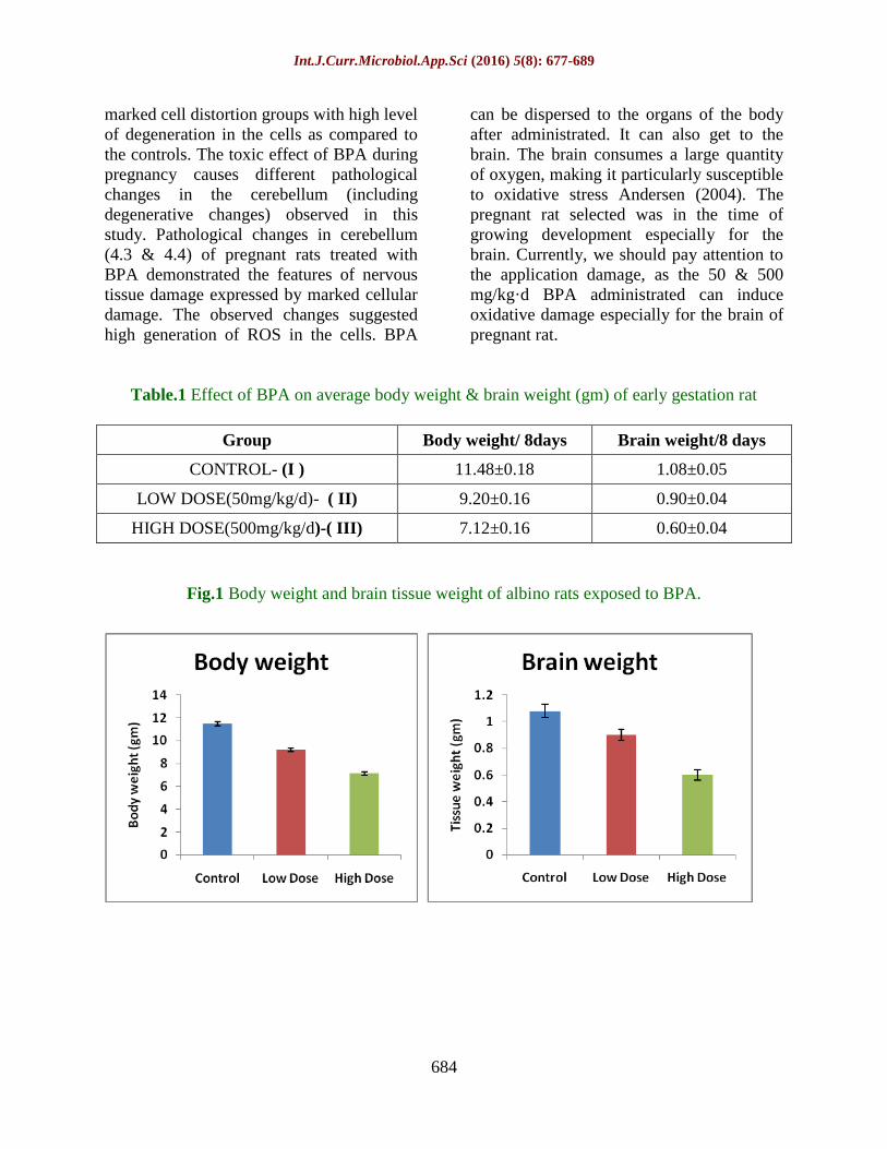
Table.1 Effect of BPA on average body weight & brain weight (gm) of early gestation rat
Group Body weight/ 8days Brain weight/8 days
CONTROL- (I ) 11.48±0.18 1.08±0.05
LOW DOSE(50mg/kg/d)- ( II) 9.20±0.16 0.90±0.04
HIGH DOSE(500mg/kg/d)-( III) 7.12±0.16 0.60±0.04
Fig.1 Body weight and brain tissue weight of albino rats exposed to BPA.

Int.J.Curr.Microbiol.App.Sci (2016) 5(8): 677-689
685
Fig.2 Superoxide dismutase, Catalase, Glutathione, Malondialdehyde activity levels in brain
tissue samples of albino rats exposed to BPA
Fig.3 Brain tissue morphology (H & E staining)
Fig-3.1: Control Fig-3.2: Low dose (50 mg/kg)
Fig-3.3: High dose (500 mg/kg)
1
2
3
1
3
2

Int.J.Curr.Microbiol.App.Sci (2016) 5(8): 677-689
686
Fig.4 Purkinjee cell layer morphology in cerebellum tissue (H & E staining)
Fig-4.1: Control cerebellum Fig-4.2: Purkinjee cell layer
Fig-4.3: Low dose Purkinjee cell layer Fig-4.4: High dose Purkinjee cell layer
Fig.5 Agarose gel electrophoresis of genomic DNA extracted from rat brain from various
treatment groups. Lane 1: DNA from the control group, Lane 2: DNA from the low dose treated
group, Lane 3: DNA from higher dose treated group, M: marker.

Int.J.Curr.Microbiol.App.Sci (2016) 5(8): 677-689
687
Lastly, effects of BPA toxicity on DNA
were also investigated (brain tissue). BPA
induction caused an increased DNA damage
as indicated by the increased fragmentation
of DNA were observed. DNA fragmentation
in this study as a consequence of BPA
exposure. BPA is known to increase the
levels of reactive oxygen species (Cavalieri
et al., 2010). This is known to cause damage
to various macromolecules and also to
DNA. Damage to DNA is one of the
markers and typical characteristic of
apoptosis (Wyllie, 1993) and BPA toxicity
can lead to faster apoptosis as seen in the
previous study which clearly revealed
disruption of cells. Further, BPA has been
shown to possess antioxidative properties
and hence may decrease the levels of free
radicals and ultimately the damage to DNA.
During critical periods of embryonic and
prenatal development, the hormonal milieu
is crucial for the correct organization of
neuroendocrine circuits that coordinate sex-
specific physiology so, the altered
expression levels of hormones at the
hypothalamus and pituitary (in brain) levels
may be the cause and/or the consequence of
the changes in gonadal steroidogenesis and
sex hormone production (Fernandez et al.,
2010). There are possible mechanistic
effects of BPA on the local regulatory
circuits of hypothalamus and pituitary (in
brain) (Mahoney and Padmanabhan, 2010).
Knobil et al., (1994) pointed out that the
decrease serum LH level by BPA may be
due to the consequence of the increase in
GnRH pulse frequency, leading to
desensitization of the pituitary. Additionally,
decrease of serum LH might cause by
estrogenic action of BPA at the level of the
HPG axis, mimicking the estrogenic
suppression of LH, FSH secretion, while it
inhibited LH, FSH secretion, suggesting that
the effect of BPA on gonadotropes is
specific to the mechanism that underlies the
control of LH, FSH secretion. In my
previous study, decreased levels of
progesterone caused by BPA could lead to
low levels of the sex hormones, which will
trigger impaired immune function and a host
of other issues associated with hormone
imbalance. BPA might disrupt the motility
of muscular layers of the oviduct and the
sequential phenomena of embryo
implantation by binding estrogen &
progesterone receptors (Uterine proliferation
and differentiation during the pre
implantation period in pregnancy have been
well characterized, and these processes are
dependent on estrogen and progesterone),
ultimately resulting in a reduction of the
number of embryos and in uterine weight
during pregnancy. It can leads to
fetoplacental and uterine growth restriction
in pregnant rats. These data indicate that
BPA may be capable of altering important
events during critical periods of brain
development.
References
Andersen, J.K. 2004. Oxidative stress in
neurodegeneration: Cause or
consequence. Med, 7, 18-25.
Bancroft, J.D. and Stevens, A. 1996. Theory
and practice of histological techniques.
4th ed.Churchill Livingstone: New
York.
Braun, J.M., Kalkbrenner, A.E., Calafat,
A.M., Yolton, K., Ye, X., Dietrich, K.
N., and Lanphear, B.P. 2011. “Impact
of early-life bisphenol A exposure on
behavior and executive function in
children,” Pediatrics, vol. 128, no. 5,
pp. 873–82.
Braun, J.M., Yolton, K., Dietrich, K.N.,
Hornung, R., Ye, X., Calafat, A.M.,
and Lanphear, B.P. 2009. “Prenatal
bisphenol A exposure and early
childhood behavior.,” Environ. Health
Perspect., vol. 117, no. 12, pp. 1945–
52.

Int.J.Curr.Microbiol.App.Sci (2016) 5(8): 677-689
688
Faroon, O., Jones, D., de Rosa, C. 2001.
Effects of polychlorinated biphenyls
on the nervous system. Toxicol. Ind
Health; 16: 305-33.
Hamre, K.M. and West, J.R. 1993.The
effects of the timing of ethanol
exposure during the brain growth spurt
on the number of cerebellar Purkinje
and granule cell nuclear profiles.
Alcohol Clin. Exp. Res., 17: 610-22.
Jacobson, J.L., Jacobson, S.W. 1996.
Intellectual impairment in children
exposed to polychlorinated biphenyls
in utero. N Engl. J. Med., 335: 7839.
Kabuto, H., Amakawa, M. and Shishibori,
T. 2004. “Exposure to bisphenol A
during embryonic/fetal life and
infancy increases oxidative injury and
causes underdevelopment of the brain
and testis in mice,” Life Sci., vol. 74,
no. 24, pp. 2931–2940.
Kawai, K., S. Murakami, E. Senba, T.
Yamanaka, Y. Fujiwara, C. Arimura,
T. Nozaki, M. Takii and C. Kubo.
2007. Changes in estrogen receptors
alpha and beta expression in the brain
of mice exposed prenatally to
bisphenol A. Regulatory Toxicol.
Pharmacol., RTP, 47: 166-170.
Kensler, T.W., Egner, P.A., Taffe, B.G., and
Trush, M.A. 1989. Role of free
radicals in tumor promotion and
progression. Prog. Clin. Biol. Res.,
298, 233-248.
Khattak, S.G., Moghtader K.K., McMartin
M., Barrera D., Kennedy D and Koren
G. 1999. Pregnancy outcome
following gestational exposure to
organic solvents: a prospective
controlled study. The J. American
Med. Assoc., 281: 1106-1109.
Korkmaz, A., Ahbab, M.A., Kolankaya, D.
and Barlas, N. 2010. Influence of
Vitamin C on Bisphenol A, nonyl-
phenol and octylphenol induced
oxidative damages in liver of male
rats. Food and Chem. Toxicol., 48:
2865-2871.
Kubo, K., Arai, O., Omura, M., Watanabe,
R., Ogata, R., and Aou, S. 2003. “Low
dose effects of bisphenol A on sexual
differentiation of the brain and
behavior in rats.” Neurosci. Res., vol.
45, no. 3, pp. 345–56.
Laure-Kamionowska, M., Mali ska, D.
2009. Calbindin positive Purkinje cells
in the pathology of human cerebellum
occurring at the time of its
development. Folia Neuropathol., 47:
300-5.
Mohamed Atif, A., Said Ahmed and Refaat
A. Eid. 2015. Effect of Bisphenol-A
on the Post-Natal Development and
Structure of Rat Cerebellum. Int. J.
Curr. Microbiol. App. Sci., 4(6): 14-
35.
Nakamura, K., Itoh K., Dai H., Han L.,
Wang X., Kato S., Sugimoto T. and
Fushiki S. 2012. Prenatal and
lactational exposure to low-doses of
bisphenol A alters adult mice
behavior. Brain and Development, 34:
57-63.
Ramos, J., Varayoud, G.J., Kass, L.,
Rodríguez, H., Costabel, L., Munoz-
De-Toro, M. and Luque, E.H. 2003.
“Bisphenol-A induces both transient
and permanent histofunctional
alterations of the hypothalamic-
pituitary-gonadal axis in prenatally
exposed male rats,” Endocrinol., vol.
144, no. 7, pp. 3206–15.
Ryan, B.C. and Vandenbergh, J.G. 2006.
“Developmental exposure to
environmental estrogens alters anxiety
and spatial memory in female mice,”
Horm. Behav., vol. 50, no. 1, pp. 85–
93.
Sui, Y., Ai, N., Park, S.H., Rios-Pilier, J.,
Perkins, J.T., Welsh, W.J. and Zhou,
C. 2012. “Bisphenol A and its
analogues activate human pregnane X

Int.J.Curr.Microbiol.App.Sci (2016) 5(8): 677-689
689
receptor.,” Environ. Health Perspect.,
vol. 120, no. 3, pp. 399–405.
Thomas, J., Goodlett, Ch R., West, J. 1998.
Alcoholinduced Purkinje cell loss
depends on developmental timing of
alcohol exposure and correlates with
motor performance. Dev. Brain Res.,
105: 159-66.
Wang, S.H. and Shih, Y.L. 2009. Cadmium
toxicity toward autophagy through
ROS-activated GSK-3beta in
mesangial cells. Toxicol. Sci., 108:
124-131.
Wyllie, A.H. 1993. Apoptosis (The 1992
Frank Rose memorial lecture). Br. J.
Cancer, 67: 205–208.
How to cite this article:
Geetharathan, T., 2016. Effect of Bisphenol- A on Brain Tissue in Pregnant Rat.
Int.J.Curr.Microbiol.App.Sci. 5(8): 677-689. doi: http://dx.doi.org/10.20546/ijcmas.2016.508.077