Effect of Adenovirus-Mediated RNA Interference on...
12
JOURNAL OF VIROLOGY, Dec. 2006, p. 12236–12247 Vol. 80, No. 24 0022-538X/06/$08.000 doi:10.1128/JVI.01205-06 Copyright © 2006, American Society for Microbiology. All Rights Reserved. Effect of Adenovirus-Mediated RNA Interference on Endogenous MicroRNAs in a Mouse Model of Multidrug Resistance Protein 2 Gene Silencing In ˜igo Narvaiza,†‡ Oscar Aparicio,† Marı ´a Vera,§ Nerea Razquin, Sergia Bortolanza, Jesu ´s Prieto, and Puri Fortes* Division of Gene Therapy and Hepatology, CIMA, University of Navarra, Pio XII 55, 31008 Pamplona, Spain Received 9 June 2006/Accepted 19 September 2006 RNA interference with viral vectors that express short hairpin RNAs (shRNAs) has emerged as a powerful tool for functional genomics and therapeutic purposes. However, little is known about shRNA in vivo process- ing, accumulation, functional kinetics, and side effects related to shRNA saturation of the cellular gene silencing machinery. Therefore, we constructed first-generation recombinant adenoviruses encoding different shRNAs against murine ATP-binding cassette multidrug resistance protein 2 (Abcc2), which is involved in liver transport of bilirubin to bile, and analyzed Abcc2 silencing kinetics. C57/BL6 mice injected with these viruses showed significant impairment of Abcc2 function for up to 3 weeks, as reflected by increased serum bilirubin levels. The lack of Abcc2 function correlated with a specific reduction of Abcc2 mRNA and with high levels of processed shRNAs targeting Abcc2. Inhibition was lost at longer times postinfection, correlating with a decrease in the accumulation of processed shRNAs. This finding suggests that a minimal amount of processed shRNAs is required for efficient silencing in vivo. This system was also used to evaluate the effect of shRNA expression on the saturation of silencing factors. Saturation of the cellular silencing processing machinery alters the accumulation and functionality of endogenous microRNAs (miRNAs) and pre-miRNAs. However, expression of functional exogenous shRNAs did not change the levels of endogenous miRNAs or their precur- sors. In summary, this work shows that adenoviral vectors can deliver sufficient shRNAs to mediate inhibition of gene expression without saturating the silencing machinery. RNA interference (RNAi) is a sequence-specific posttran- scriptional gene-silencing process originally described in Cae- norhabditis elegans and plants (53). Subsequently, RNAi has been described in many organisms where regulation of gene expression is achieved by inducing DNA heterochromatiniza- tion, mRNA cleavage, translation inhibition, or gene deletion (29). It has been calculated that RNAi controls the expression of 30% of human genes, some of which are involved in devel- opmental timing, neuronal and hematopoietic cell fate, stem cell division, and cell death and proliferation (35, 47). More- over, a clear connection between cancer and RNAi has been recently shown (8, 19, 38, 43, 46). MicroRNAs (miRNAs) are short, 22-nucleotide (nt)-long, noncoding RNAs that function as silencing effector molecules (29). miRNAs are transcribed as long primary transcripts (160 nt long or longer, called pri-miRNAs) that undergo a sequential maturation process. First, pri-miRNAs are cleaved in the nucleus by an RNase III called Drosha into imperfectly pairing stem-loop precursors of 70 nt called pre-miRNAs (29, 34). The pre-miRNAs are then exported to the cytoplasm by a Ran-GTP-dependent process mediated by exportin 5 (Exp-5) (5, 16, 30, 40, 59). Once in the cytoplasm, Dicer processes pre-miRNAs, rendering mature double-stranded miRNAs (see Fig. 2A) that interact with the RNA-induced silencing complex (RISC) (4, 29, 53). The antisense strand of the miRNA must be incorporated into RISC to mediate sequence-specific mRNA reg- ulation (53). RNAi-based technology is a powerful tool for functional genomic studies and constitutes a promising source for new therapeutic approaches (17). These goals can be achieved by molecules similar to pre-miRNAs and miRNAs called short hairpin RNAs (shRNAs) and small interfering RNAs (siRNAs), respectively. shRNAs are similar in structure to pre-miRNAs and mature following the same Exp-5/Dicer path- way into siRNAs (16, 30, 31, 59). siRNAs are 21-nt-long RNA duplexes that interact with RISC to mediate gene silenc- ing similarly to what has been described above for miRNAs (53). However, pairing between siRNAs and target sequences is generally designed to be perfect, activating a RISC-mediated cleavage of the target mRNA, while in most cases the func- tional binding size between the miRNA and the target mRNA is only 6 to 8 nt, inducing a RISC-mediated inhibition of mRNA translation or a decrease in mRNA stability (53). Delivery of siRNAs and shRNAs into mammalian cells has been achieved by several means. In cell culture, the simplest method is transfection of chemically synthesized siRNAs or plasmids expressing shRNAs (6, 11). In vivo silencing has been obtained by administration of synthetic siRNAs or by viral vectors expressing shRNAs such as lentiviruses, retroviruses, adeno-associated viruses (AAV), or adenoviruses (21, 36, 42, * Corresponding author. Mailing address: Division of Gene Therapy and Hepatology, CIMA, University of Navarra, Pio XII 55, 31008 Pamplona, Spain. Phone: 34-948-194700, ext. 4025. Fax: 34-948- 194717. E-mail: [email protected]. † I.N. and O.A. contributed equally to this work. ‡ Present address: Laboratory of Genetics, The Salk Institute for Biological Studies, La Jolla, Calif. § Present address: Department of Medicine, Mount Sinai School of Medicine, New York, N.Y. Published ahead of print on 4 October 2006. 12236
Transcript of Effect of Adenovirus-Mediated RNA Interference on...

JOURNAL OF VIROLOGY, Dec. 2006, p. 12236–12247 Vol. 80, No. 240022-538X/06/$08.00�0 doi:10.1128/JVI.01205-06Copyright © 2006, American Society for Microbiology. All Rights Reserved.
Effect of Adenovirus-Mediated RNA Interference on EndogenousMicroRNAs in a Mouse Model of Multidrug Resistance
Protein 2 Gene Silencing�
Inigo Narvaiza,†‡ Oscar Aparicio,† Marıa Vera,§ Nerea Razquin, Sergia Bortolanza,Jesus Prieto, and Puri Fortes*
Division of Gene Therapy and Hepatology, CIMA, University of Navarra, Pio XII 55, 31008 Pamplona, Spain
Received 9 June 2006/Accepted 19 September 2006
RNA interference with viral vectors that express short hairpin RNAs (shRNAs) has emerged as a powerfultool for functional genomics and therapeutic purposes. However, little is known about shRNA in vivo process-ing, accumulation, functional kinetics, and side effects related to shRNA saturation of the cellular genesilencing machinery. Therefore, we constructed first-generation recombinant adenoviruses encoding differentshRNAs against murine ATP-binding cassette multidrug resistance protein 2 (Abcc2), which is involved in livertransport of bilirubin to bile, and analyzed Abcc2 silencing kinetics. C57/BL6 mice injected with these virusesshowed significant impairment of Abcc2 function for up to 3 weeks, as reflected by increased serum bilirubinlevels. The lack of Abcc2 function correlated with a specific reduction of Abcc2 mRNA and with high levels ofprocessed shRNAs targeting Abcc2. Inhibition was lost at longer times postinfection, correlating with adecrease in the accumulation of processed shRNAs. This finding suggests that a minimal amount of processedshRNAs is required for efficient silencing in vivo. This system was also used to evaluate the effect of shRNAexpression on the saturation of silencing factors. Saturation of the cellular silencing processing machineryalters the accumulation and functionality of endogenous microRNAs (miRNAs) and pre-miRNAs. However,expression of functional exogenous shRNAs did not change the levels of endogenous miRNAs or their precur-sors. In summary, this work shows that adenoviral vectors can deliver sufficient shRNAs to mediate inhibitionof gene expression without saturating the silencing machinery.
RNA interference (RNAi) is a sequence-specific posttran-scriptional gene-silencing process originally described in Cae-norhabditis elegans and plants (53). Subsequently, RNAi hasbeen described in many organisms where regulation of geneexpression is achieved by inducing DNA heterochromatiniza-tion, mRNA cleavage, translation inhibition, or gene deletion(29). It has been calculated that RNAi controls the expressionof 30% of human genes, some of which are involved in devel-opmental timing, neuronal and hematopoietic cell fate, stemcell division, and cell death and proliferation (35, 47). More-over, a clear connection between cancer and RNAi has beenrecently shown (8, 19, 38, 43, 46).
MicroRNAs (miRNAs) are short, �22-nucleotide (nt)-long,noncoding RNAs that function as silencing effector molecules(29). miRNAs are transcribed as long primary transcripts(�160 nt long or longer, called pri-miRNAs) that undergo asequential maturation process. First, pri-miRNAs are cleavedin the nucleus by an RNase III called Drosha into imperfectlypairing stem-loop precursors of �70 nt called pre-miRNAs(29, 34). The pre-miRNAs are then exported to the cytoplasm
by a Ran-GTP-dependent process mediated by exportin 5(Exp-5) (5, 16, 30, 40, 59). Once in the cytoplasm, Dicer processespre-miRNAs, rendering mature double-stranded miRNAs (seeFig. 2A) that interact with the RNA-induced silencing complex(RISC) (4, 29, 53). The antisense strand of the miRNA must beincorporated into RISC to mediate sequence-specific mRNA reg-ulation (53).
RNAi-based technology is a powerful tool for functionalgenomic studies and constitutes a promising source for newtherapeutic approaches (17). These goals can be achievedby molecules similar to pre-miRNAs and miRNAs calledshort hairpin RNAs (shRNAs) and small interfering RNAs(siRNAs), respectively. shRNAs are similar in structure topre-miRNAs and mature following the same Exp-5/Dicer path-way into siRNAs (16, 30, 31, 59). siRNAs are �21-nt-longRNA duplexes that interact with RISC to mediate gene silenc-ing similarly to what has been described above for miRNAs(53). However, pairing between siRNAs and target sequencesis generally designed to be perfect, activating a RISC-mediatedcleavage of the target mRNA, while in most cases the func-tional binding size between the miRNA and the target mRNAis only 6 to 8 nt, inducing a RISC-mediated inhibition ofmRNA translation or a decrease in mRNA stability (53).
Delivery of siRNAs and shRNAs into mammalian cells hasbeen achieved by several means. In cell culture, the simplestmethod is transfection of chemically synthesized siRNAs orplasmids expressing shRNAs (6, 11). In vivo silencing has beenobtained by administration of synthetic siRNAs or by viralvectors expressing shRNAs such as lentiviruses, retroviruses,adeno-associated viruses (AAV), or adenoviruses (21, 36, 42,
* Corresponding author. Mailing address: Division of Gene Therapyand Hepatology, CIMA, University of Navarra, Pio XII 55, 31008Pamplona, Spain. Phone: 34-948-194700, ext. 4025. Fax: 34-948-194717. E-mail: [email protected].
† I.N. and O.A. contributed equally to this work.‡ Present address: Laboratory of Genetics, The Salk Institute for
Biological Studies, La Jolla, Calif.§ Present address: Department of Medicine, Mount Sinai School of
Medicine, New York, N.Y.� Published ahead of print on 4 October 2006.
12236
on August 28, 2012 by U
niversidad de Navarra
http://jvi.asm.org/
Dow
nloaded from
49, 52, 55, 57). Viral vectors offer several advantages for de-livery of shRNAs, including longer expressions and bettertransduction efficiencies than siRNA administration (21, 57).Several reports have described inhibition of liver messengerswith siRNAs and shRNAs, making the liver a suitable targetorgan for RNAi approaches (36, 52, 55, 57). Adenoviruses areexcellent vectors for liver gene delivery because of their highliver tropism and infection efficiency that allows massive he-patic transduction with a short-term expression profile (51, 56).However, little is known about adenovirus-delivered kinetics offunctional shRNAs. Also, RNAi amplification or cell-to-celltransmission, as happens in C. elegans, has never been evalu-ated in the liver. Finally, undesirable side effects of shRNAexpression in the liver have not been studied in detail. Sideeffects related to shRNA delivery could be associated withoff-target gene silencing, induction of interferon response, orsaturation of the endogenous RNAi machinery (23, 24, 58).
Several reports have shown that the endogenous RNAi ma-chinery is limiting. Thus, Exp-5 has been saturated in tissueculture cells expressing high levels of shRNAs and overexpres-sion of Exp-5 has been shown to increase RNAi efficiency (58).Saturation of RISC has also been demonstrated in tissue cul-ture cells (23). Also, Dicer function has been inhibited in thepresence of double-stranded RNA-binding proteins or by in-cubation with double-stranded RNAs, such as virus-associated(VA) RNAs (2, 37, 39). VA RNAs are �160-nt-long double-stranded RNA molecules expressed in adenovirus-infectedcells (41). Thus, adenovirus expressing shRNAs could saturatethe cellular silencing machinery by expression of exogenousshRNAs and VA RNAs, as well as expression of adenovirusmiRNAs, as has been recently described (2, 3, 48). Also, wild-type adenovirus (AdWT) infection has been shown to inacti-vate RISC in tissue culture (2). Inhibition of the silencingmachinery would decrease the level and functionality of en-dogenous miRNAs and therefore alter the expression of targetcellular genes. This altered expression in the cell could lead totoxicity by different mechanisms. In fact, the expression ofmiRNAs is lower in cancer cells and it has been suggested thatreduced miRNA expression leads to a cancer-specific block ofexpression that interferes with the normal development of thecells (38).
To study adenovirus-delivered shRNA kinetics and cellulareffects, we constructed first-generation recombinant adenovi-ruses encoding different shRNAs to silence the murine ATP-binding cassette subfamily C member 2 multidrug resistanceprotein 2 (Abcc2/MRP2/cMOAT) (AdshAbcc2). Abcc2 is aliver export pump located in the canalicular membrane ofhepatocytes responsible for secretion of conjugated bilirubin tobile (7, 27, 28). Mutations in the gene for Abcc2 are respon-sible for Dubin-Johnson syndrome, a hereditary disease char-acterized by conjugated hyperbilirubinemia (25, 26, 32, 54).We monitored Abcc2 silencing after intravenous administra-tion of AdshAbcc2 in C57/BL6 mice for up to 150 days. Byquantitative reverse transcription (RT)-PCR and measure-ment of serum bilirubin accumulation, we demonstrated Abcc2knockdown and functional inhibition for 3 weeks. Abcc2 inhi-bition correlated with higher levels of processed antisenseshRNAs targeting Abcc2, while lower nonfunctional accumu-lation was detected for up to 150 days. Moreover, adenovirus-mediated expression of functional shRNAs did not affect the
accumulation or processing of endogenous miRNAs such aslet-7, miR16, or miR21. These results indicate that adenovirusvectors can be used to express a sufficient level of shRNAs inmouse liver capable of silencing target genes without inhibitionof the cellular RNAi machinery.
MATERIALS AND METHODS
Plasmids. Enhanced green fluorescent protein (GFP) cDNA was digested withNotI, filled with Klenow, excised with PstI from pEGFP (Promega), and clonedinto PstI-SmaI-digested pAdLox (kind gift from S. Hardy) downstream of thecytomegalovirus promoter (18, 45) to generate pAdLoxGFP. We used pSuper(kindly given by R. Agami) to construct the plasmids that express shRNAs(6). Proper oligonucleotide sequences (Sigma) were cloned into the BglII andHindIII sites of pSuper to obtain plasmids expressing shAbcc2a and shAbcc2b(Table 1; Fig. 1A). Similarly, oligonucleotides containing Abcc2-unrelated se-quences were used to obtain a plasmid expressing shMock. These plasmids weredigested with EcoRI and XhoI, and fragments containing shRNA sequencesdownstream of the H1 promoter were cloned in the orientation opposite to thatof the adenovirus E1 gene into XhoI-EcoRI-opened pAdlox. This generatedpAdLoxshAbcc2a, pAdLoxshAbcc2b, and pAdLoxshMock. Positive clones wereverified by DNA sequencing (ABI Prism 310 genetic analyzer from Perkin-Elmer).
Cell lines. Human embryonic kidney 293 cells (adenovirus E1 transformed)and murine hepatoma cell line Hepa 1-6 were obtained from the American TypeCulture Collection. The CRE8 selective cell line was kindly provided by S.Hardy. All cell lines were cultured in Dulbecco modified Eagle medium supple-mented with 10% fetal calf serum, 2 mM L-glutamine, and 100 U/ml streptomy-cin-penicillin. Cell lines were cultured at 37°C in a 5% CO2 atmosphere. All cellculture reagents were obtained from GIBCO BRL/Life Technologies.
Adenovirus construction and cell infection. Recombinant adenoviruses ex-pressing GFP (AdGFP) and mock shRNA or shRNAs against murine Abcc2(AdshAbcc2a and AdshAbcc2b) were constructed with the Cre-lox recombina-tion system (18, 45). Briefly, CRE8 cells were cotransfected by using calciumphosphate with �5 DNA and SfiI-predigested pAdLoxGFP, pAdLoxshMock,pAdLoxshAbcc2a, or pAdLoxshAbcc2b. After 7 to 10 days, lysates from cotrans-fected cells were used to infect CRE8 cells in order to remove the �5 helpervirus contamination. Four passages were performed to ensure efficient �5 re-moval (data not shown). Lysates from the last passages were used to infect 293cells to amplify adenoviruses. Packaged viral DNA was purified as previouslydescribed (18) and analyzed in agarose gels after BsaBI digestion (Fig. 1B).These adenoviruses and an adenovirus that expresses luciferase (AdLuc; kindlyprovided by J. Wilson) were amplified in 293 cells, purified by double cesiumchloride gradient ultracentrifugation, and desalted by dialysis overnight at 4°Cagainst phosphate-buffered saline, pH 7.2 (45). Adenovirus infective unit titerswere determined by limiting dilution. Hepa 1-6 cells were infected with adeno-virus as previously described, with a multiplicity of infection (MOI) of 600 (45).This MOI was sufficient to infect 99% of the cells as determined by fluorescence-activated cell sorter analysis (data not shown).
Animal treatments and serum analysis. Five- to 6-week-old C57/BL6 mice(Harlan) received 2 � 109 infective units of adenovirus in 200 �l of saline byintravenous injection into the tail vein. Blood and liver samples were collected atthe indicated times postinfection. Mouse blood samples were drawn from theretroorbital sinus and immediately stored in darkness. After 30 to 60 min ofincubation at room temperature, samples were centrifuged at 10,000 rpm in atable microcentrifuge and serum samples were stored at �20°C. Bilirubin levelswere determined with a total bilirubin kit (ABX Diagnostics) in a Hitachi auto-analyzer (Roche Diagnostics) by following the supplier’s recommendations.When indicated, animals received an intravenous injection in the tail vein of 100�l of 100 mM NaCl and 50 mM Na2CO3 with 10 mg/kg bilirubin (Sigma) 40 minbefore blood was drawn (22). All experiments with animals were performed byfollowing guidelines from the institutional ethical commission.
Protein analysis. For liver histological analysis, mice were intravenously in-jected by the tail vein with 150 �l of AdGFP containing 2 � 109 or 109 infectiveunits. At 7 and 24 days after adenovirus administration, the animals were sacri-ficed and their livers were excised and fixed overnight in 3.7% paraformaldehydein CSK buffer (14). Samples were then embedded in Tissue-Tek OCT (Sakura),snap-frozen in liquid nitrogen, and stored at �80°C. For GFP analysis, sampleswere sliced, dried overnight at room temperature in the dark, mounted withVectashield 4�,6�-diamidino-2-phenylindole (DAPI) solution (Vector Laborato-ries), and analyzed with a Nikon fluorescence microscope.
VOL. 80, 2006 IN VIVO EFFECT OF ADENOVIRUS-DELIVERED shRNAs 12237
on August 28, 2012 by U
niversidad de Navarra
http://jvi.asm.org/
Dow
nloaded from

Preparation and analysis of RNA. Total RNA from Hepa 1-6 cells was purifiedby the guanidinium method as previously described (9). Frozen livers werehomogenized in denaturing solution (9) with an Ultraturrax (Ika-Werke) at 4°C,and total RNA was purified by the guanidinium method.
Total RNA was repurified with an RNeasy Minikit (QIAGEN) for real-timequantitative RT-PCR. The RT reaction was done with Moloney murine leukemia
virus reverse transcriptase (Promega) for 1 h at 37°C. Real-time quantitative PCRwas performed with a LightCycler system (Roche Diagnostics) with LightCyclerFaststart Master DNA SYBR green I (Roche) by following the supplier’s in-structions. Sequences of forward and reverse primers for Abcc2 mRNA andmurine actin mRNA quantification by LightCycler RT-PCR are indicated inTable 1. The amplification program consisted of 1 cycle of 95°C for 1 min,
TABLE 1. Oligonucleotides used in this study
Name Sequence Usea
pSAbcc2AS GATCCCCGATACTGGACAAGCCACAATTCAAGAGATTGTGGCTTGTCCAGTATCTTTTTGGAAA
Cloning of pSUPERshAbcc2a
pSAbcc2aAS AGCTTTTCCAAAAAGATACTGGACAAGCCACAATCTCTTGAATTGTGGCTTGTCCAGTATCGGG
Cloning of pSUPERshAbcc2a
pSAbcc2bS GATCCCCTTCTCTACCTATGCACTTGTTCAAGAGACAAGTGCATAGGTAGAGAATTTTTGGAAA
Cloning of pSUPERshAbcc2b
pSAbcc2bAS AGCTTTTCCAAAAATTCTCTACCTATGCACTTGTCTCTTGAACAAGTGCATAGGTAGAGAAGGG
Cloning of pSUPERshAbcc2b
Abcc2S197 CCAAGCAGGTGTTCGTTGTGT qPCR of Abcc2 mRNAAbcc2AS628 TGTCATACCAACTAAACGTAACGC qPCR of Abcc2 mRNAmActin5� ACTGCGCTTCTTGCCGC qPCR of murine actin mRNAmActin3� CATGACGCCCTGGTGTC qPCR of murine actin mRNAasAbcc2a AAGATACTGGACAAGCC PE of shAbcc2a, Northern blottingasAbcc2b AATTCTCTACCTATGCA PE of shAbcc2b, Northern blottingLet-7 AAAACTATACAACCTACTAC PE of Let-7, Northern blottingmiR16 AACGCCAATATTTACGTGC PE of miR16, Northern blottingmiR21 AGTCAACATCAGTCTGATAA PE of miR21, Northern blottingpre-miR16 AGAATCTTAACGCCAATATT PE of pre-miR16, Northern blottingpre-miR21 GATTCAACAGTCAACATCAG PE of pre-miR21, Northern blottingU6 snRNA TGCTAATCTTCTCTGTATCGT PE of U6 snRNA, Northern blottingLet-7Arrestor UGAGGUAGUAGACGCAG Invader quantification of Let-7Let-7Probe CCGTCGCTGCGTCTACTACCTCAGGCUUCGGCC Invader quantification of Let-7Let-7Invader GGCUUCGGCCAACTATACAACT Invader quantification of Let-7miR21Arrestor UAGCUUAUCAGAGUGCGC Invader quantification of miR21miR21Probe AACGAGGCGCACTCTGATAAGCTAGGCUUCGGCC Invader quantification of miR21miR21Invader GGCUUCGGCCTCAACATCAGG Invader quantification of miR21
a qPCR, quantitative PCR; PE, primer extension.
FIG. 1. Schematic representation of shRNAs targeting Abcc2 mRNA (Abcc2 shRNAs) and adenovirus expressing Abcc2 shRNAs (AdshAbcc2).(A) Sequence and structure of Abcc2 shRNAs. Two constructs were designed that express shRNAs targeting Abcc2 mRNA sequences. The linearsequence and the predicted folded hairpin are shown for each Abcc2 shRNA. Transcription initiation is indicated by an arrow. Sense and antisensesequences are shown in bold. (B) A first-generation adenovirus genome with E1 and E3 deleted (black boxes) is shown, and the relative positionsof BsaBI restriction sites are indicated. The 5� end of the genome is highlighted above, including the 5� internal terminal repeat (ITR), thepackaging signal (�), the loxP site, and, in the opposite direction, the H1 promoter driving the expression of the shRNA formed by sense (s) andantisense (as) sequences separated by a loop.
12238 NARVAIZA ET AL. J. VIROL.
on August 28, 2012 by U
niversidad de Navarra
http://jvi.asm.org/
Dow
nloaded from

followed by 40 cycles of 95°C for 20 s, 59°C (Abcc2) or 64°C (actin) for 5 s, and72°C for 20 s (Abcc2) or 15 s (actin). Melting curve analysis was performed at83°C (Abcc2) or 91°C (actin). The average of triplicate data of each sample wasused to calculate the relative change in gene expression after normalization toactin mRNA.
Primer extension analysis of shRNAs, miRNAs, and U6 snRNA was per-formed with total RNA isolated from Hepa 1-6 cells or from homogenized frozenliver extracts, as previously described (3, 12). Primer sequences are shown inTable 1. Primer extension conditions were set up to work in the linear range.Northern blot assays were done as previously described, with oligonucleotides inTable 1 (3, 13). Invader quantification of let-7 and miR21 was done by followingthe recommendations of the supplier (Third Wave) (1). U6 snRNA quantifica-tion was done in the same experiments as an internal control. The oligonucleo-tides used are listed in Table 1.
Statistical analysis. Results are shown as the arithmetic mean the standarderror of the mean. Statistical comparisons were made by analysis of variance.Differences were deemed significant for a real alpha of 0.05. All statisticalanalyses were carried out with SPSS v11.0 (SPSS, Inc.).
RESULTS
Adenovirus-mediated Abcc2 silencing in murine hepatomacells. Two different shRNAs, targeting Abcc2 mRNA nt �265to �283 and �311 to �329 (Abcc2a and -b shRNAs, respec-tively), were designed according to recommended properties(10) and mFold-predicted secondary structure for Abcc2mRNA (60) (data not shown; Fig. 1A). Abcc2 shRNA se-quences were cloned into pSuper (6) and then inserted intothe genome of recombinant adenovirus defective in the E1and E3 early genes (18). This generated adenovirus expressingshRNAs against Abcc2 mRNA (AdshAbcc2a and -b) tran-scribed from the H1 promoter in the direction opposite to thatof the adenovirus 5� internal terminal repeat (Fig. 1B). A
similar adenovirus was generated which expresses an shRNAunrelated to Abcc2 (AdshMock).
In AdshAbcc2-infected cells, the corresponding shRNAshould be expressed and processed by Dicer into siRNAs (Fig.2A). However, only one of the strands of a single siRNA willremain attached to the RISC complex (53). Target Abcc2mRNA expression should be inhibited if enough RISC com-plexes incorporate the antisense strand of Abcc2 siRNA. Thus,the presence of this antisense strand was analyzed by primerextension of total RNA isolated from Hepa 1-6 cells infectedwith the AdshAbcc2 viruses at 600 infective units/cell. ThisMOI guarantees that all cells have been infected (data notshown). As shown in Fig. 2B, a 21-nt band was detected byprimer extension with a specific oligonucleotide that hybridizesto the antisense strand of Abcc2a siRNA (oligonucleotideasAbcc2a, Fig. 2A and Table 1). This band was specific forAdshAbcc2a-infected cells (Fig. 2B, lane 3), as it was notdetected in mock-infected cells or in cells infected withAdshAbcc2b, AdshMock, or AdGFP (Fig. 2B, lanes 1 and 2and lanes 4 and 5). Similar experiments confirmed the pres-ence of the antisense strand of Abcc2b siRNA and the sensestrand of Abcc2a and Abcc2b siRNAs (data not shown). How-ever, we were unable to detect unprocessed Abcc2 shRNAs byNorthern blot assay or primer extension, indicating efficientprocessing by Dicer (data not shown).
The detection of the antisense siRNAs correlated with adecrease in Abcc2 mRNA. The levels of Abcc2 mRNA werequantified by LightCycler RT-PCR of RNA isolated at 72 hpostinfection from Hepa 1-6 cells mock infected or infected
FIG. 2. Hepa 1-6 cells infected with AdshAbcc2 show decreased Abcc2 mRNA levels. (A) Schematic representation of shRNA processingdetected by primer extension. An shRNA targeting Abcc2 mRNA (Abcc2a shRNA) can be processed by Dicer to an siRNA (Abcc2a siRNA). Theaccumulation of the antisense strand of Abcc2a siRNA can be detected by hybridization or extension from oligonucleotide asAbcc2a. (B) Detectionof the antisense strand of Abcc2a siRNA in AdshAbcc2a-infected cells. Buffer only (Oligo) or RNA isolated from Hepa 1-6 cells (Mock) or cellsinfected for 72 h with the AdGFP, AdshMock, or AdshAbcc2 virus was subjected to primer extension with oligonucleotide asAbcc2a. The positionsof the oligonucleotide and of the antisense strand of Abcc2a siRNA (asAbcc2a siRNA) are indicated to the left. The position of the 20-bp markeris indicated to the right. (C) Abcc2 mRNA levels are decreased in AdshAbcc2-infected cells. Total RNA described in panel B was used to quantifyAbcc2 and actin mRNA by LightCycler RT-PCR. The values shown are percentages of Abcc2 and actin mRNA expression compared to those ofmock-infected cells. Error bars indicate standard deviations obtained after quantification of three different experiments.
VOL. 80, 2006 IN VIVO EFFECT OF ADENOVIRUS-DELIVERED shRNAs 12239
on August 28, 2012 by U
niversidad de Navarra
http://jvi.asm.org/
Dow
nloaded from
with AdGFP or the AdshAbcc2 viruses. Abcc2 mRNA levels ofcells infected with AdshAbcc2a or AdshAbcc2b were 15% and31%, respectively, of the values observed in AdGFP- or mock-infected cells (Fig. 2C). However, similar amounts of the non-targeted actin mRNA were found in all cases. This ensuresintegrity of the RNA samples and specificity of Abcc2 mRNAinhibition.
Intravenous administration of the AdshAbcc2 viruses tomice causes hyperbilirubinemia and a decrease in Abcc2 mRNA.Adenoviruses have been proven to be valuable gene deliveryvectors in vivo with special tropism for the liver (51). To ana-lyze if AdshAbcc2 viruses could inhibit Abcc2 mRNA expres-sion in mouse liver, we first set up conditions to infect mostliver cells. Thus, increasing amounts of AdGFP were adminis-tered by the tail vein to three to five C57/BL6 mice per group(data not shown). Animals were sacrificed 7 to 24 days postin-fection (dpi) for histological analysis of liver samples by fluo-rescence microscopy. Under our experimental conditions, 2 �109 infective units/mouse is the minimal dose of AdGFP
needed to detect GFP expression in more than 95% of thehepatocytes after 7 dpi (Fig. 3A). According to the adenovirusshort-term expression properties, less than 5% of the hepato-cytes expressed GFP 24 days after AdGFP administration (Fig.3A) (56). In agreement with these results, a strong light emis-sion signal was observed with a charge-coupled device camerain the liver area of mice injected with AdLuc at 7 dpi but notat 24 dpi (data not shown). These experiments also showedthat adenovirus transgene expression increases up to 7 dpi withthe vector in C57/BL6 mice (data not shown; 56).
Thus, five to seven C57/BL6 mice per group were injectedintravenously with 2 � 109 infective units of AdLuc,AdshAbcc2a, AdshAbcc2b, or AdshMock. A group of micereceiving the vehicle alone (saline) was also included as anegative control. At 7 dpi, the animals were sacrificed forcollection of serum and liver samples. RNA isolated from liverextracts was analyzed by primer extension or Northern blotassay. As was observed in Hepa 1-6 cells (Fig. 2B), a 21-ntspecific band was extended from oligonucleotide asAbcc2a
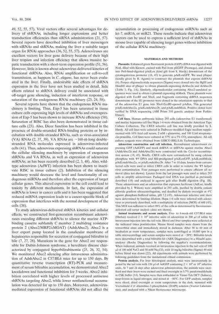
FIG. 3. AdshAbcc2 administration in mice affects Abcc2 functionality. (A) Dose required to infect more than 95% of hepatocytes. Mice weremock infected or infected with 2 � 109 infective units of AdGFP/mouse, and liver samples were processed at 7 or 24 dpi. Histological analysis ofliver samples stained with DAPI (blue) was visualized in a fluorescence microscope. GFP expression is shown in green. (B, C) The antisense strandof Abcc2 siRNAs is detected in AdshAbcc2-infected animals. Mice were mock infected or infected with AdLuc or AdshMock as controls orinfected with AdshAbcc2a or AdshAbcc2b. At 7 days posttreatment, serum and liver samples were analyzed. Buffer only (oligo) or total liver RNAwas analyzed by primer extension with oligonucleotide asAbcc2a (B) or by Northern blot assay with oligonucleotide asAbcc2b (C). The positionof the antisense strand of Abcc2a siRNA or Abcc2b siRNA (asAbcc2 siRNA) is indicated to the left. The position of the 20-bp marker is indicatedto the right. (D) Abcc2 mRNA levels are decreased in AdshAbcc2-infected animals. Total RNA described in panel B was used to quantify Abcc2and actin mRNAs by LightCycler RT-PCR. Results were plotted as indicated in Fig. 2C. (E) AdshAbcc2 administration to mice causeshyperbilirubinemia. Mice described in panel B were injected with synthetic bilirubin, and 40 min later, serum samples were collected to quantifybilirubin levels. Error bars indicate standard deviations obtained with five to seven animals.
12240 NARVAIZA ET AL. J. VIROL.
on August 28, 2012 by U
niversidad de Navarra
http://jvi.asm.org/
Dow
nloaded from

(Table 1) incubated with liver RNA from AdshAbcc2a-in-fected animals (Fig. 3B, lanes 4 and 5). A similar band wasobserved when RNA from AdshAbcc2b-infected animals washybridized with oligonucleotide asAbcc2b (Table 1; Fig. 3C,lanes 3 and 4). These bands were specific since they were notdetected in mock-infected animals or in animals infected withthe other adenoviruses tested (Fig. 3B, lanes 2 and 3 and lanes6 to 9, and C, lanes 1 and 2). The presence of the antisensestrand of Abcc2 siRNAs correlated with a decrease in Abcc2mRNA accumulation. Quantification of Abcc2 mRNA levelsby quantitative RT-PCR from liver RNA samples showed thatAbcc2 mRNA amounts were 5% and 12% of the levels of thecontrol groups in AdshAbcc2a- and AdshAbcc2b-receivingmice, respectively (Fig. 3D). This decrease was specific, assimilar levels of the nontargeted actin mRNA were found in allcases (Fig. 3D).
Abcc2 transports serum bilirubin to bile in hepatocytes. Al-terations in the Abcc2 gene are the cause of Dubin-Johnsonsyndrome, which is characterized by hyperbilirubinemia andjaundice (7, 25, 26, 32). It could then be expected that Abcc2mRNA silencing would cause a decrease in Abcc2 expression,reduced transport of bilirubin to bile, and hyperbilirubinemia.To assess whether AdshAbcc2a and AdshAbcc2b lead toAbcc2 functional inhibition in vivo, bilirubin was measured inserum samples collected from three to five mice per group in-fected as described above. We observed a significant increase inserum bilirubin in mice receiving AdshAbcc2a or AdshAbcc2b(0.90 0.66 mg/dl) compared to control animals (0.11 0.05
mg/dl). Serum bilirubin forms as a by-product of erythrocyteturnover, which, under normal conditions, does not saturatethe machinery that transports bilirubin to bile. To study theeffect of AdshAbcc2 viruses with saturating bilirubin, five toseven mice per group infected as described above received 10mg/kg of synthetic bilirubin intravenously and blood was col-lected 40 min later (22). After this time, synthetic bilirubin wascompletely cleared in control mice, while mice injected withAdshAbcc2a or AdshAbcc2b showed marked hyperbiliru-binemia (Fig. 3E).
AdshAbcc2 liver transduction impairs bilirubin clearance forup to 3 weeks. Once we detected Abcc2 functional inhibition andbilirubin clearance impairment 7 days after AdshAbcc2 injection,we analyzed in vivo Abcc2 silencing kinetics following AdshAbcc2administration. Five to seven C57/BL6 mice per group were mockinjected or injected with 2 � 109 infective units of AdshAbcc2a orwith AdshMock or AdLuc as negative controls. Liver and serumsamples were collected on different days postinfection. Serumbilirubin was quantified 40 min after synthetic bilirubin injectionon days �1, 3, 5, 9, 19, 24, and 150 post virus administration. Wefound normal levels of serum bilirubin in all animals at �1 and 3dpi (Fig. 4A and data not shown). This was expected since ex-pression from adenovirus is observed from 48 h posttransduction(56) and some time should be required from Abcc2 mRNA deg-radation to functional impairment of Abcc2 protein. Remarkably,we detected high bilirubin values from day 5 until day 19 postin-jection of AdshAbcc2a (Fig. 4A). In these animals, the increase inbilirubin was maximal at 9 dpi and was no longer significant at day
FIG. 3—Continued.
VOL. 80, 2006 IN VIVO EFFECT OF ADENOVIRUS-DELIVERED shRNAs 12241
on August 28, 2012 by U
niversidad de Navarra
http://jvi.asm.org/
Dow
nloaded from

24 after AdshAbcc2a administration (Fig. 4A). In control animalsor in all animals analyzed at 150 dpi, serum bilirubin showedvalues similar to baseline levels (Fig. 4A and data not shown).
RNA was isolated from liver samples taken from the animalsdescribed above at 7, 12, 24, and 150 dpi. Liver RNA was also
isolated at 7 dpi from three mice transduced with AdshAbcc2b.Abcc2 mRNA levels quantified by real-time RT-PCR showeda significant reduction in mice infected with AdshAbcc2b orwith AdshAbcc2a for 7 or 12 days (Fig. 4B). This result is inagreement with the functional inhibition of Abcc2 shown
FIG. 4. AdshAbcc2 liver transduction affects Abcc2 functionalityfor up to 3 weeks. (A) AdshAbcc2-infected animals show hyperbiliru-binemia for 19 days. Mice were mock injected or injected with 2 � 109
infective units of AdshMock or AdLuc as negative controls or withAdshAbcc2a. Bilirubin was quantified in serum samples collected ondays 3, 5, 9, 15, 19, and 24 post virus administration and 40 min aftersynthetic bilirubin injection. Error bars indicate standard deviations.Asterisks highlight significant differences. (B) Abcc2 mRNA is de-creased in AdshAbcc2-treated animals for up to 12 days. Liver RNAwas isolated from animals treated as described in panel A, at 7, 12,24, and 150 dpi. Liver RNA was also isolated from animals trans-duced with AdshAbcc2b for 7 days. Liver RNA was used to quantifyAbcc2 mRNA by LightCycler RT-PCR. Results are plotted as in-dicated in Fig. 2C. (C) Antisense siRNAs against Abcc2 mRNAwere detected at 150 dpi with AdshAbcc2a. Liver RNA described inpanel B or buffer (oligo) was analyzed by primer extension witholigonucleotide Abcc2aAS (top). Some of the samples were alsoanalyzed by Northern blot assay hybridized to oligonucleotideasAbcc2a (bottom). The positions of the oligonucleotide and theantisense Abcc2a siRNA (asAbcc2a siRNA) are indicated to theleft. The position of the 20-bp marker is indicated to the right.
12242 NARVAIZA ET AL. J. VIROL.
on August 28, 2012 by U
niversidad de Navarra
http://jvi.asm.org/
Dow
nloaded from

above. Control animals and all animals analyzed after 24 or 150dpi showed normal levels of Abcc2 mRNA (Fig. 4B and datanot shown). Also, the inhibition was specific for Abcc2 mRNA,as normal levels of actin mRNA were found in all cases.
The accumulation of small RNAs against Abcc2 mRNA wasanalyzed in the liver RNA samples described above by North-ern blot assay and primer extension with oligonucleotideasAbcc2a. Both techniques detected high levels of antisenseAbcc2a siRNAs in mice transduced with AdshAbcc2a at 7 and12 dpi (Fig. 4C, lanes 1 to 5). Thus, the decrease in Abcc2mRNA and the impairment of bilirubin clearance correlatedwith a strong accumulation of siRNAs against Abcc2 mRNA.Small RNAs were not detected with oligonucleotide asAbcc2ain control samples (Fig. 4C, lanes 10 to 14). Interestingly, wewere able to detect a weak signal corresponding to antisenseAbcc2a siRNA at day 24, and even day 150, postinfection (Fig.4C, lanes 6 to 9). However, these levels of small RNA were notsufficient to silence Abcc2 mRNA since bilirubin clearance andAbcc2 mRNA values were similar to those of controls (Fig. 4Aand B).
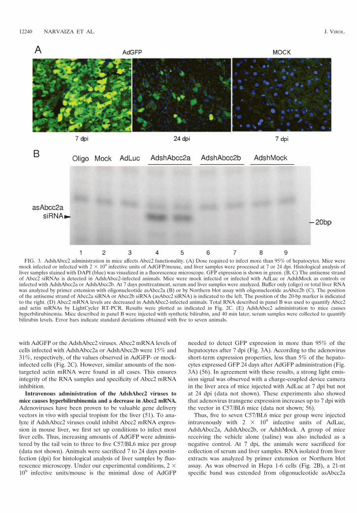
Expression of functional shRNAs does not affect processingor accumulation of endogenous liver miRNAs. BecauseshRNAs and miRNAs share the same processing and nuclearexport machinery (30, 31), we explored whether functionalexpression of shRNAs in the liver could modify endogenousmiRNA processing or accumulation. Thus, miRNAs werequantified in liver RNA samples isolated from mice mocktransduced or transduced with 2 � 109 infective units of AdLucor AdshAbcc2a at 7, 12, 24, or 150 dpi. These RNAs werenormalized to U6 snRNA, and accumulation of several endog-enous miRNAs was analyzed. Out of seven cellular miRNAschosen, only let-7, miR16, miR21, and liver-specific miR122were expressed to detectable amounts in mouse liver cells (33).Accumulation of these miRNAs was analyzed by primer ex-tension and Northern blot assay with specific oligonucleotides(Fig. 5A; Table 1; data not shown). Quantification of the re-sults obtained with primer extensions and Northern blot assaysindicated variability in the amount of endogenous miRNAs indifferent animals. However, statistical analysis revealed non-significant differences between liver extracts from AdLuc- ormock-treated mice and AdshAbcc2a-injected mice (data notshown). Also, there were no differences among controls andmice treated with AdshAbcc2b and analyzed at 7, 12, or 24days posttreatment (data not shown). The Invader techniqueto quantify endogenous miRNAs was also used to reduce themouse-to-mouse variability observed by Northern blot assay orprimer extension (1). When quantification of let-7 or miR21was carried out with Invader assays, similar results were ob-tained but with a lower variability. No differences were ob-served between controls or AdshAbcc2a-infected animals an-alyzed at 7, 12, 24, or 150 days posttreatment (Fig. 5B and C).
Competition of functional shRNAs for the silencing process-ing machinery could still induce the accumulation of miRNAprecursors (pre-miRNAs). Thus, a blockade of Exp-5 or Dicershould alter the localization and accumulation of pre-miRNAs.Therefore, accumulation of pre-miR16 and pre-miR21 wasanalyzed by primer extension (Fig. 5D) and Northern blotassay (data not shown) with specific antisense oligonucleotides(Table 1). Again, quantification of pre-miR16 or pre-miR21did not reveal significant differences between liver extracts
from control mice or AdshAbcc2a-transduced mice analyzed at7, 12, 24, or 150 days posttreatment (data not shown).
DISCUSSION
One of the major obstacles to using siRNAs in vivo is theirefficient delivery into the target tissue or organ. The experienceaccumulated in recent years in the gene therapy field has led tothe optimization of several viruses as gene delivery vectors.Thus, viral vectors expressing shRNAs have proven to be valu-able vehicles to achieve RNAi (21, 49, 55, 57). However, de-tailed studies analyzing the kinetics of inhibition or the sideeffects related to functional shRNA expression in vivo have notbeen carried out. To address these questions, we have chosenfirst-generation adenoviruses that silence the hepatocellularAbcc2 transporter in mouse liver. With these vectors, we wereable to induce Abcc2 silencing in tissue cultured liver cells (Fig.2C) and in murine liver (Fig. 3D and 4B), whereas otherapproaches such as hydrodynamic administration of plasmidcoding for shAbcc2 had failed (data not shown).
AdshAbcc2 caused Abcc2 silencing from day 5 up to day 19after vector administration (Fig. 4A). As Abcc2 transportsbilirubin into bile, silencing of Abcc2 was detected by hyper-bilirubinemia (Fig. 3D and 4A). Hyperbilirubinemia is alsodetected in patients with Dubin-Johnson syndrome, inwhom Abcc2 function is impaired (25, 26, 32, 54). Thus, ourAdshAbcc2 adenoviruses could be administered in mice togenerate transient knockdowns that could serve as mousemodels for Dubin-Johnson syndrome and for the study of thepathophysiological consequences derived from Abcc2 dysfunc-tion. Moreover, Abcc2, among other ATP-binding cassettetransporters, has important clinical implications since it is re-sponsible for excretion of drugs and xenotoxins and for resis-tance to multiple chemotherapeutic agents (20). Thus, oursystem could enable a better definition of the implication ofthis transporter in specific metabolic functions and may serveto enhance the sensitivity of liver cancer cells to chemothera-peutic agents.
Impairment of Abcc2 function correlates with a decrease inliver Abcc2 mRNA as detected by RT-PCR (Fig. 3D and 4B).Abcc2 mRNA levels decreased at days 7 and 12 after adeno-virus injection but were back to normal at day 24, when theincrease in blood bilirubin was not significant (Fig. 4A and B).This kinetics of inhibition follows a classical short-term expres-sion pattern of first-generation adenoviral vectors (Fig. 3A).Silencing is mediated by the antisense strand of processedshRNAs. These antisense small RNA mediators were detectedby Northern blot assay or primer extension up to 150 days afterAdshAbcc2 administration in mice (Fig. 4C). This suggests theexistence of an siRNA reservoir and/or the persistence of fewcopies of adenovirus genomes in liver cells. We favor the latterhypothesis since we were able to detect viral genomes by quan-titative PCR in DNA extracts from murine livers 150 days afterinjection with AdshAbcc2a (data not shown). However, thelow levels of antisense Abcc2 siRNA detected 24 or 150 dayspost AdshAbcc2 administration were not sufficient to silenceAbcc2 (Fig. 4). Only the levels detected at days 7 and 12 wereefficient inhibitors of Abcc2 (Fig. 4). These results indicate thatthere is a threshold amount of siRNAs required for efficientsilencing. Also, these functional levels need to be expressed in
VOL. 80, 2006 IN VIVO EFFECT OF ADENOVIRUS-DELIVERED shRNAs 12243
on August 28, 2012 by U
niversidad de Navarra
http://jvi.asm.org/
Dow
nloaded from
FIG. 5. Expression of functional shRNAs does not affect processing or accumulation of endogenous liver miRNAs. (A) Accumulation ofmiRNAs is not altered in AdshAbcc2-infected cells as detected by primer extension. Buffer (oligo) or liver RNA described in Fig. 5 was analyzedby primer extension with oligonucleotides that hybridize to let-7, miR16, miR21, or U6 snRNA (as a loading control). (B, C) let-7 (B) and miR21(C) accumulation is not altered in AdshAbcc2-infected cells as detected by Invader assay. RNAs described in panel A were analyzed by Invaderassay and plotted as relative units. Error bars indicate standard deviations obtained with five to seven animals. (D) Abnormal accumulation ofprecursor miRNAs is not detected in AdshAbcc2-infected cells. Buffer (oligo) or liver RNA, as described in panel A, was analyzed by primerextension with oligonucleotides that hybridize to pre-miR16, pre-miR21, or U6 snRNA (as a loading control).
12244
on August 28, 2012 by U
niversidad de Navarra
http://jvi.asm.org/
Dow
nloaded from

the majority of cells. Thus, efficient silencing was not detectedwhen we used fewer than 2 � 109 infective units of AdshAbcc2/mouse (data not shown). These data also show that mouse livercells do not support efficient amplification or spread of siRNAsignals, as has been shown in worms or plants (53).
Application of RNAi to gene therapy approaches has first toface the challenge of avoiding side effects. RNAi-induced tox-icity has been associated with three major causes: off-targetinhibition, activation of the interferon response, and saturationof the cellular silencing machinery. Several authors have ques-tioned the specificity of RNAi, as expression of some nontargetgenes may be decreased (24). We have not conducted detailedexperiments to rule out off-target inhibition in our system.However, the expression of specific cellular genes such asthat for murine actin was not altered by expression of Abcc2shRNAs (Fig. 2C and 3D; data not shown). Also, all micesurvived throughout the study and showed normal behaviorwithout apparent loss of weight or other signs of illness. Liverfunctionality was not affected, as ascites, hepatomegaly, hepa-tocyte necrosis, or liver fibrosis was not apparent after autop-sies were carried out. In addition, bilirubin levels were notaltered in mice treated with adenovirus expressing siRNAsother than the specific inhibitors of Abcc2 (Fig. 3E and 4A).Finally, some sequences of siRNAs and shRNAs or highdoses of these small RNAs can induce the interferon re-sponse (24, 44). However, we could not detect induction ofthe interferon pathway by quantitative RT-PCR of 2�5�oli-goadenylate synthetase mRNA with RNA isolated fromHepa 1-6 cells or murine livers infected with AdshAbcc2(data not shown).
Numerous authors have pointed out the need for more in-formation about shRNA toxicity by saturation of the cellularsilencing machinery (17, 29, 58). Their worries are based on thesaturability of silencing factors such as Exp-5, Dicer, and RISC(23, 39, 58). Processing and function of shRNAs require thecellular silencing machinery. Thus, overexpression of shRNAscan saturate Exp-5 in tissue culture cells and in vivo (15, 58).During the process of writing this report, a study was publishedshowing that high levels of AAV-delivered shRNAs induceliver injury in mice (15). The authors showed that overex-pressed shRNAs saturate Exp-5, leading to a decrease in theaccumulation and functionality of liver miRNAs. We hypoth-esized that a similar result could be obtained with adenovirus-delivered shRNAs. Besides, adenovirus infection could affectthe silencing machinery by additional mechanisms. On the onehand, first-generation adenoviruses express VA RNAs thatcould saturate Exp-5 (5, 16, 50; data not shown). On the otherhand, VA RNAs can inhibit Dicer function and can be pro-cessed to functional viral miRNAs that could help in saturationof the silencing machinery (2, 3, 39, 48). Finally, other adeno-virus miRNAs could be expressed from other sequences in theadenovirus genome (2, 3, 39, 48).
To address the saturation of the silencing machinery, weanalyzed the processing and accumulation of cellular miRNAsin murine livers infected with first-generation adenovirus vec-tors that express shRNAs. The cellular miRNAs chosen werelet-7, miR16, miR21, and miR122, as they are well expressed inmouse liver. These miRNAs and their pre-miRNAs were an-alyzed by Northern blot assay, primer extension, and Invader(Fig. 5 and data not shown). Even when adenovirus-mediated
shRNA delivery had inhibited the expression of the target genefor 1 week (from day 5 to day 12, when miRNAs were ana-lyzed), no differences were observed in the accumulation oflet-7, miR16, and miR21 miRNAs or pre-miRNAs. Also, wedid not observe alterations in the expression of miRNA-regu-lated genes such as that for Ras, Notch, or RelA (data notshown). These experiments were performed under conditionsin which more than 95% of the cells were expressing adenovi-rus-delivered shRNAs. This result indicates that efficient si-lencing in vivo can be achieved without altering miRNA bio-genesis or accumulation or the functionality of the RNAimachinery.
A similar result was described by Grimm et al. when theyused minimal doses of AAV expressing shRNAs (AAV/shRNA) or, in most cases, when they delivered higher doses ofthese vectors but the stem length of the shRNAs was 19 nt.These conditions are identical to our experimental settings.However, Grimm et al. also reported that doses of AAV/shRNA fivefold higher than the dose required for completeliver transduction (1012 particles of AAV/shRNA) led to tox-icity in 73% of the animals because of oversaturation of thecellular miRNA pathway. It would be of interest to analyze ifhigher levels of adenovirus expressing shRNAs can also inducetoxicity in our system. To address this question, we have triedto produce high titers of AdshAbcc2a and -b. However, all ofour attempts ended in production of adenovirus enriched ininfective particles that had lost expression of Abcc2 shRNA,probably because these sequences have been lost by recombi-nation (data not shown). Also, in our experiments, doses ofAdGFP or AdWT higher than 4 � 109 infective units permouse led to toxicity that was unrelated to inhibition of thesilencing pathway (data not shown). Therefore, we evaluatedthe effect of high levels of adenovirus expressing shRNAs in293 cells, which allow replication of first-generation adenovirusvectors. In uninfected cells, the shRNA-mediated inhibition ofa luciferase reporter was fivefold whereas the inhibitiondropped to two- to threefold in cells infected for 3 days withAdWT, AdGFP, or AdshAbcc2a (data not shown). Similarresults have been obtained by others with AdWT (2, 39). Theseresults indicate that adenovirus-infected cells may have im-paired shRNA processing or function. However, in the samecells we did not detect an alteration of the processing or func-tion of endogenous miRNAs (data not shown). We believe thatlonger times postinfection may be required to affect thesemiRNAs. In fact, a decrease in mouse liver miRNAs has onlybeen detected after 2 weeks of infection with AAV overex-pressing shRNAs (15).
In summary, we show that adenoviral vectors are suitable fordelivery of shRNAs and allow the silencing of a liver trans-porter for up to 3 weeks. The silencing of the Abcc2 RNAcorrelated with inhibition of the bilirubin export function andwith the presence of high levels of shRNAs expressed from theadenoviral vectors. Moreover, we showed the existence of athreshold in the levels of processed antisense shRNA requiredfor efficient target silencing. Importantly, the data presentedin this work indicate that adenovirus expressing functionalshRNAs may efficiently inhibit target genes without alterationor saturation of the miRNA silencing machinery.
VOL. 80, 2006 IN VIVO EFFECT OF ADENOVIRUS-DELIVERED shRNAs 12245
on August 28, 2012 by U
niversidad de Navarra
http://jvi.asm.org/
Dow
nloaded from

ACKNOWLEDGMENTS
This work was supported by CICYT (SAF2003-01804), FIS (01/1310and 01/0843), Instituto Carlos III (C03/02 and PI051098), and theEducation and Health Department of the Navarra Governmentand through the agreement between FIMA and the UTE projectCIMA. O.A. is a holder of an FPI fellowship from the Spanish Ministryof Education. I.N. and M.V. are recipients of an FIMA postdoctoralfellowship.
We are grateful to S. Hardy for the adenovirus Cre-lox System, R.Agami for pSuper, and J. Wilson for AdLuc. We thank M. Zaratieguifor helpful scientific discussions and help with statistical analysis, E.Casales for technical assistance, L. Martınez and A. Vales for bilirubinquantifications, P. Alzuguren for serum sample collection support, andP. Garces for tissue sample processing. We are also grateful to C.Smerdou, R. Hernandez-Alcoceba, M. D. Weitzman, J. Suh, and P.Gastaminza for critical readings of the manuscript.
REFERENCES
1. Allawi, H. T., J. E. Dahlberg, S. Olson, E. Lund, M. Olson, W. P. Ma, T.Takova, B. P. Neri, and V. I. Lyamichev. 2004. Quantitation of microRNAsusing a modified Invader assay. RNA 10:1153–1161.
2. Andersson, M. G., P. C. Haasnoot, N. Xu, S. Berenjian, B. Berkhout, and G.Akusjarvi. 2005. Suppression of RNA interference by adenovirus virus-as-sociated RNA. J. Virol. 79:9556–9565.
3. Aparicio, O., N. Razquin, M. Zaratiegui, I. Narvaiza, and P. Fortes. 2006.Adenovirus virus-associated RNA is processed to functional interferingRNAs involved in virus production. J. Virol. 80:1376–1384.
4. Bartel, D. P. 2004. MicroRNAs: genomics, biogenesis, mechanism, and func-tion. Cell 116:281–297.
5. Bohnsack, M. T., K. Czaplinski, and D. Gorlich. 2004. Exportin 5 is aRanGTP-dependent dsRNA-binding protein that mediates nuclear export ofpre-miRNAs. RNA 10:185–191.
6. Brummelkamp, T. R., R. Bernards, and R. Agami. 2002. A system for stableexpression of short interfering RNAs in mammalian cells. Science 296:550–553.
7. Buchler, M., J. Konig, M. Brom, J. Kartenbeck, H. Spring, T. Horie, and D.Keppler. 1996. cDNA cloning of the hepatocyte canalicular isoform of themultidrug resistance protein, cMrp, reveals a novel conjugate export pumpdeficient in hyperbilirubinemic mutant rats. J. Biol. Chem. 271:15091–15098.
8. Caldas, C., and J. D. Brenton. 2005. Sizing up miRNAs as cancer genes. Nat.Med. 11:712–714.
9. Chomczynski, P., and N. Sacchi. 1987. Single-step method of RNA isolationby acid guanidinium thiocyanate-phenol-chloroform extraction. Anal. Bio-chem. 162:156–159.
10. Dykxhoorn, D. M., C. D. Novina, and P. A. Sharp. 2003. Killing the mes-senger: short RNAs that silence gene expression. Nat. Rev. Mol. Cell Biol.4:457–467.
11. Elbashir, S. M., J. Harborth, W. Lendeckel, A. Yalcin, K. Weber, and T.Tuschl. 2001. Duplexes of 21-nucleotide RNAs mediate RNA interference incultured mammalian cells. Nature 411:494–498.
12. Fortes, P., D. Bilbao-Cortes, M. Fornerod, G. Rigaut, W. Raymond, B.Seraphin, and I. W. Mattaj. 1999. Luc7p, a novel yeast U1 snRNP proteinwith a role in 5� splice site recognition. Genes Dev. 13:2425–2438.
13. Fortes, P., Y. Cuevas, F. Guan, P. Liu, S. Pentlicky, S. P. Jung, M. L.Martinez-Chantar, J. Prieto, D. Rowe, and S. I. Gunderson. 2003. Inhibitingexpression of specific genes in mammalian cells with 5� end-mutated U1small nuclear RNAs targeted to terminal exons of pre-mRNA. Proc. Natl.Acad. Sci. USA 100:8264–8269.
14. Fortes, P., A. I. Lamond, and J. Ortin. 1995. Influenza virus NS1 proteinalters the subnuclear localization of cellular splicing components. J. Gen.Virol. 76(Pt. 4):1001–1007.
15. Grimm, D., K. L. Streetz, C. L. Jopling, T. A. Storm, K. Pandey, C. R. Davis,P. Marion, F. Salazar, and M. A. Kay. 2006. Fatality in mice due to over-saturation of cellular microRNA/short hairpin RNA pathways. Nature 441:537–541.
16. Gwizdek, C., B. Ossareh-Nazari, A. M. Brownawell, A. Doglio, E. Bertrand,I. G. Macara, and C. Dargemont. 2003. Exportin-5 mediates nuclear exportof minihelix-containing RNAs. J. Biol. Chem. 278:5505–5508.
17. Hannon, G. J., and J. J. Rossi. 2004. Unlocking the potential of the humangenome with RNA interference. Nature 431:371–378.
18. Hardy, S., M. Kitamura, T. Harris-Stansil, Y. Dai, and M. L. Phipps. 1997.Construction of adenovirus vectors through Cre-lox recombination. J. Virol.71:1842–1849.
19. He, L., J. M. Thomson, M. T. Hemann, E. Hernando-Monge, D. Mu, S.Goodson, S. Powers, C. Cordon-Cardo, S. W. Lowe, G. J. Hannon, and S. M.Hammond. 2005. A microRNA polycistron as a potential human oncogene.Nature 435:828–833.
20. Hoffmann, U., and H. K. Kroemer. 2004. The ABC transporters MDR1 and
MRP2: multiple functions in disposition of xenobiotics and drug resistance.Drug Metab. Rev. 36:669–701.
21. Hommel, J. D., R. M. Sears, D. Georgescu, D. L. Simmons, and R. J.DiLeone. 2003. Local gene knockdown in the brain using viral-mediatedRNA interference. Nat. Med. 9:1539–1544.
22. Huang, W., J. Zhang, S. S. Chua, M. Qatanani, Y. Han, R. Granata, andD. D. Moore. 2003. Induction of bilirubin clearance by the constitutiveandrostane receptor (CAR). Proc. Natl. Acad. Sci. USA 100:4156–4161.
23. Hutvagner, G., M. J. Simard, C. C. Mello, and P. D. Zamore. 2004. Se-quence-specific inhibition of small RNA function. PLoS. Biol. 2:E98.
24. Jackson, A. L., and P. S. Linsley. 2004. Noise amidst the silence: off-targeteffects of siRNAs? Trends Genet. 20:521–524.
25. Kartenbeck, J., U. Leuschner, R. Mayer, and D. Keppler. 1996. Absence ofthe canalicular isoform of the MRP gene-encoded conjugate export pumpfrom the hepatocytes in Dubin-Johnson syndrome. Hepatology 23:1061–1066.
26. Keitel, V., J. Kartenbeck, A. T. Nies, H. Spring, M. Brom, and D. Keppler.2000. Impaired protein maturation of the conjugate export pump multidrugresistance protein 2 as a consequence of a deletion mutation in Dubin-Johnson syndrome. Hepatology 32:1317–1328.
27. Keppler, D., and J. Konig. 1997. Hepatic canalicular membrane 5: expressionand localization of the conjugate export pump encoded by the MRP2(cMRP/cMOAT) gene in liver. FASEB J. 11:509–516.
28. Keppler, D., J. Konig, and M. Buchler. 1997. The canalicular multidrugresistance protein, cMRP/MRP2, a novel conjugate export pump expressedin the apical membrane of hepatocytes. Adv. Enzyme Regul. 37:321–333.
29. Kim, V. N. 2005. MicroRNA biogenesis: coordinated cropping and dicing.Nat. Rev. Mol. Cell Biol. 6:376–385.
30. Kim, V. N. 2004. MicroRNA precursors in motion: exportin-5 mediates theirnuclear export. Trends Cell Biol. 14:156–159.
31. Kim, V. N. 2005. Small RNAs: classification, biogenesis, and function. Mol.Cells 19:1–15.
32. Konig, J., A. T. Nies, Y. Cui, I. Leier, and D. Keppler. 1999. Conjugate exportpumps of the multidrug resistance protein (MRP) family: localization, sub-strate specificity, and MRP2-mediated drug resistance. Biochim. Biophys.Acta 1461:377–394.
33. Lagos-Quintana, M., R. Rauhut, W. Lendeckel, and T. Tuschl. 2001. Iden-tification of novel genes coding for small expressed RNAs. Science 294:853–858.
34. Lee, Y., C. Ahn, J. Han, H. Choi, J. Kim, J. Yim, J. Lee, P. Provost, O.Radmark, S. Kim, and V. N. Kim. 2003. The nuclear RNase III Droshainitiates microRNA processing. Nature 425:415–419.
35. Lewis, B. P., C. B. Burge, and D. P. Bartel. 2005. Conserved seed pairing,often flanked by adenosines, indicates that thousands of human genes aremicroRNA targets. Cell 120:15–20.
36. Lewis, D. L., J. E. Hagstrom, A. G. Loomis, J. A. Wolff, and H. Herweijer.2002. Efficient delivery of siRNA for inhibition of gene expression in post-natal mice. Nat. Genet. 32:107–108.
37. Li, W. X., H. Li, R. Lu, F. Li, M. Dus, P. Atkinson, E. W. Brydon, K. L.Johnson, A. Garcia-Sastre, L. A. Ball, P. Palese, and S. W. Ding. 2004.Interferon antagonist proteins of influenza and vaccinia viruses are suppres-sors of RNA silencing. Proc. Natl. Acad. Sci. USA 101:1350–1355.
38. Lu, J., G. Getz, E. A. Miska, E. Alvarez-Saavedra, J. Lamb, D. Peck, A.Sweet-Cordero, B. L. Ebert, R. H. Mak, A. A. Ferrando, J. R. Downing, T.Jacks, H. R. Horvitz, and T. R. Golub. 2005. MicroRNA expression profilesclassify human cancers. Nature 435:834–838.
39. Lu, S., and B. R. Cullen. 2004. Adenovirus VA1 noncoding RNA can inhibitsmall interfering RNA and MicroRNA biogenesis. J. Virol. 78:12868–12876.
40. Lund, E., S. Guttinger, A. Calado, J. E. Dahlberg, and U. Kutay. 2004.Nuclear export of microRNA precursors. Science 303:95–98.
41. Ma, Y., and M. B. Mathews. 1996. Structure, function, and evolution ofadenovirus-associated RNA: a phylogenetic approach. J. Virol. 70:5083–5099.
42. McCaffrey, A. P., L. Meuse, T. T. Pham, D. S. Conklin, G. J. Hannon, andM. A. Kay. 2002. RNA interference in adult mice. Nature 418:38–39.
43. Meltzer, P. S. 2005. Cancer genomics: small RNAs with big impacts. Nature435:745–746.
44. Moss, E. G., and J. M. Taylor. 2003. Small-interfering RNAs in the radar ofthe interferon system. Nat. Cell Biol. 5:771–772.
45. Narvaiza, I., G. Mazzolini, M. Barajas, M. Duarte, M. Zaratiegui, C. Qian,I. Melero, and J. Prieto. 2000. Intratumoral coinjection of two adenoviruses,one encoding the chemokine IFN--inducible protein-10 and another en-coding IL-12, results in marked antitumoral synergy. J. Immunol. 164:3112–3122.
46. O’Donnell, K. A., E. A. Wentzel, K. I. Zeller, C. V. Dang, and J. T. Mendell.2005. c-Myc-regulated microRNAs modulate E2F1 expression. Nature 435:839–843.
47. Pasquinelli, A. E., S. Hunter, and J. Bracht. 2005. MicroRNAs: a developingstory. Curr. Opin. Genet. Dev. 15:200–205.
48. Pfeffer, S., A. Sewer, M. Lagos-Quintana, R. Sheridan, C. Sander, F. A.Grasser, L. F. van Dyk, C. K. Ho, S. Shuman, M. Chien, J. J. Russo, J. Ju,G. Randall, B. D. Lindenbach, C. M. Rice, V. Simon, D. D. Ho, M. Zavolan,
12246 NARVAIZA ET AL. J. VIROL.
on August 28, 2012 by U
niversidad de Navarra
http://jvi.asm.org/
Dow
nloaded from

and T. Tuschl. 2005. Identification of microRNAs of the herpesvirus family.Nat. Methods 2:269–276.
49. Rubinson, D. A., C. P. Dillon, A. V. Kwiatkowski, C. Sievers, L. Yang, J.Kopinja, D. L. Rooney, M. M. Ihrig, M. T. McManus, F. B. Gertler, M. L.Scott, and L. Van Parijs. 2003. A lentivirus-based system to functionallysilence genes in primary mammalian cells, stem cells and transgenic mice byRNA interference. Nat. Genet. 33:401–406.
50. Sano, M., Y. Kato, and K. Taira. 2006. Sequence-specific interference bysmall RNAs derived from adenovirus VAI RNA. FEBS Lett. 580:1553–1564.
51. Smith, T., N. Idamakanti, H. Kylefjord, M. Rollence, L. King, M. Kaloss, M.Kaleko, and S. C. Stevenson. 2002. In vivo hepatic adenoviral gene deliveryoccurs independently of the coxsackievirus-adenovirus receptor. Mol. Ther.5:770–779.
52. Soutschek, J., A. Akinc, B. Bramlage, K. Charisse, R. Constien, M. Donoghue,S. Elbashir, A. Geick, P. Hadwiger, J. Harborth, M. John, V. Kesavan, G.Lavine, R. K. Pandey, T. Racie, K. G. Rajeev, I. Rohl, I. Toudjarska, G.Wang, S. Wuschko, D. Bumcrot, V. Koteliansky, S. Limmer, M. Manoharan,and H. P. Vornlocher. 2004. Therapeutic silencing of an endogenous gene bysystemic administration of modified siRNAs. Nature 432:173–178.
53. Tomari, Y., and P. D. Zamore. 2005. Perspective: machines for RNAi. GenesDev. 19:517–529.
54. Tsujii, H., J. Konig, D. Rost, B. Stockel, U. Leuschner, and D. Keppler. 1999.Exon-intron organization of the human multidrug-resistance protein 2(MRP2) gene mutated in Dubin-Johnson syndrome. Gastroenterology 117:653–660.
55. Uprichard, S. L., B. Boyd, A. Althage, and F. V. Chisari. 2005. Clearance ofhepatitis B virus from the liver of transgenic mice by short hairpin RNAs.Proc. Natl. Acad. Sci. USA 102:773–778.
56. Wolff, G., S. Worgall, N. van Rooijen, W. R. Song, B. G. Harvey, and R. G.Crystal. 1997. Enhancement of in vivo adenovirus-mediated gene transferand expression by prior depletion of tissue macrophages in the target organ.J. Virol. 71:624–629.
57. Xia, H., Q. Mao, H. L. Paulson, and B. L. Davidson. 2002. siRNA-mediatedgene silencing in vitro and in vivo. Nat. Biotechnol. 20:1006–1010.
58. Yi, R., B. P. Doehle, Y. Qin, I. G. Macara, and B. R. Cullen. 2005. Overex-pression of exportin 5 enhances RNA interference mediated by short hairpinRNAs and microRNAs. RNA 11:220–226.
59. Yi, R., Y. Qin, I. G. Macara, and B. R. Cullen. 2003. Exportin-5 mediates thenuclear export of pre-microRNAs and short hairpin RNAs. Genes Dev.17:3011–3016.
60. Zuker, M. 2003. Mfold web server for nucleic acid folding and hybridizationprediction. Nucleic Acids Res. 31:3406–3415.
VOL. 80, 2006 IN VIVO EFFECT OF ADENOVIRUS-DELIVERED shRNAs 12247
on August 28, 2012 by U
niversidad de Navarra
http://jvi.asm.org/
Dow
nloaded from