EfÞcient rejection of scattered light enables deep optical ...
11
Efficient rejection of scattered light enables deep optical sectioning in turbid media with low-numerical-aperture optics in a dual-axis confocal architecture Jonathan T. C. Liu Stanford University Department of Electrical Engineering Ginzton Laboratory and School of Medicine James H. Clark Center for Biomedical Engineering and Science Stanford, California 94305 Michael J. Mandella Stanford University Department of Electrical Engineering Ginzton Laboratory Stanford, California 94305 James M. Crawford University of Florida Department of Pathology, Immunology and Laboratory Medicine Gainesville, Florida 32610 Christopher H. Contag Stanford University School of Medicine James H. Clark Center for Biomedical Engineering and Science Stanford, California 94305 Thomas D. Wang University of Michigan Department of Internal Medicine Ann Arbor, Michigan 48109 Gordon S. Kino Stanford University Department of Electrical Engineering Ginzton Laboratory Stanford, California 94305 Abstract. Miniature endoscopic microscopes, with subcellular imag- ing capabilities, will enable in vivo detection of molecularly-targeted fluorescent probes for early disease detection. To optimize a dual-axis confocal microscope DACM design for this purpose, we use a table- top instrument to determine the ability of this technology to perform optical sectioning deep within tissue. First, we determine how tissue scattering deteriorates the diffraction-limited transverse and vertical responses in reflectance imaging. Specifically, the vertical response of a DACM to a plane reflector is measured at various depths in a scat- tering phantom and compared with diffraction theory and Monte Carlo scattering simulations. Similarly, transverse line scans across a knife-edge target are performed at various depths in a scattering phan- tom. Second, as a practical demonstration of deep-tissue fluorescence microscopy that corroborates the findings from our scattering experi- ments, 3-D fluorescence images are obtained in thick human gas- trointestinal mucosal specimens. Our results demonstrate efficient re- jection of scattered light in a DACM, which enables deep optical sectioning in tissue with subcellular resolution that can distinguish between normal and premalignant pathologies. © 2008 Society of Photo- Optical Instrumentation Engineers. DOI: 10.1117/1.2939428 Keywords: laser scanning confocal microscopy; fluorescence imaging; Monte Carlo; in vivo imaging; tissue phantom; three-dimensional microscopy; optical sectioning. Paper 07429R received Oct. 22, 2007; revised manuscript received Dec. 9, 2007; accepted for publication Jan. 21, 2008; published online Jun. 11, 2008. 1 Motivation and Background The potential utility of a miniature optical-sectioning micro- scope for biological investigation, preclinical animal studies, and clinical use has been well appreciated and described in the literature. 1–9 Our group is developing a miniature dual- axis confocal microscope DACM that can be deployed through the instrument channel of an endoscope to detect pre- cancerous lesions in the human gastrointestinal tract. In par- ticular, a near-IR fluorescence device is being constructed to enable speckle-free imaging of molecularly targeted reagents deep within the mucosal layer of the esophagus, stomach, and colon. In the development of this instrument, we seek to ob- tain a spatial resolution of a few micrometers in all three directions, and deep tissue penetration, which is desirable for the precise delineation of tumor margins through full- thickness epithelium. In this paper, we address a void in the literature by experimentally characterizing the ability of a DACM to image deeply within tissue. A tabletop DACM is used to investigate the deleterious effects of tissue scattering by imaging reflective targets in a reproducible tissue phantom. 1083-3668/2008/133/034020/11/$25.00 © 2008 SPIE Address all correspondence to Jonathan Liu, Stanford University Ginzton Labs, Box N-123, 450 Via Palou, Stanford, CA 94305; Tel: 650–723–2729; Fax: 650– 725–3890; E-mail: [email protected] Journal of Biomedical Optics 133, 034020 May/June 2008 Journal of Biomedical Optics May/June 2008 Vol. 133 034020-1 Downloaded From: https://www.spiedigitallibrary.org/journals/Journal-of-Biomedical-Optics on 30 Jan 2022 Terms of Use: https://www.spiedigitallibrary.org/terms-of-use
Transcript of EfÞcient rejection of scattered light enables deep optical ...
Efficient rejection of scattered light enables deep opticalsectioning in turbid media with low-numerical-apertureoptics in a dual-axis confocal architecture
Jonathan T. C. LiuStanford UniversityDepartment of Electrical EngineeringGinzton Laboratory
andSchool of MedicineJames H. Clark Center for Biomedical Engineering
and ScienceStanford, California 94305
Michael J. MandellaStanford UniversityDepartment of Electrical EngineeringGinzton LaboratoryStanford, California 94305
James M. CrawfordUniversity of FloridaDepartment of Pathology, Immunology and
Laboratory MedicineGainesville, Florida 32610
Christopher H. ContagStanford UniversitySchool of MedicineJames H. Clark Center for Biomedical Engineering
and ScienceStanford, California 94305
Thomas D. WangUniversity of MichiganDepartment of Internal MedicineAnn Arbor, Michigan 48109
Gordon S. KinoStanford UniversityDepartment of Electrical EngineeringGinzton LaboratoryStanford, California 94305
Abstract. Miniature endoscopic microscopes, with subcellular imag-ing capabilities, will enable in vivo detection of molecularly-targetedfluorescent probes for early disease detection. To optimize a dual-axisconfocal microscope �DACM� design for this purpose, we use a table-top instrument to determine the ability of this technology to performoptical sectioning deep within tissue. First, we determine how tissuescattering deteriorates the diffraction-limited transverse and verticalresponses in reflectance imaging. Specifically, the vertical response ofa DACM to a plane reflector is measured at various depths in a scat-tering phantom and compared with diffraction theory and MonteCarlo scattering simulations. Similarly, transverse line scans across aknife-edge target are performed at various depths in a scattering phan-tom. Second, as a practical demonstration of deep-tissue fluorescencemicroscopy that corroborates the findings from our scattering experi-ments, 3-D fluorescence images are obtained in thick human gas-trointestinal mucosal specimens. Our results demonstrate efficient re-jection of scattered light in a DACM, which enables deep opticalsectioning in tissue with subcellular resolution that can distinguishbetween normal and premalignant pathologies. © 2008 Society of Photo-Optical Instrumentation Engineers. �DOI: 10.1117/1.2939428�
Keywords: laser scanning confocal microscopy; fluorescence imaging; MonteCarlo; in vivo imaging; tissue phantom; three-dimensional microscopy; opticalsectioning.Paper 07429R received Oct. 22, 2007; revised manuscript received Dec. 9, 2007;accepted for publication Jan. 21, 2008; published online Jun. 11, 2008.
1 Motivation and BackgroundThe potential utility of a miniature optical-sectioning micro-scope for biological investigation, preclinical animal studies,and clinical use has been well appreciated and described inthe literature.1–9 Our group is developing a miniature dual-axis confocal microscope �DACM� that can be deployedthrough the instrument channel of an endoscope to detect pre-cancerous lesions in the human gastrointestinal tract. In par-ticular, a near-IR fluorescence device is being constructed to
enable speckle-free imaging of molecularly targeted reagentsdeep within the mucosal layer of the esophagus, stomach, andcolon. In the development of this instrument, we seek to ob-tain a spatial resolution of a few micrometers in all threedirections, and deep tissue penetration, which is desirable forthe precise delineation of tumor margins through full-thickness epithelium. In this paper, we address a void in theliterature by experimentally characterizing the ability of aDACM to image deeply within tissue. A tabletop DACM isused to investigate the deleterious effects of tissue scatteringby imaging reflective targets in a reproducible tissue phantom.
1083-3668/2008/13�3�/034020/11/$25.00 © 2008 SPIE
Address all correspondence to Jonathan Liu, Stanford University Ginzton Labs,Box N-123, 450 Via Palou, Stanford, CA 94305; Tel: 650–723–2729; Fax: 650–725–3890; E-mail: [email protected]
Journal of Biomedical Optics 13�3�, 034020 �May/June 2008�
Journal of Biomedical Optics May/June 2008 � Vol. 13�3�034020-1
Downloaded From: https://www.spiedigitallibrary.org/journals/Journal-of-Biomedical-Optics on 30 Jan 2022Terms of Use: https://www.spiedigitallibrary.org/terms-of-use

We also provide examples of deep 3-D fluorescence imagesections of ex vivo gastrointestinal mucosa.
The inspiration for our DACM design was provided by amethod to achieve improved resolution through off-axis illu-mination and collection of light with high-numerical-aperture�NA� objectives.10–12 Later, low-NA optics were proposed as amethod to achieve a long working distance and a large field ofview.13 More recently, we have shown that a dual-axis archi-tecture may be fiber coupled and combined with postobjectivescanning to provide scalability of the design to millimeterdimensions.9,14 Because the vertical response of the DACM isdetermined by the overlap of the input and output beams, thisconfiguration exhibits an improved axial �vertical� sectioningresponse that is comparable to the transverse response.15 Fur-thermore, in the dual-axis configuration, it is more difficult forlight randomly scattered from the path of the input beam to becollected from the output beam, especially if the NA is small.This was investigated previously through Monte Carlo scat-tering simulations, indicating superior rejection of scatteredlight using the DACM as compared to a conventional single-axis confocal microscope.16
Until now, there have been no quantitative experimentalstudies of the optical-sectioning performance of the dual-axisconfocal architecture in turbid media. While our final goal isto develop a 3-D fluorescence device for speckle-free molecu-larly targeted disease detection, a mirror-reflectance model ischosen as a well-established and repeatable method for quan-tifying the sectioning performance of the dual-axis confocalarchitecture in scattering media. Reflectance and fluorescenceimaging are physically different in a number of ways. How-ever, one can assume that the fundamental spatial-filteringmechanism of the DACM, for rejecting out-of-focus scattered
light, is similar in both cases. For fluorescence microscopy, ananalogous mirror model is difficult to realize in practice, andcomputationally challenging to simulate. Furthermore, suchresults would not enable relevant comparison with the manypublished studies of optical-sectioning technologies that ex-plore reflectance mirror models.17–19
In this paper, we experimentally investigate the ability ofdual-axis confocal microscopy to reject background scatteringby measuring the axial response to a plane mirror embeddedin a scattering phantom. Diffraction theory calculations �noscattering� and Monte Carlo scattering simulations �ignoringdiffraction� are used to validate these experimental results.Transverse line scans of a chrome-on-glass knife-edge in tur-bid media are also presented to study how image contrastdeteriorates due to scattering. Finally, as a practical demon-stration of fluorescence imaging in tissue with a DACM, weprovide 500-�m-deep fluorescence images of thick gas-trointestinal mucosal specimens. The images corroborate theobservations from our reflectance studies in turbid media thatFWHM spatial resolution is preserved deep into tissue andthat image contrast deteriorates quickly at the point wherebackground scattering overwhelms the ballistic signal fromunscattered photons.
The DACM utilizes low-NA �0.21� optics and an inexpen-sive low-power diode laser source for imaging deep withinbiological tissues �Fig. 1�. Near-IR excitation of fluorescenceat 785 nm is used to minimize tissue absorption and scatter-ing for deep penetration, as well as to minimize the autofluo-rescence background. Aberration-free postobjective scanningof the illumination and collection beams is possible due to thelong working distance afforded by the low-NA lenses, as well
x
z
y
Scan mirror
Fused-silicahemisphere(translatingsample holder)
785-nm laser source Long-passfilter
θθ
PMT detectorAmplifier
Frame grabberand monitor
x
z
SMfiber
L1
L2
L4
L3
(a) (b)Fig. 1 DACM design. Two low-NA ��=0.21 radians� focused beams intersect at a 30 deg half-angle � with laser illumination at 785 nm andoff-axis collection of fluorescence. A galvanometric scan mirror provides fast-axis �950-Hz� scanning of the beams in the y direction. A hemi-spherical sample holder is translated with a piezo actuator in the z direction for vertical sectioning at 2 frames/s. Volume imaging is accomplishedby translating the sample holder in the x direction at 1 �m/scan �2 �m/s�. �a� Simplified schematic of the entire optical circuit and �b� 3-D designdrawing of the scan head. The beams, originally oriented horizontally in the x-y plane, are reflected perpendicularly by the scan mirror for imagingin the vertical z direction.
Liu et al.: Efficient rejection of scattered light enables deep optical sectioning…
Journal of Biomedical Optics May/June 2008 � Vol. 13�3�034020-2
Downloaded From: https://www.spiedigitallibrary.org/journals/Journal-of-Biomedical-Optics on 30 Jan 2022Terms of Use: https://www.spiedigitallibrary.org/terms-of-use

as the use of a hemispherical index-matching sample holder�Sec. 2.1�, enabling a large field of view to be scanned. Unlikesingle-axis confocal microscopes, in which the diffraction-limited axial resolution is generally much inferior to trans-verse resolution for a given objective-lens NA, the DACMproduces a diffraction-limited focal volume that is relativelybalanced in all three spatial dimensions.
2 Experimental Methods2.1 Dual-Axis Optical SetupA schematic of the DACM is shown in Fig. 1. Corning HI 780fiber is utilized for single-mode �SM� transmission of laserand fluorescence radiation. The fiber-coupled laser is a SMdiode source from Micro Laser Systems, Inc. �SRT-F785S-36�with a maximum output power of 25 mW. The laser radiationpasses through a fiber-coupled optical isolator from Optics forResearch to suppress back-reflections. The maximum laserpower transmitted through the isolator is 15 mW. For imag-ing, the laser power is reduced to a maximum incident powerof 1 mW on tissue samples.
Two low-NA Gaussian beams are configured to intersect attheir focus with a crossing half-angle � of 30 deg �Fig. 1�.The beams are focused into the sample medium with a half-angle � of 0.21 rad, where the angle � is defined with respectto the lens aperture and in the sample medium. Lenses L1 andL4 have a focal length of 30 mm and a clear aperture of9 mm. Lenses L2 and L3 have a focal length of 20.3 mm anda clear aperture of 8.53 mm. The 1 /e2 intensity NA of ourSM fiber �Corning HI 780� at 785 nm is 0.11 �mode fielddiameter=4.6 �m�.
A solid immersion lens20 consisting of a fused silica �n=1.45� hemisphere is used for index matching of the illumi-nation and collection beams into tissue �n�1.4�. The wavefront curvature of the focused beams is matched to the curvedsurface of the hemisphere as it crosses the air-glass interface,thus minimizing the spherical aberration, coma, and astigma-tism that would otherwise be introduced by a flat optical in-terface at the tissue surface15 �i.e., flat glass window�. Thehemisphere also preserves the focusing half-angle � and theincident angle � of the beams, thereby increasing the effectiveNA in tissue by a factor of n�1.4 �from 0.21 to 0.29�. Theflat surface of the hemisphere acts as a 5-mm-diam circularsample holder that is translated in z for vertical sectioning andtranslated in x for volume scans. An analysis of wave frontaberrations due to dual-axis beam scans in x and y, through asimilar index-matching optic, was performed previously,21 in-dicating a maximum difference in wavefront aberration ofonly 0.30� over a 600-�m horizontal field of view.
For the reflectance experiments in turbid media, the collec-tion fiber is connected directly into a powermeter �EXFO PM-1100�. The filter, photomultiplier tube �PMT�, amplifier, andframe grabber are only used for raster-scanned fluorescenceimaging of tissues.
2.2 Measuring the Axial and Transverse Responsein Turbid Media
2.2.1 Intralipid scattering phantomAmong the most widely used and well-studied tissue phan-toms for optical imaging are lipid emulsions similar to milk.
In these studies, 20% Intralipid is used �Baxter HealthcareCorp�. Approximate values for the optical properties of In-tralipid can be found in the literature.22 For our studies, thescattering coefficient �s is measured directly during each ex-periment. The absorption coefficient �a is negligible, and theanisotropy g �defined in Sec. 2.2.3�, is assumed to be 0.75 inour simulations, based on results reported in the literature.Vigorous stirring of the Intralipid solution prior to use resultsin consistent measurements of �s.
2.2.2 Axial mirror and transverse knife edge scansA piezoelectric stage �Physik Instrumente P-783.ZL� trans-lates the dual-axis microscope’s hemispherical index-matching sample holder in the z �axial� direction. A planemirror is held at calibrated distances above the sample holderand is translated with a high-resolution ��0.1 �m� piezoelec-tric scanner �Physik Instrumente P-762.ZL�.
For the axial mirror scans, an Intralipid scattering phan-tom, or water �for zero-scatter measurements�, is injected ontop of the sample holder, as shown in Fig. 2. The sampleholder is lowered such that the dual-axis focus is located at aprecalibrated distance above the flat surface of the sampleholder. Then, the silvered mirror is lowered into the Intralipidmedia until a peak signal is found, indicating the position �z=0� at which the mirror is at the focal plane of the dual-axismicroscope. Axial scans are performed by scanning the sil-vered mirror in the vertical direction. Control signals are gen-erated in LabVIEW using a National Instruments waveformgeneration board �PCI-MIO-16E-1�. The axial scans are per-formed with a linear ramp waveform at 0.01 Hz. Laser inten-sity signals are recorded at 10 Hz onto a memory buffer in apowermeter �EXFO PM-1100�. The memory buffer is thentransferred via a general purpose interface bus �GPIB, IEEE488.2� onto a PC through a National Instruments GPIB board�PCI-GPIB� programmed with LabVIEW.
Transverse knife-edge scans are accomplished by using apiezoelectric stage �Physik Instrumente P-783.ZL� to horizon-tally translate a chrome knife-edge target imprinted on a glasssubstrate �Applied Image Group, Rochester, New York�. Thesurface of the knife-edge target is positioned axially in the
d
Mirror orknife-edgetarget
x
z
yx
z
Intralipidscatteringphantom
Sampleholder
Fig. 2 Close-up view of the sample stage for mirror reflectance studiesin a scattering phantom �Intralipid�. The thickness of the scatteringphantom is given by d. Axial mirror scans are performed by translatinga plane reflector in the z direction. Transverse line scans are per-formed by translating a chrome-on-glass knife-edge target in the xdirection.
Liu et al.: Efficient rejection of scattered light enables deep optical sectioning…
Journal of Biomedical Optics May/June 2008 � Vol. 13�3�034020-3
Downloaded From: https://www.spiedigitallibrary.org/journals/Journal-of-Biomedical-Optics on 30 Jan 2022Terms of Use: https://www.spiedigitallibrary.org/terms-of-use

focal plane of the dual-axis microscope to achieve optimalsignal strength and contrast in the line scans. Intralipid, 20%,is injected in the gap �precalibrated distance� between thehemispherical sample holder and the knife-edge target formeasurements through scattering media.
2.2.3 SimulationsDiffraction theory calculations, described previously,15 can beused to calculate the theoretical FWHM resolution of theDACM, in response to both point and plane reflectors. Nu-merical integration routines are performed in MATLAB. Inthe dual-axis system studied here, the ratio of the aperturediameter to the 1 /e2 Gaussian beam diameter is 1.3. Experi-mental parameters are �=0.785 �m, �=0.21 rad, �=30 deg, and n=1.4 �approximate index of tissue�. Calcu-lated and measured resolutions are listed in Table 1.
We performed Monte Carlo simulations on an opticalmodel that approximates our experimental setup. As describedpreviously,16 a nonsequential ray optics program �ASAP®2006, Breault Research Org.� is used to perform scatteringsimulations. The angular distribution of scattered light, fornonabsorbing spherical scatterers, is given by the Henyey-Greenstein phase scattering function, which is an approxima-tion to Mie scattering theory:23
p��� =1
4�
1 − g2
�1 + g2 − 2g cos ��3/2 , �1�
where g, the anisotropy factor, is defined as
g = �cos �� =�0
2� �0
�
�cos �� · �p���sin � d� d�� . �2�
In these simulations, a detection “pinhole” diameter of3 �m is chosen, which approximates the “top-hat equivalent”diameter24 that would yield the same throughput as aGaussian-weighted pinhole with a 1 /e2 diameter of 4.6 �m�the mode field diameter of our SM fiber�. The illuminationsource in the simulations is a collimated beam with a Gauss-ian intensity distribution that matches the beam in our experi-mental DACM. However, diffraction effects are not modeledin the Monte Carlo simulations.
As described in Sec. 3.1, the scattering coefficient �s ofour Intralipid scattering phantom is measured experimentallyand is the value used in our Monte Carlo simulations. The
anisotropy g is estimated based on published studies.22 Ourmeasured values for �s are also consistent with publishedvalues in the literature.
2.3 Dual-Axis Imaging of Gastrointestinal Mucosa
2.3.1 Volumetric scanningVertical sectioning is the primary mode of imaging for ourdevice. Postobjective horizontal �fast-y-axis� scanning is ac-complished sinusoidally at 950 Hz with a galvanometric scanmirror from GSI Lumonics �VM500�, and vertical�slow-z-axis� depth scanning is performed at 2 Hz with a pi-ezotranslation stage from Physik Instrumente �P-290�. Thevertical waveform is a sawtooth modified with smooth turn-arounds to avoid mechanical ringing. Waveforms are pro-duced with National Instruments waveform generators pro-grammed in LabVIEW®. Signals are displayed and storedwith an 8-bit frame grabber from Data Translation �DT3152�.The frame grabber is synchronized with horizontal and verti-cal trigger pulses supplied by the same National Instrumentsboards used to generate the scanning waveforms. The imagedfield of view is 800 �horizontal� by 500 �m �vertical�, ac-quired at a pixel rate of 2.26 MHz for images that are 1000�horizontal� by 420 pixels �vertical� in size. For volumetricimaging, the sample stage is translated in the third dimensionx with a computer-controlled motorized actuator from New-port Corporation �LTA-HL�. Vertical images are acquired andstored at 2 frames /s, while the sample is translated in x at aconstant velocity to obtain serial sections separated by 1 �m.The 2-D and 3-D images and animations are rendered with theAmira 3.0 software package.
2.3.2 Depth-weighted fluorescence detectionThe fluorescence microscope utilizes a Hamamatsu PMT�H7422-50� and PMT controller �C8137-02� for the detectionof incoherent single-photon fluorescence. A Semrock Ra-zorEdge® long-pass filter �LP02-785RU-25� is used to reject785-nm laser radiation prior to PMT detection. The PMT out-put is converted to a voltage signal with a current amplifierfrom FEMTO Messtechnik GmbH �DHPCA-100�. The tran-simpedence gain of the amplifier is set to 106 with a corre-sponding bandwidth of 3.3 MHz.
A large dynamic range of fluorescence signal ��40 dB� isencountered when imaging vertical sections with deep tissuepenetration. To prevent PMT detector saturation, as well as to
Table 1 DACM specifications.
TheoreticalResolution,
FWHM �PointReflector�
Theoretical AxialResolution,
FWHM �PlaneMirror Reflector�
Theoretical FocalVolume �
�4/3����x/2���y/2���z/2��
MeasuredResolution,
FWHM
DACM�=0.21
rad,�=30 deg
x=1.34 �my=1.16 �mz=2.31 �m
z=2.1 �m 1.9 �m3 x=1.45 �m�knife-edgeresponse�
z=2.40 �m�plane mirror�
Liu et al.: Efficient rejection of scattered light enables deep optical sectioning…
Journal of Biomedical Optics May/June 2008 � Vol. 13�3�034020-4
Downloaded From: https://www.spiedigitallibrary.org/journals/Journal-of-Biomedical-Optics on 30 Jan 2022Terms of Use: https://www.spiedigitallibrary.org/terms-of-use

optimize the signal levels for acquisition with an 8-bit�24-dB� frame grabber, the PMT gain is modulated with a2-Hz sawtooth waveform synchronized to the slow-axiswaveform �vertical z scan�. By adjusting the offset and am-plitude of this gain-control waveform, it is possible to com-pensate for an exponential loss of fluorescence signal withimaging depth due to absorption and scattering losses, as wellas a depth-dependent dye distribution. Due to the slow gain-settling time of the PMT �0.2 s�, it is necessary to introduce aslight delay �phase shift� between the sawtooth waveforms forimaging at 2 frames /s.
2.3.3 Tissue preparationFresh tissues were collected via pinch biopsy during standardendoscopy at the Palo Alto Veterans Administration Hospital.Patient informed consent was obtained and approved by theInstitutional Review Board of the Stanford University Schoolof Medicine. Fresh biopsy specimens were soaked for1 to 5 min in a near-IR dye from LI-COR Biosciences�IRDye® 800CW�. The dye was dissolved, at a concentrationof 0.1 mg /mL �90 �M�, in water that contained 5% DMSOto facilitate tissue penetration. Prior to imaging, excess dyewas removed by soaking and irrigating the tissues with water.After imaging with our dual-axis fluorescence confocal mi-croscope, biopsy samples were fixed in 10% formalin andsubmitted for routine histologic processing �hematoxylin andeosin �H&E� staining�.
3 Results3.1 Axial Response in Turbid MediaAs described in Sec. 2.2.2 and depicted in Fig. 2, the DACMwas used to image reflected light from a plane mirror locatedat various depths within an Intralipid scattering phantom. Ouraxial mirror response experiments were all performed in aregime where the peak signal, from a mirror placed at thefocus of our microscope, is dominated by ballistic �unscat-tered� photons. Therefore, the decay in the peak mirror signal,as a function of depth, follows Beer’s law:exp�−2 �sd /cos ��. Here we assume that absorption effectsare negligible, as compared to scattering losses. The depth ofthe mirror in the scattering media is given by d and the factorof 2 accounts for both the illumination and collection pathlengths. For the dual-axis configuration, a factor of cos � isintroduced due to the off-axis geometry of the dual-axisbeams.
Figure 3 is a normalized plot of the measured fall-off in theballistic signal �mirror at the focus� and the signal due tobackground scatter �no mirror� as a function of depth. As wecan see from the plot, the peak signal is well above the back-ground scattering level, suggesting that it is dominated��98% � by ballistic photons reflected at the mirror surface.The scattering coefficient �s may be inferred from a curve fitto the peak-signal decay as a function of depth. The back-ground signal due to scattering is measured by removing themirror from the Intralipid phantom. Monte Carlo simulations,based on the measured value for �s, yield good agreementwith experimental values: the ballistic signal falls off exactlyaccording to Beer’s law and the scattering background decaysat the same rate as the experiments. The Monte Carlo simu-
lation of the background scattering level is approximately4 dB below the experimental values. This disparity is due tothe fact that a SM fiber was used as the collection pinhole inour experimental DACM. Therefore, diffraction, slight opticalmisalignments and/or aberrations introduce losses in the peak�ballistic� signal that were not captured in the idealdiffraction-free Monte Carlo simulations.
Axial mirror scans, from the focal plane at z=0 �m to adefocus of z=20 �m, were performed at various depthswithin the scattering phantom, as displayed in Fig. 4�a�. Notethat the near-focus axial response, primarily due to ballisticphotons reflected from the mirror, remains diffraction limiteduntil the background scattering level is approached and beginsto dominate. The Monte Carlo simulations in Fig. 4�b� showgood agreement with the measured results. Figure 4�b� alsocontains results from two diffraction-theory calculations. Thefirst diffraction-theory calculation is of the axial response ofour DACM to a plane mirror in the absence of scattering. Thiscalculation matches the experimental curve, shown in Fig.4�a�, for an axial mirror scan in water �no scattering�. Thesecond diffraction-theory calculation in Fig. 4�b� is of theaxial response of a conventional single-axis confocal micro-scope with the same FWHM �−3-dB� axial resolution as ourDACM. The single-axis geometry yields a relatively slowfall-off in its diffraction-limited axial response ��1 /z2� com-pared with the dual-axis ��exp�−kz2��. More implications ofthese results are discussed in Sec. 4.
3.2 Transverse Response in Turbid MediaAs described in Sec. 2.2.2, a transverse scan, of a chromeknife-edge on glass, was performed in scattering media. The
3 4 5 6 7 8 9
-45
-40
-35
-30
-25
-20
-15
-10
-5
0
5
Normalizedsignal(dB)
Optical length, L = (2 μs d / cosθ)
Peak (ballistic) signalLinear fit: μs = 34 mm
-1
Background scatteringsignal (no mirror)Monte-carlo simulation ofbackground signal
Fig. 3 Dual-axis confocal signal and scattering background versusdepth. At these optical lengths, the peak signal is dominated by bal-listic photons reflected from a plane mirror located at the focus of thedual-axis microscope, and exhibits a Beer’s law decay ��s=34 mm−1� with respect to depth through scattering media. Signaldue to background scatter is also measured, and predicted by MonteCarlo scattering simulations, by removing the mirror from the scatter-ing phantom �20% Intralipid�. Monte Carlo simulations ignore diffrac-tion, alignment and aberration losses, leading to the �4 dB discrep-ancy between measured and simulated scattering background levels.The optical length L gives the total number of mean free paths inscattering media traversed by the ballistic dual-axis photons along theillumination and collection route.
Liu et al.: Efficient rejection of scattered light enables deep optical sectioning…
Journal of Biomedical Optics May/June 2008 � Vol. 13�3�034020-5
Downloaded From: https://www.spiedigitallibrary.org/journals/Journal-of-Biomedical-Optics on 30 Jan 2022Terms of Use: https://www.spiedigitallibrary.org/terms-of-use

reflectivity of chrome at 785 nm is approximately 40%, andthe Fresnel reflection at a water-glass interface is �0.4%.This 20-dB difference in reflectivity is seen in Fig. 5�a� forlow levels of scattering �L7.5�. As the optical length Lincreases, the contrast in signal between chrome and glass
decreases. The high reflectivity of the chrome �40%� ensuresthat ballistic photons reflected from that surface dominateover the scattering background. Therefore, the signal from thechrome surface obeys a Beer’s law decay with respect todepth, even at L=15. The weaker signal from the glass sur-face, however, is increasingly dominated by the scatteringbackground as L extends above 7.5.
In Fig. 5�a�, the transverse resolution of the knife-edge linescan remains relatively constant as a function of scatteringlength, whereas contrast deteriorates quickly at the most ex-treme scattering lengths �L�15�. A measurement of the 10 to90% intensity points of the knife-edge response confirms thatthis quantity is constant as a function of imaging depth �Fig.5�b��. This is consistent with the axial mirror scan results: theFWHM �−3-dB� spatial resolution is clearly preserved re-gardless of imaging depth in turbid media �Fig. 4�a��. How-ever, contrast degrades quickly at the point where backgroundsignal due to scattering overwhelms the ballistic signal. Fig-ure 6 shows the effect of the scattering background on thecontrast between the chrome and glass surfaces of the knife-edge, where contrast is defined as
contrast =Imax − Imin
Imax + Imin. �3�
The implications of these results are discussed in Sec. 4.
3.3 Deep Dual-Axis Fluorescence Imaging ofGastrointestinal Mucosa
Clearly, to demonstrate optical-sectioning performance in realtissues, axial and transverse mirror models, though quantita-tively informative, are poor substitutes for images obtained infresh biological tissues. Therefore, we present volumetricfluorescence images of gastrointestinal mucosa, as describedin Sec. 2.3.1. While there is much variability in biologicaltissues, reported values for �s, for esophagus and colon at785 nm, are approximately 7 and 20 mm−1, respectively.25,26
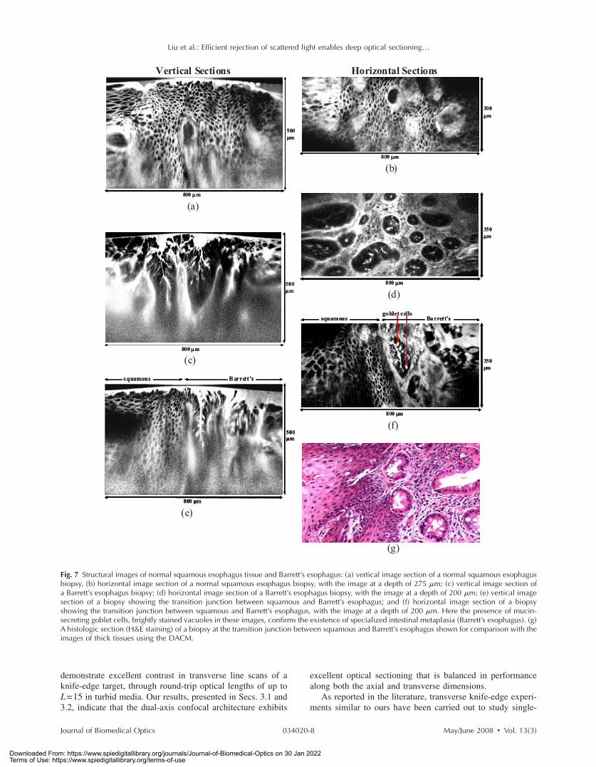
3.3.1 Diagnostic imaging of esophageal mucosaMucosal biopsies from normal esophagus were imaged fol-lowing dye application. A vertical section is shown in Fig.7�a�, demonstrating the ability to visualize squamous cells to adepth of 500 �m. Note that the PMT detector gain is variedduring each vertical image, at 2 frames /s, to compensate foran exponential decrease in fluorescence signal with depth. Ahorizontal rendering at a depth of 275 �m, of 300 verticalsections spaced by 1 �m, is shown in Fig. 7�b�.
Barrett’s esophagus is a condition in which the normalsquamous epithelium of the esophagus has been replaced byan abnormal columnar epithelium termed specialized intesti-nal metaplasia. Since Barrett’s esophagus is often indistin-guishable from normal stomach �gastric� mucosa under white-light endoscopy, biopsy and histological examination arenecessary for the accurate diagnosis of this precancerous con-dition. Vertical and horizontal images of Barrett’s esophagus,respectively, are shown in Figs. 7�c� and 7�d�. Figure 7�e�shows a vertical section of a transition junction between squa-mous and Barrett’s esophagus. A horizontal rendering, at adepth of 205 �m, is shown in Fig. 7�f� along with the corre-sponding histology �H&E staining� in Fig. 7�g�. The presence
Fig. 4 Axial response of a dual-axis microscope to a plane mirror inturbid media. The measured scattering coefficient of our Intralipidscattering phantom �s is 34 mm−1. An anisotropy of g=0.75 is as-sumed in the simulations. �a� Experimental measurements made withthe mirror located at round-trip optical lengths of L=4.7–8.2, whereL=2�sd /cos � is the total number of mean free paths traversed byballistic photons along the illumination and collection paths in turbidmedia; a �s=0 curve obtained by performing a mirror scan throughwater is also shown, and �b� diffraction theory �no scattering� andMonte Carlo simulations �no diffraction�. Diffraction simulations areprovided for our dual-axis confocal �NA=0.21 focusing lenses� aswell as for a single-axis confocal �NA=0.21 focusing lens� with anidentical FWHM �−3-dB� axial resolution.
Liu et al.: Efficient rejection of scattered light enables deep optical sectioning…
Journal of Biomedical Optics May/June 2008 � Vol. 13�3�034020-6
Downloaded From: https://www.spiedigitallibrary.org/journals/Journal-of-Biomedical-Optics on 30 Jan 2022Terms of Use: https://www.spiedigitallibrary.org/terms-of-use

of mucus-secreting goblet cells, which appear as brightlystained vacuoles among the columnar cells lining the glands�Fig. 7�f��, confirms the specialized intestinal metaplasia ofBarrett’s esophagus rather than gastric mucosa.
3.3.2 Diagnostic imaging of colonic mucosaColonic polyps are routinely biopsied and diagnosed histo-logically. Adenomatous polyps are a dysplastic transformationof colonic mucosa with precancerous potential. Figure 8�a�shows a vertical section of a colonic polyp biopsy, followingdye application. A horizontal rendering of this tissue, at adepth of 50 �m, is shown in Fig. 8�b�, along with corre-sponding histology �H&E staining� in Fig. 8�c�. This exampleof an adenoma, showing low-grade dysplasia, is easily con-trasted with and distinguished from optical sections of normalcolonic mucosa �Fig. 8�d�� as well as optical sections of ad-enoma exhibiting high-grade dysplasia �Fig. 8�f��. The corre-sponding histology �H&E staining� is shown in respective or-der �Fig. 8�e� and Fig. 8�g��. In normal mucosa, the crypts�glands� are circular and evenly spaced. In adenomatous mu-
cosa, the crypts are ellipsoidal with high eccentricity and arelined with hyperproliferative and stratified colonocytes.
4 DiscussionThe dual-axis confocal architecture has advantages over con-ventional single-axis confocal microscope designs, particu-larly for miniaturization and in vivo use. The DACM utilizeslow-NA objectives, which provide long working distances andenable aberration-free postobjective scanning over a largefield of view. Deep tissue microscopy requires efficient rejec-tion of background scattering, which has been accomplishedin a variety of ways and described in the literature. Amongpopular methods are time- and coherence-gating methods,such as optical coherence tomography, as well as nonlinearmicroscopy techniques, accomplished with high-power lasersand high-NA focusing. In this paper, we demonstrate thatsimple low-NA optics and low-power diode lasers can be uti-lized in a dual-axis confocal architecture to image deeplywithin turbid media. The DACM does not require coherentdetection, thereby making it compatible with molecularly tar-geted fluorescence imaging that is free from speckle noise.For example, we plan to utilize a miniature endoscopicDACM to image fluorescent peptides, discovered by otherresearchers within our group, that preferentially bind to cell-surface molecular biomarkers of disease in the colon.27
To quantify the optical-sectioning performance of a table-top DACM, simple reflectance experiments were performedthat may be verified with Monte Carlo scattering simulations.We previously performed diffraction-theory calculations toshow that the axial response of the DACM falls off quicker,and has a larger dynamic range, than the response of a single-axis confocal microscope with an equivalent FWHM axialresolution.15 In this study, we show that in the vicinity of thefocal plane ��20 �m�, the axial response of our DACM to aplane mirror, through up to �8 mean free paths of scatteringmedia �round-trip optical length�, is superior to thediffraction-limited �scatter-free� performance of a single-axisconfocal with an equivalent FWHM axial resolution. We also
Fig. 5 �a� Transverse line scans of a chrome-on-glass knife-edge target positioned at various round-trip optical lengths, L=2�sd /cos �, in anIntralipid scattering phantom. The knife-edge surface is positioned at the focal plane of our dual-axis microscope for maximum signal and contrast.The maximum difference in signal is �20 dB and degrades with depth as background scattered light begins to dominate. �b� Distance between the10 to 90% intensity points in our knife-edge line scans, which remains relatively constant as a function of imaging depth in turbid media �opticallength�.
0 2 4 6 8 10 12 14 160.0
0.2
0.4
0.6
0.8
1.0
Optical length, L = (2 μs d / cosθ)
Contrast
Fig. 6 Contrast in our knife-edge line scans as a function of depth. Seetext for details.
Liu et al.: Efficient rejection of scattered light enables deep optical sectioning…
Journal of Biomedical Optics May/June 2008 � Vol. 13�3�034020-7
Downloaded From: https://www.spiedigitallibrary.org/journals/Journal-of-Biomedical-Optics on 30 Jan 2022Terms of Use: https://www.spiedigitallibrary.org/terms-of-use
demonstrate excellent contrast in transverse line scans of aknife-edge target, through round-trip optical lengths of up toL=15 in turbid media. Our results, presented in Secs. 3.1 and3.2, indicate that the dual-axis confocal architecture exhibits
excellent optical sectioning that is balanced in performancealong both the axial and transverse dimensions.
As reported in the literature, transverse knife-edge experi-ments similar to ours have been carried out to study single-
Vertical Sections Horizontal Sections
Vertical Section
800 μμm
500μμm
Vertical Section
800 μμm
500μμm
(a)
800 μμm
300μμm
800 μμm
300μμm
(b)
800μμm
500μμm
800μμm
500μμm
(c)
800 μμm
350μμm
800 μμm
350μμm
(d)
800 μμm
500μμm
squamous Barrett’s
800 μμm
500μμm
800 μμm
500μμm
squamous Barrett’s
(e)
800 μμm
350μμm
goblet cellssquamous Barrett’s
800 μμm
350μμm
goblet cellssquamous Barrett’s
(f)
(g)
Fig. 7 Structural images of normal squamous esophagus tissue and Barrett’s esophagus: �a� vertical image section of a normal squamous esophagusbiopsy, �b� horizontal image section of a normal squamous esophagus biopsy, with the image at a depth of 275 �m; �c� vertical image section ofa Barrett’s esophagus biopsy; �d� horizontal image section of a Barrett’s esophagus biopsy, with the image at a depth of 200 �m; �e� vertical imagesection of a biopsy showing the transition junction between squamous and Barrett’s esophagus; and �f� horizontal image section of a biopsyshowing the transition junction between squamous and Barrett’s esophagus, with the image at a depth of 200 �m. Here the presence of mucin-secreting goblet cells, brightly stained vacuoles in these images, confirms the existence of specialized intestinal metaplasia �Barrett’s esophagus�. �g�A histologic section �H&E staining� of a biopsy at the transition junction between squamous and Barrett’s esophagus shown for comparison with theimages of thick tissues using the DACM.
Liu et al.: Efficient rejection of scattered light enables deep optical sectioning…
Journal of Biomedical Optics May/June 2008 � Vol. 13�3�034020-8
Downloaded From: https://www.spiedigitallibrary.org/journals/Journal-of-Biomedical-Optics on 30 Jan 2022Terms of Use: https://www.spiedigitallibrary.org/terms-of-use

axis confocal microscopes. The most impressive results werereported by Kempe et al.19 In that study, chrome-on-glass re-flectance gratings were imaged, yielding results that are com-parable to our own results shown in Fig. 6. However, thoseresults were obtained from a confocal microscope that had aneffective NA of only 0.28, which is similar to the effectiveNA of our DACM �Sec. 2.1�. Such a confocal, in a single-axis
arrangement, would exhibit an FWHM axial resolution of theorder of 15 �m, not to mention an extremely slow axial re-sponse, as discussed in Sec. 3.1. It has been determined thatthere is a reduction in the amount of background scatteredlight collected by a single-axis confocal as NA is reduced, atthe cost of a degradation in resolution and axial-sectioning
Vertical Section
Horizontal Sections
800 μμm
500μμm
800 μμm
500μμm
(a)
800μμm
350μμm
800μμm
350μμm
(b) (c)
(e)800μμm
300μμm
800μμm
300μμm
(d)
800 μμm
350μμm
800 μμm
350μμm
(f) (g)
Fig. 8 Structural images of colonic mucosa: �a� vertical image section of a colonic polyp biopsy, where an adenoma with low-grade dysplasia isobserved; �b� horizontal image section of a colonic polyp biopsy exhibiting low-grade dysplasia, image at a depth of 140 �m; �c� histologic section�H&E staining� of an adenomatous polyp with low-grade dysplasia, for comparison with dual-axis confocal images of thick tissues; �d� horizontalsection, at a depth of 115 �m, of a normal colon biopsy, where evenly spaced, circular crypts �glands� are observed; �e� histologic section �H&Estaining� of a normal colon biopsy; �f� horizontal section, at a depth of 115 �m, of an adenomatous polyp biopsy, exhibiting high-grade dysplasia,where irregular and elongated crypts are observed; and �g� histologic section �H&E staining� of an adenomatous polyp biopsy exhibiting high-gradedysplasia.
Liu et al.: Efficient rejection of scattered light enables deep optical sectioning…
Journal of Biomedical Optics May/June 2008 � Vol. 13�3�034020-9
Downloaded From: https://www.spiedigitallibrary.org/journals/Journal-of-Biomedical-Optics on 30 Jan 2022Terms of Use: https://www.spiedigitallibrary.org/terms-of-use

response.28 Therefore, while a low-NA single-axis confocalmicroscope may perform well for imaging a flat “presec-tioned” reflective target in scattering media, the slow axialresponse and poor axial resolution would not enable effectiveoptical sectioning in 3-D biological tissues. Note that ourstudy is by no means intended as a comprehensive compari-son between single-axis and dual-axis confocal microscopies.These two confocal architectures represent distinct modalitiesthat possess strengths and weaknesses for different applica-tions. Our primary aim was to perform experimental studies tocharacterize the DACM in turbid media, as has been donewith single-axis confocal microscopes in the past.
Of the numerous studies of optical-sectioningtechnologies,17–19,28 few studies investigate both axial andtransverse performance in scattering media. Of those studies,still fewer, if any, provide a demonstration of imaging perfor-mance in actual biological tissues as well. Since mirror andknife-edge models may be misleading, we demonstrate deep3-D fluorescence microscopy in biological tissues. Collec-tively, these results consistently demonstrate the deep optical-sectioning abilities of the DACM. In particular, for the reflec-tance studies described in Secs. 3.1 and 3.2, we observed thatFWHM spatial resolution is not significantly affected by im-aging depth �optical length�, but that image contrast quicklydegrades as the scattering background overwhelms the ballis-tic signal. This is also apparent in many of the images ofbiological tissues. For example, in Figs. 7�a� and 7�e�, themembranes of the individual squamous esophagus cells re-main well defined and resolvable deep into the sample, butquickly lose visibility at a certain point as the scattering back-ground overwhelms the ballistic signal from the cell walls.
In summary, the dual-axis confocal architecture exhibitsefficient rejection of scattered light, as demonstrated throughquantitative mirror reflectance studies in a scattering phantom,and through imaging exogenous single-photon fluorescencecontrast in human gastrointestinal biopsy samples. The resultsindicate the utility of this technology to distinguish cellularand morphological signatures of various pathologies such asBarrett’s esophagus and colonic adenomas, as well as to dis-tinguish between grades of colonic dysplasia. The 3-Doptical-sectioning ability of the DACM enables arbitrary ori-entations and locations to be viewed. Thus, a miniatureendoscope-compatible version of this device would provide avaluable tool for rapidly diagnosing and staging diseases, aswell as for guiding surgical resection.
AcknowledgmentsThis work was funded in part by grants from the NationalInstitutes of Health, including K08 DK067618 �NIDDK�, U54CA105296 �NCI�, and R33 CA109988 �NCI�. This work wasalso supported by funding through the Center for Biophoton-ics, a National Science Foundation Center managed by theUniversity of California, Davis �PHY 0120999�. Jonathan Liuis supported by a Canary Foundation / American Cancer So-ciety postdoctoral fellowship for early cancer detection. Wethank Shai Friedland, Roy Soetikno, Peyman Sahbaie, andLarry Wong for technical support.
References1. J. G. Fujimoto, M. E. Brezinski, G. J. Tearney, S. A. Boppart, B.
Bouma, M. R. Hee, J. F. Southern, and E. A. Swanson, “Optical
biopsy and imaging using optical coherence tomography,” Nat. Meth-ods 1, 970–972 �1995�.
2. G. J. Tearney, M. E. Brezinski, B. E. Bouma, S. A. Boppart, C. Pitris,J. F. Southern, and J. G. Fujimoto, “In vivo endoscopic optical biopsywith optical coherence tomography,” Science 276, 2037–2039�1997�.
3. B. E. Bouma and G. J. Tearney, Handbook of Optical CoherenceTomography, Marcel Dekker, New York �2002�.
4. M. T. Myaing, D. J. MacDonald, and X. Li, “Fiber-optic scanningtwo-photon fluorescence endoscope,” Opt. Lett. 31, 1076–1078�2006�.
5. K. Carlson, M. Chidley, K.-B. Sung, M. Descour, A. Gillenwater, M.Follen, and R. Richards-Kortum, “In vivo fiber-optic confocal reflec-tance microscope with an injection-molded plastic miniature objec-tive lens,” Appl. Opt. 44, 1792–1797 �2005�.
6. R. Kiesslich, J. Burg, M. Vieth, J. Gnaendiger, M. Enders, P.Delaney, A. Polglase, W. McLaren, D. Janell, and S. Thomas, “Con-focal laser endoscopy for diagnosing intraepithelial neoplasias andcolorectal cancer in vivo,” Gastroenterology 127, 706–713 �2004�.
7. W. Piyawattanametha, R. P. J. Barretto, T. H. Ko, B. A. Flusberg, E.D. Cocker, H. Ra, D. Lee, O. Solgaard, and M. J. Schnitzer, “Fast-scanning two-photon fluorescence imaging based on a microelectro-mechanical systems two-dimensional scanning mirror,” Opt. Lett. 31,2018–2020 �2006�.
8. J. T. C. Liu, M. J. Mandella, H. Ra, L. K. Wong, O. Solgaard, G. S.Kino, W. Piyawattanametha, C. H. Contag, and T. D. Wang, “Minia-ture near-infrared dual-axis confocal microscope utilizing a two-dimensional microelectromechanical systems scanner,” Opt. Lett. 32,256–258 �2007�.
9. R. Kiesslich, P. R. Galle, and M. Neurath, “Endoscopic surveillancein ulcerative colitis: smart biopsies do it better,” Gastroenterology133, 742–745 �2007�.
10. E. H. K. Stelzer and S. Lindek, “Fundamental reduction of the obser-vation volume in far-field light microscopy by detection orthogonal tothe illumination axis: confocal theta microscopy,” Opt. Commun. 111,536–547 �1994�.
11. E. H. K. Stelzer, S. Lindek, S. Albrecht, R. Pick, G. Ritter, N.Salmon, and R. Stricker, “A new tool for the observation of embryosand other large specimens: confocal theta fluorescence microscopy,”J. Microsc. 179, 1–10 �1995�.
12. S. Lindek and E. H. K. Stelzer, “Optical transfer functions for con-focal theta fluorescence microscopy,” J. Opt. Soc. Am. A 13, 479–482�1996�.
13. R. H. Webb and F. Rogomentich, “Confocal microscope with largefield and working distance,” Appl. Opt. 38, 4870–4875 �1999�.
14. T. D. Wang, M. J. Mandella, C. H. Contag, N. Y. Chan, and G. S.Kino, “Dual axes confocal microscope with post-objective scanningand low coherence heterodyne detection,” Opt. Lett. 28, 1915–1917�2003�.
15. J. T. C. Liu, M. J. Mandella, S. Friedland, R. Soetikno, J. H. Craw-ford, C. H. Contag, G. S. Kino, and T. D. Wang, “Dual-axis confocalreflectance microscope for distinguishing colonic neoplasia,” J.Biomed. Opt. 11, 054019 �2006�.
16. L. K. Wong, T. D. Wang, M. J. Mandella, and G. S. Kino, “Improvedrejection of multiply scattered photons in confocal microscopy usingdual-axis architecture,” Opt. Lett. 32, 1674–1676 �2007�.
17. J. A. Izatt, M. R. Hee, G. M. Owen, E. A. Swanson, and J. G.Fujimoto, “Optical coherence microscopy in scattering media,” Opt.Lett. 19, 590–592 �1994�.
18. J. M. Schmitt, A. Knuttel, and M. Yadlowsky, “Confocal microscopyin turbid media,” J. Opt. Soc. Am. A 11, 2226–2235 �1994�.
19. M. Kempe, W. Rudolph, and E. Welsch, “Comparative study of con-focal and heterodyne microscopy for imaging through scattering me-dia,” J. Opt. Soc. Am. A 13, 46–52 �1996�.
20. S. M. Mansfield, W. Studenmund, G. S. Kino, and K. Osato, “Highnumerical aperture lens system for optical storage,” Opt. Lett. 18,305–307 �1993�.
21. T. D. Wang, C. H. Contag, M. J. Mandella, N. Y. Chan, and G. S.Kino, “Confocal fluorescence microscope with dual-axis architectureand biaxial postobjective scanning,” J. Biomed. Opt. 9, 735–742�2004�.
22. B. W. Pogue and M. S. Patterson, “Review of tissue simulating phan-toms for optical spectroscopy, imaging and dosimetry,” J. Biomed.Opt. 11, 041102 �2006�.
Liu et al.: Efficient rejection of scattered light enables deep optical sectioning…
Journal of Biomedical Optics May/June 2008 � Vol. 13�3�034020-10
Downloaded From: https://www.spiedigitallibrary.org/journals/Journal-of-Biomedical-Optics on 30 Jan 2022Terms of Use: https://www.spiedigitallibrary.org/terms-of-use

23. L. Henyey and J. Greenstein, “Diffuse radiation in the galaxy,” As-trophys. J. 93, 70–83 �1941�.
24. A. E. Siegman, Lasers, University Science Books, Mill Valley, CA�1986�.
25. T. Collier, M. Follen, A. Malpica, and R. Richards-Kortum, “Sourcesof scattering in cervical tissue: determination of the scattering coef-ficient by confocal microscopy,” Appl. Opt. 44, 2072–2081 �2005�.
26. H. J. Wei, D. Xing, G. Y. Wu, H. M. Gu, J. J. Lu, Y. Jin, and X. Y. Li,“Differences in optical properties between healthy and pathologicalhuman colon tissues using a Ti:sapphire laser: an in vitro study using
the Monte Carlo inversion technique,” J. Biomed. Opt. 10, 044022�2005�.
27. P. Hsiung, J. Hardy, S. Friedland, R. Soetikno, C. B. Du, A. P. W.Wu, P. Sahbaie, J. M. Crawford, A. W. Lowe, C. H. Contag, and T. D.Wang, “Detection of colonic dysplasia in vivo using a targeted fluo-rescent septapeptide and confocal microendoscopy,” Nat. Methods14, 454–456 �2008�.
28. M. Gu, T. Tannous, and J. R. Sheppard, “Effect of an annular pupil onconfocal imaging through highly scattering media,” Opt. Lett. 21,312–314 �1996�.
Liu et al.: Efficient rejection of scattered light enables deep optical sectioning…
Journal of Biomedical Optics May/June 2008 � Vol. 13�3�034020-11
Downloaded From: https://www.spiedigitallibrary.org/journals/Journal-of-Biomedical-Optics on 30 Jan 2022Terms of Use: https://www.spiedigitallibrary.org/terms-of-use