EffectofPyrroleImplantsSynthesizedbyDifferent ... ·...
15
ARTÍCULO DE INVESTIGACIÓN REVISTA MEXICANA DE INGENIERÍA BIOMÉDICA ib Vol. 36, No. 1, Ene-Abr 2015, pp. 7-21 Effect of Pyrrole Implants Synthesized by Different Methods on Spinal Cord Injuries of Rats L. Álvarez-Mejía 1,3 , H. Salgado-Ceballos 3 , R. Olayo 2 , G.J. Cruz 4 , M.G. Olayo 4 , A. Díaz-Ruiz 5 , C. Ríos 5 , R. Mondragón-Lozano 1,5 , A. Morales-Guadarrama 1,7 , S. Sánchez-Torres 3,6 , J. Morales 2 1 Depto. de Ing. Eléctrica, 2 Depto. de Física, UAM-Iztapalapa. 3 Centro Médico Nacional Siglo XXI. 4 Depto. de Física, ININ, 5 Depto. de Neuroquímica, INNN Manuel Velasco Suárez, 6 Escuela Superior de Medicina. IPN. 7 Centro Nacional de Investigación en Imagenología e Instrumentación Médica ABSTRACT Polypyrrole (PPy) and polypyrrole/polyethylene glycol (PPy/PEG) implants synthesized by chemical, electro- chemical, and plasma polymerization methods were implanted into the injured spinal cord of rats to determine their effect on motor function recovery. Before implantation, the materials were characterized by infrared (IR) spectroscopy. An experimental model of traumatic spinal cord injury (TSCI) by complete transection at thoracic level 9, in rats was used. The polymer implants were inserted immediately after transection. Motor function recovery was evaluated once a week during 5 weeks using the Basso, Beattie and Bresnahan (BBB) motor scale. Histological evaluation was done at the end of the recovery evaluation period using hematoxylin/eosin stain. Results showed that animals implanted with polymers synthesized by plasma had a better integration into the nerve tissue, less inflammatory response and a better functional recovery than animals implanted with polymers synthesized by chemical or electrochemical methods. Keywords: fpolypyrrole implants, chemical synthesis, electrochemical synthesis, plasma synthesis, traumatic spinal cord injury. Correspondencia: Juan Morales Corona Departamento de Física, Universidad Autónoma Metropolitana Iztapalapa. Av. San Rafael Atlixco 186 Col. Vicentina Del. Iztapalapa, México D.F. C.P. 09340. Correo electrónico: [email protected] Fecha de recepción: 29 de abril de 2014 Fecha de aceptación: 21 de octubre de 2014
Transcript of EffectofPyrroleImplantsSynthesizedbyDifferent ... ·...
ARTÍCULO DE INVESTIGACIÓN REVISTA MEXICANA DE
INGENIERÍA BIOMÉDICAibVol. 36, No. 1, Ene-Abr 2015, pp. 7-21
Effect of Pyrrole Implants Synthesized by DifferentMethods on Spinal Cord Injuries of Rats
L. Álvarez-Mejía1,3, H. Salgado-Ceballos3, R. Olayo2, G.J. Cruz4, M.G. Olayo4,A. Díaz-Ruiz5, C. Ríos5, R. Mondragón-Lozano1,5, A. Morales-Guadarrama1,7,S. Sánchez-Torres3,6, J. Morales2
1Depto. de Ing. Eléctrica, 2 Depto. de Física, UAM-Iztapalapa. 3 Centro Médico Nacional Siglo XXI.4Depto. de Física, ININ, 5Depto. de Neuroquímica, INNNManuel Velasco Suárez, 6Escuela Superior de Medicina.IPN. 7Centro Nacional de Investigación en Imagenología e Instrumentación Médica
ABSTRACTPolypyrrole (PPy) and polypyrrole/polyethylene glycol (PPy/PEG) implants synthesized by chemical, electro-chemical, and plasma polymerization methods were implanted into the injured spinal cord of rats to determinetheir effect on motor function recovery. Before implantation, the materials were characterized by infrared (IR)spectroscopy. An experimental model of traumatic spinal cord injury (TSCI) by complete transection at thoraciclevel 9, in rats was used. The polymer implants were inserted immediately after transection. Motor functionrecovery was evaluated once a week during 5 weeks using the Basso, Beattie and Bresnahan (BBB) motor scale.Histological evaluation was done at the end of the recovery evaluation period using hematoxylin/eosin stain. Resultsshowed that animals implanted with polymers synthesized by plasma had a better integration into the nerve tissue,less inflammatory response and a better functional recovery than animals implanted with polymers synthesized bychemical or electrochemical methods.Keywords: fpolypyrrole implants, chemical synthesis, electrochemical synthesis, plasma synthesis,traumatic spinal cord injury.
Correspondencia:Juan Morales CoronaDepartamento de Física, Universidad Autónoma MetropolitanaIztapalapa. Av. San Rafael Atlixco 186 Col. Vicentina Del.Iztapalapa, México D.F. C.P. 09340.Correo electrónico: [email protected]
Fecha de recepción:29 de abril de 2014
Fecha de aceptación:21 de octubre de 2014
8 Revista Mexicana de Ingeniería Biomédica · volumen 36 · número 1 · Ene-Abr, 2015
RESUMENEn el presente trabajo se comparó el efecto de implantes poliméricos derivados del pirrol (polipirrol o PPy)y del copolímero polipirrol/polietilenglicol (PPy/PEG), obtenidos por diferentes métodos de síntesis: químico,electroquímico y polimerización por plasma con el propósito de determinar si el método de síntesis puede influirsobre el efecto que producen al ser implantados después de una lesión traumática de la médula espinal de ratas.Antes de realizar el implante, las características químicas y estructurales de los polímeros fueron analizadas porespectroscopia de infrarrojo (IR). Se utilizó un modelo experimental de lesión traumática de médula espinal (LTME)por sección completa en ratas. La LTME se realizó a nivel torácico 9 y el polímero fue implantado de inmediato enla zona de lesión. La recuperación de la función motora se evaluó mediante la escala Basso, Beattie y Bresnahan(BBB) una vez por semana durante 5 semanas. La evaluación histológica se realizó al término del seguimiento conla tinción de hematoxilina/eosina. Los resultados muestran que los animales implantados con polímeros sintetizadospor plasma se integraron mejor al tejido nervioso, redujeron la respuesta inflamatoria y favorecieron una mayorrecuperación funcional en comparación con los animales implantados con materiales sintetizados por métodos químicoso electroquímicos.Palabras clave: implantes de polipirrol, síntesis química, síntesis electroquímica, síntesis por plasma,lesión traumática de la médula espinal.
INTRODUCTION
Traumatic spinal cord injuries (TSCI) triggera series of secondary events that increase theoriginal damage, prevent axonal regenerationand produce different degrees of functionalimpairment below the site of injury which canlead to permanent paralysis [1].
To date, due to the complexpathophysiology of TSCI, no therapeuticstrategy has been effective to restore the lostfunctions after lesion. Many strategies havebeen suggested and attempted in order tofind a solution to this problem. One proposalin this field consists of using transplantsof tissues or cells to promote axonalregeneration and functional recovery afterTSCI [2]. Among experimental transplantsthat have produced positive results are fetaltissue [3], fresh or predegenerate peripheralnerve [4-5], Schwann cells alone or incombination with different molecules [2,6],olfactory ensheathing cells [2,7] and neuralstem cells [2,8-9].
Notwithstanding, the majority of thetransplants that have been used in thetreatment of TSCI have not been able torestore the nerve function in a significant way.Recently, research in tissue engineering hasproduced materials that have the potential ofbeing a better treatment for this pathology[10-12].
To form a viable transplant for TSCItreatment, the materials employed mustbe biocompatible with the nervous tissue(where the communication is mainly by ionexchange), and their chemical composition,hydrophobicity and electrical activity shouldbe well characterized.
Recently, polymers with intrinsicconductive properties have gained relevanceas smart materials with biologicalapplications [13]. These polymers possessthe physical and chemical properties oforganic polymers and the electrical propertiesof the metals [14]. Additionally, thesematerials have the ability to bind tovarious chemical substances (dopants) which
Álvarez-Mejía et al. Effect of pyrrole implants synthesized by different methods on spinal cord injuries of rats. 9
affect the physicochemical properties ofthe polymers, such as their conductivity.Furthermore, materials of this type have beendeveloped with the capability of supportingand modulating the growth of differentcell types which makes them suitable forbiological and biomedical applications [15].
Among biocompatible and electricallyconductive polymers, polypyrrole (PPy) isone of the most studied. PPy is easyto synthesize, thermally stable, and hashigh conductivity in comparison with otherconductive polymers [16]. PPy has beenused as biosensor for measuring cholesterol[17], glucose in blood [18], blood group,antibodies [19], and vapors of organicsolvents [20]. It has also been usedfor coating neural probes [21], devices fordrug and biomolecule release [22], andartificial muscles [23]. In addition to beingbiocompatible [24], the cytocompatibility ofPPy has been demonstrated using L929mouse fibroblast and Neuro2a neuroblastomacells [25]. Moreover, PPy has been used tosupport cell adhesion and growth of differentcell types in vitro [13, 26-34], and as guidefor regenerating rat sciatic nerve [33, 35].Our research group, has demonstrated thatPPy synthesized by plasma polymerizationpromotes neuroprotection and leads torecovery of motor and sensory functions aftera TSCI by complete section in in vivo studieswith rats [36-38]. These PPy implantsshowed no significant inflammatory responsein situ after 4 weeks of implantation [25].
Typically, PPy is synthesized by chemicaland electrochemical polymerization methods.It is also possible to synthesize it byplasma polymerization, but the productshows important differences. The chemicallysynthesized PPy is crystalline, and thereis a lack of control over the mass or thethickness of the film obtained [39]. PPysynthesized electrochemically, has similarchemical structure to that obtained bychemical methods, but can be more easilydoped to enhance its conductive properties.Also, the film properties can be controlled
directly in the course of the polymerization.Both the chemical and electrochemicalsynthetic methods use accelerators andsolvents, which modify the adhesion andhydrophobicity properties of the resultingmaterials. These extra components canbe dangerous or toxic; thus they mustbe fully removed from the polymers beforebeing applied to any biological system. Incontrast, polymerization by plasma onlyrequires the base monomer to start thereaction without introducing other chemicalcompounds. When plasma polymerization isused, the oxidation is promoted by the impactof free electrons that travel along the electricfield with monomer molecules [40]. Thematerial produced by plasma polymerizationdoes not have a regular chemical structurebut specific chemical groups are present.Plasma polymerization produces a dense filmwith a crosslinked structure [41]. Thecrosslinked structure permits the materialto retain its mechanical properties even inbiological media, while the functional groupsexposed to the surface mediate favorableinteractions with many types of cells.
Given the differences in chemical andphysical structure of the PPy materialsobtained by different synthesis methods,it is important to evaluate how thesevariations affect implants and their effecton recuperation from spinal cord injuries.The goal of the present study is to compareimplants made of pyrrole derivatives, PPyand PPy co-polymerized with polyethyleneglycol (PPy/PEG) obtained by chemical,electrochemical and plasma polymerization.We analyze the chemical structure of theprepared implants, their ability to integratewith nervous tissue, their inflammatoryresponse and their effect on the recovery ofmotor function after a TSCI in rats. In thisstudy PPy/PEG was tested because after aTSCI the neuronal membranes are rupturedand PEG has been shown to aid in the repairof membranes [42-43]; which could enhancethe benefic effect of PPy when implantedafter a TSCI.
10 Revista Mexicana de Ingeniería Biomédica · volumen 36 · número 1 · Ene-Abr, 2015
MATERIALS AND METHODS
Plasma polymerization
The preparation of iodine doped PPy (PPy/I)and PPy/PEG thin films has been previouslydescribed [36-37]. Briefly, the films wereprepared in a tubular glass reactor, 9 cmin diameter and 25 cm long, capped withstainless steel flanges with access ports. Theports were used to connect the reactor toa vacuum pump, a Pirani gauge (Edwards),and to introduce reactants. For thepreparation of PPy films, pyrrol (Aldrich,99%) vapor was used; and to synthesize thePPy/PEG copolymer, pyrrol was introducedby one port and, simultaneously, PEG(Aldrich) was introduced through anotherport. In the center of the flanges twostainless steel flat circular electrodes with 7cm of diameter and separated by 9 cm, wereinserted with the aim to create a homogenouselectric field in the reactor. The electric fieldwas generated by a Dressler Cesar 136 RFPower Generator. A field frequency of 13.5MHz and power of 18 W was used. Thepressure in the reactor was 5 × 10⁻² Torrand the synthesis time was 300 min. Thepolymers were separated from the internalreactor walls applying acetone and using athin spatula.
Electrochemical synthesis
PPy/PEG copolymer was synthesized bythe conventional electrochemical methodusing an tituanium electrode (99.99% Sigma-Aldrich) with an exposed area of 4.0 cm2.Before each experiment, the electrode surfacewas polished with silicon carbide (SiC)paper grade 2000, degreased with acetoneand rinsed with deionized water. For theelectropolymerization an aqueous solution of0.2 M pyrrol monomer in 0.2 M of oxalic acid(Ac. Ox) with 8% PEG (molecular weight600 Daltons) was prepared. All solutions wereprepared with deionized water (18.2 MΩ).
Electrochemical polymerizations wereperformed at room temperature in a
conventional cell with three electrodes: arod of Ti functioned as the active electrode,a saturated calomel electrode (SCE) thatfunctioned as the reference electrode, a rod ofplatinum (Pt) as the counter electrode. Allelectrodes were connected to a Potentiostatgalvanostat Autollab PGSTAT 302N with theGPES 4.9 electrochemical software.
The PPy/PEG film was synthesizedpotentiostatically at 0.9 V vs. SCE, andallowed to grow for 4 hours. The film wasrinsed with deionized water and removedfrom the Ti electrode surface and left to dryat a temperature of 60 oC for 24 hrs.
Chemical synthesis
Two different chemically synthesized PPywere used. A commercial PPy doped withsulphonic acid was purchased from Sigma-Aldrich (CAS 30604-81-0 and 577030-5G,Pcode 1000874358, Lot # MKB, elementalanalysis C-69.94 %; N-15.42 % (N/C=0.22),O-12.58 %; S-2.06 %.
The other PPy was synthesized in ourlaboratory by oxidation. The polymerwas synthesized by mixing 48 mL ofdodecylbenzenesulfonic acid (70 wt. %dissolved in 2-propanol), 900 mL of distilledwater, 7 mL of pyrrole, and 11.4 g ofammonium persulphate dissolved in 25 mLof water. The reaction was performed at 25
C during 24 hours.
Infrared Characterization
The polymers were analyzed by infrared(IR) spectroscopy with a Nicolette 550spectrophotometer with a 400-4000 cm−1
interval using 32 scans [36, 40].
Implants
Each polymer was pulverized and thencompressed at 9 Ton/cm2 for 10 min to forma thin tablet of 1 cm in diameter and 0.5mm thickness. Finally, the thin table was cutaccording to the diameter of the spinal cord.
Álvarez-Mejía et al. Effect of pyrrole implants synthesized by different methods on spinal cord injuries of rats. 11
Animals and surgical procedures
Female Long Evans rats with 220 to 260 g ofbody weight were maintained under standardlaboratory conditions and free access to foodand water. Animal care and the protocols foranimal use were approved by the Scientificand Ethics Committees of the InstitutoMexicano del Seguro Social.
Eighteen rats were prepared to receive acomplete spinal cord section at thoracic 9level (T-9) and then, six experimental groups(n=3 animals per group) were formed:
1. Control: animals without implant.
2. PPy: animals implanted with PPysynthesized by plasma.
3. ChPPy1: animals implanted with PPychemically-synthesized.
4. ChPPy2: animals implanted withPPy purchased from Sigma-Aldrich(chemically-synthesized).
5. PPy/PEG: animals implanted withPPy/PEG synthesized by plasma.
6. EPPy-PEG: animals implantedwith PPy/PEG electrochemically-synthesized.
Before implantation, animals wereanesthetized intramuscularly with a mixtureof ketamine and xylazine (77.5 and 12.5mg/kg). Then, an aseptic surgery undermicroscopic inspection was done by making asagittal incision on the skin from the middleback followed by a dissection of the spineparavertebral muscles. Two laminae wereremoved (T-8 and T-9) to expose the spinalcord tissue [36-38].
The meninges were longitudinally cutand spinal cord tissue was completelytransected by cutting transversally allfibers. Transection was corroborated with amicrosurgical hook to ensure that no pathwayremained connected. The correspondingimplant (approximately 10 mg) was then
inserted at the injured zone in the cavitybetween both sides of the transection. Onlythe animals in the control group did notreceive any implant. Finally, the meningeswere sutured, as well as the paravertebralmuscles and skin. Animals were treated withan anti-inflammatory drug (0.31ml/62.5mlof paracetamol into drinking water during 3days) and an antibiotic (200 µL of benzathinepenicillin, in one i.m. dose). Afterwards,rats recovered from anesthesia and surgicalprocedures in an intensive care unit for smallanimals (Schoer Manufacturing CO., KansasCity, MO, USA) and placed into individualacrylic cages with sterile sawdust for receivingfood and water ad libitum. The day afterSCI, the absence of hind limb movement wascorroborated to ensure the complete sectionof the spinal cord. Their intestine and bladderwere handled by manual expression twice aday and visual inspection was performed dayby day looking for skin irritation or decubitusulcers [36-38].
Motor function recovery
The motor function recovery of hind limbs ineach rat from the six groups were assessedweekly during five weeks using the Basso,Beattie and Bresnahan (BBB) scale [44],which has 22 points and where 0 representstotal absence of movement (paralysis) and21 represents a normal walking. The testwas applied by two observers blinded to thetreatment that the animals received.
Histological analysis
Thirty days after spinal cord transection,all animals were anesthetized as describedbefore, followed by intraperitonealadministration of 0.2 mL of heparin. Then,a wide thoracotomy was performed and200 mL of cool physiological saline solutionwas perfused transcardially at 30 mL/minfollowed by 400 mL of 4% paraformaldehydein phosphate buffer. Afterwards, 2 cm ofthe spinal cord were taken including the
12 Revista Mexicana de Ingeniería Biomédica · volumen 36 · número 1 · Ene-Abr, 2015
injury zone. The spinal cord specimens wereembedded in paraffin. Serial longitudinal10 µm thick sections were obtained andstained with hematoxylin and eosin. Imageswere obtained using a computerized systemequipped with an IM 1000 software and a 300FX digital camera [36-38].
Statistical analysis
The BBB scores were evaluated usingANOVA of repeated measures followed byDunnett’s test. Significant differences wereconsidered when p < 0.05. All analyses wereperformed using the SPSS 16.0 software.
RESULTS
Chemical structure of implants
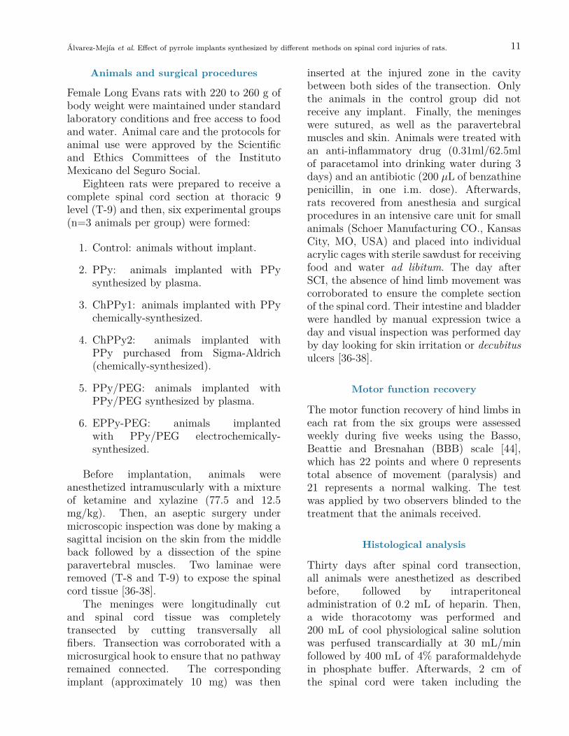
The chemical structure of polymers employedin the experiment was determined by IRspectroscopy. Figure 1 shows the similaritybetween the two chemically-synthesized PPy;in particular in the region between 1750and 4000 cm−1. Small differences in thespectra can be observed between 400 and1750 cm−1 which we believe are due tosome substitutions in the pyrrole rings. Incontrast, the same figure shows a significantdifference between the chemically-synthesizedPPy and PPy synthesized by plasma. At3355 cm−1 one can observe the differentvibrations of the primary and secondaryamine groups. Another difference is thevibration corresponding to nitrile groups at2213 cm−1.
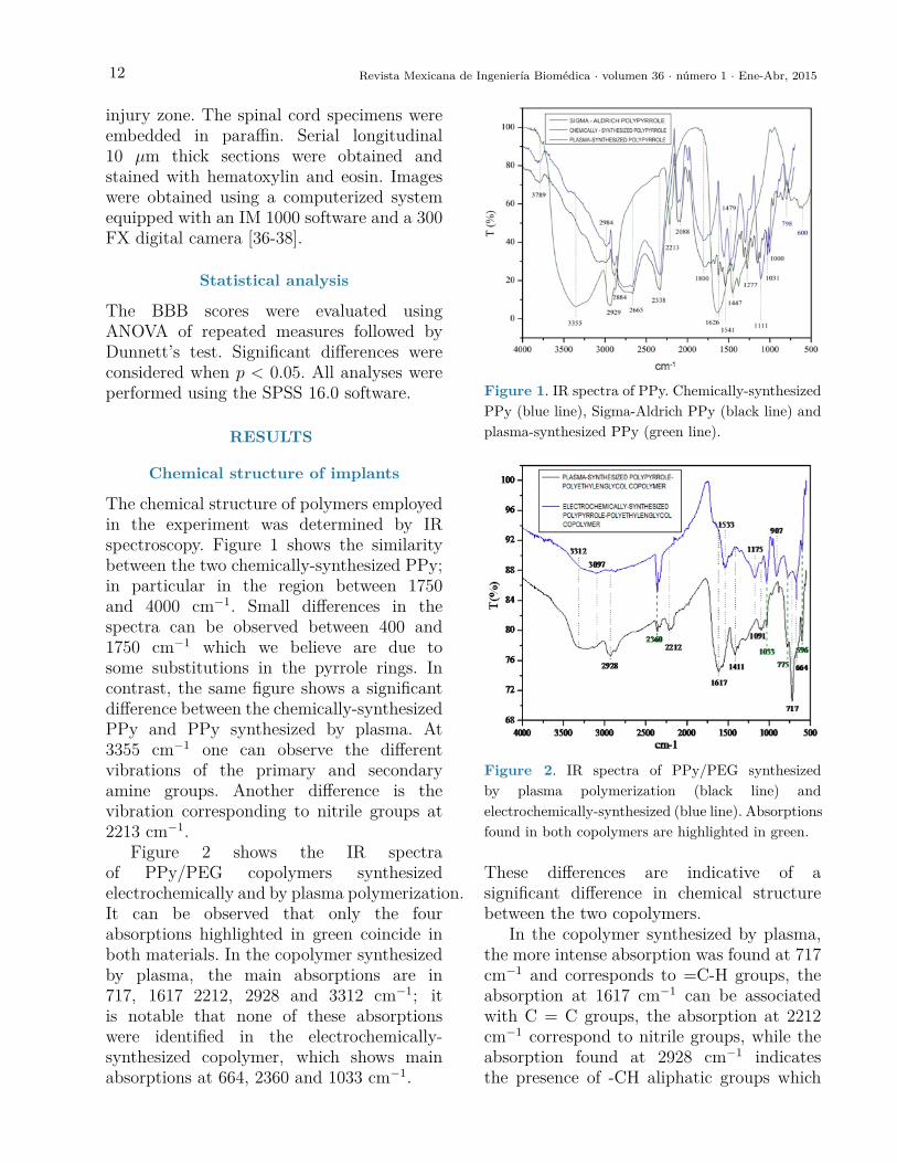
Figure 2 shows the IR spectraof PPy/PEG copolymers synthesizedelectrochemically and by plasma polymerization.It can be observed that only the fourabsorptions highlighted in green coincide inboth materials. In the copolymer synthesizedby plasma, the main absorptions are in717, 1617 2212, 2928 and 3312 cm−1; itis notable that none of these absorptionswere identified in the electrochemically-synthesized copolymer, which shows mainabsorptions at 664, 2360 and 1033 cm−1.
Figure 2 shows the IR spectra of PPy/PEG copolymers synthesized electrochemically and by plasma
polymerization. It can be observed that only the four absorptions highlighted in green coincide in both
materials. In the copolymer synthesized by plasma, the main absorptions are in 717, 1617 2212, 2928 and
3312 cm-1; it is notable that none of these absorptions were identified in the electrochemically-
synthesized copolymer, which shows main absorptions at 664, 2360 and 1033 cm-1. These differences are
indicative of a significant difference in chemical structure between the two copolymers.
In the copolymer synthesized by plasma, the more intense absorption was found at 717 cm-1 and
corresponds to =C-H groups, the absorption at 1617 cm-1 can be associated with C = C groups, the
absorption at 2212 cm-1 correspond to nitrile groups, while the absorption found at 2928 cm-1 indicates
the presence of -CH aliphatic groups which can arise from ethylene glycol segments or from fragments of
pyrrole molecules which were fractionated due to the high energy of the particles in the plasma, the
absorption at 3312 cm-1 can be associated with the presence of amine groups. These absorptions indicate
that in the copolymer synthesized by plasma, the heteroaromatic character of the pyrroles predominates.
In the case of the electrochemically-synthesized co-polymer, the more intense absorption was found in
664 cm-1 and can be assigned to N-O groups. This implies a substitution of N-H groups by N-O groups
which could be a consequence of ethylene glycol participation during the synthesis. The absorption at
1033 cm-1 corresponds to C-O groups that are found in ethylene glycol repeat units. These absorptions
indicate that in the electrochemically-synthesized copolymer, the oxygenated character of the ethylene
glycol predominates.
Figure 1. IR spectra of PPy. Chemically-synthesized PPy (blue line), Sigma-Aldrich PPy (black line) and
plasma-synthesized PPy (green line).
Figure 1. IR spectra of PPy. Chemically-synthesizedPPy (blue line), Sigma-Aldrich PPy (black line) andplasma-synthesized PPy (green line).
Figure 2. IR spectra of PPy/PEG synthesized by plasma polymerization (black line) and
electrochemically-synthesized (blue line). Absorptions found in both copolymers are highlighted in
green.
Motor function recovery
The motor function was evaluated 24 h after complete transection of the spinal cord with the aim to
corroborate if hind limb performance showed complete bilateral paralysis in all rats (BBB=0).
Afterwards, the BBB locomotor rating scale was assessed once a week during 5 weeks to evaluate the
gradual improvement of the animals. Results were as follow (Figure 3):
a) Control (3 animals alive): BBB=1, meaning that animals had slight movement of 1 or 2 joints.
b) PPy (3 animals alive): BBB=5.33, meaning that animals were able to move the 3 joints of the
hind limb (hip, knee and ankle), and in two them, the movements were extensive.
c) ChPPy1 (3 animals alive and 2 dead animals, non-evaluable): BBB=1, meaning that the animals
had slight movement of 1 or 2 joints. Although one of the animals had an improvement of 4
points in the BBB scale in the third week, score decreased with time.
d) ChPPy2 (3 animals alive and 4 dead animals, non-evaluable): BBB=1.33, meaning that the
animal had slight movement in one or two joints. But just as in the ChPPy1 group, one animal
presented improvement until the fourth week and subsequently decreased the effect of the
implant.
e) PPy-PEG (3 animals alive): BBB=3.66, meaning that animals had extensive movement in 1 or 2
joints of the hind limb and slight movement of other joint (hip, knee and ankle).
f) EPPy-PEG (3 animals alive): BBB=2.6, meaning that animals had extensive movement of 1 joint
and slight movement of 2 joints of the hind limbs.
Figure 2. IR spectra of PPy/PEG synthesizedby plasma polymerization (black line) andelectrochemically-synthesized (blue line). Absorptionsfound in both copolymers are highlighted in green.
These differences are indicative of asignificant difference in chemical structurebetween the two copolymers.
In the copolymer synthesized by plasma,the more intense absorption was found at 717cm−1 and corresponds to =C-H groups, theabsorption at 1617 cm−1 can be associatedwith C = C groups, the absorption at 2212cm−1 correspond to nitrile groups, while theabsorption found at 2928 cm−1 indicatesthe presence of -CH aliphatic groups which

Álvarez-Mejía et al. Effect of pyrrole implants synthesized by different methods on spinal cord injuries of rats. 13
can arise from ethylene glycol segments orfrom fragments of pyrrole molecules whichwere fractionated due to the high energy ofthe particles in the plasma, the absorptionat 3312 cm−1 can be associated with thepresence of amine groups. These absorptionsindicate that in the copolymer synthesized byplasma, the heteroaromatic character of thepyrroles predominates.
In the case of the electrochemically-synthesized co-polymer, the more intenseabsorption was found in 664 cm−1 and canbe assigned to N-O groups. This implies asubstitution of N-H groups by N-O groupswhich could be a consequence of ethyleneglycol participation during the synthesis. Theabsorption at 1033 cm−1 corresponds to C-O groups that are found in ethylene glycolrepeat units. These absorptions indicate
that in the electrochemically-synthesizedcopolymer, the oxygenated character of theethylene glycol predominates.
Motor function recovery
The motor function was evaluated 24 hafter complete transection of the spinalcord with the aim to corroborate if hindlimb performance showed complete bilateralparalysis in all rats (BBB=0). Afterwards,the BBB locomotor rating scale was assessedonce a week during 5 weeks to evaluate thegradual improvement of the animals. Resultswere as follow (Figure 3):
a) Control (3 animals alive): BBB=1,meaning that animals had slightmovement of 1 or 2 joints.
The dead animals did not show infection of the urinary or respiratory tract, hypertrophy of urethral
meatus, or shallow or deep wounds. Thus, the cause of death is unknown.
Animals implanted with materials synthesized by plasma demonstrated greater motor function recovery
comparing with animals implanted with materials obtained by chemical or electrochemical synthesis and
with control group animals. Animals with implants synthesized by plasma showed significant differences
with animals from the control group; PPy (p = 0.038) and PPy / PEG (p = 0.5).
Figure 3. The locomotion recovery measured by BBB open field score after traumatic spinal cord
injury was evaluated along the time. Control: animals without implant; PPy: animals implanted with
polypirrole (PPy) synthesized by plasma; ChPPy1: animals implanted with PPy chemically-
synthesized ; ChPPy2: animals implanted with PPy purchased from Sigma-Aldrich (chemically-
synthesized); PPy/PEG: animals implanted with PPy copolymerized with polyethylene glycol
(PPy/PEG) and synthesized by plasma; EPPy/PEG: animals implanted with electrochemically-
synthesized PPy/PEG co-polymer. Results are expressed as means ±SE. ANOVA of repeated
measures followed by Dunnett´s test. *Control group different from PPy and PPy/PEG (p < 0.038,
and 0.05, respectively).
Histological analysis
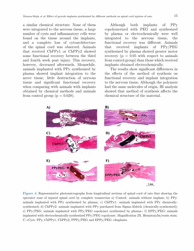
Four weeks after SCI, animals were sacrificed to analyze the integration of implants to the spinal cord
tissue and the inflammatory response. The control group (Fig. 4A) showed greater tissue destruction
compared with implanted animals. Analysis of implants integration to the spinal cord tissue showed that
PPy implants synthesized by plasma polymerization were well integrated to the tissue and that the
surrounding tissue showed little destruction (Fig. 4B). The chemically-synthesized implants ChPPy1 and
Figure 3. The locomotion recovery measured by BBB open field score after traumatic spinal cord injurywas evaluated along the time. Control: animals without implant; PPy: animals implanted with polypirrole(PPy) synthesized by plasma; ChPPy1: animals implanted with PPy chemically-synthesized; ChPPy2: animalsimplanted with PPy purchased from Sigma-Aldrich (chemically-synthesized); PPy/PEG: animals implanted withPPy copolymerized with polyethylene glycol (PPy/PEG) and synthesized by plasma; EPPy/PEG: animalsimplanted with electrochemically-synthesized PPy/PEG co-polymer. Results are expressed as means ±SE.ANOVA of repeated measures followed by Dunnett’s test. *Control group different from PPy and PPy/PEG(p < 0.028, and 0.05, respectively).
14 Revista Mexicana de Ingeniería Biomédica · volumen 36 · número 1 · Ene-Abr, 2015
b) PPy (3 animals alive): BBB=5.33,meaning that animals were able to movethe 3 joints of the hind limb (hip,knee and ankle), and in two them, themovements were extensive.
c) ChPPy1 (3 animals alive and 2dead animals, non-evaluable): BBB=1,meaning that the animals had slightmovement of 1 or 2 joints. Although oneof the animals had an improvement of4 points in the BBB scale in the thirdweek, score decreased with time.
d) ChPPy2 (3 animals alive and 4 deadanimals, non-evaluable): BBB=1.33,meaning that the animal had slightmovement in one or two joints. But justas in the ChPPy1 group, one animalpresented improvement until the fourthweek and subsequently decreased theeffect of the implant.
e) PPy-PEG (3 animals alive): BBB=3.66,meaning that animals had extensivemovement in 1 or 2 joints of the hindlimb and slight movement of other joint(hip, knee and ankle).
f) EPPy-PEG (3 animals alive):BBB=2.6, meaning that animals hadextensive movement of 1 joint and slightmovement of 2 joints of the hind limbs.
The dead animals did not showinfection of the urinary or respiratory tract,hypertrophy of urethral meatus, or shallowor deep wounds. Thus, the cause of death isunknown.
Animals implanted with materialssynthesized by plasma demonstrated greatermotor function recovery comparing withanimals implanted with materials obtainedby chemical or electrochemical synthesisand with control group animals. Animalswith implants synthesized by plasma showedsignificant differences with animals from thecontrol group; PPy (p = 0.028) and PPy /PEG (p = 0.05).
Histological analysis
Four weeks after SCI, animals were sacrificedto analyze the integration of implants tothe spinal cord tissue and the inflammatoryresponse. The control group (Fig. 4A) showedgreater tissue destruction compared withimplanted animals. Analysis of implantsintegration to the spinal cord tissue showedthat PPy implants synthesized by plasmapolymerization were well integrated to thetissue and that the surrounding tissue showedlittle destruction (Fig. 4B). The chemically-synthesized implants ChPPy1 and ChPPy2(Fig. 4C and 4D respectively) had similaramount of tissue destruction and completelack of implant integration, with a great cystat the injury epicenter and a complete loss ofhistological architecture.
Regarding the inflammatory response,morphometric analysis showed the presenceof 142 polymorphonuclear cells per every4 microns in the animals with ChPPy1implant and 128 polymorphonuclear cellsin the animals with ChPPy2 implant(obtained from Sigma-Aldrich). In contrast,the morphometric analysis of samples withimplants synthesized by plasma for bothPPy and PPy/PEG showed about 74inflammatory cells at the tissue surroundingthe implant, and if PPy was doped withiodine (PPy/I) 95 inflammatory cells werefound in the tissue that surrounded theimplant.
DISCUSSION AND CONCLUSION
The present work compared the effect ofdifferent implants derived from pyrrole(PPy and PPy/PEG) and obtained bystandard chemical, electrochemical, andplasma polymerizations. The implantswere analyzed according to their chemicalstructure, integration with the nervous tissue,and their effect on functional recovery in ratswith complete section of the spinal cord.
The PPy implants that were chemicallysynthesized as ChPPy1 and ChPPy2 have
Álvarez-Mejía et al. Effect of pyrrole implants synthesized by different methods on spinal cord injuries of rats. 15
a similar chemical structure. None of themwere integrated to the nervous tissue, a largenumber of cysts and inflammatory cells werefound on the tissue around the implants,and a complete loss of cytoarchitectureof the spinal cord was observed. Animalsthat received ChPPy1 or ChPPy2 showedsome functional recovery between the thirdand fourth week post injury. This recovery,however, decreased afterwards. Meanwhile,animals implanted with PPy synthesized byplasma showed implant integration to thenerve tissue, little destruction of nervoustissue and significant functional recoverywhen comparing with animals with implantsobtained by chemical methods and animalsfrom control group (p = 0.028).
Although both implants of PPycopolymerized with PEG and synthesizedby plasma or electrochemically were wellintegrated to the nervous tissue, thefunctional recovery was different. Animalsthat received implants of PPy/PEGsynthesized by plasma showed greater motorrecovery (p = 0.05 with respect to animalsfrom control group) than those which receivedimplants obtained electrochemically.
The results show significant differences inthe effects of the method of synthesis onfunctional recovery and implant integrationto the nervous tissue. Although the polymershad the same molecules of origin, IR analysisshowed that method of synthesis affects thechemical structure of the material.
Figure 4. Representative photomicrographs from longitudinal sections of spinal cord of rats that showing theepicenter zone of injured spinal cord by complete transection a) Control: animals without implant; b) PPy:animals implanted with PPy synthesized by plasma; c) ChPPy1: animals implanted with PPy chemically-synthesized; d) ChPPy2: animals implanted with PPy purchased from Sigma-Aldrich (chemically-synthesized);e) PPy/PEG: animals implanted with PPy/PEG copolymer synthesized by plasma-; f) EPPy/PEG: animalsimplanted with electrochemically-synthesized PPy/PEG copolymer. Magnification 2X. Hematoxylin/eosin stain.C→Cyst, PPy, ChPPy1, ChPPy2, PPPy/PEG and EPPy/PEG→Implants.
16 Revista Mexicana de Ingeniería Biomédica · volumen 36 · número 1 · Ene-Abr, 2015
Wangh et al., showed that the chemicalstructure of PPy and of polythiophenesynthesized by plasma are different fromthe ones chemically synthesized because thematerials obtained by plasma are highlycrosslinked and branched [45] and they canform three-dimensional network which cansupport and promote neural cells grown whilethe presence of heteroaromatic amine groupsand nitrile groups can favor neuroprotectionafter an injury in the central nervous system.Furthermore, the polymers synthesized byplasma are insoluble, thermally stable andchemically inert [46], characteristics thatmake them more desirable for implants in abiological system.
Pyrrole belongs to a class of heterocycliccompounds that are in various naturalcomponents such as the heme group,chlorophyll and vitamin B12 [47] and havebeen used in various applications of chemicalmedicine as anti-inflammatory, antibacterialand antihypertensive agents, as well as agentsfor tyrosine kinase inhibition[48].
Currently, PPy is mainly synthesizedby chemical oxidation or electrochemicalpolymerization [16]. Nevertheless, forbiomedical applications, plasma synthesismight be a better option because theunique characteristics of PPy generatedby plasma that allow improvement of thecellular microenvironment and favor theattachment and growth of cells. The lowrate of degradation of the material maintainsthe cell adhesion, promotes morphologicalmaturation and allows preservation of itsproperties [34]. It has been shown incultures of nerve cells on surfaces treatedwith PPy, that the adhesion, proliferation,attachment, viability and number of synapsesis increased compared with that observed incultures performed on surfaces treated withcompounds as Poly DL-Ornithine / Laminin[34]. Furthermore, our research group hasdemonstrated neuroprotective activity ofPPy synthesized by plasma, as well as greaterfunctional recovery in animals that received
this type of implant after a TSCI, comparedwith those who did not receive it [36-38].
The above may be due to the chemicalstructure of PPy obtained by plasmapolymerization. IR spectroscopy of thismaterial shows the presence of a variety ofchemical groups including primary amines,nitriles and aliphatic sections [34], and it hasbeen shown that the structures consistingmainly of methyl-, hydroxyl-, amino- andcarboxyl- functional groups, which are foundin natural biological surfaces, favor thegrowth of cells [49].
The adaptability of tissues to materialssuch as the implants studied in this workbegins with the absorption of solutions atthe surface of the material. The hydrophilicproperties of PPy, which can be increasedby increasing the ionization capability of thematerial, allow one to store solutions andfavor interaction with cells [50-51], whichgenerates optimal sites for cell attachment.
Nerve cells carry out their function bygenerating electrical activity. The nervoussystem thus responds to electric fieldsand the key component of the neuralcommunication is the action potentialgenerated in the synapse. This implies thatthe ideal biomaterial to implant in thissystem must introduce electrical stimulatorsto promote neuronal growth and nerveregeneration [13]. The material must promoteregeneration of the nervous tissue at theinterface by attraction or rejection of ionsand polar groups between the cells and thematerial. In addition to this, it has beenshown that electrical stimulation alters theabsorption of proteins and the interactionwith the nerve cells [52], which couldalso favor nerve regeneration processes.Although the breaking of rings in theplasma polymerization process results ina polymer complex of low conductivity,when is introduced into a biological systemits sensitivity to humidity increases itsconductivity [40]. It is known that theelectric conductivity of conductive polymers
Álvarez-Mejía et al. Effect of pyrrole implants synthesized by different methods on spinal cord injuries of rats. 17
synthesized by chemical or electrochemicalmethods oscillates within a range of 10−10 to10−5 S cm−1 [39]. The electric conductivityof the plasma-synthesized PPy measured at30 % relative humidity is around 10−12 Scm−1, while at 90 % of relative humidity it is10−9 S cm−1 [40]. The PPy/PEG copolymerhas a conductivity of 10−12 S cm−1 at 30% of relative humidity [36] and of 10−9 to10−8 S cm−1 when it is dampened with ionicsolutions [51].
Due to their physical and chemicalproperties, the PPy does not alter thebiological functions of the cell cultures andprovides better cell attachment and anincreased rate of proliferation, which may bedue to the accumulation of amino groups(-NH2) and interaction with other groupsgenerated during the process of plasmapolymerization [34], which may explain thebetter results obtained when using PPysynthesized by plasma vs the PPy synthesizedby conventional chemical or electrochemicalmethods.
ACKNOWLEDGMENTS
The work was supported by CONACyT grantNo. 155239 and by IMSS grant No. FOFOI2005/1/I/149. Ana Laura Álvarez receiveda scholarship from CONACyT, No. 172211.The authors want to thank to María delCarmen Baltazar for her technical assistance.
REFERENCES
1. C.A. Oyinbo, “Secondary injurymechanisms in traumatic spinal cordinjury: a nugget of this multiplycascade,” Acta Neurobiol Exp, vol. 71,pp. 281-299, 2011.
2. W. Tetzlaff, E.B. Okon, S. Karimi-Abdolrezaee, C.E. Hill, J.S. Sparling,J.R. Plemel, W.T. Plunet, E.C.Tsai, D. Baptiste, L.J. Smithson,M.D. Kawaja, M.G. Fehlings, B.K.Kwon “A systematic review of cellular
transplantation therapies for spinal cordinjury,” J Neurotrauma., vol. 28, no. 8,pp. 1611-1682, 2011.
3. S. Venkatachalam, “Fetal neuraltissue transplantation for spinal cordinjury repair,” Chapter 23 in N.Bhattacharya, P. Stubblefield (eds.),Human Fetal Tissue Transplantation.Springer London, pp. 297-305, 2013.
4. H. Salgado-Ceballos, I. Grijalva,G. Guizar-Sahagun, A. L. Espitia,A. Martínez, A. Feria-Velasco,“Predegenerated peripheral nerve graftwith and without methylprednisoloneadministration after traumatic spinalcord injury in adult rats,” Neurosci.Res. Comm., vol. 33, no. 2, pp. 77-85,2003.
5. M.P. Côté, A.A. Amin, V.J. Tom, J.D.Houle “Peripheral nerve grafts supportregeneration after spinal cord injury,”Neurotherapeutics, vol. 8, no. 2, pp.294-303, 2011.
6. H. Kanno, Y. Pressman, A. Moody,R. Berg, E.M. Muir, J.H. Rogers,H. Ozawa, E. Itoi, D.D. Pearse,M.B. Bunge, “Combination ofengineered Schwann cell grafts tosecrete neurotrophin and chondroitinasepromotes axonal regeneration andlocomotion after spinal cord injury,” J.Neurosci., vol. 34, no. 5, pp. 1838-1855, 2014.
7. Y.J. Rao, W.X. Zhu, Z.Q. Du, C.X.Jia, T.X. Du, Q.A. Zhao, X.Y. Cao,Y.J. Wang “Effectiveness of olfactoryensheathing cell transplantation fortreatment of spinal cord injury,” Genet.Mol. Res., vol. 13, no. 2, pp. 4124-4129, 2014.
8. Z.A, Sobani, S.A. Quadri, S.A. Enam,“Stem cells for spinal cord regeneration:Current status,” Surg Neurol Int., vol.1, pp. 93, 2010.
18 Revista Mexicana de Ingeniería Biomédica · volumen 36 · número 1 · Ene-Abr, 2015
9. H. Nakajima, K. Uchida, RodriguezA. Guerrero, S. Watanabe, D. Sugita,N. Takeura, A. Yoshida, G. Long,K. Wright, E. Johnson, H. Baba,“Transplantation of mesenchymal stemcells Promotes the alternative pathwayof macrophage activation and functionalrecovery after spinal cord injury,” J.Neurotrauma., vol. 29, no. 8, pp. 1614-1625, 2012.
10. J. Ai, A. Kiasat-Dolatabadi, S.Ebrahimi-Barough, A. Ai, N.Lotfibakhshaiesh, A. Norouzi-Javidan,H. Saberi, B. Arjmand, HR. Aghayan,“Polymeric scaffolds in neural tissueengineering: A review,” Arch. NeuroSci., vol. 1, no. 1, pp. 15-20, 2013.
11. V. Krishna, S. Konakondla, J. Nicholas,A. Varma, M. Kindy, X. Wen,“Biomaterial-based interventions forneuronal regeneration and functionalrecovery in rodent model of spinal cordinjury: A systematic review,” J SpinalCord Med., vol. 36, no. 3, pp. 174-190,2013.
12. M. Wang, P. Zhai, X. Chen, D.J.Schreyer, X. Sun, F. Cui “Bioengineeredscaffolds for spinal cord repair,” TissueEng. Part B Rev., vol. 17, no. 3, pp.177-194, 2011.
13. L. Ghasemi-Mobarakeh, M.P.Prabhakaran, M. Morshed, M.H. Nasr-Esfahani, H. Baharvand, S. Kiani, S.S.Al-Deyab, S. Ramakrishna “Applicationof conductive polymers, scaffolds andelectrical stimulation for nerve tissueengineering,” J. Tissue Eng Regen Med,vol. 5, no. 4, 17-35, 2011.
14. I.S. Chronakis, S. Grapenson, A. Jakob,“Conductive polypyrrole nanofibersvia electrospinning: electrical andmorphological properties,” Polymer,vol. 47, no. 5, pp. 1597-1603, 2006.
15. Q. Zhang, Y. Yan, S. Li, et al.,“The synthesis and characterizationof a novel biodegradable andelectroactive polyphosphazene for nerveregeneration,” Mater. Sci. Eng. C, vol.30, no. 1, pp. 160-166, 2010.
16. M.A. Chougule, S.G. Pawar, P.R.Godse, R.N. Mulik, S. Sen, V.B.Patil, “Synthesis and characterizationof polypyrrole (PPy) thin films,” SoftNanoscience Letters, vol. 1, no. 1, pp.6-10, 2011.
17. J.C. Vidal, E. Garcia, J.R. Castillo“In situ preparation of a cholesterolbiosensor: entrapment of cholesteroloxidase in an overoxidized polypyrrolefilm electrodeposited in a flow system:determination of total cholesterol inserum,” Analytica Chimica Acta, vol.385, no. 1-3, pp. 213-222, 1999.
18. E. Lopez-Crapez, T. Livache, J.Marchand, J. Grenier “K-ras mutationdetection by hybridization to apolypyrrole DNA chip,” Clin Chem.,vol. 47, no. 2, pp. 186-194, 2001.
19. T.E. Campbell, A.J. Hodgson, G.G.Wallace “Incorporation of eythrocytesinto polypyrrole to form the basis of abiosensor to screen for rhesus (D) bloodgroups and rhesus (D) antibodies,”Electroanalysis, vol. 11, no. 4, pp. 215-222, 1999.
20. R.H.M. van de Leur, A. van derWaal, “Gas and vapour detection usingpolypyrrole,” Synthetic Metals, vol.102, no. 1-3, pp. 1330-1331, 1999.
21. A.B. Sanghvi, K.P. Miller, A.M.Belcher, C.E. Schmidt, “Biomaterialsfictionalization using a novel peptidethat selectively binds to a conductingpolymer,” Nat Mater, vol. 4, no. 6, pp.496-502, 2005.
22. J.R. Reynolds, H. Ly, F. Selampinar,P.J. Kinlen, “Controlled drug and
Álvarez-Mejía et al. Effect of pyrrole implants synthesized by different methods on spinal cord injuries of rats. 19
biomolecule release from electroactivehost polymer systems,” PolymerPreprints, vol. 40, no. 1, pp. 307,1999.
23. T.F. Otero, M.T. Cortés, “Artificialmuscles with tactile sensitivity,” Adv.Mater., vol. 15, no. 3, 279-289, 2003.
24. X. Wang, X. Gu, C. Yuan, S. Chen, P.Zhang, T. Zhang, J. Yao, F. Chen, G.Chen, “Evaluation of biocompatibilityof polypyrrole in vitro and in vivo,” JBiomed Mater Res A, vol. 68A, no. 3,pp. 411-422, 2004.
25. R.L. Williams, P.J. Doherty, “Apreliminary assessment of poly(pyrrole)in nerve guide studies,” J. Mater Sci-Mater M., vol. 5, no. 6-7, pp. 429-433,1994.
26. H. Castano, E.A. O’Rear, P.S.McFetridge, V.I. Sikavitsas “Polypyrrolethin films formed by admicellarpolymerization support the osteogenicdifferentiation of mesenchymal stemcells,” Macromol Biosci, vol. 4, no. 8,pp. 785-794, 2004.
27. H.K. Song, B. Toste, K. Ahmann,D. Hoffman-Kim, G.T. Palmore,“Micropatterns of positive guidancecues anchored to polypyrrole dopedwithpolyglutamic acid: a new platformfor characterzing neurite extension incomplex environments,” Biomaterials,vol. 27, no. 3, pp. 473-484, 2006.
28. D.D. Ateh, P. Vadgama, H.A.Navasaria, “Polypyrrole-based conductingpolymers and interactions withbiological tissues,” J. R. Soc. Interface,vol. 3, no. 11, pp. 741-752, 2006.
29. N. Gomez, C.E. Schmidt, “Nervegrowth factor-immobilized polypyrrole:bioactive electrically conductingpolymer for enhanced neuriteextension,” J. Biomed. Mater. Res,vol. 81, no. 1, pp. 135-149. 2007.
30. J.Y. Lee, C.A. Bashur, A.S. Goldstein,C.E. Schmidt, “Polypyrrole-coatedelectrospun PLGA nanofibers for neuraltissue applications,” Biomaterials, vol.30, no. 26, pp. 4325-4334, 2009.
31. D. Kai, M.P. Prabhakaran, G.Jin, S. Ramakrishna, “Polypyrrole-contained electrospun conductivenanofibrous membranes for cardiactissue engineering,” J. Biomed. Mater.Res. A, vol. 99, no. 3, pp. 376-385,2011.
32. A.D. Bendrea, L. Cianga, I. Cianga,“Review paper: progress in the fieldof conducting polymers for tissueengineering applications,” J. Biomater.Appl., vol. 26, no. 1, pp. 3-84, 2011.
33. Z. Zhang, M. Rouabhia, Z. Wang,C. Roberge, G. Shi, P. Roche, J.Li, L.H. Dao, “Electrically conductivebiodegradable polymer compositefor nerve regeneration: electricity-stimulated neurite outgrowth and axonregeneration,” Artif Organs, vol. 31, no.1, pp. 13-22, 2007.
34. E. Zuñiga-Aguilar, R. Olayo, O.Ramírez-Fernández, J. Morales, R.Godínez, “Nerve cells culture fromlumbar spinal cord on surfaces modifiedby plasma pyrrole polymerization,” JBiomater Sci Polym Ed., vol. 25, no.7, pp. 729-747, 2014.
35. X. Wang, X. Gu, C. Yuan, S. Chen, P.Zhang, T. Zhang, J. Yao, F. Chen, G.Chen “Evaluation of biocompatibilityof polypyrrole in vitro and in vivo,” JBiomed Mater Res A, vol. 68, no. 3,pp. 411-422, 2004.
36. R. Olayo, C. Ríos, H. Salgado-Ceballos,G. Cruz, J. Morales, G. Olayo, L.Alvarez, R. Mondragón, A. Morales-Guadarrama, G. Guizar-Sahagun, A.Diaz-Ruiz, “Tissue spinal cord responsein rats after implants of polypyrrole and
20 Revista Mexicana de Ingeniería Biomédica · volumen 36 · número 1 · Ene-Abr, 2015
polyethylene glicol obtained by plasma,”J Mater Sci:Mater Med, vol. 19, no. 2,pp. 817-826, 2008.
37. G.J. Cruz, R. Mondragón-Lozano, A.Diaz-Ruiz, J. Manjarrez, R. Olayo,H. Salgado-Ceballos, M.G. Olayo, J.Morales, L. Alvarez-Mejía, A. Morales,M. Méndez-Armenta, N. Plascencia, M.Fernandez, C. Ríos “Plasma polypyrroleimplants recover motor function in ratsafter spinal cord transection,” J MaterSci:Mater Med, vol. 23, no. 10, pp.2583-2592, 2012.
38. A. Morales-Guadarrama, H. Salgado-Ceballos, J. Morales, C. Ríos, G.J.Cruz, A. Diaz-Ruiz, M.G. Olayo, L.Alvarez-Mejia, R. Mondragón-Lozano,R. Olayo, “CAT and MRI studies ofspinal cord injured rats implanted withPPy/I,” Revista Mexicana en IngenieríaBiomédica, vol. 34, no. 2, pp. 145-155,2013.
39. T.V. Vernitskaya, O.N. Efimov,“Polypyrrole: a conducting polymer; itssynthesis, properties and applications,”Russian Chemical Reviews, vol. 66, no.5, pp. 443-457, 1997.
40. G.J. Cruz, J. Morales, R. Olayo, “Filmsobtained by plasma polymerization ofpyrrole,” Thin solid films, vol. 342, no.1-2, pp. 119-126, 1999.
41. J. Morales, E. Pérez-Tejada, C.R.Montiel, V.H. Torres, R. Olayo,“Modificación superficial por plasmaaplicada a biomateriales [Surfacemodification by plasma applied tobiomaterials,” In: La Física Biológicaen México: Temas Selectos 2 [BiologicalPhysics in Mexico:Selected Items 2].México D. F.: Colegio Nacional, pp.241-257, 2008.
42. J. Luo, R. Borgens, R. Shi,“Polyethylene glycol immediatelyrepairs neuronal membranes and
inhibits free radical production afteracute spinal cord injury,” J Neurochem,vol. 83, no. 2, pp. 471-480, 2002.
43. R. Shi “Polyethylene glycol repairsmembrane damage and enhancesfunctional recovery: a tissue engineeringapproach to spinal cord injury,”Neurosci Bull, vol. 29, no. 4, 460-466,2013.
44. D.M. Basso, M.S. Beattie, J.C.Bresnahan “A sensitive and reliablelocomotor rating scale for open fieldtesting in rats,” J. Neurotrauma, vol.12, pp. 1-21, 1995.
45. J. Wang, K.G. Neoh, E.T. Kang,“Comparative study of chemicallysynthesized and plasma polymerizedpyrrole and thiophene thin films,” ThinSolid Films, vol. 446, no. 2, pp. 205-217, 2004.
46. M.M. Kamal, A.H. Bhuiyan,“Structural and optical characterizationof plasma polymerized pyrrolemonolayer thin films,” Advances inOptoelectronic Materials (AOM), vol.1, no. 2, pp. 11-17, 2013.
47. C.Y. De Leon, B. Garem, “A NewApproach to porphobilinogen and itsanalogs,” Tetrahedron, vol. 23, no. 9,pp. 7731-7752, 1997.
48. A.M. Manning, D.J. Davis, “TargetingJNK for therapeutic benefit: from junkto gold?,” Nat Rev Drug Discov., vol. 2,no. 7, pp. 554-565, 2003.
49. P. Roach, D. Eglin, K. Rohde, CC.Perry, “Modern biomaterials: a review- bulk properties and implications ofsurface modifications,” J Mater SciMater Med., vol. 18, no. 7, pp. 1263-1277, 2007.
50. L.M. Gómez, M.G. Olayo, G.J.Cruz, M. González-Torres, O.G.López, C. De Jesús, “Interaction of
Álvarez-Mejía et al. Effect of pyrrole implants synthesized by different methods on spinal cord injuries of rats. 21
plasma polypyrrole particles with ionicsolutions,” Macromolecular Symposia,vol. 325-326, no. 1, pp. 112-119, 2013.
51. E. Colín, M.G. Olayo, G.J. Cruz,L. Carapia, J. Morales, R. Olayo,“Affinity of amine-functionalizedplasma polymers with ionic solutionssimilar to those in the human body,”
Progress in Organic Coatings, pp.64322-326, 2009.
52. A. Kotwal, C.E. Schmidt, “Electricalstimulation alters protein adsorptionand nerve cell interactions withelectrically conducting biomaterials,”Biomaterials vol. 22, no. 10, pp. 1055-1064, 2001.
ib