edoc.unibas.ch Seidl_The... · 2020. 5. 6. · Created Date: 3/13/2020 7:22:00 PM
Novel Therapeutic Approaches for
Neuromuscular Diseases
Inauguraldissertation
zur
Erlangung der Würde eines Doktors der Philosophie
vorgelegt der
Philosophisch-Naturwissenschaftlichen Fakultät
der Universität Basel
von
Ruben Herrendorff
aus Wimmis (BE)
Basel, 2015
Originaldokument gespeichert auf dem Dokumenteserver der Universität Basel
edoc.unibas.ch
Genehmigt von der Philosophisch-Naturwissenschaftlichen Fakultät
auf Antrag von
Prof. Dr. Michael Sinnreich
Prof. Dr. Markus Rüegg
Prof. Dr. Beat Ernst
Basel, den 11. November 2014
Prof. Dr. Jörg Schibler
Dekan
Acknowledgements I
Acknowledgements
“It is good to have an end to journey toward; but it is the journey that matters, in the end.”
(Ernest Hemingway)
On the PhD journey, which I have been pursuing during the last four years, many lovely
people accompanied me and I would like to express my sincerest thanks to some of them on
this occasion.
I would like to especially thank Prof. Michael Sinnreich for providing me with a very interest-
ing drug discovery project for DM1, for his support, guidance and motivation. I would also
like to thank Prof. Beat Ernst and Prof. Andreas J. Steck for initiating the anti-MAG neuropa-
thy project and for having supported me as great mentors since my Master’s. I admire the
fascination for science and medicine of all three of you. Many thanks also go to Prof. Markus
Rüegg for valuable feedback and for taking time to look at my thesis.
I would like to particularly thank all of the members of the Neuromuscular Research group,
where is spent most of my PhD time: Dr. Jon Ashley, Dr. Bilal Azakir, Marielle Brockhoff,
Dr. Perrine Castets, Dr. Sabrina Di Fulvio, Beat Erne, Frances Kern, Dr. Jochen Kinter, Dr.
San Pun, Adeline Stiefvater and Dr. Tatiana Wiktorowicz. Thank you Jochen for supervising
my project, for many interesting discussions, and for sharing many valuable insights. Frances,
thank you so much for the warm welcome to the Sinnreich group. In addition, I would like to
thank all members of the Institute of Molecular Pharmacy, where I started the PhD. You all
ensured a stimulating and interesting working environment, many laughs, and a good time.
I would like to thank all our collaborators for making the DM1 project possible: Maria Teresa
Faleschini, Prof. Matthias Hamburger, Dr. Olivier Potterat; Prof. David Boykin, Prof. Reto
Brun, Dr. Tanja Wenzler; Jacqueline Bezençon and many others who helped and assisted me.
I am incredibly thankful to my friends and family for all their love and support. You’re great!
Unending thanks go to my dear parents, my sister and her fiancé, my parents-in-law, my
godfather, my good old buddies, and also to my lovely wife. You’re awesome!
Last but not least I would like to thank the Almighty for his protection, love and mercy. I
would like to express my deepest gratitude to you for my life, which is nothing but a gift, with
all its opportunities, e.g. the privilege to work in research. May I never forget you and may I
never take all this for granted.
Summary II
Summary
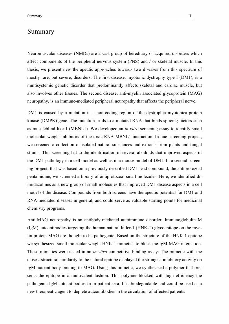
Neuromuscular diseases (NMDs) are a vast group of hereditary or acquired disorders which
affect components of the peripheral nervous system (PNS) and / or skeletal muscle. In this
thesis, we present new therapeutic approaches towards two diseases from this spectrum of
mostly rare, but severe, disorders. The first disease, myotonic dystrophy type I (DM1), is a
multisystemic genetic disorder that predominantly affects skeletal and cardiac muscle, but
also involves other tissues. The second disease, anti-myelin associated glycoprotein (MAG)
neuropathy, is an immune-mediated peripheral neuropathy that affects the peripheral nerve.
DM1 is caused by a mutation in a non-coding region of the dystrophia myotonica-protein
kinase (DMPK) gene. The mutation leads to a mutated RNA that binds splicing factors such
as muscleblind-like 1 (MBNL1). We developed an in vitro screening assay to identify small
molecular weight inhibitors of the toxic RNA-MBNL1 interaction. In one screening project,
we screened a collection of isolated natural substances and extracts from plants and fungal
strains. This screening led to the identification of several alkaloids that improved aspects of
the DM1 pathology in a cell model as well as in a mouse model of DM1. In a second screen-
ing project, that was based on a previously described DM1 lead compound, the antiprotozoal
pentamidine, we screened a library of antiprotozoal small molecules. Here, we identified di-
imidazolines as a new group of small molecules that improved DM1 disease aspects in a cell
model of the disease. Compounds from both screens have therapeutic potential for DM1 and
RNA-mediated diseases in general, and could serve as valuable starting points for medicinal
chemistry programs.
Anti-MAG neuropathy is an antibody-mediated autoimmune disorder. Immunoglobulin M
(IgM) autoantibodies targeting the human natural killer-1 (HNK-1) glycoepitope on the mye-
lin protein MAG are thought to be pathogenic. Based on the structure of the HNK-1 epitope
we synthesized small molecular weight HNK-1 mimetics to block the IgM-MAG interaction.
These mimetics were tested in an in vitro competitive binding assay. The mimetic with the
closest structural similarity to the natural epitope displayed the strongest inhibitory activity on
IgM autoantibody binding to MAG. Using this mimetic, we synthesized a polymer that pre-
sents the epitope in a multivalent fashion. This polymer blocked with high efficiency the
pathogenic IgM autoantibodies from patient sera. It is biodegradable and could be used as a
new therapeutic agent to deplete autoantibodies in the circulation of affected patients.
Table of Contents III
Table of Contents
Acknowledgements ............................................................................................................ I
Summary .......................................................................................................................... II
List of Abbreviations ....................................................................................................... IV
List of Schemes ............................................................................................................... IX
List of Tables .................................................................................................................... X
List of Figures ................................................................................................................. XI
Preface ........................................................................................................................... XII
1 Introduction to Neuromuscular Diseases .................................................................... 1
2 Myotonic Dystrophy Type I ........................................................................................ 2 2.1 Introduction ..................................................................................................... 2 2.2 Molecular Disease Mechanism ....................................................................... 2 2.3 Current Treatments ......................................................................................... 6 2.4 New Therapeutic Approaches ......................................................................... 6
2.4.1 Antisense oligonucleotides ............................................................ 6 2.4.2 Enzymatic degradation of DMPK transcripts ................................ 7 2.4.3 Gene therapy .................................................................................. 8 2.4.4 Small molecules ............................................................................. 9 2.4.5 Aim of our study ............................................................................ 9
2.5 Manuscript I .................................................................................................. 10 2.6 Manuscript II ................................................................................................. 39 2.7 Discussion ..................................................................................................... 64 2.8 Conclusion and Outlook ............................................................................... 69
3 Anti-Myelin Associated Glycoprotein Neuropathy ................................................. 73 3.1 Introduction ................................................................................................... 73 3.2 Molecular Disease Mechanism ..................................................................... 75 3.3 Current Therapeutic Approaches .................................................................. 80
3.3.1 Aim of our study .......................................................................... 82 3.4 Manuscript III ............................................................................................... 82 3.5 Discussion ................................................................................................... 116 3.6 Outlook ....................................................................................................... 119
4 Closing Words ......................................................................................................... 120
References ..................................................................................................................... 121
List of Abbreviations IV
List of Abbreviations
AAE Adeno-associated virus
AICAR 5-Aminoimidazole-4-carboxamide ribonucleotide
AIDP Acute inflammatory demyelinating polyneuropahty
AMPK Adenosine monophosphate-activated protein kinase
AON Antisense oligonucleotide
ASRE Artificial site-specific RNA endonuclease
ATE1 Arginyltransferase 1
ATP Adenosine triphosphate
AUC Area under the curve
BCA assay Bicinchoninic acid assay
BSA Bovine serum albumin
Cavg Average (plasma) concentration
CD20 B-lymphocyte antigen CD20 (MS4A2: membrane-spanning 4-
domains, subfamily A, member 1)
cDNA Complementary DNA
CELF1 (=CUGBP1) CUGBP, elav-like family member 1
CIDP Chronic inflammatory demyelinating polyneuropathy
CIDP Chronic inflammatory demyelinating polyneuropathy
CLCN1 / CLC1 Chloride channel, voltage-sensitive 1
Chloride channel 1, skeletal muscle
Cmax Maximum (plasma) concentration
CNS Central nervous system
COSY (NMR) Correlated spectroscopy (NMR)
CRDs Carbohydrate recognizing domains
CUGBP1 CUG triplet repeat, RNA binding protein 1
Cy3 Cyanine 3
DAPI 4,6 diamino-2-phenylindole dihydrochloride
DBU Diazabicycloundecen
DCM Dichloromethane
DEPC Diethylpyrocarbonate (diethyl dicarbonate)
DHB Dihydroberberine

List of Abbreviations V
DM1 Myotonic dystrophy type 1
DM2 Myotonic dystrophy type 2
DMAP 4-dimethylamino-pyridine
DMEM Dulbecco's Modified Eagle's Medium
DMF N,N-dimethylformamide
DMPK Dystrophia myotonica-protein kinase
DMSO Dimethyl sulfoxide
DNA Desoxyribonucleic acid
DNAse Desoxyribonuclease
DTT Dithiotreitol
ECL Electrochemiluminescence
EDA Ethylenediamine
ELISA Enzyme linked immunosorbent assay
EMSA Electrophoretic mobility gel shift assay
ESI-MS Electron spray ionization MS
ETR3 Elav-type RNA-binding protein 3
FBS Fetal bovine serum
FHL1 Four and a half LIM domains 1
FISH Fluorescence in situ hybridization
FT-IR Fourier transform IR
Gal Galactose
GBS Guillain-Barré syndrome
GlcA Glucuronic acid
GlcNAc N-acetylglucosamine
GM1, GM1b, GD1a,
GD1b, GT1a, GQ1b, etc.
(general: GXnx)
G = ganglioside, X = number of sialic acids, n = characterizes
the carbohydrate chain length and the sequence of migration on
TLC, x = indicates the type of carbohydrate linkage
GST Glutathione S-transferase
HEPES 4-(2-hydroxyethyl)-1-piperazineethanesulfonic acid
HIS Polyhistidin (hexahistidine, His6)
HIV Human immunodeficiency virus
HNK-1 Human natural killer-1
HPLC High performance liquid chromatography
HRMS High resolution MS
List of Abbreviations VI
HRP Horseradish peroxidase
HSA (LR / SR) Human skeletal actin (long repeat / short repeat)
HSQC (NMR) Heteronuclear single quantum coherence (NMR)
IC50 / Tox IC50 Half maximal inhibitory concentration / toxicity IC50
ICNMD International congress on neuromuscular diseases
IgG Immunoglobulin G
IgM Immunoglobulin M
INCAT Inflammatory neuropathy cause and treatment
INSR Insulin receptor
IR Infrared (spectroscopy)
IVIg Intravenous immunoglobulin
KD Dissociation constant
L-MAG Long MAG
LC-MS Liquid chromatography-mass spectrometry
LD50 Median lethal dose
LNA Locked nucleic acid
log D7.4 Distribution co-efficient at pH 7.4
MAG Myelin associated glycoprotein
MAO Monoamino oxidase
MBNL1 Muscleblind-like 1
Me Methyl
MeCN Acetonitrile
MGUS IgM monoclonal gammopathy of unknown significance
minHNK-1 epitope Minimal HNK-1 epitope
MMN Multifocal motor neuropathy
MOE 2’-O-Methoxyethyl
mRNA Messenger RNA
MS Mass spectrometry
MYOD (=MYOD1) Myoblast determination protein 1 or myogenic differentiation 1
NMD Neuromuscular disease
NMR Nuclear magnetic resonance (spectroscopy)
NP-40 Tergitol-type NP-40, nonyl phenoxypolyethoxylethanol
Nucleobases A, C, T, G, U Adenine, cytosine, thymine, guanine, uracil
OD Optical density

List of Abbreviations VII
p-MeOPh p-methoxyphenyl
P0 Myelin protein zero
PAMPA Parallel artificial membrane permeability assay
PBS Phosphate buffered saline
PCR Polymerase chain reaction
Pe Effective permeation
PE Plasma exchange
pKa Negative decadic logarithm of the acid dissociation constant
PKC Protein kinase C
PL(minHNK-1)x Polylysine loaded with x% minimal HNK-1 epitope
PMO Morpholino AON
PMP22 Peripheral myelin protein 22
PNS Peripheral nervous system
PPB Plasma protein binding
PPMO Peptide-linked morpholino AON
PROMM Proximal myotonic myopathy
PS Phosphorothioate
Py/pyr Pyridine
qPCR Quantitative PCR
RIPA Radio-immunoprecipitation assay
RNA Ribonucleic acid
RNAi RNA interference
RNase H Ribonuclease H
rt Room temperature
RT-PCR Reverse transcription PCR
RT-qPCR Reverse transcription quantitative PCR
S1 Screening study 1 (natural compounds)
S2 Screening study 2 (synthetic compounds)
SDS Sodium dodecyl sulfate
SDS-PAGE Sodium dodecyl sulfate polyacrylamide gel electrophoresis
SERCA1 Sarcoplasmic/endoplasmic reticulum Ca2+ ATPase 1
SGLPG Sulfoglucuronyl lactosaminyl paragloboside
SGPG Sulfoglucuronyl paragloboside
shRNA Small hairpin RNA or short hairpin RNA
List of Abbreviations VIII
Siglec Sialic acid-binding immunoglobulin-like lectin
S-MAG Short MAG
SSC Saline-sodium citrate
TAE Tris/acetate/EDTA (ethylenediaminetetraacetic acid)
TAR RNA Trans-activation response element RNA
TBE Tris/Borate/EDTA (ethylenediaminetetraacetic acid)
TEA Triethylamine
Tf Trifluormethanesulfonyl
TFA Trifluoroacetic acid
THF Tetrahydrofuran
TLC Thin layer chromatography
TMB Tetramethylbenzidine
TMSOTf Trimethylsilyl trifluoromethanesulfonate
TNNT2 Cardiac troponin T type 2
TREDs Triple nucleotide repeat expansion diseases
tRNA Transfer RNA
Ts p-toluylsulfonyl
Tween-20 Polyoxyethylene (20) sorbitan monolaurate
UTR Untranslated region
UV-Vis Ultraviolet-visible (spectroscopy)
WT Wild type
ZNF9 Zinc finger protein 9

List of Schemes IX
List of Schemes
Scheme 1 (II 1): Synthesis of DB1796 and DB1973. Reagents and conditions. ..................... 62 Scheme 2 (III 1): Synthesis of the minimal HNK-1 epitope in sulfated (1a,b) and unsulfated
form (2). Reagents and conditions. ............................................................................... 111 Scheme 3 (III 2): Synthesis of the minHNK-1 polymer 25. Reagents and conditions. ......... 112
List of Tables X
List of Tables
Table 1 (II 1): Physicochemical and pharmacokinetic parameters of di-imidazolines and the lead compound pentamidine. .......................................................................................... 63
Table 2: In vivo pharmacokinetic parameters of DB1796 and DB175. ................................... 69 Table 3: Diphenylfuran derivatives and their stabilization of poly(A)•(U) and poly(A)•(T)
(93). ................................................................................................................................. 71 Table 4 (III 1): IC50 values of compounds 1a, 1b, 2 and the minHNK-1 polymer (25) for the
four patient sera MK, DP, KH and SJ. .......................................................................... 115 Table 5 (III 2): IC50 values of PL(minHNK-1)10-44 for patient sera KH, MK and SJ, and the
monoclonal HNK-1 antibody (119). ............................................................................. 115
List of Figures XI
List of Figures
Figure 1: Pathomechanism of DM1.. ......................................................................................... 3 Figure 2 (I 1): Screening for small molecules of natural origin that disrupt the MBNL1-CUG78
complex in vitro. ............................................................................................................. 30 Figure 3 (I 2): HPLC-based activity profiling of the methanol extract of Peganum harmala. 30 Figure 4 (I 3): Representative RT-qPCR and RT-PCR splicing data for WT and DM1 control
cells as well as treated DM1 human myoblast cells. ...................................................... 31 Figure 5 (I 4): FISH detection of CUGn RNA and immunofluorescence detection of MBNL1
in human WT and DM1 myoblasts. ................................................................................ 34 Figure 6 (I 5): Representative in vivo RT-qPCR splicing data for vehicle treated WT and
HSALR mice as well as compound treated HSALR mice (quadriceps muscle). ............... 35 Figure 7 (I 6): Western immunoblot detection and quantification of CLCN1 protein levels in
mouse quadriceps muscle. .............................................................................................. 37 Figure 8 (II 1): Screening for nucleic acid binding small molecules that disrupt the MBNL1-
CUG78 RNA complex in vitro. ....................................................................................... 57 Figure 9 (II 2): Representative RT-qPCR and RT-PCR splicing data for WT and DM1 control
cells as well as treated DM1 human myoblast cells. ...................................................... 58 Figure 10 (II 3): Di-imidazolines do not affect alternative splicing of MBNL1-independently
regulated ATE1 and FHL1 pre-mRNAs in treated DM1 myoblasts, compared to untreated WT and DM1 control myoblasts. .................................................................... 60
Figure 11 (II 4): FISH detection of CUGn RNA and immunofluorescence detection of MBNL1 in human WT and DM1 myoblasts. ................................................................. 61
Figure 12: Mean concentration in mouse plasma over time profiles for DB1796 and DB175.68 Figure 13: The HNK-1 trisaccharide epitope. .......................................................................... 76 Figure 14: IgM autoantibodies target the HNK-1 epitope on MAG and other myelin
components. .................................................................................................................... 78 Figure 15 (III 1): Representative competitive binding ELISA. ............................................. 113 Figure16 (III 2): A-D: Binding curves for compounds 1a,b and 2, and polymer 25. ............ 114 Figure 17 (III 3): Inhibition curves of PL(minHNK-1)10-44 with the HNK-1 reference antibody
(119). Inhibitory activity of polymers approaches a maximum with increasing epitope loading. ......................................................................................................................... 114
Figure 18 (III 4): Control polymer PL(minHNK-1)0 without minHNK-1 epitope loading does not inhibit IgM antibody binding from patient sera MK, KH, and SJ to MAG. .......... 115

Preface XII
Preface
“It is an exciting time to work in the field of neuromuscular diseases”. This was one of the
closing remarks that Prof. Charles Thornton addressed to the younger researches in the audi-
ence at the International Conference for Neuromuscular Diseases (ICNMD) 2014 in Nice,
France during his keynote speech (10.07.2014). Prof. Thornton is an expert for neuromuscular
diseases and one of the leading researchers in the field of myotonic dystrophy type I (DM1).
Indeed, over the past decades many pathomechanisms have been elucidated for neuromuscu-
lar disorders. Thus, new targets for potential therapies have been identified and new therapies
are being developed and evaluated in clinical trials.
Neuromuscular disorders are mostly rare, but they often impose a significant socio-economic
burden on the patients as well as their families, relatives, friends etc. Therefore, identification
of therapies for affected individuals is imperative. Indeed, for both of the here addressed
neuromuscular diseases, DM1 and anti-MAG neuropathy, new therapies are needed and for
both diseases currently new therapeutic agents are being evaluated in clinical trials.
The aim of this thesis is to evaluate novel therapeutic approaches for both of these diseases.
The thesis describes the design and identification of new chemical agents and discusses their
therapeutic potential. The work for the DM1 project was performed in the research group of
Prof. Michael Sinnreich, Neuromuscular Research Group, Departments of Neurology and
Biomedicine, University Hospital Basel. The work for the anti-MAG neuropathy project was
performed in the research group of Prof. Beat Ernst, Molecular Pharmacy, University of
Basel, and under the supervision of Prof. Andreas J. Steck, Departments of Neurology and
Biomedicine, University Hospital Basel.
The thesis is written in a manuscript format. It contains two manuscripts addressing DM1 and
one manuscript focusing on anti-MAG neuropathy. The manuscripts are currently un-
published, but ready for submission. A patent on several substances from the DM1 drug
discovery project is pending. The anti-MAG neuropathy work has been published in a patent
(EP14159528), March 2014. The basic structure of the thesis is given in the following part.
XIII
Chapter 1: The introductory part of the thesis gives a definition for neuromuscular diseases
and outlines commonly encountered symptoms as well as the overall medical need.
Chapter 2: This chapter gives an introduction to the two known forms of myotonic dystrophy
and then focuses on the more common form DM1. Its disease mechanism, clinical features
and current therapeutic approaches are outlined. Subsequently, two manuscripts describe our
drug discovery efforts for DM1. Manuscript I describes the screening of natural substances
and the identification of alkaloids, whereas manuscript II describes the screening of a library
of synthetic antiprotozoal small molecules and the identification of di-imidazolines. The
therapeutic potential of the identified natural and synthetic small molecules is discussed.
Chapter 3: This chapter is dedicated to anti-MAG neuropathy. First, an introduction to im-
mune-mediated neuropathies is given. Within this group of disorders, anti-MAG neuropathy
is outlined in detail with regard to its pathomechanism and clinical aspects. Furthermore,
current therapeutic approaches are outlined. Thereafter, manuscript III describes the synthesis
and biological evaluation of new therapeutic agents and a new therapeutic approach with a
glycopolymer. This new therapeutic approach for anti-MAG neuropathy is then discussed
with respect to its potential clinical application.
Chapter 4: This chapter rounds up the described therapeutic approaches with final comments
and an outlook into the future of treatments for the two addressed neuromuscular disorders.
1 Introduction to Neuromuscular Diseases 1
1 Introduction to Neuromuscular Diseases
Neuromuscular diseases (NMDs) are a large group of hereditary or acquired disorders which
affect the peripheral nervous system and / or the muscle apparatus. These diseases originate in
dysfunctions of the peripheral nerves, the neuromuscular junctions, and the skeletal muscle
(1, 2).
Degeneration and wasting of peripheral nerve or skeletal muscle tissue can lead to severe
disability of affected individuals. In advanced disease states NMD patients often need special-
ized care. Symptoms frequently associated with NMDs are weakness (when motor neurons or
muscles are affected) (1, 3, 4); stiffness and spasticity (when upper motor neurons are affect-
ed) (1); numbness, tingling and pain (when sensory nerves are affected) (1, 5); ataxia (when
sensory or cerebellar neurons are involved), and a welter of other symptoms (1).
Muscular dystrophies, are characterized by degeneration and weakness of skeletal muscle (6)
and muscle loss can cause life-threatening disability in muscular dystrophy patients. Degener-
ation of respiratory muscles (intercostal and diaphragmatic muscle) and cardiac muscle leads
to respiratory and cardiac dysfunction in the two most common forms of muscular dystrophy,
i.e. Duchenne muscular dystrophy and myotonic dystrophy type I (DM1). Respiratory failure
is the most common cause of death in both of these muscular dystrophies. Another cause of
death in DM1 is sudden cardiac failure through heart block or ventricular arrhythmia (7).
With respect to degeneration of peripheral nerve tissue, besides motor neurons also sensory
neurons can be affected. Anti-MAG neuropathy is an example for a disorder primarily affect-
ing sensory neurons (8) via an autoimmune attack of the axon-surrounding myelin sheaths.
Even though many NMDs are rare, a fact that dampens economic interest in the development
of new therapies, there is a huge medical need in this area, which calls for new and better
treatments. The following two chapters describe and discuss novel therapeutic approaches for
DM1 and anti-MAG neuropathy, two disorders from the vast spectrum of NMDs.

2 Myotonic Dystrophy Type I 2
2 Myotonic Dystrophy Type I
2.1 Introduction
Two types of myotonic dystrophy
There are two types of myotonic dystrophies that can be distinguished on the basis of clinical
characteristics and genetic determinants (loci). Type I (DM1, Curschmann-Steinert) is caused
by CTG triple nucleotide expansion in the 3’ untranslated region (UTR) of the DMPK gene
(9), whereas type II (DM2, proximal myotonic myopathy, PROMM) is caused by a CCTG
expansion in intron 1 of the zinc finger protein 9 (ZNF9) gene (10).
Clinical aspects of DM1
DM1 is the more common form of the two variants and is in fact the most common form of
muscular dystrophy among the adult population with an estimated prevalence of 1:8’000. The
disease is inherited in an autosomal dominant manner and affects multiple organ systems,
most visibly the skeletal muscle with wasting, weakness and an inability to relax (myotonia).
Conduction defects in cardiac muscle lead to arrhythmia and may cause sudden death. Be-
sides cataracts, endocrine and gastrointestinal disturbances, as well as insulin resistance, the
CNS is also affected, leading to intellectual impairment and daytime somnolence. Compared
to other muscular dystrophies, DM1 is somewhat unique with regard to its multisystemic
nature, the occurrence of myotonia, and the phenomenon of anticipation. Anticipation means
that affected members in succeeding generations show earlier disease onset and have more
severe disease courses. Morbidity is high in DM1 with a significant mortality due to respirato-
ry and cardiac dysfunction (7, 11).
2.2 Molecular Disease Mechanism
Location and characteristics of the CTG trinucleotide expansion
In contrast to the majority of genetic diseases, DM1 is not caused by mutated protein(s) but
by a toxic effect of an RNA molecule (Fig 1B) (12, 13). A sequence of CTG triplet repeats,
located in the 3’ UTR of the DMPK gene, is abnormally elongated in patients (9, 14). Disease
severity and age of disease onset correlate with the number of triplet repeat expansions (15,
16) (Fig 1A). Thus, four severity grades of DM1 are distinguished, late-onset/asymptomatic,

2 Myotonic Dystrophy Type I 3
adult-onset, childhood-onset, and congenital, each correlating with increasing CTG expansion
size (11). Germline instability of the genomic CTG expand is thought to be responsible for the
anticipation phenomenon. Besides germline repeat expansion, also somatic expansion takes
place which over a lifespan leads to a mosaicism, with very long expands occurring in muscle
tissue compared to other tissues (17). The expansions probably take place during DNA repair
(18).
Figure 1: Pathomechanism of DM1. A) The molecular basis of DM1 is an expansion of an unstable repeat sequence in the noncoding part of the DMPK gene. Severity of disease is correlated with repeat expansion size. B) In DM1 the mutation is located in a noncoding region and does not alter the protein sequence, but leads to toxic RNA hairpins. C) Example of a splicing defect in DM1: The sequestration of the alternative splicing factor MBNL1 by toxic RNA leads to altered splicing of the muscle-specific CLCN1 chloride channel. The channel is lost, leading to symptoms of myotonia (Figure courtesy of Dr. J. Kinter).

2 Myotonic Dystrophy Type I 4
A new paradigm of RNA toxicity
Although studies showed decreased DMPK levels in DM1 (19), the phenotype of DMPK
deficient mice was not reminiscent of the DM1 disease phenotype (20, 21). These mice only
developed a late onset and rather mild myopathy, indicating that altered DMPK levels might
be responsible at most for a minor part of the DM1 disease features. To determine a potential-
ly direct toxic effect of the DMPK-CUGn RNA, the group of Prof. Charles Thornton designed
and compared two mouse lines with a muscle-specific transgene. The HSALR mouse line
contained a long CTG250 repeat within the 3’ UTR of a human skeletal actin transgene,
whereas the HSASR contained a shorter CTG5 repeat (12). The muscle phenotype of these
HSALR mice was shown to recapitulate major features of the DM1 disease phenotype. Inter-
estingly, the disease evocation seemed to be independent of the genetic locus of the expanded
repeat (HSA instead of DMPK locus). These in vivo experiments were the first to provide
evidence of the toxicity of expanded transcripts such as DMPK-CUGn RNA in DM1. Subse-
quently analysis of other mouse models (DM300, DMSXL) showed that expression of ex-
panded DMPK transcripts in the context of the human DM1 locus also recapitulated the skele-
tal muscle DM1 characteristics, and furthermore recapitulated cardiac, CNS and other DM1
features (22, 23). All of these mouse models—HSALR, DM300, DMSXL—are suitable for
therapeutic studies (23).
Molecular effects of toxic DMPK-CUGn transcripts
How does this toxic DMPK-CUGn RNA exert its toxic effect? The CTGn expansion in the
human DM1 locus leads to RNA transcripts with expanded CUG repeats. These fold into
double stranded, A-form DNA-like hairpins that sequester splicing factors, such as MBNL1
(24, 25). The mutant DMPK RNA transcripts are retained in the nuclei of DM1 cells where
they form aggregates with MBNL1, so-called foci (26-28). The resulting lack of available
MBNL1, due to RNA sequestration, leads to mis-regulated alternative splicing of a multitude
of pre-mRNAs in various tissues, such as the skeletal muscle chloride channel (CLCN1) (29-
31) (Fig. 1C), the insulin receptor (INSR) (32, 33), sarcoplasmic/endoplasmic reticulum Ca2+
ATPase 1 (SERCA1) (34) and cardiac troponin T type 2 (TNNT2) (35). The multitude of
genes mis-spliced in DM1 accounts for the multisystemic nature of DM1 (11).
Correlation of mis-splicing and specific disease symptoms
The mis-splicing of a specific gene and its correlation with a specific disease symptom can be
well explained with the example of the chloride channel CLCN1 (Fig. 1C). The chloride
channel CLCN1 is developmentally regulated in skeletal muscle, such that fetal muscle cells

2 Myotonic Dystrophy Type I 5
do not express this channel (31). The fetal mRNA of CLCN1 includes a “developmental”
exon (exon 7a). Inclusion of this exon in the mRNA causes a shift in the open reading frame
leading to premature termination of translation and to the translation of a truncat-
ed/nonfunctional CLCN1 protein that is sent for degradation. It is only after removal of this
“fetal” exon, through developmentally-regulated, MBNL1-dependent alternative splicing, that
the CLCN1 transcript can yield a functional protein in mature muscle. In DM1 patients, the
splicing factor MBLN1 is sequestered by CUGn-RNAs and cannot exert its function on target
pre-mRNAs, including the pre-mRNA of CLCN1 (29, 30). CLCN1 is present at the plasma
membrane of skeletal muscle and is responsible for maintaining the resting potential of the
muscle cell at a precise level. Absence of CLCN1 alters the chloride conductance of the mus-
cle plasma membrane and leads to myotonia (36). Along the same lines, the mis-splicing of
the insulin receptor pre-mRNA leads to increased expression of a receptor isoform with de-
creased insulin-sensitivity, which accounts for symptoms of diabetes (32).
MBNL1 and other splicing factors involved in DM1
The importance of MBNL1 sequestration for the DM1 pathomechanism was shown by means
of a MBNL1 knockout mouse model (37). The phenotype of this mouse model recapitulated
major DM1 defects such as mis-splicing, myotonia, myopathy and faithfully mimicked the
multisystemic nature of DM1. Overexpression of MBNL1 in the HSALR mouse model suc-
cessfully reversed mis-splicing and myotonia (38). These findings indicate that the toxic
effect of expanded DMPK-CUGn transcripts are mainly mediated trough MBNL1 sequestra-
tion.
MBNL1 and other members of the MBNL family are major regulators of alternative splicing
and are involved in a shift from fetal to post-natal splicing patterns (39). The human MBNL
genes are homologous with the Drosophila muscleblind gene, which is substantially involved
in eye and muscle differentiation (24). Alternative splicing occurs in about 41 – 60% of genes
in the human genome and accounts for an enormously rich protein diversity via different
splicing isoforms for a specific gene (40). Besides MBNL proteins, other splicing factors
involved in the DM1 pathomechanism are members of the CUG-BP and ETR3-like factor
(CELF) proteins (31). The CUG binding protein 1 (CUGBP1, CELF1) and MBNL1 work
antagonistically in the splicing regulation of the INSR and TNNT2 pre-mRNA (33, 35). The
steady-state levels of CUGBP1 are augmented in nuclei of DM1 cells/tissues via PKC-
mediated hyperphosphorylation and stabilization of CUGBP1 (41).
2 Myotonic Dystrophy Type I 6
The interaction of the toxic DMPK transcripts with splicing factors, the resulting formation of
foci and disruption of splicing are the best-studied molecular features of DM1. However, the
toxic RNA transcripts might affect many other cellular pathways, e.g. the mentioned PKC
activation. According to comments of Prof. C. Thornton at the ICNMD 2014, as yet undetect-
ed translation products of the CUGn expansions could contribute to the DM1 pathomecha-
nism.
2.3 Current Treatments
There is no causal treatment available for DM1 patients to date. Patients are currently treated
symptomatically, such as with cardiac monitoring with pacemaker implantation, if required,
and the removal of cataracts (7, 11). Pain, e.g. muscle or abdominal pain, can be treated with
analgesics; diabetic symptoms with anti-diabetic drugs such as glitazones (42). Daytime
sleepiness is treated with the CNS stimulant Modafinil in some patients (43).
2.4 New Therapeutic Approaches
Now that the molecular pathomechanism for DM1 is known, it is possible to design causal
treatments. The toxic DMPK-CUGn transcript has been identified as the culprit of the disease
and has therefore been the molecular target of interest in most studies evaluating new thera-
peutic approaches. Therapeutic approaches aimed at either at displacement of splicing factors
from the DMPK transcripts, in particular MBNL1, or at the degradation of toxic DMPK RNA
transcripts. Seminal therapeutic studies had shown that, either by overexpression of MBNL1
(38) or displacement of MBNL1 from CUGn-RNA (13), the pathomechanism of DM1 can be
reversed. Following this direction, many therapeutic approaches have been investigated and
are briefly mentioned in this section.
2.4.1 Antisense oligonucleotides
RNA interference (RNAi) as well as antisense oligonucleotide (AON) therapeutics have been
successfully used to interfere with the DM1 pathomechanism in mouse models of the disease.
Two RNAi studies showed that RNAi are effective in silencing toxic DMPK transcripts, these
studies are discussed in more detail in section 2.4.3 as these studies made use of viral vectors
and can thus be regarded as RNAi-based gene therapy (44, 45). AON therapeutics have been
2 Myotonic Dystrophy Type I 7
used to either liberate splicing factors from toxic DMPK transcripts (13, 46) or degrade the
toxic transcripts (47, 48). The study of Wheeler et al. was performed with an intramuscularly
injected CAG25 morpholino AON (PMO) in HSALR mice (13). Splicing, foci formation and
myotonia was corrected in injected and electroporated muscles. However, PMOs are not
suited for systemic delivery due to insufficient cell-permeability. Therefore, a peptide-linked
morpholino AON (PPMO) with increased permeability was synthesized. It was delivered
systemically (intravenously) to HSALR mice and successfully restored aspects of the DM1
pathology, i.e. splicing, foci and myotonia (46). However, there are unsolved issues regarding
toxicity of cationic peptides and suitable peptides have to be screened as clinical candidate
compounds.
The degradation of DMPK transcripts is particularly promising as nuclear-retained DMPK
CUGn transcripts (and other nuclear-retained transcripts) were shown to be unusually vulner-
able to antisense silencing in studies by Lee et al. and Wheeler et al. (47, 48). Both studies
used gapmer AONs either targeting the CUGn repeats (47) or targeting DMPK sequences up-
or downstream of the CUGn repeats (48). Gapmer AONs contain modified nucleotides with
increased RNA affinity and resistance to nucleases at both ends and a central gap of unmodi-
fied nucleotides or RNase H-tolerated phosphorothioate (PS) nucleotides to attract RNase H.
RNase H recognizes RNA-DNA duplexes and mediates cleavage as well as decay of the
target RNA. Lee et al. used locked nucleic acid (LNA) or 2’-O-Methoxyethyl (MOE) nucleic
acids for the ends and PS nucleotides for the gap. Wheeler et al. used MOE nucleotides at the
ends, unmodified nucleotides for the gap region and a PS nucleotide at both gap borders.
These and other antisense chemistries are currently being investigated with respect to nucle-
ase resistance and membrane permeability. Delivery of AONs remains a big challenge. A first
AON therapeutic, a gapmer, is moving towards clinical trials. ISIS Pharmaceuticals is cur-
rently preparing a clinical phase I trial with their ISIS-DMPKRx development candidate. One
drawback of AONs and other therapeutic agents designed to degrade the mutant DMPK tran-
scripts is that they also reduce DMPK protein expression, which might cause therapeutic side
effect. Nevertheless, DMPK deficient mice have shown only a very mild and late-onset mus-
cle phenotype, thus DMPK might be partly dispensable (20, 21).
2.4.2 Enzymatic degradation of DMPK transcripts
Apart from AON therapeutics designed to degrade the toxic DMPK transcripts, enzymatic
degradation approaches have been investigated, i.e. degradation by ribozymes and exonucle-
2 Myotonic Dystrophy Type I 8
ases. Ribozyme target sites were detected in the 3’UTR of the DMPK transcript, thus
Langlois et al. designed a nuclear-retained hammerhead ribozyme, expressed under a
tRNAmeti promoter, to cleave DMPK transcripts (49). This approach reduced the DMPK
transcript by 63% in transfected DM1 myoblasts and furthermore reduced nuclear foci. Fol-
lowing a similar path, Zhang et al. designed artificial site-specific RNA endonucleases
(ASREs) to bind and cleave the mutant DMPK transcripts (50). This approach resulted in
reduction of nuclear foci and splicing restoration of several genes in transfected myoblasts.
Both ribozymatic and enzymatic degradation of toxic CUGn RNA present gene therapeutic
approaches; they were discussed in this subchapter due to their uniqueness. More gene thera-
py studies are discussed in the following subchapter.
2.4.3 Gene therapy
Delivery of exogenous MBNL1 is a therapeutic approach which was successfully tested in the
HSALR mouse model (38). Delivery was achieved by intramuscular injection with an adeno-
associated virus (AAV). The overexpression of MBNL1 was able to saturate the CUG binding
sites of the toxic CUGn transcripts and excess free MBNL1 reversed the mis-splicing and
myotonia in the mouse model. Another study by D. Furling et al., used retroviral in vitro
transfection of human DM1 myoblasts with an antisense RNA complementary to CUG13 and
to 110bp following the repeat tract (44). This study showed that antisense RNA delivery with
a retrovirus decreased mutant RNA and ameliorated CUGBP1 levels and aspects of myoblast
function. A study by Langlois et al. also investigated an RNAi-based gene therapy approach
using lentiviral delivery of shRNA to fetal DM1 myoblasts to silence DMPK transcripts (45).
Thus, DMPK RNA was successfully down-regulated. At the ICNMD 2014 conference D.
Furling presented yet unpublished data on a new gene therapy with an engineered “mini”
MBNL1 that displaces native MBNL1 from toxic RNA transcripts. Native MBNL1 can thus
perform its proper splicing function.
So far, no such gene therapy has been tested in DM1 patients. Issues with systemic delivery,
inadequate transfection efficiency and the immunogenicity of viral vectors still hamper the
use of gene therapy in the clinics.
2 Myotonic Dystrophy Type I 9
2.4.4 Small molecules
Following in the footsteps of the proof of concept study by Wheeler et al. (13), which had
initially described the reversibility of the DM1 pathomechanism by displacement of MBNL1
from CUGn-RNA by means of an AON, several studies aimed to identify small molecules
which are able to work similarly as the AON. Small molecules, compared to AONs and gene
therapy approaches (e.g. RNAi, MBNL1 overexpression, enzymes to cleave DMPK RNA),
have the potential to readily penetrate into a multitude of tissues, including the CNS. A varie-
ty of small molecules has been described, which interfere with the MBNL1 CUGn RNA com-
plex and improve DM1-associated molecular defects in vitro, and in some cases also in vivo:
Several approaches successfully yielded small molecules, such as screening of known nucleic
acid binders (51), RNA structure-based rational design of small molecules (52), rational
design of oligomers of CUGn-RNA binders by modular assembly (53, 54), combinatorial
chemistry (55, 56), and high throughput screening (57, 58). Furthermore, small molecules
have been identified that act on other targets than the MBNL1 CUGn RNA complex. These
molecules include gene transcription modulators (59), kinase modulators (60, 61), or Ras
farnesyltransferase inhibitors (62).
The challenge with small molecules is to find compounds with reasonable target selectivity
and low toxicity. Targeting RNA is a challenge due to (i) smaller structural variability of
RNA and (ii) high flexibility of RNA, both compared to protein targets (63). Besides RNA,
other cellular targets, such as kinases, are of interest with respect to DM1 small molecule drug
discovery. These protein targets might pose less of a hurdle regarding selectivity and toxicity.
2.4.5 Aim of our study
Our study aimed to identify of novel small molecules to target the MBNL1 CUGn RNA com-
plex with potential ameliorating effects on DM1-associated defects. We screened small mole-
cules of natural and of synthetic origin. Our first study (manuscript I) is the first to specifical-
ly focus on natural compounds drug discovery in the DM1 field and includes the screening of
an extract library. The second study (manuscript II) is based on the lead compound pentami-
dine (51) and describes the screening of synthetic pentamidine-related antiprotozoal com-
pounds. Both studies describe the discovery of new small molecules with therapeutic potential
for DM1.

2 Myotonic Dystrophy Type I 10
2.5 Manuscript I
Identification of Plant-derived Alkaloids with Therapeutic Potential for Myotonic Dystrophy Type I
Ruben Herrendorff1, Jochen Kinter1, Frances Kern1, Maria Teresa Faleschini2, Matthias Ham-
burger2, Olivier Potterat2, Michael Sinnreich1*
1 Neuromuscular Research Group, Departments of Neurology and Biomedicine, University
Hospital Basel, Switzerland
2 Division of Pharmaceutical Biology, Department of Pharmaceutical Sciences, University of
Basel, Klingelbergstrasse 50, CH-4056 Basel, Switzerland
* To whom correspondence should be addressed.

2 Myotonic Dystrophy Type I 11
Abstract
Myotonic dystrophy type I (DM1) is a disabling neuromuscular disease affecting multiple
organ systems, predominantly skeletal muscle, with no causal treatment available. This dis-
ease is caused by expanded CTG triplet repeats in the 3’ UTR of the Myotonic Dystrophy
Protein Kinase (DMPK) gene, and the disease severity is roughly correlated to the repeat
expansion size. On the RNA level these expanded CUG repeats (CUGn) form hairpin struc-
tures, which lead to ribonuclear inclusions. More specifically, the RNA with expanded CUG
repeats sequesters splicing-factors, such as muscleblind-like 1 (MBNL1). Lack of available
MBNL1 leads to mis-regulated alternative splicing of many target pre-mRNAs, leading to the
multisystemic symptoms in DM1.
To date, many studies aiming to identify small molecules targeting the MBNL1-CUGn com-
plex have been focusing on synthetic molecules. In an effort to identify new small molecules
that liberate sequestered MBNL1 from CUGn-RNAs we focused specifically on small mole-
cules of natural origin. Natural products remain an important source for drug substances and
play a significant role in providing novel leads and pharmacophores for medicinal chemistry.
In a new DM1 mechanism-based biochemical assay, we screened a collection of isolated
natural compounds, and a library of over 2100 extracts from plants and fungal strains. HPLC-
based activity profiling in combination with spectroscopic methods were used to identify the
active principles in the extracts. Bioactivity of the identified compounds was investigated in a
human cell model of DM1, as well as in a mouse model of DM1. We identified several alka-
loids, including the beta-carboline harmine and the isoquinoline alkaloid berberine, which
rescued certain aspects of the DM1 pathology in these models. Alkaloids as a compound class
may have potential for drug discovery in other rare RNA-mediated diseases like myotonic
dystrophy type II, fragile-X tremor ataxia syndrome and different types of spinocerebellar
ataxias.
2 Myotonic Dystrophy Type I 12
Introduction
Myotonic dystrophy type I (DM1) is one of the most common neuromuscular diseases with
relatively high prevalence numbers of about 1:8’000 in the adult population (11). This auto-
somally dominant inherited disease affects multiple organs, most prominently the skeletal
muscle, with wasting, weakness and an inability to relax (myotonia) (11). Currently, there is
no effective treatment for this disabling disease. The pathomechamism of DM1 is linked to a
CTGn expansion in the 3’ UTR of the Myotonic Dystrophy Protein Kinase (DMPK) gene (9,
14) leading to a toxic gain-of-function RNA (12, 13). The mutant DMPK transcript is en-
trapped within nuclei of affected cells, where it forms aggregates (foci) with splicing factors
such as muscleblind-like 1 (MBNL1) (26, 27). Bound to mutant DMPK CUGn RNA, MBNL1
is no longer available for correct splicing of its target pre-mRNAs (24, 25). Thus, the splicing
of a multitude of pre-mRNAs is mis-regulated, including the skeletal muscle chloride channel
(CLCN1), the insulin receptor (INSR), sarcoplasmic/endoplasmic reticulum Ca2+ ATPase 1
(SERCA1) and cardiac troponin T type 2 (TNNT2) pre-mRNA (29, 30, 32, 34, 35, 39, 64).
Interestingly, the mis-splicing of some pre-mRNAs can directly be linked to a certain disease
symptom, e.g. in the case of the CLCN1 pre-mRNA. It is developmentally regulated by
MBNL1; after birth MBNL1 promotes the exclusion of the alternative exon 7a from the
CLCN1 pre-mRNA. Hence, MBNL1 sequestration by CUGn RNA causes inclusion of exon
7a, leading to a shift in the open reading frame and to premature termination of translation
(29, 30). As a result of this, functional CLCN1 protein is decreased and resting chloride con-
ductance is reduced, which leads to myotonic discharges characteristic of DM1 (65).
To date, most therapeutic strategies towards DM1 focused either on the development of
agents degrading the toxic RNA or blocking its pathogenic interaction with proteins; these
strategies are reviewed in ref. (66). Antisense oligonucleotides targeting the DMPK-CUGn
transcripts have been shown to reverse the toxic RNA effect in vitro and in vivo (13, 47).
Viral overexpression of MBNL1 also resulted in a reversal of the toxic RNA effect in vivo
(38). Compared to the antisense oligonucleotide and gene-therapy approaches, an advantage
of a suitable small molecule drug is its potential to penetrate into all tissues affected in DM1
patients, as well as its likely oral bioavailability. A variety of small molecules have been
described that interfere with the MBNL1-CUGn RNA complex and improve DM1 associated
molecular defects in vitro and in some cases also in vivo. Several approaches were successful
in identifying small molecules, such as screening of known nucleic acid-binders (51), rational
2 Myotonic Dystrophy Type I 13
design of small molecules based on the structure of CUGn RNA (52), rational design of oli-
gomers of CUGn RNA binders by modular assembly (53, 54), combinatorial chemistry (55,
56), and high throughput screening (57, 58). Increasingly, molecules are identified that act on
other targets than the MBNL1-CUGn RNA complex. These molecules include gene transcrip-
tion modulators (59), kinase modulators (60, 61, 67), or Ras farnesyltransferase inhibitors
(62).
With respect to the small molecule strategy, most DM1 drug discovery studies have been
focusing on synthetic small molecules to target the CUGn RNA. Only a few small molecules
of natural origin have been described to target the toxic repeat RNA, such as neomycin B (51)
and lomofungin (57). To our knowledge, our study is the first to focus on small molecules of
natural origin and represents the first screening of natural extracts in a DM1 drug discovery
effort. Natural products represent a rich source of structurally diverse entities and remain an
important source for novel leads and pharmacophores in medicinal chemistry (68). We de-
scribe here the screening of isolated natural compounds and extracts from plants and fungal
strains in a novel DM1 RNA-MBNL1 inhibition assay, using a DMPK-CUG78 RNA. We
identified several alkaloids as MBNL1-CUGn RNA complex inhibitors. Testing their bioac-
tivity in a human myoblast model of DM1, as well as in the HSALR mouse model of DM1,
showed that the alkaloids ameliorated certain aspects of the DM1 pathology.
Results
Identification of small molecules of natural origin, which disrupt the MBNL1-CUG78 RNA complex in vitro
A collection of 70 isolated natural compounds and a library containing 2128 extracts from
plants and fungi were screened with a novel in vitro CUG78-MBNL1 inhibition assay (Fig. I
1A). For the screening assay we used in vitro transcribed CUG78 RNA and purified recombi-
nant MBNL1-HIS (25). Compounds were co-incubated with recombinant MBNL1-HIS in 96-
well plates containing immobilized CUG78 RNA, and then MBNL1-CUG78 complex inhibi-
tion was measured by antibody detection of RNA-bound MBNL1-HIS. From the compound
library we identified the isoquinoline alkaloid berberine (Fig. I 1B) as a complex formation
inhibitor with an IC50 of 86.3 ± 5.8 µM (Fig. I 1C). Another alkaloid, isaindigotone, showed
weak inhibitory activity at 100 µM concentration. From the extract library we identified 21

2 Myotonic Dystrophy Type I 14
extracts that inhibited MBNL1-CUG78 complex formation by at least 40%, as compared to no-
compound controls, at a concentration of 100 µg/mL. In order to identify the active principles
in the extracts we used an approach referred to as HPLC-based activity profiling. It combines
separation of complex mixtures with spectroscopic data recorded on-line, and with biological
information obtained in parallel from time-based microfractionation and subsequent bioassay
(69). In addition, off-line microprobe NMR analysis was used to fully establish the structure
of active compounds. The ten extracts with the strongest inhibitory activity were fractionated
using this approach, and the resulting 29 or 30 fractions per extract were retested in the
CUG78-MBNL1 inhibition assay. The alkaloid harmine (Fig. I 1B) was identified as an active
constituent in a methanolic extract from roots of Peganum harmala (Nitrariaceae) (Fig. I 2).
Besides, two closely related diterpenquinones, methylenetanshinquinone and 1,2-
dihydrotanshinquinone, were detected in the active fractions of an ethyl acetate extract from
roots of Salvia miltiorrhiza (Lamiaceae). A commercial sample of harmine had an IC50 of
132.4 ± 9.3 µM (Fig. I 1C), whereas the two diterpenquinones, also commercially obtained,
showed weak inhibitory activity at 100 µM concentration. The inhibitory activity of the re-
maining eight extracts could be assigned to tannins, since the activity was lost after filtration
of the extracts through a polyamide cartridge (data not shown) (70). Based on the structure of
the isoquinoline alkaloid berberine, the most active compound from our screening, we
searched for structural analogues with higher inhibitory activity. In this effort, we identified
the synthetic alkaloid coralyne (Fig. I 1B), a fully planar berberine derivative, as a strong
MBNL1-CUG78 complex inhibitor with an IC50 of 17.8 ± 0.2 µM, (Fig. I 1C). As reference
compounds, Hoechst 33258 and neomycin B, two known nucleic acid-binders, were tested
and had IC50 values of 195.5 ± 3.0 µM and 5.3 ± 0.6 µM, respectively. Pentamidine, a previ-
ously described lead compound for DM1 (51), had no inhibitory effect on complex formation,
even at a concentration as high as 500 µM.
Identified alkaloids improve splicing in a human myoblast cell model of DM1
The alkaloids identified in our in vitro screening assay were tested for their ability to reverse
mis-splicing in human DM1 myoblasts. We investigated the alternative splicing of the insulin
receptor (INSR) and cardiac troponin T type 2 (TNNT2) pre-mRNA in two human fibroblast
cell lines containing a doxycycline inducible MYOD construct (71). The wild type (WT) cell
line contained a CUG5 repeat in the 3’ UTR of the DMPK gene, whereas the DM1 cell line
contained a CUG1300 repeat. These cell lines were differentiated into myoblasts by addition of

2 Myotonic Dystrophy Type I 15
doxycycline, which induced MYOD expression. As indicators of differentiation, MYOD and
desmin expression were confirmed with IHC staining (data not shown). Differentiated DM1
myoblasts were treated for one day with the identified alkaloids, followed by RNA isolation,
reverse transcription, and quantitative PCR (qPCR). DMSO-treated WT and DM1 cells
served as controls. For the quantification of alternative splicing, a qPCR-based method was
developed that makes use of two primer pairs. One primer pair detected all alternative splic-
ing variants of a pre-mRNA and the other primer pair detected only those variants which
included an investigated alternative exon. Berberine rescued the splicing of the TNNT2 pre-
mRNA but had a negative effect on the INSR pre-mRNA splicing (Fig. I 3A). TNNT2 pre-
mRNA splicing was rescued by 62.1 ± 3.2% (20 µM), 75.1 ± 2.8% (40 µM) and 86.2 ± 0.8%
(80 µM) through berberine treatment (Fig. I 3A). In contrast to berberine, harmine improved
the splicing of the TNNT2 pre-mRNA by 53.3 ± 3.5% (20 µM), 76.8 ± 1.6% (40 µM) and
66.1 ± 1.2% (80 µM) (Fig. I 3B). Furthermore, it also improved splicing of the INSR pre-
mRNA by 55.4 ± 3.3% at a concentration of 80 µM (Fig. I 3B). The synthetic berberine de-
rivative coralyne and the two diterpenequinones, identified together with harmine during the
extract screening, showed no effect on splicing in the DM1 cell model (data not shown). The
alternative splicing results obtained with the qPCR method were confirmed with classical RT-
PCR and visualization of two alternatively spliced isoforms of the INSR and TNNT2 pre-
mRNA on 3% agarose gels (Fig. I 3C,D). To investigate the selectivity of berberine and
harmine, we tested their effect on alternative splicing of two genes known to be alternatively
spliced, but independently of MBNL1, i.e. ATE1 and FHL1 (Fig I 3E,F) (72). We analyzed
exon 7 inclusion in the ATE1 pre-mRNA, which was close to 33% for the WT cell line and
close to 40% in the DM1 cell line. Exon 5 inclusion in the FHL1 pre-mRNA was close to 0.1
- 0.2% for both the WT and the DM1 cell line. Compared to untreated WT and DM1 control
cells, treatment of DM1 cells with berberine and harmine (both at 20 µM and 80 µM) did not
show any significant difference in alternative splicing of both genes.
Harmine reduces CUGn repeat foci formation in a human myoblasts cell model for DM1
To examine whether the alkaloids reduced the sequestration of MBNL1 by CUGn RNA, foci
formation was investigated in the same human cell lines used in the cellular splicing assay.
Immunofluorescence staining in both the WT and the DM1 cell line showed that MBNL1 was
mainly localized to the nuclei. In the DM1 cells, punctuate staining of MBNL1 within the

2 Myotonic Dystrophy Type I 16
nucleus could be co-localized in foci with Cy3-CAG10 probe staining. Upon addition of 80
µM harmine, we observed a clear reduction in the intensity and quantity of the foci (Fig. I
4A). However, 40 µM concentration of harmine was not sufficient to reduce foci formation.
Berberine did not reduce foci formation, neither at 40 µM, nor at 80 µM (Fig I 4A). Quantifi-
cation of foci in a total of 150 randomly chosen nuclei per condition showed a significant
decrease in foci number for 80 µM harmine-treated DM1 myoblasts to 0.9 ± 0.2 foci per
nucleus, compared to DMSO-treated DM1 myoblasts with 4.7 ± 0.3 foci per nucleus (Fig. I 4
B). MBLN1 staining gave a rather diffuse, nuclear and cytoplasmic localization, indicative of
the release of MBNL1 from the toxic CUGn RNA and its redistribution. 80 µM berberine
treatment in contrast seemed to increase the number of foci to 5.8 ± 0.9 foci per nucleus (Fig. I
4B).
Cellular toxicity evaluation of berberine and harmine
Before testing the identified alkaloids in vivo, we evaluated their cellular toxicity in a cell
viability assay. C2C12 mouse myoblasts were treated with the alkaloids at concentrations
ranging from 1 to 900 µM and the concentrations were determined at which half of the cells
remained viable after two days of compound incubation, i.e. toxicity IC50 (Tox IC50) values.
Berberine yielded a Tox IC50 value of 212.1 ± 18.3 µM, whereas harmine gave a value of
123.3 ± 4.6 µM. The IC50 values from the CUG78-MBNL1 inhibition assay (Fig. I 1A) and the
Tox IC50 values were relatively close to each other for both alkaloids. However, both of the
compounds showed an effect on alternative splicing in our cell model at concentrations signif-
icantly lower than the IC50 and Tox IC50 values, e.g. at 20 µM (Fig I 3). Mitomycin C was
measured as a reference compound and yielded a Tox IC50 of 20.4 ± 1.6 µM.
Identified alkaloids ameliorate splicing of the CLCN1 pre-mRNA in the HSALR DM1 mouse model
We treated HSALR mice, a DM1 model containing a CTG250 repeat expressed under an actin
promoter, with the identified alkaloids (12). We administered the compounds in a short treat-
ment protocol, consisting of 2 injections at an interval of 12h. 2-4 hours after the second
injection, mice were sacrificed, and quadriceps muscle dissected for splicing and protein
analysis. Groups of a minimum of three mice were either treated with vehicle or with com-

2 Myotonic Dystrophy Type I 17
pounds at two or three dose levels. The compounds were tested for their ability to restore
splicing of the CLCN1 (29, 30) and SERCA1 (34) pre-mRNA. MBNL1 promotes the exclu-
sion of the alternatively spliced exon 7a of the CLCN1 pre-mRNA and promotes the inclusion
of exon 22 of the SERCA1 pre-mRNA (29, 30, 34). WT mice at the age of 10 to 12 weeks
showed a CLCN1 pre-mRNA exon 7a inclusion of 5.0 ± 0.5%, whereas in the HSALR line the
inclusion level was elevated to 40.9 ± 2.5%. The level of SERCA1 pre-mRNA exon 22 inclu-
sion in the WT mice was at 83.9 ± 2.9%. In the HSALR mice exon 22 inclusion was decreased
to 24.8 ± 2.7%. Splicing was analyzed by qPCR, analogously to the splicing analysis in hu-
man myoblasts. Compared to vehicle-treated control mice, treatment of HSALR mice with 20
mg/kg berberine i.p. showed a clear effect on CLCN1 pre-mRNA as well as on SERCA1 pre-
mRNA splicing, but was highly toxic to treated mice. Treatment with 5 mg/kg and 10 mg/kg
was better tolerated but did not result in any significant splicing improvement (data not
shown). We therefore tested two close derivatives of berberine, dihydroberberine (DHB) and
palmatine, with higher reported LD50 values, i.e. lower toxicity. DHB improved the splicing
of the CLCN1 pre-mRNA at the higher dose of 10 mg/kg by 32.5% (P=0.0008, Student’s t-
test), whereas 5 mg/kg showed no statistical significant effect (Fig. I 5A). The CLCN1 splic-
ing improvement by 10 mg/kg DHB was confirmed by classical RT-PCR and analysis of
splicing isoforms on a 3% agarose gel (Fig. I 5D). Palmatine treatment improved the CLCN1
pre-mRNA splicing at a dose of 40 mg/kg by 34.8% (P=0.0009), and at a dose of 25 mg/kg
by 25.3% (P=0.0017), whereas 10 mg/kg did not show a significant effect (Fig. I 5B).
Harmine treatment at a dose of 40 mg/kg decreased CLCN1 pre-mRNA exon 7a inclusion by
31.2% (P=0.0003) and did not significantly improve splicing at a dose of 20 mg/kg (Fig. I
5C). DHB, palmatine and harmine had no significant effect on SERCA1 pre-mRNA splicing
(Fig. I 5). Only the initially tested 20 mg/kg berberine had shown an improvement of
SERCA1 pre-mRNA splicing.
CLCN1 protein levels in quadriceps muscle of WT, HSALR, and treated HSALR mice.
We examined by Western immunoblot analysis whether the high dose alkaloid treatments,
which had ameliorated CLCN1 pre-mRNA splicing, also increased protein levels of function-
al full-length CLCN1 channel in vivo. Using a rabbit polyclonal antibody against the N-
terminus of full-length CLCN1 (30), the CLCN1 channel was detected at the expected size of
130 kD in nuclear/membrane fractions of quadriceps muscle of WT and HSALR mice. As a
2 Myotonic Dystrophy Type I 18
protein loading control, we used Lamin B. CLCN1 protein levels in quadriceps muscle of four
vehicle-treated HSALR mice were decreased by 29.1 ± 12.4% (p=0.023, Student’s t-test, n=4,
three immunoblots) compared to four vehicle-treated WT mice (Fig. I 6). Treatment of
HSALR mice with 10 mg/kg DHB raised CLCN1 protein levels by 27.1 ± 16.1% (p=0.095,
n=4), compared to levels of vehicle treated HSALR mice (Fig I 6). Although the CLCN1
protein levels of DHB-treated HSALR mice were close to WT levels, the effect did not reach
statistical significance. Both the palmatine and harmine high dose treatments of 40 mg/kg did
not increase the CLCN1 protein levels in HSALR mice (Fig I 6).
Discussion
Drug discovery for RNA targets is an emerging field, and a variety of compounds have been
described to bind to specific RNA secondary structural elements (63). Interestingly, many
known RNA-binding drugs are of natural origin, such as antibiotics that target the bacterial
ribosomal RNA (73). Hence, it is of interest to investigate compounds of natural origin in
drug discovery efforts for RNA-mediated diseases such as DM1. Furthermore, as RNA is still
a relatively unexploited drug target, natural products present a rich source of new and diverse
chemical scaffolds for medicinal chemistry programs (68).
In our in vitro screening we identified aromatic alkaloids that inhibited the formation of the
MBNL1-CUG78 RNA complex: Berberine, coralyne and harmine. Aromatic alkaloids might
be of particular interest for RNA drug discovery as they can interact with RNA via stacking,
hydrogen-bonding, or electrostatic interaction (63), and have been previously shown to bind
to double stranded RNA (74). To our knowledge, in the context of DM1, no alkaloids have
been described hitherto as therapeutic compounds. Although, an in vitro screening study by
Chen et. al. yielded six hits of which four were alkaloids or alkaloid derivatives of the opioid
and ergot alkaloid-type, none of these alkaloids could be confirmed in a second screening
assay (58). From our three identified alkaloids, berberine and harmine displayed restorative
effects in the DM1 models used in this study. Interestingly, both have been described before
to bind to specific RNA structures, such as double stranded RNAs, tRNA, and polyA RNA
(74-77). In spite of the weak IC50 values in the CUG78-MBNL1 inhibition assay, both com-
pounds ameliorated distinct molecular defects in the human DM1 myoblasts. Berberine
strongly improved TNNT2 splicing, however, it worsened the INSR splicing (Fig. I 3A,C)
and increased the number of foci in the nuclei of DM1 myoblasts (Fig. I 4A,B). Both undesir-
able effects might be due to compound binding to the natural target pre-mRNAs of MBNL1

2 Myotonic Dystrophy Type I 19
and interference with MBNL1 splicing. MBNL1-targeted pre-mRNAs and the toxic DMPK-
CUGn transcripts share the same MBNL1 binding motifs (78). Harmine in contrast improved
the splicing of both the TNNT2 and INSR pre-mRNA (Fig. I 3B,D) and nearly completely
eliminated foci formation in the DM1 myoblasts at 80 µM concentration.
One major obstacle in RNA drug discovery is to transfer in vitro activity into cellular and in
vivo activity (63). We found that our in vitro assay selects for two types of strong in vitro
complex inhibitors: (i) Highly charged molecules, such as neomycin B, with unfavorable
pharmacokinetic profiles that hamper membrane permeation and (ii) unselective nucleic-acid
binders such as coralyne. The aminoglycoside neomycin B was identified as a MBNL1-CUG4
complex inhibitor by Warf et al.; it did not show any splicing amelioration in a DM1 cell
model (51). We confirmed the strong complex inhibition of neomycin B in our in vitro assay
(IC50 5.3 ± 0.6 µM). Coralyne also displayed strong complex inhibition in our in vitro assay
(IC50 17.81 ± 0.2 µM, Fig. I 1B), but failed to improve the splicing in DM1 myoblasts. The
fully planar coralyne has been described as a complete intercalator for double stranded RNA
and tRNA (74); its insufficient selectivity for the CUGn RNA might be the reason for it lack-
ing bioactivity in our study. It is understandable that screening hits with (i) low membrane
permeability and/or (ii) unselective nucleic acid-binding easily fail to transfer their in vitro
activity into cellular or in vivo activity. Berberine and harmine, despite moderate in vitro
inhibitory activity, are membrane permeable and also show a degree of selectivity for
MBNL1-dependent alternative splicing events (Fig. I 3E,F). Their binding to the CUGn RNA
might, however, involve an intercalative component, as both are rather planar. Indeed, berber-
ine has been described as a partial intercalator for double-stranded RNA (74).
We also tested berberine and harmine in the HSALR mouse model for DM1. We suggest that
the time point of muscle dissection is crucial to detect a potential splicing effect and that the
splicing effect is dependent on the pharmacokinetic profile, especially the in vivo half-life
time, of the tested compound. Performing a short treatment protocol and muscle dissection 2-
4 h after the second injection, both alkaloids led to improvement of CLCN1 splicing in
HSALR mice at high doses, i.e. 40 mg/kg harmine (Fig. I 5) and 20 mg/kg berberine. At these
doses, both compounds showed side effects. Harmine is described as a tremorogenic sub-
stance (79). We indeed observed tremors in the HSALR mice after administration of harmine,
and an adaptation effect after a few repeated doses (80). Berberine was highly toxic to mice
treated with 20 mg/kg, we therefore tested it at lower doses (5 and 10 mg/kg) that were better
tolerated but no longer improved splicing. Berberine has been described as an inhibitor of

2 Myotonic Dystrophy Type I 20
complex I in the mitochondrial respiratory chain (81); hence, decreased metabolic activity
might account for the side effects we observed, notably the reduced activity and decreased
body temperature. The effect of berberine on thermoregulation in mice has been previously
described by Jiang et al. (82). Toxicity and off-target effects of berberine- or harmine-type
alkaloids will have to be modified by means of medicinal chemistry in future studies. Con-
cerning off-target effects, berberine is a strong AMPK activator (83) and harmine has been
described as a DYRK kinase inhibitor (84). Both, berberine and harmine furthermore exert
CNS effects, given that both are monoamine oxidase (MAO)-inhibitors (85, 86). On the one
hand, this is a side effect that requires modulation; on the other hand, the fact that both alka-
loids penetrate the blood brain barrier stirs hope for a future small molecule therapy that also
ameliorates the CNS pathology associated with DM1. To address toxicity of berberine we
tested less toxic derivatives in HSALR mice. The structurally similar dihydroberberine (DHB)
significantly improved splicing of the CLCN1 pre-mRNA at a dose of 10 mg/kg, as did pal-
matine at 25 and 40 mg/kg (Fig. I 5). 10 mg/kg DHB treatment furthermore caused a trend
towards increased protein levels of correctly spliced CLCN1 channel in quadriceps muscle of
HSALR mice, as shown by Western immunoblot analysis (Fig. I 6). Alkaloids, such as berber-
ine-types, might therefore ameliorate the myotonia caused by CLCN1 pre-mRNA mis-
splicing.
In conclusion, we report on the discovery of several plant-derived alkaloids as novel bioactive
small molecules with therapeutic potential for DM1. Through inhibition of the CUGn-MBNL1
complex, presumably by intercalating toxic CUGn RNA, the alkaloids ameliorate certain
aspects of the DM1 pathology. Most interestingly, two alkaloids of the berberine-type and the
alkaloid harmine significantly improve the splicing of the CLCN1 pre-mRNA in the HSALR
mouse model. The identified alkaloids are not suited for therapeutic application themselves;
the relatively low potency and toxicity are issues that require medicinal chemistry optimiza-
tion. However, these molecules help to further understand the characteristics of small mole-
cules that interact with toxic CUGn RNA and provide new chemical scaffolds for medicinal
chemistry studies, thus contributing to further progress in small molecule drug discovery for
this disabling neuromuscular disease.
2 Myotonic Dystrophy Type I 21
Experimental
Compounds
Compounds and extracts screened in this study were part of natural product libraries estab-
lished at the Division of Pharmaceutical Biology of the University of Basel. One library con-
tained 70 pure natural compounds as 10 mM solutions in DMSO and a second library consist-
ed of 2128 extracts from plants and fungi archived as 10 mg/mL solutions in DMSO (69).
Harmine hydrochloride was purchased TCI Europe (#H0002). Berberine chloride (#B3251),
palmatine chloride hydrate (#361615), coralyne sulfoacetate (#S424536) were ordered from
Sigma Aldrich. Dihydroberberine (#80429) was purchased from PhytoLab GmbH. Meth-
ylenetanshinquinone (#QP-393) and 1,2-dihydrotanshinquinone (#QP-1166) were obtained
from Quality Phytochemicals LLC, East Brunswick, NJ,
MBNL1 preparation
MBNL1 cDNA (isoform with amino acids 1–382) was kindly provided by Maurice Swanson
(25), University of Florida, USA. The pGEX-6P-MBNL1-N-His (amino acids 1–253) con-
struct used in this study was cloned according to Yuan et al. (25). Using BL21 expression
cells, protein expression was induced with 0.1 mM isopropyl-β-D-thiogalactosid (IPTG) at an
OD600 = 0.8–1, for 3–4 h at 30 °C and 180 rpm. Bacteria were pelleted at 7000 rpm for 10 min
at 4 °C, the supernatant was discarded and the pellet frozen. The pellet was lysed in lysis
buffer (50 mM Tris [pH 8.5], 150 mM NaCl, 5 mM dithiotreitol (DTT) and 10% glycerol)
containing 1 mg/mL of lysozyme, 10 µg/mL DNAse, and protease inhibitors 0.5 µg/mL apro-
tinin, 1 µg/mL pepstatin, 2 µg/mL leupeptin and 1 mM phenylmethanesulfonylfluoride
(PMSF). N-octyl-β-D-glucopyranoside was added to a final concentration of 7.3 mg/mL and
the lysate was rocked for 15 min at room temperature (rt). After three times of freezing in
liquid nitrogen and thawing in hand-hot water, the cell extract was centrifuged at 4 °C for 10
min at 13000 rpm and the supernatant, containing GST-MBNL1-HIS, was collected. GST-
MBNL1-HIS protein was bound to His-Select Nickel Affinity Gel beads (Sigma) overnight at
4 °C in equilibration buffer (50 mM Tris [pH 7.4], 150 mM NaCl, 1 mM DTT, 10% glycerol).
Beads were washed 1x with equilibration buffer and 1x with equilibration buffer containing

2 Myotonic Dystrophy Type I 22
10 mM imidazole. Protein was eluted from beads with elution buffer (equilibration buffer +
500 mM imidazole).
Eluted protein was then bound to Glutathione Sepharose 4B beads (GE Healthcare) by incu-
bation in equilibration buffer for 4 h at 4 °C. Beads were washed 1x with equilibration buffer.
MBNL1-HIS was cleaved from beads in cleavage buffer (10 mM Tris [pH 7.4], 50 mM NaCl,
1 mM DTT) overnight at 4 °C with PreScission Protease (GE Healthcare). The eluate was
collected, protein concentration determined with the NanoDrop spectrophotometer and a BCA
assay (Sigma). The purity of MBNL1-HIS was evaluated by means of SDS-PAGE. MBNL1-
HIS aliquots were snap frozen in liquid nitrogen and stored at -70°C.
CUG78 RNA preparation
For this study we used a CAG78 repeat from a blood sample of a patient, who gave informed
consent. Genomic DNA was isolated from the patient sample with the Puregene Blood Core
Kit B (Qiagen) according to the manufacturer’s protocol. A genomic fragment located in the
DMPK gene, containing the CAG78 repeat, was PCR amplified with HOT Start DNA Poly-
merase (Solis BioDyne) by addition of Solution S. The following primers were used for PCR:
DMPK forward - 5’CAG CTC CAG TCC TGT GAT CC3’ and DMPK reverse – 5’CTG GCC
GAA AGA AAG AAA TG3’. The amplicon was agarose gel purified by means of the QI-
Aquick Gel Extraction Kit (Qiagen). The purified DNA fragment was cloned by TA-cloning
with the pCR II-TOPO plasmid vector in Dh5α bacterial cells. The plasmid was purified with
the QIAprep Spin Miniprep Kit (Qiagen) and used as template for PCR amplification with the
following primers: T7 CUG forward - 5’TAA TAC GAC TCA CTA TAG GCA GCT CCA
GTC CTG TGA TCC3’ and T7 CUG reverse - 5’TAA TAC GAC TCA CTA TAG GCT GGC
CGA AAG AAA GAA ATG3’. The amplified DNA was purified with the QIAquick PCR
Purification Kit (Qiagen) and the concentration measured with the NanoDrop spectrophotom-
eter. 200 ng of DNA was used as a template for in vitro RNA transcription by means of the
MEGAscript T7 transcription kit (Ambion). CUG78 RNA was biotin-labeled by addition of
1.875 mM biotin-14-CTP (37.5 nmol), 6.625 mM CTP (112.5 nmol), and 7.5 mM ATP, GTP
and UTP to the transcription reaction. RNA quality and purity was tested by visualization of
RNA on denaturing 8 M urea/TBE 5% polyacrylamide gels.

2 Myotonic Dystrophy Type I 23
Screening assay: CUG78-MBNL1 inhibition assay
All wash steps were performed at rt with 150 µL wash buffer per well (25 mM Tris [pH 7.4],
80 mM NaCl, 1 mM MgCl2, 0.5 mM DTT, 0.05% Tween-20, 1.5 mg/mL BSA, DEPC-treated
water). The incubation steps were performed with 50 µL incubation buffer per well (wash
buffer + 25 U/mL RNasin) at 30 °C on a BIOSAN plate shaker at 300 rpm. Reacti-Bind Neu-
trAvidin coated 96-well plates (Pierce) were prewashed once. Wash buffer was removed and
of incubation buffer containing 25 ng of biotinylated CUG78 RNA was added per well. After
incubation for 1 h the plates were washed twice. Wash buffer was removed and 45 µL incuba-
tion buffer containing 300 ng of MBNL1-HIS was added followed by the addition of 5 µL of
10x concentrated compound or 5 µL of DMSO/water for controls. After washing wells twice,
mouse anti-HIS antibody (1:2000, GE Healthcare, #27-4710-01) was incubated for 1 h. An-
other two wash steps preceded 1 h incubation with goat anti-mouse-HRP antibody (1:8000,
Jackson ImmunoResearch Laboratories, #115-035-174). After two final wash steps, 70 µL of
TMB substrate (Thermo Scientific) was added to the wells at rt. The colorimetric reaction was
performed at 30 °C, 700 rpm for 3 to 5 min and stopped with 70 µL 0.15 M H2SO4 per well.
The optical density was read at 450 nm wavelength with a Molecular Devices plate reader.
Inhibition curves were fitted with Prism® software and the IC50 values determined.
HPLC-based activity profiling of extracts
HPLC-based activity profiling was performed on a Waters 2695 Alliance Separation Module
equipped with a Waters 996 Photodiode Array (PDA) detector and a C18 SunFire column
(3.0 x 150 mm; 3.5 µm; Waters). Mobile phase consisted of 0.1% formic acid in H2O (A) and
MeCN (B), and a gradient of 5 to 95% B in 30 min was applied. The flow rate was 0.5
mL/min. 900 µg of extract were injected in three portions and time-based microfractions were
collected into a deep well 96-well microtiter plate (30 fractions of 60 s each). The microtiter
plate was then dried in a Genvac EZ-2 evaporating system at 35 °C overnight. For screening,
the dried fractions were taken up in 16 µL of DMSO, and 1 or 2 µL were used in the screen-
ing assay.
2 Myotonic Dystrophy Type I 24
Cell splicing assay
The two used human fibroblast cell lines (WT and DM1) with an inducible MyoD construct
for differentiation into myoblasts were kindly provided by Denis Furling, Université Pierre et
Marie Curie-Paris, Paris, France (71). These cell lines were grown in monolayers in growth
medium (DMEM with GlutaMAX, 10% FBS, 30 mM HEPES, 50 µg/mL gentamycin) at 37
°C under 5% CO2. 50’000 cells per well were seeded in 0.1% gelatin-coated 6-well plates and
grown for 2.5 to 3 days. Cells were washed 1x with DMEM, then differentiation medium (=
growth medium + 5 µg/mL Doxycycline) was added. After 24 h of differentiation the cells
were washed with DMEM. The myoblast culture was then treated for 24 h with the com-
pounds of interest or DMSO in differentiation medium. The cells were washed 1x with PBS
and total RNA was extracted with TRI reagent (Sigma) according to the manufacturer’s pro-
tocol. The resulting RNA pellet was taken up in 20 µL of RNase free water and the concentra-
tion of the isolated RNA was determined with a NanoDrop spectrophotometer. 500 ng of
RNA was used for reverse transcription, which was performed with the SuperScript III First-
Strand Synthesis System for RT-RCR (Invitrogen) according to the manufacturer’s protocol
with random hexamer primers. Of the resulting cDNA 1 µL was used as template for PCR,
with gene specific primers, with a Tm of 61 °C, and 40 amplification cycles. Two PCR meth-
ods were used to quantify the splicing of marker genes:
(1) RT-PCR: In this protocol PCR was performed as described above with the following
primer pairs: INSR forward - 5'CCA AAG ACA GAC TCT CAG AT3' and reverse - 5'AAC
ATC GCC AAG GGA CCT GC3'; TNNT2 forward - 5'ATA GAA GAG GTG GTG GAA
GAG TAC3' and reverse - 5'GTC TCA GCC TCT GCT TCA GCA TCC3'. Amplification was
performed with HOT Start DNA Polymerase (Solis BioDyne) and an Applied Biosystems
PCR machine. The PCR products were run on 3% agarose gels stained with RedSafe (iN-
tRON). Gels were imaged with a Gel Doc XR+ (Bio-Rad) and the band densities were quanti-
fied with the ImageJ software.
(2) RT-qPCR: In this protocol PCR was performed as described above with two primer pairs
for each gene, one primer pair only amplifying those splicing variants which contain an inves-
tigated alternative exon (exon) and the other primer pair amplifying all possible splicing
variants (pan): INSR forward - 5’GAC CTG GTC TCC ACC ATT CG3’ and reverse (exon) - 5’CAC CAG TGC CTG AAG AGG TT3’
and reverse (pan) - 5’ACG AAA ACC ACG TTG
TGC AG3’; TNNT2 forward - 5’CTG CTG TTC TGA GGG AGA GC3’ and reverse (exon) - 5’TCG TCC TCT CTC CAG TCC TC3’ and reverse (pan) - 5’CTG CTC CTC CTC CTC GTA
2 Myotonic Dystrophy Type I 25
CT3’. As gene expression control ß-actin was used with the following primers: forward - 5’CCA ACC GCG AGA AGA TGA3’ and reverse - 5’CCA GAG GCG TAC AGG GAT AG3’.
Amplification was performed with HOT FIREPol EvaGreen qPCR Mix (Solis BioDyne) on
an Applied Biosystems qPCR machine. The amplification efficiencies of the individual reac-
tions were determined via standard curves and the splicing quantification was done with
Excel® software.
MBNL1-independent splicing
The effect of the tested alkaloids on MBNL1-independent splicing events was analyzed using
the same RT-qPCR protocol (2), with primers for two alternatively spliced genes, ATE1 and
FHL1 (72). ATE1 forward - 5'GGG TTT CCA GGC TCA AGG TC3' and reverse (exon) - 5'TGA ACT GCG AAC TTG GTG GA3' and reverse (pan) - 5'TGT GTG ATG CAT TCT
CTG GTA A3'. FHL1 forward - 5'ATG CCG ATT GCT TTG TGT GT3' and reverse (exon) - 5'CTG GGT GGC TCA CTC TTG AC3' and reverse (pan) - 5'TCT TGC ATC CAG CAC ACT
TCT3'.
FISH and immunofluorescence
Human fibroblasts were seeded on 0.3% gelatin-coated coverslips in 24-well cell culture
dishes (15000 cells/well) and grown for up to 24 h in growth medium at 37 °C under 5% CO2.
After a wash step with DMEM, cells were differentiated in differentiation medium for 24 h.
Another wash step with DMEM preceded the compound addition in differentiation medium
and incubation for 24 h. Cells were washed in PBS and fixed for 10 min in 4% paraformalde-
hyde phosphate buffered solution. Cells were washed 3x in PBS for 5 min and permeabilized
for 5 min in PBS, 0.2% Triton X-100. For the probing with Cy3-CAG10 coverslips were pre-
washed in 2xSSC, 30% formamide for 10 min at rt. Hybridization was performed for 2 h at 37
°C with 1 ng/µL Cy3-CAG10 in incubation buffer (30% formamide, 2X SSC, 2 µg/mL BSA,
66 µg/mL yeast tRNA, 2 mM vanadyl complex, 0.05% Triton X-100). Coverslips were then
washed once in 2xSSC, 30% formamide buffer for 30 min at 37°C, once with 1xSSC for 30
min at rt and twice with PBS, 0.05% Triton X-100 for 10 min each.
Coverslips were then incubated for 15 min at rt in blocking buffer (3% BSA, PBS, 0.05%
Triton X-100) followed by overnight incubation at 4 °C with 1:5000 diluted MBNL1 antibody
(A2764, kind gift from C. Thornton) in PBS, 0.05% Triton X-100 and washed twice in PBS
0.05% Triton X-100 for 10 min. Goat anti-rabbit DyLight 488 antibody (Jackson Imu-
2 Myotonic Dystrophy Type I 26
noresearch Laboratories, #111-485-144) was added at a dilution of 1:750 in PBS for 1 h in
PBS 0.05% Triton X-100. Coverslips were then washed twice in PBS 0.05% Triton X-100 for
10min. Cells were stained with 1:20000 DAPI (4,6 diamino-2-phenylindole dihydrochloride)
for 5min in PBS 0.05% Triton X-100 and washed twice in PBS 0.05% Triton X-100 for 10
min. Coverslips were then mounted on glass slides with FluorSave reagent (Calbiochem) and
dried for at least one day before imaging. For quantification the amount of foci was counted
for 150 randomly chosen nuclei from three experiments, 50 nuclei per experiment and condi-
tion.
Cell viability assay
C2C12 mouse myoblasts were plated in 96-well plates in growth medium, 4000 cells in 100
µL/well, and grown overnight at 37 °C under 5% CO2. 65 µL of medium was removed and 35
µL of 2x concentrated compound in growth medium added and incubated for 48 h. Com-
pound concentrations ranged from 1 to 900 µM. As a reference compound mitomycin C
(#M0440, Sigma) was used. After compound incubation 14 µL of CellTiter-Blue reagent
(Promega), containing the dye resazurin, was added per well and incubated at 37 °C under 5%
CO2 for 1.5 h. Viable cells convert resazurin to resorufin, which is fluorescent. The fluores-
cence was then measured by means of an Infinite F500 plate reader (Tecan), with an excita-
tion wavelength of 535 nm and an emission wavelength of 590 nm. Signal-concentration
curves were fitted with Prism® software to determine Tox IC50 values.
Treatment in mice
HSALR transgenic mice in line 20b were kindly provided by Charles Thornton, University of
Rochester, USA. FVB/N WT control mice were obtained from the animal facility of the
Department of Biomedicine, University Hospital Basel, Switzerland. Age- and gender-
matched groups of WT or HSALR mice were treated by intraperitoneal (i.p.) injections of
compounds or vehicle. We used male mice at an age of 10 to 12 weeks. Considering the short
in vivo half-life times of isoquinoline alkaloids such as berberine (87) and beta-carboline
alkaloids such as harmine (88), we performed a short in vivo treatment protocol with two i.p.
injections at an interval of 12 h and sacrificed the mice 2-4 h after the second injection.
Harmine hydrochloride was administered in saline, dihydroberberine and palmatine hydro-

2 Myotonic Dystrophy Type I 27
chloride in 5% DMSO/PBS. The quadriceps, tibialis anterior and gastrocnemius muscles of
mice were dissected for analysis. For splicing and protein analysis quadriceps muscle was
powdered after freezing in liquid nitrogen and aliquots stored at -70°C. Animal studies were
conducted in accordance with the Animal Research Authorities of the canton of Basel, Swit-
zerland.
Mouse splicing assay
Powdered quadriceps muscle tissue was taken up in TRI reagent (Sigma) and was grinded
with a rotor-stator Polytron® (Kinematica) for 30 s at 4 °C. Extracellular matrix material and
debris was then removed by a centrifugation step at 12000 rpm, 4 °C for 10 min. The superna-
tant was transferred into a new tube and RNA was extracted according to the TRI reagent
(Sigma) manufacturer’s protocol. Reverse transcription, PCR, and splicing analysis was per-
formed as described earlier for the cell splicing assay. In the RT-PCR protocol (1) the follow-
ing primers were used: CLCN1 forward - 5’GGA ATA CCT CAC ACT CAA GGC C3’ and
reverse - 5’CAC GGA ACA CAA AGG CAC TGA ATG T3’; SERCA1 forward - 5’GCT CAT
GGT CCT CAA GAT CTC AC3’ and reverse - 5’GGG TCA GTG CCT CAG CTT TG3’. In
the RT-qPCR protocol (2) the following primer pairs were used: CLCN1 forward (exon) 5’GGG CGT GGG ATG CTA CTT TG3’ and forward (pan) - 5’CTG ACA TCC TGA CAG
TGG GC3’ and reverse - 5’AGG ACA CGG AAC ACA AAG GC3’; SERCA1 forward - 5’GCC CTG GAC TTT ACC CAG TG3’ and reverse (exon) - 5’ACG GTT CAA AGA CAT
GGA GGA3’ and reverse (pan) - 5’CCT CCA GAT AGT TCC GAG CA3’.
Western immunoblot detection of CLCN1 protein from mouse muscle
Proteins were extracted from powdered mouse quadriceps muscle according to Dimauro et.
al. (89) to obtain the nuclear/membrane fraction. Instead of using the NET buffer from the
Dimauro protocol (89), RIPA+ buffer was used (50 mM Tris HCL [pH 8.0], 150 mM NaCl,
1% NP-40, 0.5% sodium deoxycholate, 1% Triton X-100, 0.1% SDS, 10% glycerol), contain-
ing protease and phosphatase inhibitor tablets (Roche). Protein concentrations were deter-
mined with a BCA assay (Sigma). 10 ug per sample were separated by Tris-glycine SDS-
PAGE on a 8% gel and analyzed by Western immunoblot using Protran BA85 nitrocellulose
membranes (GE Healthcare), rabbit polyclonal anti-CLCN1 antibody (1:1000, kind gift from
Thomas Cooper, Baylor College of Medicine, USA) (30) and HRP-tagged goat anti-rabbit

2 Myotonic Dystrophy Type I 28
secondary antibody (1:10000, Jackson Immunoresearch, #11-035-003). To detect Lamin B,
goat polyclonal anti-Lamin B antibody (1:1000, Santa Cruz Biotechnology, #sc-6216) and
HRP-tagged swine anti-goat antibody (1:10000, Life Technologies, #ACI3404) were used.
All antibodies were incubated in TBS, 3% BSA, 0.1% Tween-20, 0.08% SDS. Protein bands
were detected with the ECL technique using LumiGLO (KPL) chemiluminescent substrate.
Membranes were exposed to Super RX films (Fuji).
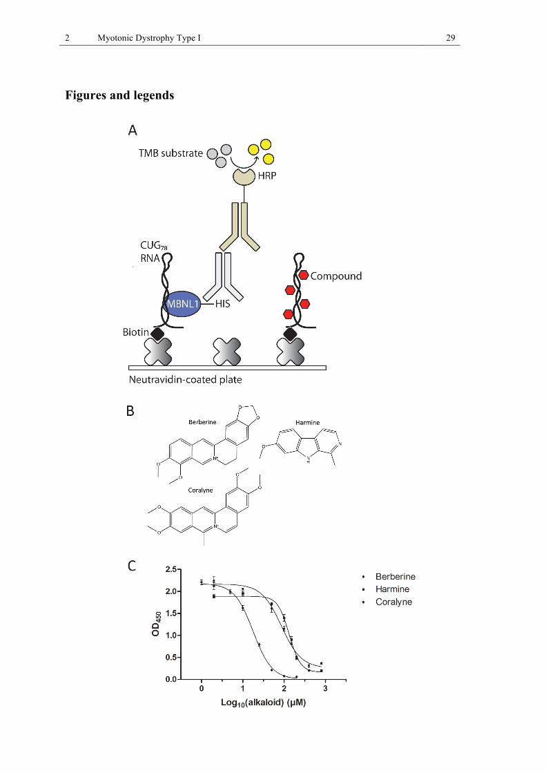
2 Myotonic Dystrophy Type I 29
Figures and legends

2 Myotonic Dystrophy Type I 30
Figure 2 (I 1): Screening for small molecules of natural origin that disrupt the MBNL1-CUG78 com-plex in vitro.
Fig. I 1A: CUG78-MBNL1 displacement screening assay: Biotinylated CUG78 RNA is immobilized and incubated with MBNL1 and compounds. RNA-bound MBNL1 is detected via a primary anti-HIS and a secondary anti-mouse-HRP antibody. A substrate is added to give an optical signal, correlating to the amount of RNA-bound MBNL1. In between all five incubation steps, washing is performed.
Fig. I 1B: Molecular structure of the identified alkaloids berberine, harmine, and coralyne.
Fig. I 1C: MBNL1-CUG78 RNA complex inhibition curves with berberine, harmine, and coralyne.
Figure 3 (I 2): HPLC-based activity profiling of the methanol extract of Peganum harmala. Shown on the y-axis are the online HPLC-UV trace at 254 nm (black) and the OD450 signal from the CUG78-MBNL1 inhibition assay (red) for each of the 29 fractions (min 1 to 30, time on x-axis). The activity of the fractions at minutes 8 and 9 could be assigned to the alkaloid harmine. Based on on-line MS and UV spectroscopic data, the other major peak eluting at 6.6 min was tentatively assigned to harmol. The activity in the early eluting fraction 2-3 min did not correspond to any peak and was not further investigated.
0
500
1000
1500
2000
2500
3000
0.00 2.00 4.00 6.00 8.00 10.00 12.00 14.00 16.00 18.00 20.00 22.00 24.00 26.00 28.00 30.00
Intensity [mAU]
Time [min]
OD450
0
0.5
1
1.5
2
2.5
3
OD450
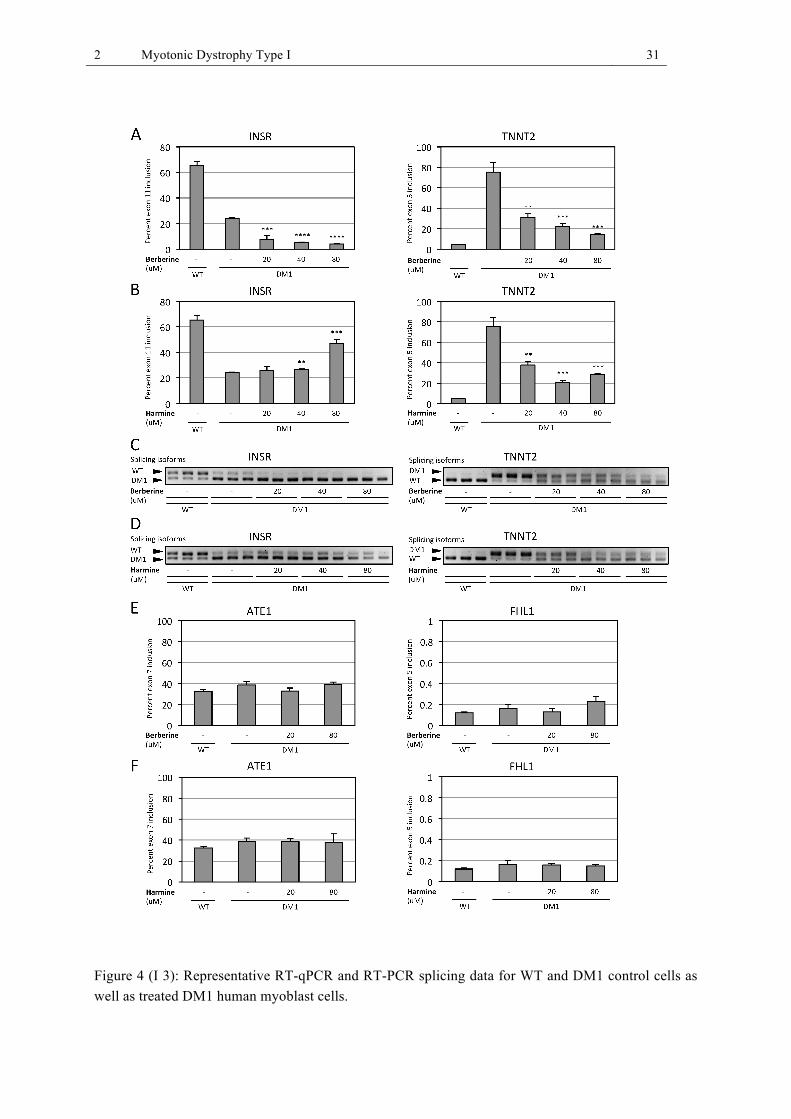
2 Myotonic Dystrophy Type I 31
Figure 4 (I 3): Representative RT-qPCR and RT-PCR splicing data for WT and DM1 control cells as well as treated DM1 human myoblast cells.

2 Myotonic Dystrophy Type I 32
Fig. I 3A: Berberine improves the splicing of the TNNT2 pre-mRNA but has a detrimental effect on the INSR pre-mRNA splicing.
Fig. I 3B: Harmine improves splicing of the TNNT2 pre-mRNA and also of the INSR pre-mRNA at the highest concentration.
Fig. I 3C,D: Alternative splicing of INSR and TNNT2 pre-mRNA analyzed by RT-PCR and visual-ized on 3% agarose gels. Shown are the two alternative splicing isoforms for both genes in untreated WT and DM1 control cells and DM1 cells treated with berberine (3 C) and harmine (3 D). The WT splicing isoform represents the predominant mature splicing variant, whereas the DM1 isoform repre-sents the fetal splicing variant.
Fig. I 3E,F: Berberine and harmine do not affect alternative splicing of MBNL1-independently regu-lated ATE1 and FHL1 pre-mRNAs in treated DM1 myoblasts, compared to untreated WT and DM1 control myoblasts.

2 Myotonic Dystrophy Type I 33
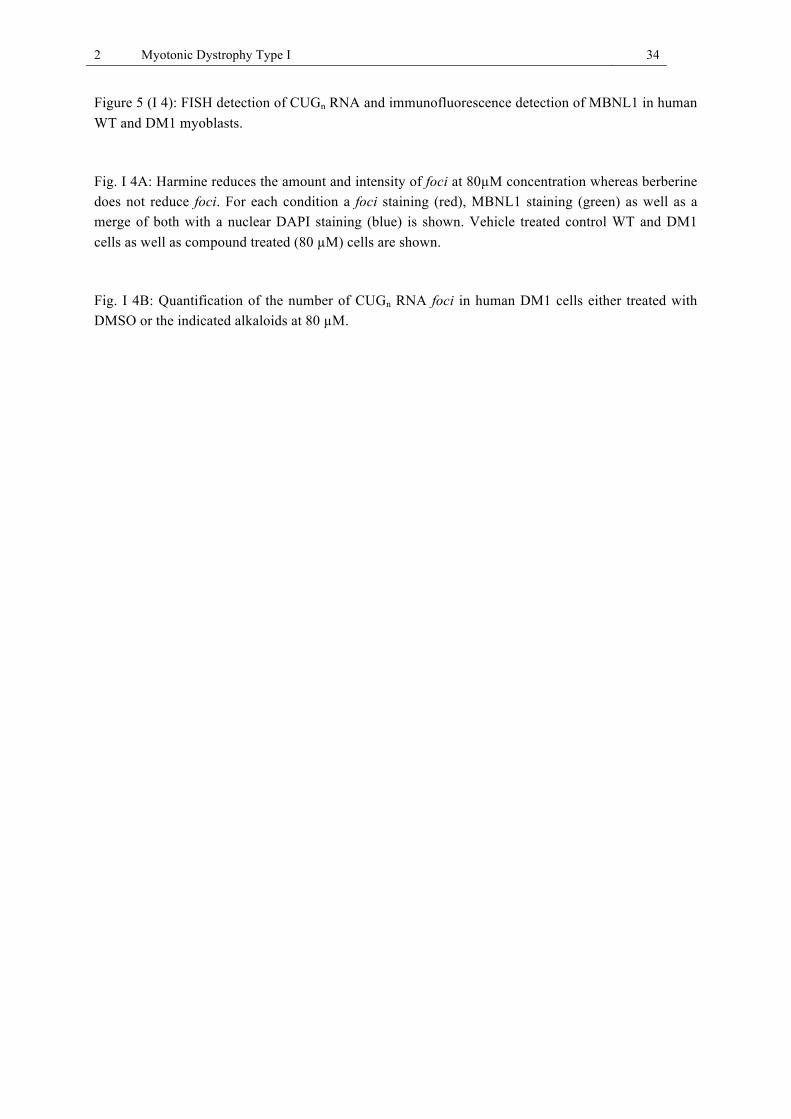
2 Myotonic Dystrophy Type I 34
Figure 5 (I 4): FISH detection of CUGn RNA and immunofluorescence detection of MBNL1 in human WT and DM1 myoblasts.
Fig. I 4A: Harmine reduces the amount and intensity of foci at 80µM concentration whereas berberine does not reduce foci. For each condition a foci staining (red), MBNL1 staining (green) as well as a merge of both with a nuclear DAPI staining (blue) is shown. Vehicle treated control WT and DM1 cells as well as compound treated (80 µM) cells are shown.
Fig. I 4B: Quantification of the number of CUGn RNA foci in human DM1 cells either treated with DMSO or the indicated alkaloids at 80 µM.

2 Myotonic Dystrophy Type I 35
Figure 6 (I 5): Representative in vivo RT-qPCR splicing data for vehicle treated WT and HSALR mice as well as compound treated HSALR mice (quadriceps muscle). Shown are the splicing of the CLCN1 channel and SERCA1. All three alkaloids improve the splicing of the CLCN1 channel whereas the splicing of SERCA1 is not improved.
Fig. I 5A: Dihydroberberine (DHB) treatment.
Fig. I 5B: Palmatine treatment.

2 Myotonic Dystrophy Type I 36
Fig. I 5C: Harmine treatment.
Fig. I 5D: Alternative splicing of the CLCN1 pre-mRNA analyzed by RT-PCR and visualized on 3% agarose gels. Shown are the two alternative splicing isoforms in untreated WT FVB/N and DM1 HSALR control mice and HSALR mice treated with 10 mg/kg dihydroberberine (DHB).
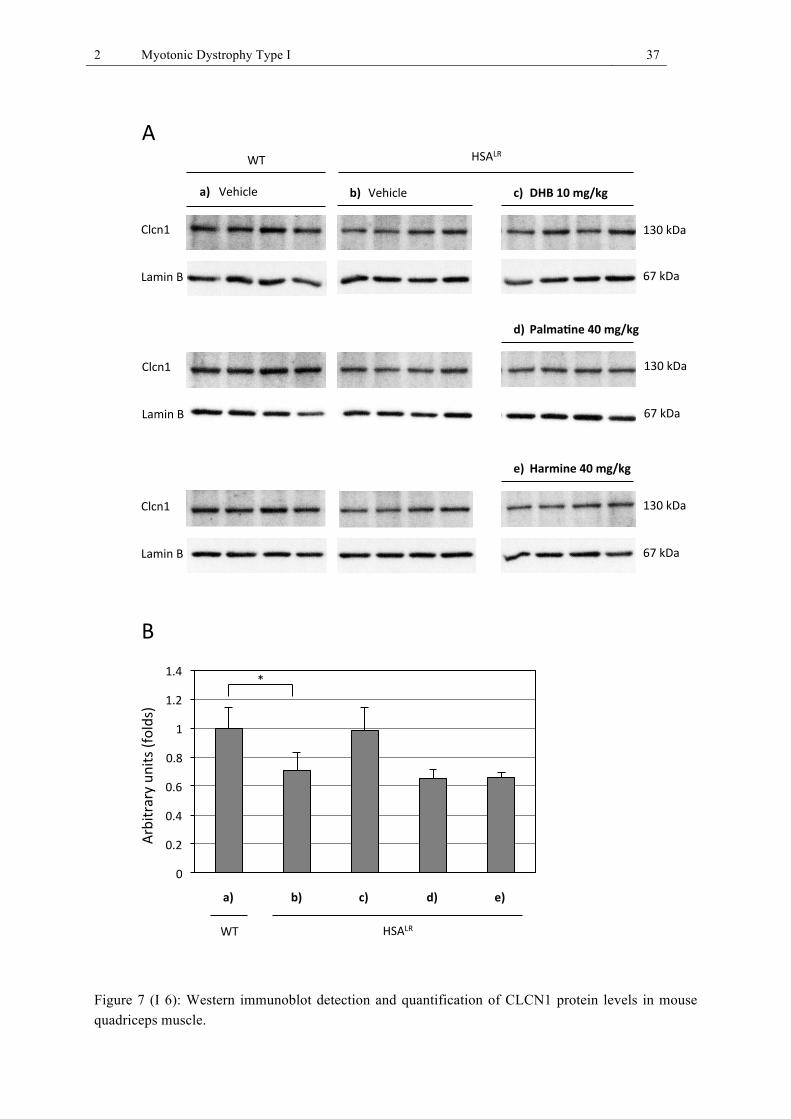
2 Myotonic Dystrophy Type I 37
Figure 7 (I 6): Western immunoblot detection and quantification of CLCN1 protein levels in mouse quadriceps muscle.
!"
!#$"
!#%"
!#&"
!#'"
("
(#$"
(#%"
)*+,(" (-!"./0"
1023,"4" &5"./0"
)*+,(" (-!"./0"
1023,"4" &5"./0"
)*+,(" (-!"./0"
1023,"4" &5"./0"
67" 89:1;"
<=>3+*="<=>3+*="!"# $"## %&'#()#*+,-+#."##
/!0*!123#4)#*+,-+#5"##
&!6*723#4)#*+,-+#3"##
:"
4"
:?@3A?0?B"C,
3AD"EFG*HDI"
89:1;"67"
!" ########$" ###############." ######5" #############3"#
J"
2 Myotonic Dystrophy Type I 38
Fig. I 6A: Western immunoblot showing CLCN1 protein levels of vehicle treated WT mice (a), vehi-cle treated HSALR mice (b), and compound treated HSALR mice (c-e). Lamin B was used as loading control.
Fig. I 6B: Quantification of CLCN1 band intensities normalized to Lamin B. The means of replicates for each condition including the standard deviation are shown.
2 Myotonic Dystrophy Type I 39
2.6 Manuscript II
Discovery of Di-imidazoline Small Molecules as Inhibitors of the MBNL1-CUGn RNA Complex with Therapeutic Potential for Myotonic Dystrophy Type I
Ruben Herrendorff1, Jochen Kinter1, Frances Kern1, Jacqueline Bezençon2, Beat Ernst2, Tanja
Wenzler3, Reto Brun3, Arvind Kumar4, Abdelbasset A. Farahat4,5, Martial Say4, David W.
Boykin4, Michael Sinnreich1*
1 Neuromuscular Research Group, Departments of Neurology and Biomedicine, University
Hospital Basel, Switzerland
2 Institute of Molecular Pharmacy, Pharmacenter, University of Basel, Switzerland
3 Medical Parasitology & Infection Biology, Swiss Tropical and Public Health Institute, and
University of Basel, Switzerland
4 Department of Chemistry, Georgia State University, Atlanta, Georgia 30303, USA
5 Department of Pharmaceutical Organic Chemistry, Faculty of Pharmacy, Mansoura Univer-
sity, Mansoura 35516, Egypt
* To whom correspondence should be addressed.
2 Myotonic Dystrophy Type I 40
Abstract
Myotonic Dystrophy Type I (DM1) is the most common muscular dystrophy among the adult
population with a prevalence of about 1:8000. To this date, there is no causal treatment avail-
able for this disabling disease. A genetic mutation in the non-coding 3’ region of the Myoton-
ic Dystrophy Protein Kinase (DMPK) gene lies at the root of this disease’s pathomechanism.
A trinucleotide expansion of CTG repeats within this locus leads to a pathogenic CUGn pre-
mRNA that sequesters splicing factors, such as muscleblind-like 1 (MBNL1), leading to mis-
regulated alternative splicing of a plethora of pre-mRNAs.
Several studies were performed, which aimed at identifying small molecules that displace
MBNL1 from the pathogenic RNA to restore alternative splicing. Pentamidine was described
as an inhibitor of the MBNL1-CUGn RNA complex; it restored molecular defects in a cell
model of DM1, and partially in a mouse model of DM1. However, pentamidine is not suited
for long-term treatment, mainly due to its toxicity. Our study comprised the screening of 434
pentamidine-related small molecules in an in vitro CUG78-MBNL1 inhibition assay. Here, we
describe the synthesis, the biological and pharmacokinetic evaluation of three di-imidazolines
with improved properties compared to pentamidine. Testing of the di-imidazolines in human
DM1 myoblasts led to the identification of a new lead compound DB175, which fully restored
the splicing of the INSR and TNNT2 pre-mRNAs, which are both mis-spliced in DM1. Di-
imidazolines comprise a new compound class with therapeutic potential for DM1 and possi-
bly other RNA-mediated diseases.

2 Myotonic Dystrophy Type I 41
Introduction
Myotonic Dystrophy Type I (DM1) is the most common neuromuscular disorder among the
adult population (11). This severe disease currently has no treatment but, due to its relatively
well-understood pathomechanism, provides several opportunities for therapeutic intervention.
The pathomechamism of DM1 has been linked to a mutation in the non-coding 3’ region of
the Myotonic Dystrophy Protein Kinase (DMPK) gene (9, 14). The mutant DMPK transcript
presents a toxic gain-of-function RNA that binds splicing factors, such as muscleblind-like 1
(MBNL1) (24, 25, 37, 38), which are retained within the nuclei of affected cells (26-28). This
leads to mis-regulated alternative splicing of a variety of pre-mRNAs, including the skeletal
muscle chloride channel (CLCN1) (29-31), the insulin receptor (INSR) (32), sarcoplas-
mic/endoplasmic reticulum Ca2+ ATPase 1 (SERCA1) (34) and cardiac troponin T type 2
(TNNT2) (35).
Among different therapeutic approaches towards DM1, reviewed in ref. (66), much effort has
been put in the search for small molecules as potential therapeutics. Compared to antisense
oligonucleotide and gene therapy approaches, the small molecule approach has the advantage
of systemic tissue penetration. Several studies led to the identification of small molecules that
displaced MBNL1 from the toxic DMPK CUGn transcript and thus restored DM1-associated
molecular defects. Identified small molecules include pentamidine and neomycin B (51), an
acridine-triazole conjugate (52), modularly assembled polyvalent derivatives of Hoechst
33258 (53), kanamycin A (90), the antimicrobial lomofungin and its derivative dilomofungin
(57), as well as peptides and peptide-derivatives (55, 91). From these compounds pentamidine
and the peptide ABP1 have been reported to partially restore splicing in vivo (51, 91).
Pentamidine was discovered through screening of a collection of nucleic acid binders in an
electrophoretic mobility gel shift assay (EMSA) (51). Parkesh et al. employed chemical simi-
larity searching, with pentamidine and Hoechst 33258 as starting points, to find stronger
MBNL1-CUGn RNA complex inhibitors (92). The screening of nucleic acid binders (51) and
usage of identified complex inhibitors as chemical starting points for stronger inhibitors (92)
has been validated as a drug discovery approach in DM1.
Due to toxicity issues, pentamidine itself is not suited as a drug for DM1 (51). Using the
antiprotozoal drug pentamidine as a lead substance, we chose a collection of 434 pentami-
dine-related compounds from a library of small molecules that was generated during a drug

2 Myotonic Dystrophy Type I 42
discovery program for Trypanosomiasis. These aromatic, mostly dicationic small molecules
were screened in a CUG78-MBNL1 inhibition assay, using a DMPK CUG78 transcript ampli-
fied from DNA of a DM1 patient. In this study we identified three di-imidazolines DB1796,
DB1973 and DB175 that demonstrated improved properties compared to the lead substance
pentamidine with respect to complex inhibitory activity in vitro, amelioration of molecular
defects in a DM1 cell model, and cellular toxicity. DB1796 and DB1973 have not been de-
scribed before, we therefore also report their synthesis.
Results
Discovery of di-imidazoline small molecules that disrupt the MBNL1-CUG78 RNA complex in vitro
A collection of 434 pentamidine-related small molecules was screened with an in vitro
CUG78-MBNL1 inhibition assay (Fig. II 1A). Compounds were co-incubated with recombi-
nant MBNL1-His in 96-well plates containing immobilized CUG78 RNA, and then MBNL1-
CUG78 complex inhibition was measured. We identified 36 hits at 0.5 µM and 6 hits at 0.125
µM compound concentration (> 80% inhibition of MBNL1 binding to CUG78 RNA). Among
the hits were imidazolines, amidines, tetrahydropyrimidines or amines, with the majority of
hits being of the di-imidazoline type. The six strongest complex inhibitors comprised the
diphenylfuran imidazoline DB60, previously described by Wilson et al. (93), three structural-
ly close derivatives thereof, a dibenzimidazolfuran imidazoline and a dibenzimidazolfuran
amidine. The CUG78-MBNL1 inhibition assay was used to determine the IC50 values for three
most potently bioactive di-imidazolines: DB1796, DB1796 and DB175 (Fig. II 1B). DB 1796
had an IC50 value of 1.5 ± 0.1 µM, DB 1973 an IC50 of 6.2 ± 0.6 µM and DB 175 an IC50 of
5.0 ± 0.1 µM (Fig. II 1C). Pentamidine, the lead compound underlying this study, had no
inhibitory effect on complex formation whatsoever, even at concentrations as high as 500 µM.
As additional reference compounds, we tested Hoechst 33258 and neomycin B, two previous-
ly described complex inhibitors (51, 92), which had IC50 values of 195.5 ± 3.0 µM and 5.3 ±
0.6 µM, respectively.
2 Myotonic Dystrophy Type I 43
Synthesis of the di-imidazolines DB1796, DB1973 and DB175
The synthesis of DB175, a dicationic carbazole, has been described by Patrick et al. (94). The
syntheses of both DB1796 and DB1973 are outlined in scheme II 1. The first step of the
preparation of DB1796 employs a Stille coupling reaction between 2-bromo-5-
cyanothiophene and 1-(tert-butoxycarbonyl)-2-(trimethylstannyl)-1H-indole-6-carbonitrile
(95), which yielded the coupled product. We detected significant loss of the Boc-protecting
group during the reaction and/or work-up; consequently we treated the crude product with
Boc-anhydride to simplify the purification by column chromatography. The bis-nitrile was
converted into DB1796 using Pinner methodology to form the imidate ester followed by
subsequent reaction with ethylene-diamine. The first step in the synthesis of DB1973 involves
the oxidative cycloaddition reaction between 3,4-diaminobenzonitrile and 2-cyano-5-
formylthiophene using sodium bisulfite as the oxidizing agent to form the corresponding bis-
nitrile. We employed the process described for DB1796 to convert the bis-nitrile into
DB1973.
Identified di-imidazolines show reversal of mis-splicing of pre-mRNAs in a human myoblast cell model for DM1
We subsequently tested the compounds ability to improve splicing defects in a human my-
oblast model for DM1 (71). The wild type (WT) cell line contained a short CUG5 repeat; the
DM1 cell line contained a long CUG1300 repeat. DM1 myoblasts were treated for 24 h with
the identified compounds, followed by RNA isolation and reverse transcription. DMSO-
treated WT and DM1 myoblasts served as controls. For the quantification of alternative splic-
ing, a qPCR-based method was used. This method employs two primer pairs; one primer pair
detects all occurring splicing variants of a pre-mRNA and another primer pair only detects
those pre-mRNA variants that include an investigated alternative exon. The six strongest
complex inhibitors did not improve splicing in the DM1 myoblasts at 20 or 40 µM concentra-
tion; the diphenylfuran imidazoline DB60 and three close derivatives thereof were toxic to the
DM1 myoblasts at these concentrations and if tested at lower, more tolerable concentrations
the compounds did not show any splicing amelioration (data not shown). Three structurally
similar di-imidazolines emerged as the most potently bioactive compounds: DB 1796, DB
1973 and DB 175. All three compounds improved the splicing of two pre-mRNAs, the insulin
receptor (INSR) and the cardiac troponin T type 2 (TNNT2) pre-mRNA (Fig. II 2 A-C). In
2 Myotonic Dystrophy Type I 44
WT cells close to 60% of INSR transcripts included exon 11, whereas in DM1 cells exon
inclusion was reduced to about 10-25%. In WT cells, TNNT2 exon 5 inclusion was around
5%; whereas in DM1 cells inclusion was increased to at least 45% of transcripts. DB 1796
improved the alternative splicing of the INSR pre-mRNA by 20.0 ± 2.9% (10 µM), 34.7 ±
1.4% (20 µM) and 13.1 ± 1.0% (40 µM), whereas the splicing for the TNNT2 pre-mRNA was
improved by 14.6 ± 0.8% (10 µM), 25.7 ± 0.7% (20 µM) and 64.8 ± 0.2% (40 µM). Also DB
1973 improved the alternative splicing of the INSR pre-mRNA by 38.8 ± 2.3% (10 µM), 38.5
± 3.8% (20 µM) and 34.6 ± 3.1% (40 µM), and the splicing for the TNNT2 pre-mRNA was
improved by 12.9 ± 2.5% (10 µM), 17.4 ± 0.8% (20 µM) and 28.8 ± 2.3% (40 µM). DB175
effectively improved the splicing of both pre-mRNAs in a concentration-dependent manner
and splicing was fully restored at 20 µM concentration. Splicing of the INSR pre-mRNA was
improved by 13.2 ± 2.3% (5 µM), 60.8 ± 1.3% (10 µM) and 87.8 ± 6.2% (20 µM), whereas
the TNNT2 splicing was improved by 59.9 ± 1.8% (5 µM), 91.7 ± 0.4% (10 µM) and 101.2 ±
0.4% (20 µM). Splicing restoration by DB175 was confirmed with RT-PCR and visualization
of the two alternative splicing isoforms of the INSR and TNNT2 pre-mRNA on 3% TAE
agarose gels (Fig. II 2D). Pentamidine did not improve splicing of the INSR pre-mRNAs (Fig
II 2C). Only the highest pentamidine concentration used (40 µM) showed a small but signifi-
cant improvement of 17.4 ± 3.8% in TNNT2 splicing (Fig. II 2C). To investigate the selectivi-
ty of the three identified di-imidazolines, we tested whether they affected MBNL1-
independent splicing events in the same DM1 cell line (Fig. II 3A-C). We therefore analyzed
the alternative splicing of two genes, ATE1 and FHL1, which are known to be alternatively
spliced, but independent of MBNL1 (72). Exon 7 inclusion in the ATE1 pre-mRNA was close
to 33% for the WT cell line and close to 48% in the DM1 cell line. Exon 5 inclusion in the
FHL1 pre-mRNA was close to 0.1% for both the WT and the DM1 cell line. Compared to the
splicing of ATE1 and FHL1 in untreated WT and DM1 control cells the compound-treated
DM1 cells did not show any significant difference in alternative splicing of both genes.
Identified di-imidazolines reduce nuclear CUGn RNA foci formation in a human myoblasts cell model for DM1 and change the distribution pattern of MBNL1
Nuclear aggregates of MBNL1 and CUGn RNA, called foci, are a further hallmark of DM1.
The identified imidazolines were tested for their ability to reduce foci formation (Fig. II 4A)
using the same two cell lines that were used for the alternative splicing analysis. Only the
DM1 cell line showed CUGn RNA staining and co-localization with MBNL1 staining. The
2 Myotonic Dystrophy Type I 45
MBNL1 staining showed a predominantly nuclear staining in the WT cells, whereas in the
DM1 cells MBNL1 was concentrated mainly in nuclear foci. At 80 µM concentration of
DB1796 and DB1973, the number of foci was reduced (Fig. II 4B). MBNL1 staining was
more distributed in the nuclei of treated cells, indicating the release of MBNL1 from the toxic
CUGn transcripts. Treatment with DB175 at 40 µM concentration also reduced foci formation
(Fig. II 4B), resulting in a diffuse MBNL1 staining. MBNL1 was detectable in the nucleus as
well as in the cytoplasm of treated cells, indicating increased nuclear export. CUGn RNA
staining was found to be decreased in the nuclei of DB175 treated cells, at the same time the
staining was increased in the cytoplasm of treated cells and co-localized partly with MBNL1.
DB175 might therefore increase nuclear export of toxic DMPK transcripts and MBNL1, but
might also stabilize the RNA transcripts. Quantification of foci in a total of 150 randomly
chosen nuclei per condition showed a decrease of foci number per nucleus, from 4.7 ± 0.3 in
DMSO-treated DM1 control cells to 3.1± 0.4 (80 µM DB1796), to 2.1 ± 0.3 (80 µM
DB1973), and to 3.2 ± 0.1 (40 µM DB175) following compound treatments.
Cellular toxicity of the identified di-imidazolines
A cell viability assay with C2C12 mouse myoblasts was performed to evaluate the cellular
toxicity of the three identified di-imidazolines and two reference compounds, pentamidine
and mitomycin C. We incubated C2C12 myoblasts with compound concentrations ranging
from 1 – 900 µM and determined the toxicity IC50 (Tox IC50) values, whereby half of the cells
were still viable after two days of compound incubation. DB1796 had a Tox IC50 of 129.8 ±
3.7 µM, DB1973 had a Tox IC50 of 205.9 ± 10.3 µM and DB175 one of 130.0 ± 1.3 µM. The
factor between the activity and toxicity IC50 value (IC50 / Tox IC50) is 86.5 for DB1796, 33.2
for DB 1973 and 26.0 for DB 175. This value can be regarded as a rough selectivity index.
Pentamidine had a Tox IC50 of 53.5 ± 0.8 µM. Mitomycin C was measured as a reference
compound and gave a Tox IC50 of 20.4 ± 1.6 µM.
In vitro pharmacokinetic analysis of the identified di-imidazolines
To evaluate the di-imidazolines’ potential for application in vivo, we analyzed their physico-
chemical parameters such as lipophilicity (distribution coefficient log D7.4), pKa values, and
permeability (Table II 1). In addition, mouse plasma protein binding (PPB) was determined
2 Myotonic Dystrophy Type I 46
(Table II 1). DB1973 was shown to be strongly hydrophilic (logD7.4 < -1.5). For estimating
the influence of the pKa on lipophilicity and permeability we experimentally determined the
pKa value for DB1973 by the 1H-NMR method described in ref. (96). The experimental pKa
values of the two imidazoline groups of DB1973 could not be distinguished and showed a
singular pKa value of 7.83 ± 0.35; the pKa of the benzimidazole was 0.61 ± 0.07. In addition,
the pKa values for the three di-imidazolines and the reference compound pentamidine were
calculated in silico (Marvin 5.6.0.2, ChemAxon, 2011). The predicted pKa values for the
imidazoline group (DB1796, DB1973, DB175) were in the range of 7 to 9.5, whereas the pKa
of the more basic amidine group (pentamidine) was above 11.5. This indicates less charge-
neutral species of pentamidine at physiological pH 7.4 compared to the di-imidazolines.
However, the low lipophilicity (< -1.5 for DB 1973) and the presence of a charged state at the
physiological pH explain the low passive permeability through an artificial membrane using
the PAMPA (Parallel Artificial Membrane Permeability Assay) (97) for all three di-
imidazolines and additionally pentamidine (logPe < -7.3). Moreover, the in vitro mouse PPB
for DB1796 (69.5%) and for DB175 (97.1%) were higher than the reported PPB of pentami-
dine (41%) (98).
Discussion
There is a fast growing interest in RNA as a drug target and DM1 is at the center of attention
in the field of RNA drug discovery. Several RNAs, owing to distinct secondary structural
elements, have been investigated intensely, e.g. the bacterial ribosome or the TAR RNA of
the human immunodeficiency virus type 1 (HIV-1) (63, 99). Also RNA triple nucleotide
repeat expansions represent distinct secondary structural elements and are being investigated
as drug targets (63, 66, 99). Triple nucleotide repeat expansions are the characteristic molecu-
lar hallmark for the group of triple nucleotide repeat expansion diseases (TREDs) including
fragile X syndrome, myotonic dystrophy type I and II, Huntington’s disease and spinocerebel-
lar ataxias (66). Among the TREDs, DM1 presents a model disease due to its relatively well-
studied pathomechanism and the theoretical reversibility thereof by therapeutic intervention,
e.g. with suited small molecules that inhibit the disease-characteristic MBNL1-CUGn RNA
complex.
The antiprotozoal drug pentamidine was the first small molecule described as an inhibitor of
this complex and it improved molecular defects of in a cell model and a mouse model of DM1
(51). As there are several drawbacks to pentamidine as a DM1 drug, notably its toxicity, the
2 Myotonic Dystrophy Type I 47
aim of the present study was to identify pentamidine-related small molecules with improved
properties. The screening of 434 antiprotozoal small molecules in an in vitro CUG78 MBNL1
inhibition assay yielded three potent di-imidazolines with partly improved properties com-
pared to pentamidine, i.e. DB1796, DB1973 and DB175. Their characteristic imidazoline
group is a cyclic and planar derivative of the amidine group, characteristic for the diamidines
such as pentamidine. The six strongest complex inhibitors from our screening inhibited the
formation of the MBNL1-CUGn RNA complex at 125 nM concentration. Five of these com-
pounds were di-imidazolines, only one was a diamidine. Interestingly, the strongest complex
inhibitor identified in our screening, the diphenylfuran imidazoline DB60, has been described
previously as a strong intercalator for double stranded RNA (93, 100). From the six strongest
inhibitors the diphenylfuran imidazoline DB60 and three structurally close derivatives were
toxic to the DM1 myoblasts at 20 or 40 µM concentration and did not improve splicing at
lower concentrations, likely due to unspecific nucleic acid intercalation. The two other inhibi-
tors of the dibenzimidazolfuran type did not improve splicing in the DM1 myoblasts, proba-
bly due to their high polarity, with four ionizable groups, and resulting poor membrane per-
meability. Overall, in vitro inhibitory activity of the six strongest MBNL1-CUGn RNA com-
plex inhibitors did not seem to correlate with a biological effect in our DM1 cell model.
We therefore tested screening hits with lower inhibitory activity (IC50 ~ 1 - 10 µM) in our
DM1 myoblast model, which successfully yielded bioactive di-imidazolines. Three of these
di-imidazolines with in vitro IC50 values in the lower µM range (1.5 – 6.2 µM) showed re-
markable effects on splicing and foci formation in our DM1 cell model. DB175 fully restored
the mis-regulated alternative splicing of the INSR and TNNT2 pre-mRNAs in the DM1 my-
oblasts at 20 µM concentration and reduced nuclear foci at 40 µM. FISH and immunofluores-
cence showed, that MBNL1 was redistributed in DM1 myoblasts treated with 40 µM DB175.
But also CUGn RNA staining was stronger in the cytoplasm compared to untreated control
cells, indicating increased nuclear export of the toxic DMPK transcripts. The increased RNA
staining might reflect stabilization, and thus decreased decay of the toxic DMPK transcripts in
the cytoplasm of these cells, similar to what has been described for di-lomofungin (57).
DB175 treatment however decreased nuclear foci, which were greatly increased by di-
lomofungin. DB1796 and DB1973 also significantly improved splicing in the DM1 myoblasts
and reduced the amount of foci. The lower in vitro inhibitory activity of these three com-
pounds might be compensated by better membrane permeability or target selectivity.
2 Myotonic Dystrophy Type I 48
We found that di-imidazolines not only were the most potent MBNL1-CUGn RNA complex
inhibitors, but also performed better than their diamidine counterparts in the cellular splicing
assay. A reason for the stronger biological effect might be better membrane permeability of
di-imidazolines compared to diamidines. Indeed, the in silico predicted pKa values of our
identified di-imidazolines (pKa 7 – 9) and the experimentally determined pKa of the DB1973
imidazoline groups (pKa 7.83 ± 0.35) indicate a lower pKa for the imidazoline group com-
pared to the amidine group of pentamidine, (pKa > 11). Thus, more uncharged species of the
imidazolines is present at physiological pH, which can potentially pass the membrane barrier.
These findings might be comparable to arylimidamides with pKa values of 7-8 and better
membrane permeability than amidines (101). However, PAMPA (97) experiments show low
passive membrane permeability of our di-imidazolines and for pentamidine at pH 7.4, which
can be explained by their low lipophilicity (< -1.5 for DB1973) and high presence of charged
state at physiological pH. Nevertheless, the compounds have shown biological activity in the
DM1 myoblasts and therefore, another major permeability mechanism (e.g. active transport)
might be involved in the uptake of these compounds into the myoblast, which needs further
investigation.
Another reason for a stronger biological effect might be stronger target RNA binding. The
planarity of the imidazoline group has been shown to enable better RNA binding by intercala-
tion than the amidine group, which is not planar (100). For the most part, the three identified
di-imidazolines DB1796, DB1973 and DB175 are planar. The planarity of the compounds
indicates intercalation as a possible binding mode. The aromatic core of the substances might
intercalate into the double stranded CUGn hairpin RNA, forming stacking interactions with
the RNA bases, as well as electrostatic interaction via the imidazoline groups with the nega-
tively charged phosphate backbone of the RNA. The three substances seem to share the same
pharmacophore. DB1796 with an indole core and DB1973 with a benzimidazole core are
structurally very similar and display the two imidazoline groups in the same fashion. DB175
has a carbazole core and displays the two imidazoline moieties in a similar way compared to
the two other DB compounds, but conformationally more restricted, which might be benefi-
cial with respect to smaller entropy cost upon binding to the target RNA.
Comparison of the three newly identified di-imidazolines with the lead compound pentami-
dine reveals several advantages of the di-imidazolines, but also disadvantages, with respect to
suitability as DM1 drugs. The newly identified compounds are much stronger binders of the
CUGn MBNL1 complex compared to pentamidine, as we did not detect any inhibition of

2 Myotonic Dystrophy Type I 49
MBNL1-CUGn RNA complex formation by pentamidine. In fact, another mode of action
recently described for pentamidine is transcription inhibition of the toxic DMPK transcripts
by DNA binding (59). In our DM1 cell model, the three di-imidazolines performed better than
pentamidine. Pentamidine did not improve splicing in DM1 myoblasts, except for a minute
improvement at the highest tested concentration of 40 µM, which was already very close to
the Tox IC50 concentration of 53.5 ± 0.8 µM. Regarding toxicity, all of the three di-
imidazolines had higher Tox IC50 values than Pentamidine, all above 120 µM. The three di-
imidazolines were less toxic to C2C12 mouse myoblasts and also to human myoblasts. Fur-
thermore, the di-imidazolines showed splicing selectivity, they did not affect the alternative
splicing of two MBNL1-independently spliced pre-mRNAs (ATE1 and FHL1). The di-
imidazolines, compared to pentamidine, have partly improved pharmacokinetic properties
which might lead to better membrane permeability, e.g. the earlier discussed lower pKa val-
ues. However, with respect to the pharmacokinetic profile, the three di-imidazolines will need
further improvement by suited chemical modification prior to an in vivo application. The
logD7.4 value of DB1973, lower than -1.5, indicates high polarity of the di-imidazolines. A
higher logD7.4 of 0.144 ± 0.003 was reported for pentamidine (98). In a recent study heptami-
dine, a pentamidine derivative with increased lipophilicity, proved to be more active in DM1
models compared to pentamidine (59). A balanced activity-pharmacokinetic profile will
eventually be needed for effective small molecules to treat DM1. In this respect, drug discov-
ery on the CUGn RNA target remains a challenge as high charge is needed for target binding,
but unwanted for membrane permeability of compounds. Overall, the di-imidazolines provide
a few advantages but also disadvantages compared to pentamidine regarding pharmacokinetic
parameters.
In conclusion, we show in this study that aromatic di-imidazolines are an promising group of
compounds for DM1 and RNA drug discovery. We have identified three di-imidazoline sub-
stances as inhibitors of the CUGn MBNL1 complex with distinct positive effects on the DM1
pathomechanism in human DM1 myoblasts. The three di-imidazolines could contribute to
DM1 drug discovery projects as new medicinal chemistry starting points. Foremost the phar-
macokinetic profile of these di-imidazolines will require further medicinal chemistry optimi-
zation. Currently, these substances are being evaluated in vivo.
2 Myotonic Dystrophy Type I 50
Experimental
Compound library
The compound library screened in this study was part of a compound library established at the
Division of Medical Parasitology & Infection Biology, Swiss Tropical and Public Health
Institute, Switzerland. The compounds were synthesized at the Department of Chemistry,
Georgia State University, Atlanta, Georgia 30303, USA and the Department of Pathology &
Laboratory Medicine, University of North Carolina, Chapel Hill, NC 27599-7525, USA. Most
of the 434 compounds were synthesized during a drug discovery program for Trypanosomia-
sis.
Synthesis
Melting points were determined on a Mel-Temp 3.0 melting point apparatus, and are uncor-
rected. TLC analysis was carried out on silica gel 60 F254 precoated aluminum sheets using
UV light for detection. 1H and 13C NMR spectra were recorded on a Bruker 400 MHz spec-
trometer using indicated solvents. Mass spectral data was obtained from the Georgia State
University Mass Spectrometry Laboratory, Atlanta, GA. Elemental analysis were performed
by Atlantic Microlab Inc., Norcross, GA and are within ± 0.4 of the theoretical values. The
compounds reported as salts frequently analyzed correctly for fractional moles of water and/or
other solvents; in each case 1H NMR spectra were consistent with the analysis. All chemicals
and solvents were purchase from Aldrich Chemical Co., VWR International, or Combi-
Blocks, Inc.
Synthesis of DB1796 and DB1973
1-N-tbutoxycarbonyl-2-(5-cyanothienyl)-6-cyanoindole
Tetrakis(triphenylphosphine)palladium(0) (0.23 g, 0.0002 mole, 4 mole %) was added to a
stirred mixture of the 1-(tert-butoxycarbonyl)-2-(trimethylstannyl)-1H-indole-6-carbonitrile
(95) (2.02 g, 0.005 mole) and 2-bromo-5-cyanothiophene (0.94 g, 0.005 mole) in deaireated
dioxane (20 mL) under nitrogen. The reaction mixture was heated at reflux for 24 h (TLC
monitored), concentrated under reduced pressure, suspended in 30 mL THF, Boc anhydride

2 Myotonic Dystrophy Type I 51
(1.09 g, 0.005 mole) and a few crystals of DMAP were added and stirred for 6 h, concentrated
under reduced pressure, ice water was added, the precipitate was filtered, washed with water,
dried and chromatographed on silica gel using hexane/ethyl acetate (70/30, v/v) giving a
white solid (1.13 g, 65%), mp 274-5 °C dec., 1H NMR (DMSO-d6): 8.25 (s, 1H), 8.29 (d, 2H,
J = 8.4 Hz), 8.11 (d, 2H, J = 8.4 Hz), 7.79 (d, 1H, J = 3.6 Hz), 7.23 (s, 1H), 7.12 (d, 1H, J =
3.6 Hz), 1.46 (s, 9H); 13C NMR (DMSO-d6): 150.2, 148.1, 138.5, 128.7, 127.8, 126.7, 124.8,
124.7, 119.0, 115.8, 112.8, 112.2, 111.4, 105.9, 85.4, 27.1 ; ESI-MS: 350 (M++1); Anal. Calc.
for C19H15N3O2S: C, 65.31; H, 4.33; N, 12.03. Found: C, 65.44; H, 4.21; N, 12.13.
2-(5-(2-imidazolinyl)-thienyl)-6-(2-(imidazolinyl)-1H-indole dihydrochloride (DB 1796)
The imidate ester dihydrochloride (0.349 g, 0.001 mole) from the above dicyano compound
and 1,2-diaminoethane (0.18 g, 0.003 mole) in 15 mL dry ethanol was allowed to react and
worked up as described for DB 1973 which yielded a pale yellow solid (0.34 g, 83%), mp
>320 °C dec., 1H NMR (DMSO-d6/D2O): 8.27 (d, 1H, J = 1.5 Hz), 7.10 (d, 1H, J = 3.9 Hz),
7.67-7.64 (m, 2H), 7.23 (brs, 1H), 7.22 (d, 1H, J = 3.9 Hz) 3.98 (s, 4H), 3.97 (s, 4H); 13C
NMR (DMSO-d6/D2O): 166.3, 154.9, 152.2, 140.5, 137.4, 130.0, 128.1, 123.5, 123.2, 122.8,
114.3, 113.2, 110.3, 103.4, 44.9, 44.7; ESI-MS: 336 (M++1); Anal. Calc. for C18H17N5S-
2HCl-0.25H2O: C, 52.48; H, 4.77; N, 17.01. Found: C, 52.51; H, 4.59; N, 16.96.
2-(5-(2-cyano)-thienyl)-5(6)-cyan-1H-benzimidazole
Sodium bisulfite (0.78 g, 0.0075 mole) was added to a stirred solution of 2-cyano-5-formyl
thiophene (0.685 g, 0.005 mole) and 4-cyano-1,2-phenylenediamine (0.665 g, 0.005 mole) in
8 mL dry DMF. The reaction mixture was heated at 135 °C for 15 h, cooled and triturated
with ice-water and the precipitated solid filtered, washed with water and dried in air. The
yellow solid was purified by column chromatography on silica gel using hexane/ethyl acetate
(70/30, v/v) to yield an off-white solid (0.74 g, 59%), mp 213-214 °C dec., 1H NMR (DMSO-
d6): 14.0 (s, 1H), 8.19 (s, 1H), 8.09 (d, 1H, J = 4 Hz), 7.97 (d, 1H, J = 4 Hz), 7.79 (d, 1H, J = 8
Hz), 7.65 (d, 1H, J = 8 Hz); 13C NMR (DMSO-d6): 148.2, 141.6, 140.9, 139.2, 132.7, 129.3,
128.7, 126.9, 122.2, 116.1, 114.6, 110.9, 105.7; ESI-MS: 251 (M++1); Anal. Calc. for
C13H6N4S: C, 62.39; H, 2.42; N, 22.39. Found: C, 62.55; H, 2.61; N, 22.21.
2-(5-(2-imidazolinyl)-thienyl)-5(6)-(2-(imidazolinyl)-1H-benzimidazole trihydrochloride (DB 1973)
A suspension of the above dicyano compound (0.5 g, 0.002 mole) in 25 mL ethanol was
saturated with dry HCl(g) at 0 °C and stirred in a closed flask for 7 days, the solid suspension
2 Myotonic Dystrophy Type I 52
was triturated with dry ether and filtered, washed with ether and dried under reduced pressure
for 4 h to yield the yellow imidate ester trihydrochloride 0.78 g (86%). 1,2-Diaminoethane
(0.18 g, 0.003 mole) was added to a suspension of 0.451 g (0.001 mole) the imidate ester in
15 mL dry ethanol, the reaction mixture was heated at reflux for 12 h, concentrated in reduced
pressure, dissolved in water, basified with 2N NaOH, the precipitated solid was filtered,
washed with water, dried and converted to HCl salt with ethanolic-HCl to yield a pale yellow
solid (0.34 g, 82%), mp >300 0C dec., 1H NMR (DMSO-d6/D2O): 8.10 (s, 1H), 7.87 (brs, 1H),
7.85 (s, 1H), 7.10 (d, 1H, J = 4 Hz), 7.65 (d, 1H, J = 4 Hz), 3.96 (s, 4H), 3.80 (s, 4H); 13C
NMR (DMSO-d6): 166.2, 160.0, 148.9, 148.0, 141.1,136.6, 129.4, 129.0, 125.7, 123.7, 123.6,
116.9, 116.1, 45.1, 41.0; ESI-MS: 337 (M++1); Anal. Calc. for: C17H16N6S-3HCl-2H2O-
0.25C2H6O: C, 42.60; H, 5.00; N, 17.03. Found: C, 42.90; H, 4.71; N, 16.69.
MBNL1 preparation
CUG78 RNA preparation
Screening assay: CUG78-MBNL1 inhibition assay
Cell splicing assay
FISH and immunofluorescence
Cell viability assay
These six procedures were performed according to the experimental section of Manuscript I.
2 Myotonic Dystrophy Type I 53
Determination of physicochemical and in vitro pharmacokinetic properties
DMSO and 1-octanol were purchased from Sigma-Aldrich (St. Louis, MI, USA). Deuterium
oxide was purchased from Armar Chemicals (Switzerland). PRISMA HT universal buffer,
GIT-0 Lipid Solution, and Acceptor Sink Buffer were ordered from pIon (Woburn, MA,
USA). Acetonitrile (MeCN) and methanol (MeOH) were ordered from Acros Organics (Geel,
Belgium).
pKa determination
Experimental pKa determination for DB1973 was performed as described in ref (96), using
NMR chemical shift measurement of reporter protons close to ionizable centers. 100 µM
solutions of the analyte were prepared in D2O. The analyte was pre-dissolved in DMSO (10
mM), which was taken as the internal standard (at 3.75 ppm). Plotting chemical shifts of
reporter protons against the pH values of several analyte samples (of varying pH), and curve
fitting by means of Prism®, yielded the pKa values at the inflection points of the sigmoidal
curves. In silico prediction of pKa values was performed with the MarvinSketch® software
(version 5.6.0.2, ChemAxon, 2011).
log D7.4 determination
log D7.4 determination was performed as described in ref. (102). For the hydrophilic
di-imidazolines two different ratios (volume of 1-octanol to volume of phosphate buffer
[0.1 M, pH 7.4]) were used for measurement, i.e. 30:140 and 40:130. The compounds were
diluted in saturated buffer to a concentration of 1 µM and transferred into 96-well plates. The
analyte solutions were then covered with saturated 1-octanol according to the chosen volume
ratio. The measurements for each ratio were performed in triplicates. After 2 h of incubation
with shaking and subsequent centrifugation, the phosphate buffer phase was transferred to a
second 96-well plate for analysis of compound concentration by liquid chromatography-mass
spectrometry (LC-MS).
LC–MS measurements (log D7.4)
Analyses were performed using an 1100/1200 Series HPLC System coupled to a 6410 Triple
Quadrupole mass detector (Agilent Technologies, Inc., Santa Clara, CA, USA) equipped with
electrospray ionization. The column used was an Atlantis T3 C18 column (2.1 x 50 mm, 3
mm particle size) (Waters Corp., Milford, MA, USA). Solvent A (Ammonium formate buffer,
2 Myotonic Dystrophy Type I 54
10 mM, pH 3, 5% MeCN) and solvent B (MeCN, containing 0.1% formic acid, v/v) were
both delivered at 0.6 mL/min. The gradient was ramped from 95% A/5% B to 5% A/95% B
over 1 min, then held at 5% A/95% B for 0.1 min, and brought back (4 min total). MS param-
eters were optimized and the molecular ion was followed for each analyte. The concentrations
of the analytes were quantified with the Agilent Mass Hunter Quantitative Analysis software
(version B.01.04).
Parallel artificial membrane permeability assay (PAMPA)
PAMPA (97) permeability (log Pe) was performed in a 96-well format as described in ref.
(102). For each compound, measurements were performed at three pH values (3.0, 5.0, and
7.4) in quadruplicate. Twelve wells of a deep well plate, i.e. four wells per pH-value, were
filled with 650 µL of PRISMA HT universal buffer adjusted to pH 3.0, 5.0, and 7.4. Samples
(150 µL) were withdrawn from each well to determine the blank spectra by UV-spectroscopy
(SpectraMax 190, Molecular Devices, Silicon Valley, CA, USA). DMSO-dissolved analyte
was added to the remaining buffer to yield 50 µM solutions. Samples (150 µL) were with-
drawn to determine the reference spectra. Further 200 µL were transferred to each well of the
donor plate of the PAMPA sandwich (pIon, P/N 110 163). The filter membranes at the bottom
of the acceptor plate were infused with 5 µL of GIT-0 Lipid Solution and 200 µL of Acceptor
Sink Buffer was filled into each acceptor well. The sandwich was assembled, placed in the
GutBoxTM, and left undisturbed for 16 h. Then, it was disassembled and samples (150 µL)
were transferred from each donor and acceptor well to UV-plates. Quantification was done by
both UV-spectroscopy. Effective permeability (log Pe) was calculated from the compound
flux, the filter area, and the initial sample concentration in the donor well with the aid of the
PAMPA Explorer Software (pIon, version 3.7).
Plasma protein binding
In vitro PPB analysis of DB1796 and DB175 was performed at Charles River Laboratories
(MA, USA). It was performed at a two concentration levels, i.e. 500 ng/mL and 5000 ng/mL.
Compounds were diluted (in triplicate) in lithium heparin containing mouse plasma to the
indicated concentrations and equilibrated with buffer using rapid equilibrium dialysis at 37
°C. Mouse plasma extraction and LC-MS measurements (see below) of compound concentra-
tion in both plasma extract and buffer were performed at three time points, i.e. 2, 4, and 24 h.
The ratio of the means (mean buffer/mean plasma) was utilized to calculate the free fraction
of compound in %, and from this the plasma protein binding was deduced in %. For plasma
extraction 10 µL plasma samples and 10 µL each of control blanks, matrix (mouse plasma in
2 Myotonic Dystrophy Type I 55
lithium heparin) blanks and ten matrix calibration standards (containing compounds of inter-
est) were pipetted into 96-well plates. 50 µL of the internal standard atenolol was added to all
samples, except the matrix blanks, which were added 50 µL of MeCN. The plate was covered,
vortexed, centrifuged for 10 min at 3000 rpm to precipitate proteins, and 40 µL of supernatant
was transferred into a new 96-well plate. 40 µL of Milli-Q water was added to each well and
the plate was covered and vortexed. LC-MS analysis was performed using a Perkin Elmer
Series 200 MicroPump LC system equipped with a Chromolith C18 RP-e (3.0 x 100 mm)
column and a Leap CTC PAL autosampler. 1 µL of sample was injected for analysis. The
mobile phase consisted of solvent A (0.1 % formic acid in water) and solvent B (0.1 % formic
acid in MeCN). A 1.40 min gradient was utilized going from 5% to 98% of solvent B with a
total run time of 3.50 min and a flow rate of 1 mL/min. Parent ions of the two analytes and
internal standard were followed using a AB Sciex QTRAP 6500 instrument, coupled to the
LC system. Concentrations of the analytes and controls (atenolol, naproxen) were then calcu-
lated with the help of the calibration standard curves.
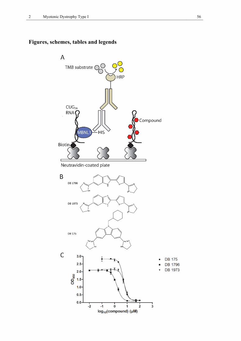
2 Myotonic Dystrophy Type I 56
Figures, schemes, tables and legends

2 Myotonic Dystrophy Type I 57
Figure 8 (II 1): Screening for nucleic acid binding small molecules that disrupt the MBNL1-CUG78 RNA complex in vitro.
Fig. II 1A: CUG78-MBNL1 inhibition screening assay: Biotinylated CUG78 RNA is immobilized and incubated with MBNL1 and compounds. RNA-bound MBNL1 is detected via a primary anti-His and a secondary anti-mouse-HRP antibody. A substrate is added to give an optical signal, correlating to the amount of RNA-bound MBNL1. In between all five incubation steps, washing is performed.
Fig. II 1B: Molecular structure of the identified MBNL1-CUG78 RNA complex inhibitors DB 1796, DB 1973 and DB 175.
Fig. II 1C: MBNL1-CUG78 RNA complex inhibition curves with DB 1796, DB 1973 and DB 175.
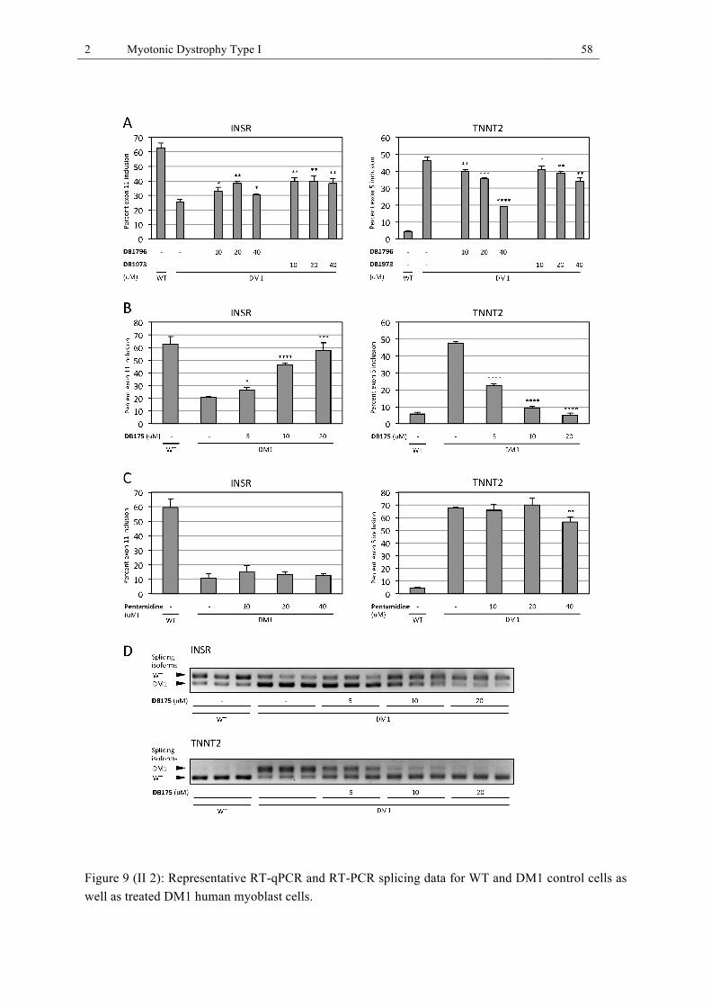
2 Myotonic Dystrophy Type I 58
Figure 9 (II 2): Representative RT-qPCR and RT-PCR splicing data for WT and DM1 control cells as well as treated DM1 human myoblast cells.

2 Myotonic Dystrophy Type I 59
Fig. II 2A,B: Di-imidazolines improve the splicing of the TNNT2 and INSR pre-mRNA. DB175 fully restores the splicing of both pre-mRNAs at 20 µM concentration.
Fig. II 2C: Pentamidine has no effect on splicing of the INSR and TNNT2 pre-mRNA, except for an improvement of TNNT2 splicing at the highest concentration of 40 µM.
Fig. II 2D: Alternative splicing of INSR and TNNT2 pre-mRNA analyzed by RT-PCR and visualized on 3% agarose gels. Shown are the two alternative splicing isoforms for both genes in untreated WT and DM1 control cells and DM1 cells treated with DB175. The WT splicing isoform is the predomi-nant mature splicing variant, whereas the DM1 isoform is the predominant fetal splicing variant.
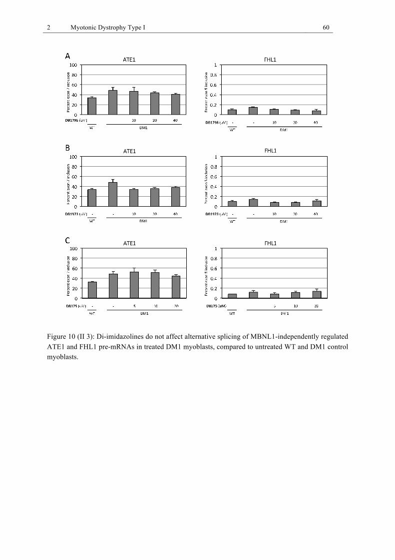
2 Myotonic Dystrophy Type I 60
Figure 10 (II 3): Di-imidazolines do not affect alternative splicing of MBNL1-independently regulated ATE1 and FHL1 pre-mRNAs in treated DM1 myoblasts, compared to untreated WT and DM1 control myoblasts.

2 Myotonic Dystrophy Type I 61
Figure 11 (II 4): FISH detection of CUGn RNA and immunofluorescence detection of MBNL1 in human WT and DM1 myoblasts.
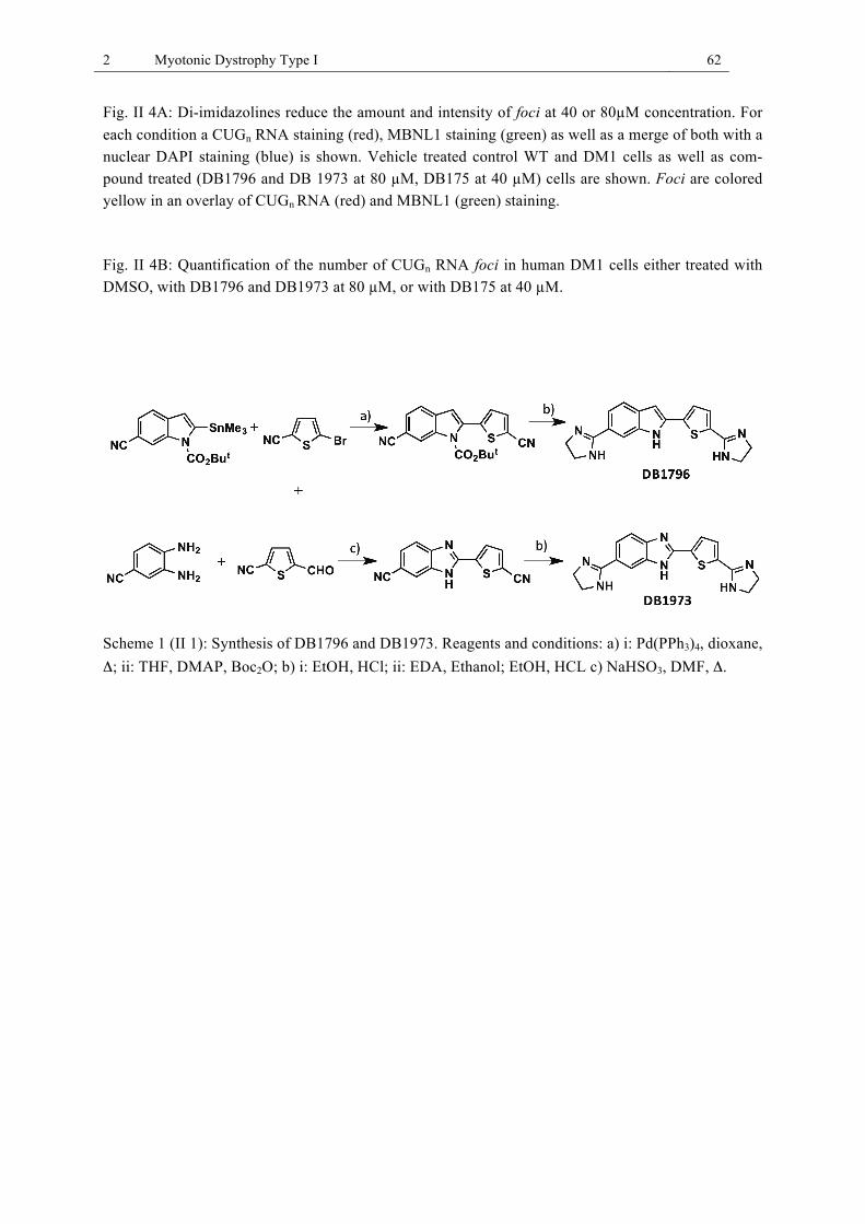
2 Myotonic Dystrophy Type I 62
Fig. II 4A: Di-imidazolines reduce the amount and intensity of foci at 40 or 80µM concentration. For each condition a CUGn RNA staining (red), MBNL1 staining (green) as well as a merge of both with a nuclear DAPI staining (blue) is shown. Vehicle treated control WT and DM1 cells as well as com-pound treated (DB1796 and DB 1973 at 80 µM, DB175 at 40 µM) cells are shown. Foci are colored yellow in an overlay of CUGn RNA (red) and MBNL1 (green) staining.
Fig. II 4B: Quantification of the number of CUGn RNA foci in human DM1 cells either treated with DMSO, with DB1796 and DB1973 at 80 µM, or with DB175 at 40 µM.
Scheme 1 (II 1): Synthesis of DB1796 and DB1973. Reagents and conditions: a) i: Pd(PPh3)4, dioxane, Δ; ii: THF, DMAP, Boc2O; b) i: EtOH, HCl; ii: EDA, Ethanol; EtOH, HCL c) NaHSO3, DMF, Δ.

2 Myotonic Dystrophy Type I 63
Table 1 (II 1): Physicochemical and pharmacokinetic parameters of di-imidazolines and the lead
compound pentamidine.
Compound log D7.4
pKa PAMPA logPe [log10-6 cm/s] / pH PPB [%] pKa
(in silico, Marvin)
pKa (experimental, 1H-NMR)
DB1796 n.d. 9.21 7.09 -
-10 ± 0.0 / 3 -7.3 ± 0.5 / 5
-7.5 ± 1.6 / 7.4 69.5
DB1973 < -1.5 9.02 6.63 3.56
7.83 ± 0.35 -
0.61 ± 0.07
-9.4 ± 1.2 / 3 -9.4 ± 1.3 / 5
-9.4 ± 1.3 / 7.4 n.d.
DB175 n.d. 8.85 9.50 -
-9.5 ± 1.1 / 3 -9.3 ± 1.5 / 5
-10 ± 0.0 / 7.4 97.1
Pentamidine 0.144 ± 0.003 (98) 12.13 11.53 -
-9.1 ± 1.1 / 3 -9.2 ± 1.2 / 5
-9.4 ± 0.8 / 7.4 41 ± 5 (98)
2 Myotonic Dystrophy Type I 64
2.7 Discussion
As both of our studies are discussed in detail in the individual manuscripts, the scope of this
general discussion will be to give an overview over both studies, to compare them, and to
draw the main conclusions, while providing some complementary information. In the follow-
ing, the study with natural compounds (manuscript I) will be referred to as study 1 (S1)
whereas the study with synthetic antiprotozoal compounds (manuscript II) will be referred to
as study 2 (S2).
Screening assay
The development of a novel in vitro CUG78-MBNL1 inhibition assay, using DMPK-CUG78
RNA transcribed from DNA of a DM1 patient and recombinant MBNL1-HIS, permitted
successful screening of compound libraries to identify CUG78-MBNL1 complex inhibitors.
Initially, we developed a thermal shift assay, based on the Differential Scanning Fluorimetry
(DSF)/Thermofluor method (103). MBNL1 thermodenaturation was monitored with an envi-
ronmentally sensitive fluorescent dye (SYPRO Orange), which becomes fluorescent upon
binding to hydrophobic patches of the unfolding protein. CUG78-RNA stabilized MBNL1 and
resulted in a shifted MBNL1 thermodenaturation curve of 3 – 4 °C higher Tm values. Success-
ful CUG78-MBNL1 complex inhibitors would decrease MBNL1 ΔTm values. This assay was
eventually deemed unsuitable for screening, because compound-intrinsic fluorescence inter-
fered with the fluorescent readout of the assay. As nucleic acid binders are often aromatic,
with extended π-electron systems, they are prone to this kind of interference. We developed
an ELISA-type assay instead, i.e. the CUG78 MBNL1-inhibition assay. By washing, com-
pounds were removed before the readout of a photometric signal that correlates to the amount
of CUG78-bound MBNL1. Compared to a similar assay described by Pushechnikov et al. (53),
instead of binding CUGn RNA to an immobilized capture DNA oligonucleotide (biotinylat-
ed), we directly immobilized CUG78 RNA (biotinylated during in vitro transcription) to neu-
travidin plates. Direct immobilization of RNA is simpler and might allow for straightforward
assay development for other RNA-protein targets. Besides the CUG78 MBNL1-inhibition
assay we also developed an electrophoretic mobility gel shift assay (EMSA), similar to the
one used in the screening study of Warf et al. (51). The EMSA was based on different elec-
trophoretic mobility in agarose gel of both CUG78-RNA and MBNL1, either in free form or in
complexed form. The EMSA was suitable as a secondary assay to confirm complex inhibi-
tors, but not for screening and for quantification of IC50 values.
2 Myotonic Dystrophy Type I 65
Screening hits
The S1 screening of natural substances at 100 µM, or extracts at 100 µg/ml, yielded a low
number of hits compared to the screening of synthetic compounds in S2. This finding makes
sense as the library in S2 contained compounds designed to bind to protozoan DNA. Thus, S2
screening at concentrations of 10 µM yielded a high number of hits that bound CUG78 RNA.
Lowering the concentration to 1 µM or lower allowed for the identification of the best bind-
ers, which inhibited MBNL1 binding to CUG78 RNA at nanomolar concentrations. These
were exclusively of the imidazoline or amidine type, predominantly imidazolines. The three
di-imidazolines DB1796, DB1973 and DB175, which eventually proved to be the most effec-
tive compounds in a DM1 cell model, were not among the best screening hits and had moder-
ate IC50 values between 1 and 6 µM. The weaker inhibitory activity is likely compensated by
better membrane permeability or target selectivity. The S1 screening, in contrast, led to the
identification of mainly alkaloids, with the best complex inhibitors being berberine and
harmine (IC50 values close to 100 µM) and coralyne (IC50 ~ 20 µM). Regarding the biological
effect, the weaker in vitro complex inhibitory activity is likely compensated by good cell
permeability; the two alkaloids are membrane permeable and even pass the blood-brain barri-
er into the CNS where they inhibit monoamino oxidase (MAO) (85, 86). The results from the
two screenings underline the strong influence of the library content, i.e. the type of screened
molecules and chemical diversity, on hit number. The library in S1 was of high structural
diversity and yielded only a few hits, even though the library was much larger than in S2.
Indeed, as the 2128 extracts were compound-mixtures, the actual number of screened mole-
cules was much greater. The screening in S2, in contrast, was a focused screening with mole-
cules belonging to a constricted chemical space, close to the lead compound pentamidine, and
thus yielded a high number of hits. In this case stringent selection by lower screening concen-
trations and further selection in a biological system, such as a DM1 cell model, proved to be
of utmost importance.
Evaluation of hits in human DM1 myoblasts and C2C12 myoblasts
Screening hits from both studies were further analyzed for their ability to improve DM1
associated molecular defects, i.e. mis-splicing and nuclear foci formation. Our primary assay
to test bioactivity was a splicing assay in DM1 myoblasts (DMPK-CUG1300) and wild type
control myoblasts (DMPK-CUG5). We checked whether compound-treated DM1 myoblasts
showed a restoration of splicing of the INSR and the TNNT2 pre-mRNA, two MBNL1-
regulated alternative splicing events (35). A secondary assay analyzed nuclear foci number.
2 Myotonic Dystrophy Type I 66
For splicing analysis we developed a new RT-qPCR method, using two primer pairs for an
alternatively spliced gene. One primer pair detects all alternative splicing variants of a gene
(“pan” primer pair) and the other primer pair detects only those variants including an investi-
gated alternatively spliced exon (“exon” primer pair), by placing either the forward or the
reverse primer directly into the alternative exon. Thereby the amount of the exon-containing
variant can be quantified relative to the total amount of all variants. Compared to classical
RT-PCR, the new method allows for more accurate and convenient quantification of exon
inclusion/exclusion by fluorescence readout with a real-time qPCR machine. RT-PCR, as an
end-point PCR, is heavily dependent on choosing the optimal PCR cycle number (amplifica-
tion time), which should lie in the exponential amplification phase for the splicing variants to
be analyzed. In addition, analysis of DNA band densities on agarose gels is a source of exper-
imental inaccuracy. Lastly, different amplicons (splicing variants) are amplified with different
effectivity, which is not accounted for in classical RT-PCR.
S1 yielded the three alkaloids, berberine, harmine and coralyne as the strongest CUG78-
MBNL1 complex inhibitors. The first two partially improved splicing in the DM1 myoblasts
whereas coralyne, probably owing to unselective nucleic-acid intercalation, showed no effect
on splicing of the INSR and TNNT2 pre-mRNA. Interestingly, the strongest inhibitors from
S2, with much lower IC50 values were mostly either not active in the cell-splicing assay or
toxic to the DM1 myoblasts. Testing of compounds with intermediate inhibitory activity (1 –
10 µM) led to the identification of the most potent bioactive compounds from S2: DB1796,
DB1973 and DB175, three di-imidazolines. DB175 fully restored splicing of both the INSR
and TNNT2 pre-mRNA at 20 µM concentration and might be an interesting new lead com-
pound for DM1 drug discovery. Overall, imidazolines were more potent in the splicing assay
compared to amidines, e.g. the lead compound pentamidine, which we suggest is due to fa-
vorable RNA-binding and favorable pharmacokinetic properties of the imidazoline group
compared to amidine (both discussed below). Structure analysis of the strongest inhibitors
showed that the in vitro screening assay selects strong intercalators such as coralyne (74),
with poor target selectivity, and highly charged molecules with poor cell permeability. Such
compounds failed to show a splicing effect in DM1 myoblasts. To circumvent the issues
linked to our in vitro screening assay, high-content cell-based phenotypic screening assays
could be used as primary screening assays, to directly select suitable small molecules in cells
(60). Some of our identified complex inhibitors were toxic at the concentrations used in cell
culture (20, 40 µM), indicating nonselective RNA/DNA binding. Some compounds also
showed a worsening effect on alternative splicing, e.g. berberine on INSR splicing, which is

2 Myotonic Dystrophy Type I 67
likely caused by their binding to nucleotide sequence motifs shared between the toxic CUGn
RNA and the target pre-mRNAs of MBNL1. MBNL1 binds to pyrimidine-embedded CG
motifs (YGCY) present in both types of RNA (78, 104). MBNL1-binding to these motives
upstream of an alternative exon promotes exon exclusion, while binding downstream of the
alternative exon promotes its inclusion (78, 104). Some compounds might therefore also bind
to MBNL1 natural target motifs and interfere with alternative splicing. Selectivity has been
best addressed by modular assembly of CUG-binding small molecules, with the resulting
oligovalent compounds binding more strongly to the toxic DMPK CUGn repeats with multiple
binding sites compared to MBNL1 target pre-mRNAs with fewer binding sites (90). These
polymers are, however, not suitable as therapeutics due to their membrane impermeability in
vivo. Our identified small molecules were not tested for in vitro binding of MBNL1 target
pre-mRNAs and other off-target RNA binding. CUG78-MBNL1 complex inhibition by these
compounds in vitro was not impaired by addition of an excess of yeast tRNA (200 nM). Fur-
thermore, we analyzed the compounds’ selectivity for MBNL1-dependent splicing events in
the DM1 myoblast model. The alkaloids as well as the di-imidazolines did not affect the
alternative splicing of two MBNL1-idependently spliced genes ATE1 and FHL1 (72), com-
pared to both wild-type and DM1 untreated control myoblasts. All of the identified bioactive
small molecules from S1 and S2 were furthermore evaluated in a cell viability assay with
C2C12 mouse myoblasts to determine Tox IC50 values which were all above 120 µM. The
compound concentrations used in the DM1 myoblast splicing assay (20, 40 µM) were below
the Tox IC50 values but still relatively close. The rather narrow therapeutic window is an issue
with the identified compounds.
Physicochemical and pharmacokinetic properties of identified compounds
The alkaloids berberine, its derivatives palmatine and dihydroberberine, and harmine are cell
permeable, even known to cross the blood brain barrier (85, 86). In spite of weaker in vitro
IC50 values than the di-imidazolines from S2, the alkaloids readily improved molecular de-
fects in the DM1 myoblast model at the same concentrations (20, 40 µM). They apparently
make up for their weaker CUGn RNA affinity with better cell permeability. We therefore did
not investigate physicochemical/pharmacokinetic parameters for the alkaloids. Such parame-
ters were investigated for the synthetic compounds from S2, which were more problematic,
due to their positive charges. Their in vitro analysis revealed little to no cell permeability (low
log Pe values) and high hydrophilicity (low log D7.4 values). Still, the lower pKa values of the
imidazoline group compared to the amidine group predict a small amount of unprotonated,
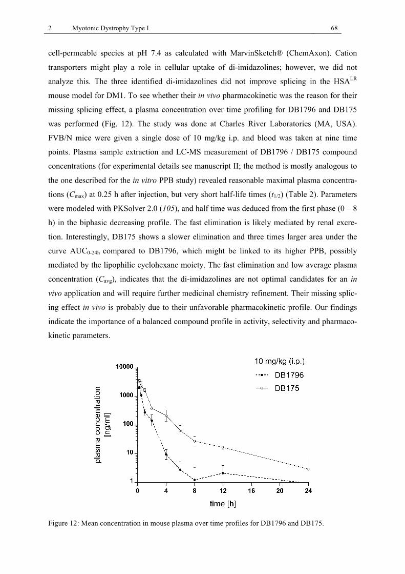
2 Myotonic Dystrophy Type I 68
cell-permeable species at pH 7.4 as calculated with MarvinSketch® (ChemAxon). Cation
transporters might play a role in cellular uptake of di-imidazolines; however, we did not
analyze this. The three identified di-imidazolines did not improve splicing in the HSALR
mouse model for DM1. To see whether their in vivo pharmacokinetic was the reason for their
missing splicing effect, a plasma concentration over time profiling for DB1796 and DB175
was performed (Fig. 12). The study was done at Charles River Laboratories (MA, USA).
FVB/N mice were given a single dose of 10 mg/kg i.p. and blood was taken at nine time
points. Plasma sample extraction and LC-MS measurement of DB1796 / DB175 compound
concentrations (for experimental details see manuscript II; the method is mostly analogous to
the one described for the in vitro PPB study) revealed reasonable maximal plasma concentra-
tions (Cmax) at 0.25 h after injection, but very short half-life times (t1/2) (Table 2). Parameters
were modeled with PKSolver 2.0 (105), and half time was deduced from the first phase (0 – 8
h) in the biphasic decreasing profile. The fast elimination is likely mediated by renal excre-
tion. Interestingly, DB175 shows a slower elimination and three times larger area under the
curve AUC0-24h compared to DB1796, which might be linked to its higher PPB, possibly
mediated by the lipophilic cyclohexane moiety. The fast elimination and low average plasma
concentration (Cavg), indicates that the di-imidazolines are not optimal candidates for an in
vivo application and will require further medicinal chemistry refinement. Their missing splic-
ing effect in vivo is probably due to their unfavorable pharmacokinetic profile. Our findings
indicate the importance of a balanced compound profile in activity, selectivity and pharmaco-
kinetic parameters.
Figure 12: Mean concentration in mouse plasma over time profiles for DB1796 and DB175.

2 Myotonic Dystrophy Type I 69
Table 2: In vivo pharmacokinetic parameters of DB1796 and DB175.
Compound AUC0-24h [ng/ml*h]
Cmax
[ng/ml] Cavg [ng/ml]
t1/2
[min] PPB [%]
DB1796 1464.5 2123.3 61.0 28.6 75.7
DB175 4467.8 3513.3 186.2 46.1 92.6
In vivo evaluation of bioactive compounds in the HSALR mouse model
Three alkaloids from S1 showed a partial improvement of DM1 characteristic mis-splicing of
the chloride channel CLCN1 in the HSALR model, i.e. the two close berberine derivatives
dihydroberberine (DHB) and palmatine, as well as harmine. DHB also caused a trend towards
increased levels of CLCN1 protein. However, the alkaloids induced toxicity in mice, an issue
which will have to be addressed in future studies with these or similar compounds. In vivo
experiments with the three di-imidazolines from S2 showed no splicing improvement. These
findings are in line with the in vitro pharmacokinetic analysis, indicating low membrane
permeability, and the in vivo pharmacokinetic profile for DB1796 and DB175, showing low
average plasma concentrations and fast elimination from mouse plasma (Fig. 12).
2.8 Conclusion and Outlook
Our drug discovery effort for DM1 led to the identification of several alkaloids in S1 and di-
imidazolines in S2. These compound classes are of interest for future DM1 drug discovery
studies and studies in other RNA-mediated diseases. The identified small molecules can serve
as valuable starting points for medicinal chemistry programs. In addition, these compounds
could be used to synthesize high-affinity/selectivity polymers that account for the repeating
structure of the binding sites on CUGn RNA, as was performed in the study of Lee et al. (90).
Taken together, the results from both of our studies show that a balanced activity, selectivity
(toxicity) and pharmacokinetic profile is key for an interesting lead compound in DM1 and
RNA drug discovery in general. As nucleic acid binders often contain positive charges at
physiological pH the refinement of the pharmacokinetic profile to allow for cell membrane
permeation is crucial. The compounds identified in our studies have therapeutic potential, but
also clear deficits that will have to be addressed in future studies.
2 Myotonic Dystrophy Type I 70
Alkaloids are an interesting compound class for DM1 drug discovery
The three strongest MBNL1-CUG78 complex inhibitors identified in the natural compound
screening (S1) were alkaloids. berberine, harmine and coralyne have all been described before
to bind to certain RNA targets, i.e. tRNA, double stranded RNA, or polyA RNA (74-77). In
the context of the DM1 characteristic CUGn RNA, only the in vitro screening study by Chen
et. al. yielded alkaloids as hits; out of six hits, four were alkaloids or alkaloid derivatives of
the opioid and ergot alkaloid type, but none of these alkaloids was confirmed in a second
screening assay (58). Altogether, the mentioned findings and our results support the notion
that alkaloids are of interest in RNA drug discovery due to certain chemical attributes that
enable RNA-binding. Possible interactions include stacking with nucleobases via aromatic
groups, hydrogen bonding with the Watson-Crick or Hoogsteen face of nucleic acids, either
as acceptors (nitrogen and oxygen groups) or as donors (hydroxyl and amine groups), and
electrostatic interaction with the negatively charged phosphate backbone of RNA through
positively charged nitrogen groups of the alkaloids. Isochinolin, beta-carboline, indole, opioid
and ergot alkaloids etc. might be of considerable valuable for RNA drug discovery, as poten-
tial therapeutic molecules or as lead compounds.
Di-imidazolines – an interesting alternative to diamidines
The focused screening of synthetic pentamidine-related substances in S2 led to the identifica-
tion of three di-imidazolines that outperformed the lead compound pentamidine in several
aspects: notably in target RNA binding, bioactivity in the DM1 myoblasts, lower toxicity and
probably improved membrane permeation. The imidazoline group seems to enable strong
RNA-binding and was superior to amidines, tetrahydropyrimidines, arylimidamides and other
related compounds in our in vitro and cellular assays. Also Parkesh et al. described a few
imidazolines as good CUGn-RNA binders (92). The molecular rationale behind strong RNA
binding of the imidazoline group lies in its planarity. Thermal melting studies on poly(A)•(U)
RNA with three aromatic diphenylfuran compounds (93), i.e. a diphenylfuran imidazoline (1),
a diphenylfuran amidine (2), and a diphenylfuran tetrahydropyrimidine (3) had shown that
RNA binding/stabilization, quantified via ∆Tm, was in the order (1) > (2) > (3) (Table 3).
Molecular modeling studies with the three compounds and double stranded A8•U8 RNA
showed that these compounds intercalate via the major RNA groove, and that steric clashes of
all three diphenylfuran substituents impaired binding via the minor groove (100). The NCN
bond angle was found to be smaller in the five-membered imidazole (1) compared to the
torsion angle in compounds (2) and (3). The planar imidazoline (1) slid more deeply into the
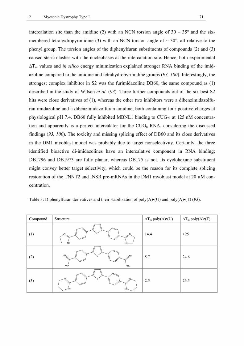
2 Myotonic Dystrophy Type I 71
intercalation site than the amidine (2) with an NCN torsion angle of 30 – 35° and the six-
membered tetrahydropyrimidine (3) with an NCN torsion angle of ~ 30°, all relative to the
phenyl group. The torsion angles of the diphenylfuran substituents of compounds (2) and (3)
caused steric clashes with the nucleobases at the intercalation site. Hence, both experimental
∆Tm values and in silico energy minimization explained stronger RNA binding of the imid-
azoline compared to the amidine and tetrahydropyrimidine groups (93, 100). Interestingly, the
strongest complex inhibitor in S2 was the furimidazoline DB60, the same compound as (1)
described in the study of Wilson et al. (93). Three further compounds out of the six best S2
hits were close derivatives of (1), whereas the other two inhibitors were a dibenzimidazolfu-
ran imidazoline and a dibenzimidazolfuran amidine, both containing four positive charges at
physiological pH 7.4. DB60 fully inhibited MBNL1 binding to CUG78 at 125 nM concentra-
tion and apparently is a perfect intercalator for the CUGn RNA, considering the discussed
findings (93, 100). The toxicity and missing splicing effect of DB60 and its close derivatives
in the DM1 myoblast model was probably due to target nonselectivity. Certainly, the three
identified bioactive di-imidazolines have an intercalative component in RNA binding;
DB1796 and DB1973 are fully planar, whereas DB175 is not. Its cyclohexane substituent
might convey better target selectivity, which could be the reason for its complete splicing
restoration of the TNNT2 and INSR pre-mRNAs in the DM1 myoblast model at 20 µM con-
centration.
Table 3: Diphenylfuran derivatives and their stabilization of poly(A)•(U) and poly(A)•(T) (93).
Compound Structure ∆Tm poly(A)•(U) ∆Tm poly(A)•(T)
(1)
14.4 >25
(2)
5.7 24.6
(3)
2.5 26.5
O
HN
NN
NH
O NHHN
H2N NH2
ON
NH HN
N
2 Myotonic Dystrophy Type I 72
The di-imidazolines DB1796, DB1973 and DB175 provide interesting compounds for RNA
targeting and valuable starting points for medicinal chemistry studies. These compounds’
selectivity and pharmacokinetic profile pose the biggest challenge for development as poten-
tial DM1 drugs. For further studies, we suggest to synthesize analogs of the identified imidaz-
olines, e.g. imidazole, triazole or tetrazole analogs, all with lower pKa values and thus lower
charge and better membrane permeability. Whether RNA binding will be maintained is to be
evaluated. A synthesized pyrazole analogue of DB175 (DB2323) was inactive in the DM1
myoblast model, indicating the importance of the H-bond donating NH group of imidazoline.
In addition, other liphophilic substituents than cyclohexane should be investigated for the lead
compound DB175. Interestingly, DB175 from S2 and harmine from S1 can be regarded as
derivatives. Therefore, two different screening studies yielded related compounds.

3 Anti-Myelin Associated Glycoprotein Neuropathy 73
3 Anti-Myelin Associated Glycoprotein Neuropathy
3.1 Introduction
Anti-MAG neuropathy is a rare disorder of the peripheral nervous system characterized by
slowly progressive demyelinating, predominantly sensory neuropathy (8). Patients have high
serum levels of monoclonal immunoglobulin M (IgM) autoantibodies that react with the
human natural killer 1 (HNK-1) epitope on the myelin-associated glycoprotein (MAG) and on
other myelin components. These antibodies are thought to be pathogenic (8, 106). The disease
prevalence is not exactly known, but is expected to be similar to the prevalence of a related
form of neuropathy called multifocal motor neuropathy (MMN), which affects about 1 – 2
persons in 100’000 people (107). Anti-MAG neuropathy affects mostly men over 50 years old
(108). Patients develop sensory loss starting in the toes and fingers, sensory ataxia, and tremor
in the hands and legs. As the disease progresses, the tremor becomes more severe and the
ataxia steadily worsens. Due to extensive muscle weakness some patients become severely
disabled and wheelchair-bound (106).
Classification of anti-MAG neuropathy
The peripheral nerve disorder anti-MAG neuropathy can be categorized as a member of the
spectrum of immune-mediated peripheral neuropathies / polyneuropathies. There are acute
and chronic types of immune-mediated peripheral neuropathies, whereby anti-MAG neuropa-
thy is of the chronic type. This disease develops in patients with IgM monoclonal gammopa-
thy of unknown significance (MGUS) and is characterized by autoreactivity against a carbo-
hydrate epitope on MAG. Monoclonal gammopathy or paraproteinemia describes the pres-
ence of excessive amounts of a monoclonal immunoglobulin. Monoclonality is thought to
arise from a somatically hypermutated B cell clone, acquiring IgM secretory activity (109,
110). In the following, a short overview will be given on immune-mediated peripheral neu-
ropathies for which anti-carbohydrate antibodies have been identified that target various
components of myelin. This overview is based on a recent review written by three leading
experts in the field of peripheral neuropathy: Andreas Steck, Nobuhiro Yuki and Francesc
Graus (111).

3 Anti-Myelin Associated Glycoprotein Neuropathy 74
Acute immune-mediated neuropathies with anti-carbohydrate antibodies
In the group of acute immune-mediated neuropathies, the Guillain-Barré syndrome (GBS)
encompasses several disease conditions that often involve autoantibodies against endogenous
nerve glycoepitopes. The two major subgroups among GBS are the acute motor axonal neu-
ropathy (AMAN) and the acute inflammatory demyelinating polyneuropathy (AIDP), with
AMAN predominantly affecting motor nerves compared to AIDP. Both AMAN and AIDP
can involve autoantibodies against gangliosides such as GM1, GM1b, GD1a and GalNAc-
GD1a. Another clinically distinct subgroup of GBS is the Fisher syndrome, which is mainly
associated with antibodies against the GQ1b or the GT1a epitope. The pathogenic autoanti-
bodies in the group of acute neuropathies are mostly of the IgG isotype (111).
Chronic immune-mediated neuropathies with anti-carbohydrate antibodies
In contrast, in the group of chronic neuropathies, mostly IgM autoantibodies are detected.
With a prevalence as high as 9 in 100’000 (112) the chronic inflammatory demyelinating
polyneuropathy (CIDP) is the most common form of chronic demyelinating polyneuropathy.
CIDP can be considered as the chronic counterpart of the acute GBS. GBS is post-infectious,
while CIDP is an autoimmune disorder. CIDP is usually not associated with anti-carbohydrate
antibodies. The two other major disease groups among the chronic inflammatory neuropathies
are the anti-MAG neuropathy and multifocal motor neuropathy (MMN). The anti-MAG
neuropathy mainly involves autoantibodies against the HNK-1 epitope, present on multiple
myelin antigens such as MAG, SGPG, SGLPG, P0 and PMP22. MMN patients often show
autoantibodies against the ganglioside GM1. Other, less frequent, chronic neuropathies en-
compass the chronic sensory axonal neuropathy with anti-sulfatide antibodies, the chronic
motor neuropathy with GD1a or GD1b antibodies, and the CANOMAD (chronic ataxic neu-
ropathy, opthalmoplegia, M-protein, agglutination, disialosyl antibodies) with antibodies
against disialosyl gangliosides, such as GQ1b and GD1b. Another subgroup of rare chronic
neuropathies involves IgM autoantibodies against chondroitin or heparan sulfate (111).
Neuropathies associated with IgM monoclonal gammopathy
Among patients with IgM monoclonal gammopathy (M-protein) the prevalence of neuropathy
is estimated to be between 5 to 31% (113, 114). About 50% of IgM antibodies in patients with
IgM gammopathy-associated neuropathy react with the HNK-1 epitope; other targets include
gangliosides, sulfatide, chondroitin and heparan sulfate (108). IgM monoclonal gammopathy
and also neuropathy can be caused by IgM-secreting lympho-proliferative diseases, e.g. Wal-
denström’s macroglobulinemia, lymphoma, myeloma, or chronic lymphocytic leukemia.

3 Anti-Myelin Associated Glycoprotein Neuropathy 75
However, most neuropathies associated with IgM gammopathy are not linked to one of these
lymphoproliferative disorders and are denoted as MGUS (115). In MGUS patients, neuropa-
thy is normally the only clinical symptom caused by the underlying hematologic disorder
(108). The IgM antibodies in most of the MGUS patients react with the HNK-1 epitope,
implying that anti-MAG neuropathy is the most common form of neuropathy in the MGUS
group. Nevertheless, the cause of its underlying lymphoproliferation is not known (8, 116).
3.2 Molecular Disease Mechanism
Discovery of IgM autoantibodies and their antigen on myelin
Anti-MAG neuropathy was first characterized and described as a distinct clinical entity in the
early 80s. Back then, it was known that monoclonal IgM autoantibodies from patients with
paraproteinemia and peripheral neuropathy react with a myelin protein component of the PNS
and an analogous component of the CNS. Studies by Braun et al. and Steck et al. described
for the first time the specific antigen for the monoclonal IgM antibodies: the myelin protein
MAG (117, 118). At the same time McGarry et al. showed that the HNK-1 antibody, which
was raised against a membrane antigen of T cells, cross-reacted with MAG (119, 120). Sub-
sequently, it was shown that the epitope for the patients’ IgM antibodies resided in the carbo-
hydrate component of MAG, as shown by loss of antibody reactivity upon deglycosylation of
human MAG (121). The same study by Ilyas et al. had furthermore described cross-reactivity
of patient IgM antibodies with glycolipids in the ganglioside fraction of human peripheral
nerve. These glycolipids were later on described to lack sialic acid and to contain 3-O-
sulfoglucuronic acid (SO3-3GlcA) as a specific epitope hallmark (122, 123). Finally, the
structure of two immunoreactive glycolipids of the peripheral nervous system was described:
sulfoglucuronyl paragloboside (SGPG) and sulfoglucuronyl lactosaminyl paragloboside
(SGLPG) (124). Interestingly, Chou et al. had also shown that both IgM antibodies from
neuropathy patients and the HNK-1 antibody bind to these glycolipids (122, 123). Taken
together, these findings indicated a shared carbohydrate epitope on MAG, SGPG and SGLPG
for IgM antibodies from patients and the HNK-1 antibody.
In 1996, the same carbohydrate epitope was also identified on the myelin glycoprotein zero
(P0) and its structure was elucidated from bovine P0 (125). The structural overlap between
the pentasaccharide of SGPG, the heptasaccharide of SGLPG and the epitope of P0 was re-
stricted to the terminal trisaccharide at the non-reducing end of these polysaccharides. Hence,

3 Anti-Myelin Associated Glycoprotein Neuropathy 76
the structure of the HNK-1 epitope was defined as SO3-3-GlcA(β1-3)Gal(β1-4)GlcNAc-OH
(Fig. 13).
Figure 13: The HNK-1 trisaccharide epitope.
Microheterogeneity of anti-MAG antibodies
Evidence from several studies underline that the specificities of the patient IgM antibodies
and the HNK-1 antibody are similar; that is to say that they react with glycoconjugates of
peripheral myelin that share the HNK-1 epitope, e.g. the glycoproteins MAG, P0, peripheral
myelin protein-22 (PMP22), as well as the PNS-specific glycolipids SGPG and SGLPG (111,
126). Although the HNK-1 epitope is established as the common antigenic determinant for
IgM antibodies of patients with anti-MAG neuropathy, there is microheterogeneity in binding
strength and structural requirements for binding of anti-MAG antibodies (127). A study by
Ilyas et al. on IgM antibody binding to SGPG and derivatives thereof had shown previously
that the antibodies differ in their structural requirements for binding (128). The study showed
that for SGPG binding some anti-MAG IgM antibodies absolutely required the sulfate but not
the carboxyl group of the HNK-1 glucuronic acid, whereas for others it was vice versa, and a
third group of antibodies bound to the glycolipid as long as one of the two negatively charged
groups was present. All of the antibodies, including the HNK-1 antibody, bound best to natu-
ral SGPG, containing both negatively charged groups (128). Two other studies emphasized
the importance of the carbohydrate chain length for antibody binding (129, 130). IgM anti-
bodies were shown to bind more strongly to glycolipids with longer carbohydrate chains. The
two terminal sugars SO3-3-GlcA(β1-3)Gal seemed to be the minimum requirement for IgM
antibody binding. Whether the additional monosaccharides towards the reducing end of the
oligosaccharide chain are involved in binding or merely serve as a spacer could not be deter-
mined. Idiotype heterogeneity is another characteristic of anti-MAG antibodies (131). There-
fore, development of an anti-idiotype antibody for a patients’ IgM yielded a therapeutically
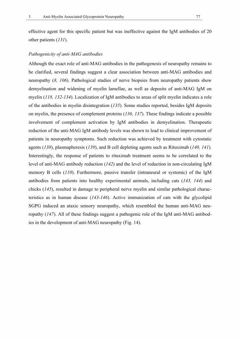
3 Anti-Myelin Associated Glycoprotein Neuropathy 77
effective agent for this specific patient but was ineffective against the IgM antibodies of 20
other patients (131).
Pathogenicity of anti-MAG antibodies
Although the exact role of anti-MAG antibodies in the pathogenesis of neuropathy remains to
be clarified, several findings suggest a clear association between anti-MAG antibodies and
neuropathy (8, 106). Pathological studies of nerve biopsies from neuropathy patients show
demyelination and widening of myelin lamellae, as well as deposits of anti-MAG IgM on
myelin (118, 132-134). Localization of IgM antibodies to areas of split myelin indicates a role
of the antibodies in myelin disintegration (135). Some studies reported, besides IgM deposits
on myelin, the presence of complement proteins (136, 137). These findings indicate a possible
involvement of complement activation by IgM antibodies in demyelination. Therapeutic
reduction of the anti-MAG IgM antibody levels was shown to lead to clinical improvement of
patients in neuropathy symptoms. Such reduction was achieved by treatment with cytostatic
agents (138), plasmapheresis (139), and B cell depleting agents such as Rituximab (140, 141).
Interestingly, the response of patients to rituximab treatment seems to be correlated to the
level of anti-MAG antibody reduction (142) and the level of reduction in non-circulating IgM
memory B cells (110). Furthermore, passive transfer (intraneural or systemic) of the IgM
antibodies from patients into healthy experimental animals, including cats (143, 144) and
chicks (145), resulted in damage to peripheral nerve myelin and similar pathological charac-
teristics as in human disease (143-146). Active immunization of cats with the glycolipid
SGPG induced an ataxic sensory neuropathy, which resembled the human anti-MAG neu-
ropathy (147). All of these findings suggest a pathogenic role of the IgM anti-MAG antibod-
ies in the development of anti-MAG neuropathy (Fig. 14).

3 Anti-Myelin Associated Glycoprotein Neuropathy 78
Figure 14: IgM autoantibodies target the HNK-1 epitope on MAG and other myelin components. Graphic modified from Van der Pol WL et al., J. Clin. Immunol. 30, 79-83 (2010).
The role of MAG in anti-MAG neuropathy
The glycoprotein MAG belongs to the family of sialic acid-binding immunoglobulin-like
lectins (siglecs) and is classified as siglec-4 (148, 149). It is an I-type lectin, preferentially
binding α2,3-linked N-acetylneuraminic acid (Neu5Ac, sialic acid) (126). Besides its capacity
to bind carbohydrate receptors, MAG is heavily glycosylated, whereby 30% of the molecule’s
weight constitutes heterogeneous N-linked oligosaccharides (150). All eight N-glycosylation
sites can carry the HNK-1 carbohydrate epitope involved in the anti-MAG neuropathy (151,
152). Of all proteins expressing this epitope, MAG has the highest HNK-1 density, which
could explain the strong binding of patients’ IgM antibodies compared to other myelin gly-
coconjugates (153). In accordance, Dalakas reports a 10 to 100 fold stronger binding of IgM
antibodies to MAG compared to SGPG, which has implications for diagnostic detection of
low-affinity anti-MAG antibodies that can be missed in SGPG-based assays (106). MAG is
located mainly in periaxonal membranes of oligodendroglial cells in the CNS and Schwann
cells in the PNS (154). Structural details of rat MAG revealed five extracellular immuno-
globulin-like domains, a single transmembrane domain and a cytoplasmic domain, which
occurs in a short and a long isoform (S-MAG, L-MAG), due to alternative splicing (155-157).
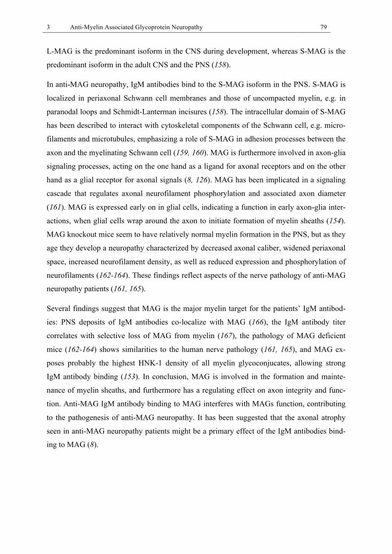
3 Anti-Myelin Associated Glycoprotein Neuropathy 79
L-MAG is the predominant isoform in the CNS during development, whereas S-MAG is the
predominant isoform in the adult CNS and the PNS (158).
In anti-MAG neuropathy, IgM antibodies bind to the S-MAG isoform in the PNS. S-MAG is
localized in periaxonal Schwann cell membranes and those of uncompacted myelin, e.g. in
paranodal loops and Schmidt-Lanterman incisures (158). The intracellular domain of S-MAG
has been described to interact with cytoskeletal components of the Schwann cell, e.g. micro-
filaments and microtubules, emphasizing a role of S-MAG in adhesion processes between the
axon and the myelinating Schwann cell (159, 160). MAG is furthermore involved in axon-glia
signaling processes, acting on the one hand as a ligand for axonal receptors and on the other
hand as a glial receptor for axonal signals (8, 126). MAG has been implicated in a signaling
cascade that regulates axonal neurofilament phosphorylation and associated axon diameter
(161). MAG is expressed early on in glial cells, indicating a function in early axon-glia inter-
actions, when glial cells wrap around the axon to initiate formation of myelin sheaths (154).
MAG knockout mice seem to have relatively normal myelin formation in the PNS, but as they
age they develop a neuropathy characterized by decreased axonal caliber, widened periaxonal
space, increased neurofilament density, as well as reduced expression and phosphorylation of
neurofilaments (162-164). These findings reflect aspects of the nerve pathology of anti-MAG
neuropathy patients (161, 165).
Several findings suggest that MAG is the major myelin target for the patients’ IgM antibod-
ies: PNS deposits of IgM antibodies co-localize with MAG (166), the IgM antibody titer
correlates with selective loss of MAG from myelin (167), the pathology of MAG deficient
mice (162-164) shows similarities to the human nerve pathology (161, 165), and MAG ex-
poses probably the highest HNK-1 density of all myelin glycoconjucates, allowing strong
IgM antibody binding (153). In conclusion, MAG is involved in the formation and mainte-
nance of myelin sheaths, and furthermore has a regulating effect on axon integrity and func-
tion. Anti-MAG IgM antibody binding to MAG interferes with MAGs function, contributing
to the pathogenesis of anti-MAG neuropathy. It has been suggested that the axonal atrophy
seen in anti-MAG neuropathy patients might be a primary effect of the IgM antibodies bind-
ing to MAG (8).
3 Anti-Myelin Associated Glycoprotein Neuropathy 80
3.3 Current Therapeutic Approaches
Patients with anti-MAG neuropathy are treated with immunosuppressive or immunomodula-
tory drugs. Treatments aim at reducing IgM antibody titers by either antibody removal or
reduction of its production by targeting of the expanded monoclonal B cell clone, or blockage
of the assumed effector mechanisms, e.g. complement activation. Paraproteinemic polyneu-
ropathies, such as anti-MAG neuropathy, are potentially treatable, and for some of these
diseases there are treatment options (106). For anti-MAG neuropathy, however, to date no
treatment has been shown to be satisfactorily effective, hence there is a clear need for new and
better treatments. In the following section, a compilation of the evaluated treatments will be
given with a specific focus on the currently used therapies.
Chemotherapeutic / cytotoxic drugs
Chemotherapy and cytotoxic drugs have been used since the 80s and a few patients were
reported to improve clinically under treatment, whereas the majority of patients were non-
responders (138). Cytostatic drugs used in treatment are fludarabine (168) and cladribine
(169), which are both purine analogues. Cyclophosphamide has been used in combination
with plasma exchange (PE) (170), and in combination with Rituximab (171). In the case of an
underlying lymphoproliferative disorder, such as Waldenström’s macroglobulinemia, agents
like bendamustine, bortezomib and thalidomide-based therapies have been used in combina-
tion with Rituximab (172). A general issue with cytostatic therapeutics is their side effects
and non-specific immunosuppression; severe and even fatal adverse effects have been report-
ed for some patients (173). There are no controlled trials with any of these treatments (174).
Plasma exchange / plasmapheresis
Plasma exchange (PE) and plasmapheresis has been reported to ameliorate neuropathy in
patients either alone or in combination therapy (139, 170). Therapy aims to lower the IgM
antibody load by removing circulating antibodies and replacing the patients’ plasma with
clean substitute plasma. Case reports show benefits lasting from 8 to 36 month, but there are
no studies showing a significant benefit of PE (174).
Intravenous immunoglobulin / IVIg
Immunoglobulin of healthy donors has been used in treatment with the aim to “normalize” the
immunoglobulin of patients. According to two placebo-controlled trials, one double-blind,
there may be a short term benefit of IVIg treatment of a few weeks after infusion (174, 175).
3 Anti-Myelin Associated Glycoprotein Neuropathy 81
Corticosteroids
Intravenous dexamethasone and oral prednisolone, both immunosuppressive glucocorticoids,
have been used in monotherapy, but also in combination therapy with PE or with cytotoxic
drugs, e.g. azathioprine, cyclophosphamide, and chlorambucil. There is, however, no data
showing statistically significant clinical improvements under corticosteroid treatment (174).
Interferon alpha-2a
Controlled trials with interferon alpha-2a have reported contradictory results. An open trial
with an IVIg treated control group had shown a benefit of interferon over IVIg (176). A new-
er, placebo-controlled, double-blind trial with interferon, however, did not show a significant
benefit of interferon (177).
Rituximab
The antibody rituximab has been discussed as being the most promising treatment for anti-
MAG neuropathy over the last years; it causes antibody-dependent cell and complement-
mediated cytotoxicity of CD20+ B-cells (8, 106, 140). Placebo-controlled trials with rituxi-
mab showed marked reduction of CD20+ B-cells and also in the anti-MAG antibody titers.
The Dalakas study showed significant clinical improvement. After 8 months, 4 of 13 rituxi-
mab-treated patients improved by ≥ 1 INCAT (Inflammatory Neuropathy Cause and Treat-
ment) score compared with 0 of 13 placebo-treated patients (p = 0.096). Excluding one ritux-
imab-randomized patient who had normal INCAT score at entry, and thus could not improve,
the results were significant (p = 0.036). The time to walk 10 meters was significantly reduced
in the rituximab group (p = 0.042). Clinically, walking improved in 7 of 13 rituximab-treated
patients (178). The more recent Leger study failed to show significant improvement in prima-
ry outcome measures (179). Nevertheless, significant improvement in secondary outcome
measures was reported. Interestingly, clinical improvement has been correlated to the effec-
tiveness of reduction of B cells and IgM antibodies (173), whereas a high load of IgM
memory B cells and the persistence of these cells has been correlated with a poor clinical
response (110). Clinical worsening has also been described for a few patients treated with
rituximab (180, 181). This paradoxical worsening might be linked to an “IgM flare”, a release
of paraprotein (anti-MAG IgM) due to rituximab-induced B cell lysis (182). Nevertheless,
most patients with anti-MAG neuropathy are currently treated with rituximab. According to
comments by Prof. M.C. Dalakas at the ICNMD 2014, it will be crucial to define relevant
primary endpoints for future trials with Rituximab or one of its successors, e.g. Eculizumab
(Soliris®), Belimumab (Benlysta®), Ocrelizumab, Ofatumumab (Arzerra®), Obinutuzumab
3 Anti-Myelin Associated Glycoprotein Neuropathy 82
(Gazyvaro/Gazyva®), which might be more efficient in inhibiting B-cells or the complement
system. According to comments by J.-M. Leger at the ICNMD 2014, painful paresthesias and
sensory ataxia (loss of sensory input in the control of movement) are the most commonly
observed disease symptoms, but sensory ataxia is not captured by the scales used in clinical
trials, such as INCAT. He suggested better outcome measures to capture improvement in
sensory ataxia, e.g. a “time for 10 meters walk” test together with additional biomarkers such
as measurement of anti-MAG IgM titers.
3.3.1 Aim of our study
The present work is focused on a new therapeutic and diagnostic approach for the anti-MAG
neuropathy, evaluating the first potentially “antigen-specific” therapy. The HNK-1 carbohy-
drate epitope has attracted considerable interest as a target in anti-MAG neuropathy and com-
pounds binding and blocking specifically the anti-MAG IgM antibodies might be therapeuti-
cally of considerable value. Based on the structure of the HNK-1 epitope, carbohydrate mi-
metics were synthesized that bind to anti-MAG IgM antibodies. Their potential application in
the diagnosis, as well as in the treatment, of anti-MAG neuropathy is of considerable medical
interest.

3 Anti-Myelin Associated Glycoprotein Neuropathy 83
3.4 Manuscript III
Anti-MAG Neuropathy - a Carbohydrate Polymer Effectively Blocks Pathogenic Anti-MAG Antibodies
Ruben Herrendorff1, Fan Yang1, Nicole Schaeren-Wiemers2, Andreas J. Steck2, Beat Ernst1*
1Institute of Molecular Pharmacy, Pharmacenter, University of Basel, Klingelbergstrasse 50,
4056 Basel, Switzerland
2Department of Biomedicine, University Hospital Basel, Hebelstrasse 20, 4031 Basel, Swit-
zerland
* To whom correspondence should be addressed:
To be submitted to Angewandte Chemie.
3 Anti-Myelin Associated Glycoprotein Neuropathy 84
Abstract
Anti-myelin-associated glycoprotein neuropathy is an antibody-mediated demyelinating
peripheral neuropathy. The disease is caused by immunoglobulin M (IgM) autoantibodies
recognizing the human natural killer-1 (HNK-1) carbohydrate epitope. A specific hallmark of
this epitope is an unusual sulfated glucuronic acid. This glyco-epitope is highly expressed in
adhesion molecules, such as the myelin-associated glycoprotein (MAG), which is present in
myelinated nerve fibers. The pathogenicity of the anti-MAG autoantibodies is well estab-
lished. Therefore, antagonists that block the antibody binding to MAG are of therapeutic and
diagnostic interest.
Our approach was to design carbohydrate ligands that block the IgM antibody binding sites by
mimicking the natural epitope. Thus, a minimal HNK-1 epitope, consisting of a sulfated
disaccharide, was synthesized. In a competitive binding assay, it successfully inhibited the
binding of pathogenic IgM antibodies from patient sera to MAG, although only at micromolar
concentrations. A comparison with the unsulfated disaccharide indicated the prerequisite of
the sulfate moiety for antibody binding. To mimic the multivalent nature of the MAG - IgM
antibody interaction, a polylysine polymer presenting the minimal HNK-1 epitope in a multi-
valent manner was prepared. Binding affinity was thereby increased by a factor of at least
34000, with inhibitory activity now being in the low nanomolar range. Potential applications
of this glycopolymer are in the diagnosis of, as well as the treatment of, anti-MAG neuropa-
thy.

3 Anti-Myelin Associated Glycoprotein Neuropathy 85
Introduction
Anti-MAG neuropathy is a demyelinating peripheral neuropathy, caused by monoclonal IgM
autoantibodies recognizing the antigenic HNK-1 carbohydrate epitope. This epitope is found
on MAG and other glycoconjugates of the peripheral nervous system (PNS). The clinical
picture is characterized by a slowly progressive demyelinating, predominantly sensory, neu-
ropathy (8, 117, 118). Cardinal clinical symptoms are sensory ataxia with impaired gait, distal
muscle weakness, and tremors (106). Evidence is strong for a pathogenic role of the anti-
MAG antibodies in demyelination and neuropathy development (8, 106). Histopathological
studies on nerve biopsies from patients show demyelinating lesions and widening of myelin
lamellae, as well as deposits of anti-MAG IgM within myelin sheaths (118, 132). Localization
of anti-MAG IgM antibodies to areas of split myelin indicates a role of the antibodies in
myelin disintegration (135). Besides IgM deposits, some studies reported the presence of
complement proteins on myelin, suggesting a potential inflammatory element in demye-
lination (136, 137). Therapeutic reduction of serum anti-MAG IgM antibody serum titers
leads to clinical improvement of patients in neuropathy symptoms (106, 140) and therapeutic
response seems to be correlated to the level of anti-MAG antibody reduction (142). Passive
transfer of patients’ anti-MAG antibodies to healthy experimental animals resulted in periph-
eral nerve myelin damage (143, 145). Active immunization of cats with the HNK-1 contain-
ing glycolipid SGPG induced autoantibodies against the HNK-1 epitope and caused an ataxic
sensory neuropathy, resembling the human disease (147).
The myelin glycoconjugates that contain the antigenic HNK-1 epitope include the glycopro-
teins MAG, protein zero (P0), peripheral myelin protein 22, as well as the glycolipids sul-
foglucuronyl paragloboside (SGPG) and sulfoglucuronyl lactosaminyl paragloboside
(SGLPG) (126). Several observations suggest MAG as the main target for the IgM antibodies:
(i) Deposits of patients’ antibodies to PNS sites are co-localized with MAG (166), (ii) MAG
is selectively lost from PNS myelin in patients with high antibody titers (167), (iii) the pathol-
ogy of MAG-knockout mice (162, 163) shows similarities to the human nerve pathology
(161, 165), and (iv) MAG has the highest HNK-1 epitope density of all myelin glycoconju-
cates, allowing strong antibody binding (153).
MAG belongs to the family of sialic acid-binding immunoglobulin-like lectins (Siglecs) (148,
149). It is located mainly in periaxonal membranes of oligodendroglial cells in the CNS and
Schwann cells in the PNS (154). It is involved in adhesion and signaling processes at the
3 Anti-Myelin Associated Glycoprotein Neuropathy 86
axon-glia interface, it has been implicated in the formation and maintenance of myelin, and it
furthermore shows a regulatory effect on axonal integrity (126). MAG is strongly glycosylat-
ed; 30% of its molecular weight are contributed by heterogeneous N-linked oligosaccharides
(150). All eight N-glycosylation sites of MAG can carry the HNK-1 epitope (152). Structural
analysis of the HNK-1 epitope from SGPG and SGLPG (123, 124) and from bovine glycopro-
tein P0 (125) revealed an unusual 3-O-sulfoglucuronic acid (SO3-3GlcA) as a specific epitope
hallmark and led to the characterization of the complete epitope structure, which was defined
as trisaccharide SO3-3-GlcA(β1-3)Gal(β1-4)GlcNAc-OH.
Although the anti-MAG IgM antibodies from different patients share the same epitope, there
is idiotypic heterogeneity (131) and therefore variability in structural requirements for binding
and binding affinity (127). A study with SGPG derivatives showed that the IgM antibodies
place different importance on the carboxyl and the sulfate group of GlcA. However, intact
SGPG, containing both negatively charged groups, was reported as optimal epitope (128).
Other studies analyzed the importance of the carbohydrate chain length for antibody recogni-
tion and the SO3-3-GlcA(β1-3)Gal disaccharide of the HNK-1 epitope was reported to be the
minimal requirement for antibody binding (129, 130).
Our study describes the design and biological evaluation of new synthetic carbohydrate lig-
ands that bind to anti-MAG IgM antibodies. The potential applications of these ligands are in
the diagnosis as well as in the treatment of anti-MAG neuropathy.
Results
The aim of our study was to design carbohydrate ligands that compete with the endogenous
HNK-1 epitope and block the binding sites of anti-MAG IgM antibodies by mimicking the
endogenous HNK-1 epitope. We were particularly interested in a potential diagnostic and
therapeutic application of carbohydrate ligands that inhibit the antigen-antibody interaction.
Based on a study by Tokuda et al. (130), reporting that IgM antibodies from anti-MAG neu-
ropathy patients, and also the HNK-1 antibody, bound to SGPG glycolipid derivatives with a
minimum of two sugars (SO3-3-GlcA(β1-3)Gal), we synthesized disaccharide ligands for
anti-MAG antibodies of patients. In this effort, we designed the minimal HNK-1 epitope
(Scheme III 1, 1a).

3 Anti-Myelin Associated Glycoprotein Neuropathy 87
Synthesis of disaccharide HNK-1 mimetics
Disaccharide HNK-1 mimetics were prepared in the sulfated (1a,b) and the non-sulfated (2)
form. The synthesis is depicted in scheme III 1. The selectively protected Gal acceptors 4a,b
(183) were glycosylated by trimethylsilyl trifluoromethanesulfonate (TMSOTf)-catalysis with
the GlcA trichloro-acetimidate donor 3 (184), affording the GlcA(β1-3)Gal disaccharides
5a,b. Deprotection of the ester groups with LiOH in tetrahydrofurane (THF)/water (H2O)
yielded 6a,b. Disaccharide 2 was obtained by catalytic hydrogenation of 6a. The 3’OH-
unprotected disaccharides 7a,b were prepared via a lactonization/methanolysis procedure
(185). Subsequent sulfation with the sulfate-pyridine complex (SO3·Py) in N,N-
dimethylformamide (DMF) gave 3-O-sulfated disaccharides 8a,b. Catalytic hydrogenation
followed by hydrolysis and treatment with Na+ cation-exchange resin afforded disaccharides
1a,b.
Biological evaluation of disaccharide HNK-1 mimetics
The synthesized compounds 1a,b and 2 were tested in the anti-MAG ELISA kit sold by
Bühlmann Laboratories AG, (Schönenbuch, Switzerland). This kit is used for the measure-
ment of anti-MAG antibody titers in patient serum samples for the purpose of diagnosis or
therapy monitoring. We applied this ELISA as a competitive binding assay, to test the inhibi-
tory activity of the synthesized HNK-1 mimetics on anti-MAG antibody binding to MAG
(Fig. III 1). The synthesized compounds and patient serum samples, containing anti-MAG
IgM antibodies, were co-incubated in MAG-coated 96 well plates. Thus, the endogenous
HNK-1 epitopes on MAG and the synthesized HNK-1 mimetics competed for binding to the
anti-MAG IgM antibodies. After a wash step, MAG-bound IgM antibodies were detected with
a horseradish peroxidase-labeled anti-human IgM antibody, followed by a colorimetric reac-
tion. Successful competition of the synthesized compounds with MAG led to diminished
MAG-binding of patients’ IgM antibodies and therefore to a decrease in measured OD450 nm
(OD: optical density) compared to no-compound controls. Serum samples from four patients
(MK, DP, KH, SJ), with an anti-MAG neuropathy diagnosis and confirmed high anti-MAG
IgM antibody titers, were chosen for our study. In preliminary experiments we determined
appropriate serum dilutions, to give OD450 nm values close to 1.0 during standard assay proce-
dure. Serum dilutions used were: DP 1:2500, KH 1:3000, SJ 1:7500, and MK 1:23000. Two
sera from patients without anti-MAG neuropathy served as controls and showed no binding to

3 Anti-Myelin Associated Glycoprotein Neuropathy 88
MAG at a dilution of 1:1000. Dilution series of the synthesized compounds were co-incubated
with the appropriately diluted patient sera to determine IC50 values (IC50: half maximal inhibi-
tory concentration). The HNK-1 mimetic 1a inhibited antibody binding to MAG in the mi-
cromolar range with the following IC50 values: MK: 124.2 ± 9.5 µM, DP: 536.1 ± 23.5 µM:
KH: 614.2 ± 20.1 µM and SJ: 793.1 ± 24.0 µM. IC50 values are listed in table III 1, and inhi-
bition curves are shown in figure III 2A. The non-sulfated analogue 2 inhibited antibody
binding in the millimolar range with following IC50 values: MK: 29.0 ± 0.5 mM and SJ: 10.0
± 1.0 mM (Fig. III 2B). For curve fitting we included an artificial high concentration of 500
mM with assumed 0% antibody binding because even at the highest tested concentration of 2
(50 mM) the inhibition of antibody binding was not complete. Suspecting a potential role of
the terminal para-methoxyphenyl group of compounds 1a and 2 in binding to the pathogenic
anti-MAG antibodies, we synthesized and tested the non-aromatic analogue of compound 1a,
i.e. the methyl-analogue 1b. The IC50 values for compound 1b in the ELISA were 860.0 ±
58.2 µM for serum MK and 1237.0 ± 56.0 µM for serum SJ (Fig. III 2C). Consequently, we
defined the best binding HNK-1 mimetic 1a as the minimal HNK-1 (minHNK-1) epitope.
Synthesis of minHNK-1 polymers
Having confirmed the inhibitory activity of the minHNK-1 epitope 1a on antibody binding to
MAG, we hypothesized that its multivalent presentation might substantially increase binding
affinity to the pathogenic antibodies, given that the IgM antibody – MAG interaction is multi-
valent in nature (153). To test our hypothesis, we synthesized the minHNK-1 carbohydrate
polymer 25, using polylysine as a backbone. The synthesis is outlined in scheme III 2. We
had to introduce a linker, which enabled the coupling of the minHNK-1 epitope to the poly-
mer backbone. 4-(2-azidoethyl)phenol (9) was obtained by amine-azide interconversion (186)
from tyrosine. It was galactosylated with the trichloro-acetimidate donor 10 (187), affording
galactoside 11. Deacetylation under Zemplén conditions (→ 12), followed by the formation of
the 3,4-isopropylidene derivative 13, dibenzylation under phase transfer catalysis using 50%
aqueous NaOH/DCM and 18-crown-6 ether (→ 14), acid-catalyzed cleavage of the acetonide
(→ 15), and mono-benzoylation yielded the selectively protected galactoside 16. To obtain
disaccharide 21, we used a reaction procedure analogous to the one described above for 1a,b.
The amino group of 21 was reacted with thiobutyrolactone and triethylamine (TEA) in DMF
to give the linker-containing minHNK-1 epitope 22, ready for coupling to the polylysine
polymer via its thiol group. Commercially available polylysine polymer 23, with an average
3 Anti-Myelin Associated Glycoprotein Neuropathy 89
degree of polymerization n around 250 and MW of 50 kDa, was acylated (→ 24) before it
was coupled to a substochiometric amount of 22 (0.4 eq) (188). To improve the water solubil-
ity of the glycosylated polylysine polymer, the remaining chloroacetamide groups were
capped with an excess of thioglycerol (188). For the purification, ultrafiltration was used
(Sartorius Stedim Vivaspin 6, MW cutoff, 5000), which yielded the glycopolymer 25. To
evaluate the influence of minHNK-1 epitope density on anti-MAG antibody binding, we
subsequently synthesized polylysine polymers with various degrees of carbohydrate epitope
(22) loading and gave them the nomenclature PL(minHNK-1)x, with x defining the percent-
age of epitope loading in %. Polymers with x=10, 25, 31, and 44% were prepared and tested
in the ELISA with the same three patient sera that polymer 25 was tested with. PL(minHNK-
1)0 with 0% epitope loading was prepared as a control polymer and was also tested with the
same three sera.
Analysis and biological evaluation of minHNK-1 polymers
Carbohydrate loading of the polylysine polymer 25 was determined by 1H NMR spectrosco-
py. According to spectral analysis, approximately 50% of the lysine side chains were coupled
to the minHNK-1 epitope. We then tested the minHNK-1 polymer 25 in the competitive
ELISA assay, using the sera from patients KH, MK and SJ (Fig. III 2D). The polymer 25
inhibited antibody binding to MAG in the low nanomolar range with the following IC50 val-
ues: MK: 2.5 ± 0.1 nM, KH: 18.3 ± 2.2 nM and SJ: 14.8 ± 0.6 nM (Table III 1). The IC50
values for the polymers PL(minHNK-1)10-44 were determined with the sera of patients MK,
KH and SJ and the monoclonal mouse HNK-1 IgM antibody (119) (Table III 2). The data
showed that increasing epitope loading correlated with higher binding affinity towards the
anti-MAG antibodies (Fig. III 3). The control polymer PL(minHNK-1)0 did not inhibit the
binding of anti-MAG antibodies from patient sera KH, MK and SJ to MAG, even at concen-
trations as high as 10 mM (Fig. III 4).
Discussion
The concept of anti-MAG neuropathy as an antibody-mediated autoimmune disease is well
established and therapies are aimed at reducing the autoantibody levels or the levels of auto-
antibody-producing B cell clones (8, 106). Conventional immunosuppressive therapy is how-
3 Anti-Myelin Associated Glycoprotein Neuropathy 90
ever a heavy burden to patients with respect to treatment side effects, thus therapies that
modulate the immune system more selectively are desired.
Intrigued by the idea of an antibody-specific treatment, Page et al. raised an anti-idiotypic
antibody against the variable regions of a monoclonal IgM antibody of an anti-MAG neuropa-
thy patient (131). This antibody successfully inhibited the binding of the individual patients’
IgM antibodies to MAG in vitro; however, it was ineffective at inhibiting the binding of 20
other patients’ IgM antibodies to MAG, indicating idiotypic heterogeneity among patient
autoantibodies. Furthermore, variable binding affinities of antibodies from different patients
to MAG have been described (127). The fact that antibodies from different patients share the
same trisaccharide HNK-1 epitope, despite their microheterogeneity, suggests that all antibod-
ies might be targeted by mimetics of their shared epitope. Fully recreated antigenic HNK-1
containing molecules, e.g. the penta- and heptasaccharide glycolipids SGPG/SGLPG, could
be used to target the antibodies. The full synthesis of these two glycolipids has been described
by Nakano et al. (189). Interestingly, glycolipids with shorter carbohydrate chains have been
shown to retain binding affinity to the anti-MAG antibodies of patients, with a minimum
requirement of two sugars (SO3-3-GlcA(β1-3)Gal) for binding (130). In addition, pure carbo-
hydrates without ceramide aglyca have been prepared, such as the full SO3-3-GlcA(β1-
3)Gal(β1-4)GlcNAc trisaccharide HNK-1 epitope (190) and also its SO3-3-GlcA(β1-3)Gal
disaccharide fragment (185). Tsvetkov et al. studied the binding of an HNK-1 pentasaccha-
ride, analogous to the glycon part of SGPG, to the monoclonal HNK-1 antibody (119) by
saturation transfer difference (STD) NMR (190). Not surprisingly, the terminal trisaccharide
SO3-3-GlcA(β1-3)Gal(β1-4)GlcNAc, defined as the HNK-1 epitope, had the strongest inter-
action with the HNK-1 antibody, whereas the terminal disaccharide SO3-3-GlcA(β1-3)Gal
was the most important moiety for binding. Besides the synthesis of full or partial HNK-1
carbohydrate epitopes, also non-carbohydrate, glycomimetic compounds have been described,
such as a cyclic peptide (191) or a HNK-1 receptor antagonist described by Nelson et al.
(192). Overall, the HNK-1 carbohydrate epitope and mimetics thereof have attracted consid-
erable interest as potential therapeutic agents.
Based on the findings of Tokuda et al. (130), and Tsvetkov et al. (190), pointing out the im-
portance of the terminal HNK-1 disaccharide for antibody binding, we synthesized the disac-
charide ligands 1a, 1b and 2 (Scheme III 1). Their binding to anti-MAG antibodies of patients
was evaluated in a competitive ELISA (Fig. III 1). Results showed variable affinities of four
different patients’ anti-MAG antibodies to our ligands, confirming their microheterogeneity

3 Anti-Myelin Associated Glycoprotein Neuropathy 91
(127). Furthermore, the sulfated disaccharide 1a bound more strongly to the pathogenic anti-
bodies than the unsulfated analogue 2 (Table III 1), which is in line with previous findings
with SGPG derivatives (128). Our data indicates the crucial role of the sulfate group; it was
required for ligand binding to all four patients’ IgM antibodies at micromolar concentration.
Nevertheless, it is not equally important to all sera. Serum MK showed a high requirement for
the sulfate with an approximately 230-fold weaker binding to the unsulfated disaccharide 2,
compared to 1a; whereas serum SJ only showed a 12.6-fold decrease in binding affinity to the
unsulfated disaccharide 2 (Table III 1). Furthermore, we found that the aromatic para-
methoxyphenyl group at the reducing end of disaccharide 1a provided better binding to the
pathogenic IgM autoantibodies. Upon exchange of the para-methoxyphenyl group with a
methyl group (1b), the compounds’ affinity for serum MK dropped by a factor of 6.9 and for
serum SJ by a factor of 1.6 (Table III 1). The aromatic moiety might act as a mimetic of the
third GlcNAc sugar belonging to the HNK-1 epitope. This might be achieved by imitating the
hydrophobic face (193) of GlcNAc and by potential cation-π interaction or π-π stacking in the
antibody binding sites. Aromatic HNK-1 mimetics such as 1a have not been described before.
A nice example of terminal (reducing-end) aromatic groups, adding substantially to binding
affinities of disaccharides, was illustrated with galectin inhibitors (194). We therefore postu-
lated that compound 1a is a pseudotrisaccharide, mimicking the natural trisaccharide, and we
termed 1a “minHNK-1 epitope”.
Due to the hurdle that the synthesis of complex carbohydrates poses, a minimal structure like
compound 1a, which is easier to prepare than the whole epitope and retains affinity to the
IgM antibodies, is advantageous. However, a problem with natural carbohydrates is their low
affinities to their binding partners. Polyvalent display of carbohydrates—or of the carbohy-
drate recognizing domains (CRDs) of carbohydrate-binding proteins—potentiates binding
affinity (195). This is also the case for the interaction between HNK-1 epitopes on MAG and
patients’ anti-MAG IgM antibodies. MAG presents up to eight HNK-1 epitopes on its extra-
cellular domains (151, 152), whereas the pentameric IgM antibodies are decavalent ligands.
The multivalent nature of the antibody-antigen interaction for MAG and the IgM antibodies
was previously shown (153). Ogino et al. measured dissociation constants (KD) in the range of
2.5 x 10⁻¹⁰ M and 2.1 x 10⁻⁷ M for the binding of IgM antibodies to MAG in solution. Diges-
tion of MAG into monovalent HNK-1 glycopeptides or the breakdown of anti-MAG IgM
pentamers into their monomers, led to substantially decreased binding affinity due to the loss
of the multivalent binding effect. The outlined findings were the rationale underlying our next
step: the preparation of glycopolymer 25, which displayed the minHNK-1 pseudotrisaccha-
3 Anti-Myelin Associated Glycoprotein Neuropathy 92
ride 1a multivalently. Polylysine was chosen as polymer backbone as it is biodegradable and
therefore suitable for therapeutic application (188). The synthesized minHNK-1 polymer 25
(Scheme III 2), with approximately 50% epitope loading, showed a massive increase in bind-
ing affinity towards the pathogenic anti-MAG IgM antibodies (Table III 1). The inhibitory
activity, now being in the low nanomolar range, was increased by a factor of at least 34000
compared to the monomer (serum KH). The affinity increase obtained for serum MK and SJ
was approximately 50000. These findings clearly demonstrated the multivalent nature of the
antigen-antibody interaction. We furthermore showed that the amount of epitope loading
(epitope density) strongly affects the inhibitory activity of the polymer. This was tested with
four polymer analogues with minHNK-1 loadings ranging from 10 to 44%, using sera from
anti-MAG neuropathy patients or the monoclonal HNK-1 antibody (119). Inhibitory activity
of the polymers on antibody binding to MAG approached a maximum inhibition with increas-
ing epitope density (Fig. III 3, Table III 2). This is best exemplified with serum MK, for
which maximum binding is reached with 31% loading. Too dense epitope loading would
likely result in reduced antibody binding.
Glycopolymers, such as compound 25, might be used as therapeutic agents to deplete patho-
genic anti-carbohydrate antibodies in vivo. A proof of concept for this therapeutic approach
has been performed in vitro and in vivo for the glycopolymer GAS914 (188). GAS914 and the
minHNK-1 polymer 25 use the same polylysine backbone, but are loaded with different car-
bohydrate epitope warheads. GAS914 carries the α(1,3)-galactosidic carbohydrate epitope. In
a non-human primate animal model, xenoantibodies against this antigenic carbohydrate
epitope were successfully depleted by immuneapheresis with GAS914 or by i.v. injection.
Interestingly, upon injection a sustained antibody down-regulation was caused by the glyco-
polymer, which was attributed to its binding to antibody-producing B cells. As these were
xenoantigenic antibodies, it will be interesting to see whether the same concept also works for
autoantigenic antibodies in vivo, such as anti-MAG antibodies. The minHNK-1 polymer 25,
or a similar glycopolymer, are therefore of considerable clinical interest as a potential diag-
nostic and therapeutic agent. In fact, polymer 25 has been successfully immobilized on micro-
titer plates and reliably detected anti-MAG antibodies in patient sera (data not shown). Re-
garding its therapeutic use, we can reasonably assume that the minHNK-1 carbohydrate pol-
ymer 25 could (i) deplete circulating anti-MAG antibodies and (ii), upon binding to the anti-
MAG antibody producing B cells, could furthermore lead to sustained down-regulation of
anti-MAG antibody production. It would be the first antigen-specific treatment agent for anti-

3 Anti-Myelin Associated Glycoprotein Neuropathy 93
MAG neuropathy. In a next step, the polymer 25 will have to be evaluated in an animal model
for anti-MAG neuropathy.
Experimental
General methods
NMR spectra were obtained on a Bruker Avance DMX-500 (500 MHz) spectrometer. As-
signment of 1H and 13C NMR spectra was achieved using 2D methods (COSY and HSQC).
Chemical shifts are expressed in ppm using residual CHCl3, CHD2OD or HDO as references.
Optical rotations were measured on a Perkin-Elmer polarimeter 341. IR spectra were recorded
using a Perkin-Elmer Spectrum One FT-IR spectrometer. Electron spray ionization mass
spectra (ESI-MS) were obtained on a Waters micromass ZQ. HRMS analysis was carried
using an Agilent 1100 LC equipped with a photodiode array detector and a Micromass QTOF
I equipped with a 4 GHz digital-time converter. Reactions were monitored by TLC using
glass plates coated with silica gel 60 F254 (Merck) and visualized by using UV light and/or by
charring with mostain (a 0.02 M solution of ammonium cerium sulfate dihydrate and ammo-
nium molybdate tetrahydrate in aq 10% H2SO4). Column chromatography was performed on
silica gel (Fluka C60 40/60) or RP-18 (Merck LiChroprep® RP-18 40/60). Methanol (MeOH)
was dried by refluxing with sodium methoxide and distillation. Pyridine was dried over acti-
vated molecular sieves (4 Å). Dimethylformamide (DMF) was purchased from Acros (99.8%,
extra dry, over molecular sieves). Dichloromethane (DCM), toluene and hexane were dried by
filtration over Al2O3 (Fluka, type 5016A basic). Molecular sieves (4 Å) were activated in
vacuo at 500 °C for 1h immediately before use. Centrifugations were carried out with an
Eppendorf Centrifuge 5804 R.
Patient sera
The sera of four patients (three men and one woman) with positive anti-MAG activity were
used in the competitive binding assay (see below). They all were tested positive for a mono-
clonal IgM gammopathy and were diagnosed with anti-MAG neuropathy at the University
Hospital of Basel (Basel, Switzerland). Serum anti-MAG antibody titers were determined by
an ELISA assay (Bühlmann Laboratories, Schönenbuch, Switzerland). Sera from two patients
3 Anti-Myelin Associated Glycoprotein Neuropathy 94
with a monoclonal IgM gammopathy and negative anti-MAG activity served as negative
control in the competitive binding assay. Use of sera was approved by the ethics committee of
the University Hospital of Basel.
Competitive binding assay
For the biological evaluation of all synthesized compounds we used the anti-MAG ELISA kit
sold by Bühlmann Laboratories AG (Schönenbuch, Switzerland). The assay was performed
according to the protocol of the manufacturer. The kit provided all buffers, antibodies, the
tetramethylbenzidine/hydrogen peroxide substrate and the 0.25 M sulfuric acid stop solution.
The 96 well plates, coated with purified MAG from human brain, were washed four times
with washing buffer (300 µl/well) before adding a dilution series of the synthesized carbohy-
drate ligands (0.05 - 50 mM for the monomers 1a and 2, 0.02 - 20 mM for the monomer 1b,
and 0.05 - 5000 nM for the polymer 25 (25 µl/well). The patient sera containing anti-MAG
IgM antibodies were added in the following dilutions (25µl/well): DP 1:2500, KH 1:3000, SJ
1:7500, MK 1:23000. The two sera that served as negative controls were used at a dilution of
1:1000. The mouse HNK-1 antibody (119) was diluted 1:1000. The plate was covered with a
plate sealer and incubated for 2 h at 5 °C. The wells were washed four times with washing
buffer (300 µl/well) before the anti-human IgM antibody conjugated to horseradish peroxi-
dase was added (100 µl/well). The mouse HNK-1 antibody was detected with a 1:10000
diluted anti-mouse horseradish peroxidase-labeled anti-mouse IgM antibody (Sigma Aldrich,
A 8786) (100 µl/well). The plate was incubated for 2 h at 5 °C. After washing the wells four
times with washing buffer (300 µl/well), a substrate solution of tetramethylbenzidine and
hydrogen peroxide was added (100 µl/well) and the plate incubated for further 30 min at
800 rpm and room temperature (rt), protected from light. The colorimetric reaction was
stopped by the addition of a 0.25 M sulfuric acid (100 µl/well) and the optical density (OD)
was measured at 450 nm with a microplate reader (Spectramax 190, Molecular Devices,
California, USA). The IC50 values (half maximal inhibitory concentration) for the tested com-
pounds were calculated using Prism® software (GraphPad Software, Inc, La Jolla, USA). The
measurements for each compound were repeated at least three times.

3 Anti-Myelin Associated Glycoprotein Neuropathy 95
Synthesis
4-Methoxyphenyl (methyl 2,3,4-tri-O-benzoyl-β-D-glucopyranuronate)-(1→3)-4-O-benzoyl-2,6-di-O-benzyl-β-D-galactopyranoside (5a).
Under argon 3 (1.12 g, 1.68 mmol), 4a (800 mg, 1.40 mmol) and activated 4 Å molecular
sieves (1.2 g) were suspended in DCM (30 mL). The mixture was stirred for 1 h at rt and then
cooled to 0 °C. TMSOTf (38.1 µL, 0.21 mmol) was added dropwise. The reaction mixture
was allowed to warm to rt overnight, and was then neutralized with TEA (100 µL) and con-
centrated. The residue was purified by chromatography (petroleum ether/EtOAc, 9:1 to 7:3) to
yield 5a (1.21 g, 1.13 mmol, 81%) as a white solid.
[α]D20 +28.4 (c 1.01, CHCl3); 1H NMR (500 MHz, CDCl3): δ 3.59 (dd, J = 7.2, 10.1 Hz, 1H,
H-6a), 3.65 (s, 3H, OMe), 3.69 (dd, J = 4.8, 10.1 Hz, 1H, H-6b), 3.74 (s, 3H, OMe), 3.93 (dd,
J = 7.8, 9.5 Hz, 1H, H-2), 3.96 (dd, J = 5.1, 6.9 Hz, 1H, H-5), 4.12 (d, J = 9.8 Hz, 1H, H-5’),
4.20 (dd, J = 3.5, 9.6 Hz, 1H, H-3), 4.46 (A, B of AB, J = 11.5 Hz, 2H, CH2Ph), 4.51, 4.90
(A, B of AB, J = 10.5 Hz, 2H, CH2Ph), 4.94 (d, J = 7.8 Hz, 1H, H-1), 5.36 (d, J = 7.5 Hz, 1H,
H-1’), 5.44 (dd, J = 7.5, 9.2 Hz, 1H, H-2’), 5.66 (t, J = 9.6 Hz, 1H, H-4), 5.72-5.79 (m, 2H, H-
3’, H-4’), 6.76, 7.00 (AA’, BB’ of AA’BB’, J = 9.1 Hz, 4H, C6H4), 7.19-7.44, 7.47-7.51,
7.54-7.61, 7.75-7.78, 7.87-7.91, 8.03-8.08 (m, 30H, 6 C6H5); 13C NMR (126 MHz, CDCl3): δ
52.88, 55.64 (2 OMe), 69.07 (C-6), 70.01 (C-4), 70.05 (C-4’), 71.76 (C-2’), 72.17 (C-3’),
72.90 (C-5’), 73.54 (C-5), 73.72, 75.23 (2 CH2Ph), 76.16 (C-3), 79.86 (C-2), 100.29 (C-1’),
102.73 (C-1), 114.57, 118.19 (4C, C6H4), 127.69, 127.78, 128.00, 128.09, 128.30, 128.37,
128.43, 128.59, 128.71, 128.89, 129.05, 129.58, 129.77, 129.82, 129.92, 130.11, 132.91,
133.08, 133.27, 133.39, 137.88, 137.90 (36C, 6 C6H5), 151.33, 155.33 (2C, C6H4), 164.45,
165.00, 165.52, 165.63, 167.15 (5 CO); ESI-MS: m/z: calcd for C62H56NaO17 [M+Na]+:
1095.35, found: 1095.48.
Methyl (methyl 2,3,4-tri-O-benzoyl-ß-D-glucopyranuronate)-(1à3)-4-O-benzoyl-2,6-di-O-benzyl-β-D-galactopyranoside (5b).
Donor 3 (916 mg, 1.38 mmol) and acceptor 4b (660 mg, 1.38 mmol) were dissolved in dry
DCM (10 mL) in a flask containing activated 4 Å molecular sieves (1.2 g) with the protection
of argon. The mixture was stirred for 1 h at rt and then cooled to 0 °C. TMSOTf (30.0 µL,
0.166 mmol) was added dropwise. The reaction mixture was allowed to warm to rt overnight
and was then neutralized with TEA (0.1 mL), concentrated and the residue was purified by

3 Anti-Myelin Associated Glycoprotein Neuropathy 96
flash chromatography (petroleum ether/EtOAc 10:0 – 7:3) to yield disaccharide 5b (1.13 g,
1.15 mmol, 83.5%) as a white solid.
[α]D20 +31.9° (c 1.0, EA); 1H-NMR (500 MHz, CDCl3): δ 8.04 (d, J = 7.9, 2H, Ar), 7.90 (d, J
= 7.9, 2H, Ar), 7.78 (d, J = 8.0, 2H, Ar), 7.37 (m, 25H, Ar), 5.75 (m, 2H), 5.67 (t, J = 9.5,
1H), 5.42 (m, 1H), 5.39 (d, J = 7.5, 1H), 4.78 (d, J = 10.7, 1H), 4.48 (m, 2H), 4.42 (d, J =
10.7, 1H), 4.35 (d, J = 7.7, 1H), 4.14 (m, 2H), 3.87 (t, J = 6.0, 1H), 3.62 (m, 9H); 13C-NMR
(125 MHz, CDCl3): δ167.19, 165.63, 165.46, 164.99, 164.40, 138.21, 137.87, 133.34, 133.21,
132.97, 132.77, 130.07, 129.80, 129.76, 129.55, 129.10, 128.95, 128.77, 128.55, 128.40,
128.28, 128.22, 127.97, 127.85, 127.71, 104.77, 100.26, 80.26, 75.93, 74.95, 73.74, 73.11,
72.92, 72.22, 71.78, 70.16, 70.10, 69.02, 57.34, 52.81. ESI-MS calcd for C56H52NaO16+
[M+Na]+: 1003.32, Found: 1003.39.
4-Methoxyphenyl (β-D-glucopyranuronate)-(1→3)-2,6-di-O-benzyl-β-D-galactopyranoside (6a).
Compound 5a (810 mg, 0.76 mmol) was suspended in THF (7 mL) and the suspension was
cooled to -10 °C. Then 2 M aq LiOH (5 mL) was added dropwise. The reaction mixture was
stirred overnight and allowed to warm to rt. The solvents were evaporated, the residue was
taken up in THF/H2O (2:3, 8 mL) and treated with TFA (4 mL) for 30 min. The mixture was
evaporated to dryness and the residue was purified by reversed-phase chromatography (RP-
18, MeOH/water, 0:1 to 2:1) to give 6a (0.47 g, 0.73 mmol, 97%) as a white solid.
[α]D20 -43.2 (c 1.00, MeOH); 1H NMR (500 MHz, CD3OD): δ 3.30-3.41 (m, 2H, H-2’, H-3’),
3.49 (t, J = 8.9 Hz, 1H, H-4’), 3.66 (s, 3H, OMe), 3.68 (d, J = 5.9 Hz, 2H, H-6a, H-6b), 3.72
(d, J = 9.7 Hz, 1H, H-5’), 3.76 (d, J = 5.9 Hz, 1H, H-5), 3.79 (dd, J = 3.3, 9.9 Hz, 1H, H-3),
3.87 (m, 1H, H-2), 4.00 (d, J = 2.7 Hz, 1H, H-4), 4.48 (s, 2H, CH2Ph), 4.70 (d, J = 7.4 Hz,
1H, H-1’), 4.84 (d, J = 7.7 Hz, 1H, H-1), 4.87 (s, 2H, CH2Ph), 6.73, 6.97 (AA’, BB’ of
AA’BB’, J = 9.0 Hz, 4H, C6H4), 7.17-7.28 (m, 8H, 2 C6H5), 7.38 (d, J = 7.1 Hz, 2H, 2 C6H5); 13C NMR (126 MHz, CD3OD): δ 56.10 (OMe), 70.37 (C-4), 70.72 (C-6), 73.35 (C-4’), 74.37
(CH2Ph), 74.85 (C-2’), 75.00 (C-5), 76.22 (C-5’), 76.46 (CH2Ph), 77.35 (C-3’), 80.11 (C-2),
82.20 (C-3), 103.87 (C-1), 105.59 (C-1’), 115.58, 119.23 (4C, C6H4), 128.66, 128.76, 128.79,
129.31, 129.41, 129.77, 139.76, 139.96 (12C, 2 C6H5), 153.05, 156.67 (2C, C6H4), 173.01
(CO); ESI-MS: m/z: calcd for C33H38NaO13 [M+Na]+: 665.23, found: 665.23.
3 Anti-Myelin Associated Glycoprotein Neuropathy 97
4-Methyl (ß-D-glucopyranuronate)-(1à3)-2,6-di-O-benzyl-β-D-galactopyranoside (6b).
Disaccharide 5b (1.26 g, 1.28 mmol) was suspended in THF (7 mL) and the suspension was
cooled to -10 °C, then 2M LiOHaq (5mL) was added dropwise with the protection of argon.
The reaction mixture was stirred overnight and allowed to warm to rt. The solvents were
evaporated, and the residue was taken up in THF/H2O (2:3, 8 mL) and treated with TFA (4
mL) for 30 min. The mixture was evaporated to dryness and the residue was purified by re-
versed-phase chromatography (RP-18, MeOH/water 0:1 – 2:1) to give 6b (650 mg,
1.10 mmol, 86%) as a white solid.
[α]D20 -23.5° (c 0.3, MeOH:H2O 3:2); 1H-NMR (500 MHz, CD3OD): δ 7.32 (m, 10H, Ar),
4.82 (d, J = 10.5, 1H), 4.77 (d, J = 10.4, 1H), 4.71 (d, J = 7.6, 1H), 4.57 (m, 2H), 4.30 (d, J =
7.7, 1H), 4.03 (d, J = 3.3, 1H), 3.73 (m, 5H), 3.63 (dd, J = 9.6, 7.8, 1H), 3.54 (s, 3H), 3.50 (t, J
= 9.3, 1H), 3.38 (t, J = 9.0, 1H), 3.34 (d, J = 7.6, 1H); 13C-NMR (125 MHz, CD3OD): δ
172.97, 140.10, 139.67, 129.58, 129.41, 129.22, 128.83, 128.69, 128.62, 106.06, 105.48,
101.39, 82.07, 80.31, 77.34, 75.89, 74.87, 74.57, 74.41, 73.31, 70.53, 70.31, 57.34; ESI-MS
calcd for C27H34NaO12+
[M+Na]+: 573.19, Found: 573.08.
4-Methoxyphenyl (sodium β-D-glucopyranuronate)-(1→3)-β-D-galactopyranoside (2).
Compound 6a (205 mg, 0.31 mmol) and Pd(OH)2/C (42 mg, 20%) were suspended in
MeOH/H2O (10:1, 5 mL) under argon. The mixture was stirred overnight under an atmos-
phere of hydrogen (1 atm), then the catalyst was filtered off through a pad of Celite. The
Celite was washed with a MeOH/H2O gradient (6 × 10 mL, 10:0, 8:2, 6:4, 4:6, 2:8, 0:10). The
filtrate was concentrated and passed over a Dowex 50X8 (Na+) ion-exchange column. After
concentration the residue was purified by reversed-phase chromatography (RP-18, water)
followed by P2 size-exclusion chromatography to give 2 (148 mg, 0.31 mmol, 96%) as a
white solid.
[α]D20 -40.7 (c 1.00, H2O); 1H NMR (500 MHz, D2O): δ 3.43 (t, J = 8.3 Hz, 1H, H-2’), 3.48-
3.56 (m, 2H, H-3’, H-5’), 3.67-3.81 (m, 7H, H-5, H-6, H-4’, OMe), 3.83 (dd, J = 2.9, 9.8 Hz,
1H, H-3), 3.90 (dd, J = 8.0 Hz, 1H, H-2), 4.22 (d, J = 2.5 Hz, 1H, H-4), 4.68 (d, J = 7.7 Hz,
1H, H-1’), 4.95 (d, J = 7.9 Hz, 1H, H-1), 6.94, 7.09 (AA’, BB’ of AA’BB’, J = 9.0 Hz, 4H,
C6H4); 13C NMR (126 MHz, D2O): δ 55.71 (OMe), 60.70 (C-6), 67.94 (C-4), 69.63 (C-2),
71.73 (C-3’), 73.09 (C-2’), 75.05 (C-5’), 75.25 (C-5), 76.18 (C-4’), 82.37 (C-3), 101.29 (C-1),

3 Anti-Myelin Associated Glycoprotein Neuropathy 98
103.61 (C-1’), 114.96, 118.10, 150.84, 154.61 (6C, C6H4), 175.92 (CO); HRMS: m/z: calcd
for C19H26NaO13 [M + H]+: 485.1271, found: 485.1276.
4-Methoxyphenyl (methyl 2,4-di-O-acetyl-β-D-glucopyranuronate)-(1→3)-4-O-acetyl-2,6-di-O-benzyl-β-D-galactopyranoside (7a).
A solution of 6a (470 mg, 0.73 mmol) in Ac2O (10 mL) was stirred at 80 °C for 90 min and
then cooled to rt. Pyridine (6 mL) and DMAP (15 mg) were added and the reaction mixture
was stirred for 3 d. The solvents were co-evaporated with toluene (5 × 5 mL). The residue
dissolved in DCM (50 mL) and extracted with brine (50 mL) and water (50 mL). The organic
phase was dried over Na2SO4 and filtered through cotton wool. After evaporation of the sol-
vent the residue was dissolved in dry MeOH (14 mL) and anhydrous NaOAc (90 mg) was
added. The mixture was stirred overnight, neutralized with Amberlyste 15 (H+) ion-exchange
resin and filtered. The filtrate was concentrated and the residue purified by flash chromatog-
raphy (petroleum ether/EtOAc, 2:1 to 1:1) to yield 7a (334 mg, 0.43 mmol, 57%) as a yellow-
ish solid.
[α]D20 +34.3 (c 1.00, CHCl3); 1H NMR (500 MHz, CDCl3): δ 1.92, 2.01, 2.04 (3s, 9H, 3
OAc), 3.48 (dd, J = 7.1, 10.1 Hz, 1H, H-6a), 3.55 (dd, J = 4.8, 10.1 Hz, 1H, H-6b), 3.60 (m,
1H, H-3’), 3.66, 3.69 (2s, 6H, 2 OMe), 3.77 (dd, J = 5.4, 7.0 Hz, 1H, H-5), 3.80 (d, J = 9.8
Hz, 1H, H-5’), 3.83 (dd, J = 7.6, 9.7 Hz, 1H, H-2), 3.89 (dd, J = 3.5, 9.7 Hz, 1H, H-3), 4.43
(A, B of AB, J = 11.6 Hz, 2H, CH2Ph), 4.64 (A of AB, J = 11.5 Hz, 1H, CH2Ph), 4.81 (d, J =
7.6 Hz, 1H, H-1), 4.83-4.87 (m, H-1’, H-2’), 4.97 (B of AB, J = 11.5 Hz, 1H, CH2Ph), 5.06 (t,
J = 9.5 Hz, 1H, H-4’), 5.36 (d, J = 3.2 Hz, 1H, H-4), 6.72, 6.96 (AA’, BB’ of AA’BB’, J =
9.1 Hz, 4H, C6H4), 7.18-7.31 (m, 10H, 2 C6H5); 13C NMR (126 MHz, CDCl3): δ 20.72, 20.76,
20.80 (3 COCH3), 52.81, 55.63 (2 OMe), 68.95 (C-6), 69.33 (C-4), 71.87 (C-4’), 72.54 (C-5),
73.04 (C-5’), 73.26 (C-3’), 73.70 (CH2Ph), 73.79 (C-2’), 75.31 (CH2Ph), 77.24 (C-3), 79.26
(C-2), 100.15 (C-1’), 102.65 (C-1), 114.56, 118.24 (4C, C6H4), 127.76, 127.83, 127.98,
128.04, 128.41, 128.53, 137.87, 138.00 (12C, 2 C6H5), 151.35, 155.35 (2C, C6H4), 167.46,
170.15, 170.36, 170.38 (4 CO); ESI-MS: m/z: calcd for C40H46NaO16 [M+Na]+: 805.28,
found: 805.34.
4-Methyl (methyl 2,4-di-O-acetyl-ß-D-glucopyranuronate)-(1à3)-4-O-acetyl-2,6-di-O-benzyl-β-D-galactopyranoside (7b).
A solution of 6b (560 mg, 0.73 mmol) in Ac2O (10 mL) was stirred at 80 °C for 90 min with
the protection of argon and then cooled to rt. Dry pyridine (6 mL) and DMAP (15 mg,
3 Anti-Myelin Associated Glycoprotein Neuropathy 99
0.123mmol) were added and the reaction mixture was stirred for 72 h with the protection of
argon. The solvents were co-evaporated with toluene (5 x 5 mL) at 30 °C. The residue was
taken up in DCM (50 mL) and extracted with sadt aq NaCl (50 mL) and water (50 mL). The
organic phase was dried over Na2SO4 and filtered through celite. After evaporation of the
solvent at 30 °C the residue was dissolved in dry MeOH (14 mL) and anhydrous NaOAc (90
mg) was added with the protection of argon. The mixture was stirred overnight, followed by
neutralization with Amberlyste 15 (H+) cation-exchange resin. The resin was then filtered off,
the filtrate was concentrated in vacuum and the residue was purified by flash chromatography
(petroleum ether/EtOAc 2:1 – 1:1) to yield 7b (250 mg, 0.362 mmol, 49.6%) as an off-white
solid.
[α]D20 -30.5° (c 0.67, EA); 1H-NMR (500 MHz, CD3CN): δ 7.37 (m, 10H, Ar), 5.30 (d, J =
3.3, 1H, H-Gal-4), 4.91 (d, J = 8.1, 1H, H-Glu-1), 4.86 (t, J = 9.2, 1H, H-Glu-4), 4.84 (d, J =
10.2, Bn), 4.73 (dd, J = 9.5, 8.1, 1H, H-Glu-2), 4.65 (d, J = 11.0, 1H, Bn), 4.51 (q, J = 11.8,
2H, Bn), 4.34 (d, J = 7.8, 1H, H-Gal-1), 3.90 (dd, 1H, J = 9.6, 3.6, H-Gal-3), 3.89 (d, J = 10.0,
H-Glu-5), 3.80 (t, J = 6.0, 1H, H-Gal-5), 3.77 (d, J = 5.3, 1H, Glu-OH), 3.69 (m, 1H, H-Glu-
4), 3.67 (s, 3H, COOMe), 3.51 (s, 3H, 1-OMe), 3.50 (m, 3H, H-Gal-6a, H-Gal-6b, and H-Gal-
2), 2.05 (s, 3H, OAc), 2.03 (s, 3H, OAc), 1.95 (s, 3H, OAc); 13C-NMR (125 MHz, CD3CN): δ
169.55 (C=O in Ac), 169.48(C=O in Ac), 169.06(C=O in Ac), 167.47(C=O in COOH),
138.54(tert-C in Bn), 138.18(tert-C in Bn), 127.98(Ar), 127.93(Ar), 127.43(Ar), 127.23(Ar),
127.18(Ar), 116.98(C-Gal-1), 103.95(C-Glu-1), 99.78, 99.63, 79.30, 76.91, 74.10, 72.60,
72.53, 71.88, 71.71, 71.48, 71.25, 69.19, 68.34, 56.04(1-OMe), 51.82(COOMe), 19.79(Ac),
19.59(Ac); ESI-MS calcd for C34H42NaO15+
[M+Na]+: 713.24, Found: 713.27.
4-Methoxyphenyl (methyl 2,4-di-O-acetyl-3-O-sulfo-β-D-glucopyranuronate)-(1→3)-4-O-acetyl-2,6-di-O-benzyl-β-D-galactopyranoside, sodium salt (8a).
Compound 7a (334 mg, 0.43 mmol) was dissolved in DMF (5 mL) and SO3·Py (370 mg, 2.34
mmol) was added. The mixture was stirred for 2 h at rt, then the reaction was quenched by
stirring with NaHCO3 (320 mg, 3.77 mmol) for 2 h. The solid was filtered off and the filter
was washed with MeOH. The filtrate was passed over a Dowex 50X8 (Na+) ion-exchange
column, concentrated and the residue was purified by flash chromatography (DCM/MeOH,
1:0 to 9:1) to give 8a (237 mg, 0.28 mmol, 65%) as a yellowish solid. During concentration
after the flash chromatography a few drops of 0.1 M aq NaOH were added.
[α]D20 -10.4 (c 1.01, MeOH); 1H NMR (500 MHz, CD3OD): δ 1.89, 2.03, 2.06 (3s, 9H, 3
OAc), 3.48 (dd, J = 7.4, 10.4 Hz, 1H, H-6a), 3.59 (dd, J = 4.4, 10.5 Hz, 1H, H-6b), 3.69, 3.72
3 Anti-Myelin Associated Glycoprotein Neuropathy 100
(2s, 6H, 2 OMe), 3.77 (dd, J = 7.8, 9.6 Hz, 1H, H-2), 3.98 (dd, J = 4.5, 7.4 Hz, 1H, H-5), 4.03
(m, 1H, H-3), 4.05 (d, J = 10.2 Hz, 1H, H-5’), 4.46, 4.49 (A, B of AB, J = 11.6 Hz, 2H,
CH2Ph), 4.60 (t, J = 9.3 Hz, 1H, H-3’), 4.73, 4.92 (A, B of AB, J = 11.8 Hz, 2H, CH2Ph),
4.94 (d, J = 7.5 Hz, 1H, H-2’), 4.96 (d, J = 7.9 Hz, 1H, H-1’), 4.99 (d, J = 8.0 Hz, 1H, H-1),
5.06 (m, 1H, H-4’), 5.40 (d, J = 3.6 Hz, 1H, H-4), 6.77, 7.00 (AA’, BB’ of AA’BB’, J = 9.1
Hz, 4H, C6H4), 7.23-7.35 (m, 8H, 2 C6H5), 7.39 (d, J = 7.2 Hz, 2H, 2 C6H5); 13C NMR (126
MHz, CD3OD): δ 19.32, 19.23, 19.64 (3 COCH3), 51.68, 54.52 (2 OMe), 68.73 (C-6), 69.50
(C-4), 69.80 (C-4’), 71.36 (C-2’), 71.91 (C-5’), 72.52 (C-5), 72.83, 74.69 (2 CH2Ph), 77.50
(C-3’), 78.57 (C-3), 78.59 (C-2), 100.04 (C-1), 102.01 (C-1’), 114.03, 117.64 (4C, C6H4),
127.14, 127.23, 127.36, 127.57, 127.81, 127.89, 138.02, 138.23 (12C, 2 C6H5), 151.29,
155.26 (2C, C6H4), 167.77, 170.07, 170.17, 170.64 (4 CO); ESI-MS: m/z: calcd for
C40H46O19S [M]+: 862.24, found: 862.42.
Methyl (methyl 2,4-di-O-acetyl-3-O-sulfo-ß-D-glucopyranuronate)-(1à3)-4-O-acetyl-2,6-di-O-benzyl-β-D-galactopyranoside, sodium salt (8b).
Compound 7b (90 mg, 0.130 mmol) was dissolved in dry DMF (1 mL) and SO3·Py (114 mg,
0.717 mmol) was added. The mixture was stirred for 2 h to give a clean conversion monitored
by TLC (DCM/MeOH 9/1). The reaction was quenched by addition of NaHCO3 (100 mg,
1.19 mmol), and then was stirred for 2 h. The solid was filtered off and the filter was washed
with DCM (30 mL) and then with MeOH (30 mL). The filtrate was passed through a Dowex
50 X 8 (Na+) cation-exchange column, and was then concentrated and the residue was puri-
fied by flash chromatography (DCM/MeOH 1:0 – 9:1) to give 8b (78 mg, 0.098 mmol, 76%)
as a yellowish solid. During concentration after the flash chromatography a drop of 0.1M
NaHCO3 aqueous solution was added.
[α]D20 +4.47° (c 1.0, CHCl3); 1H-NMR (500 MHz, CDCl3): δ 7.30 (m, 10H, Ar), 5.37 (m,
2H), 5.04 (m, 2H), 4.84 (d, J = 10.6, 1H), 4.57 (m, 2H), 4.52 (d, J = 11.8, 1H, CH2 in Bn),
4.46 (d, J = 11.8, 1H, CH2 in Bn), 4.30 (d, J = 7.8, 1H), 4.04 (d, J = 8.6, 1H), 3.89 (dd, J = 9.6,
3.4, 1H), 3.71 (m, 3H), 3.56 (m, 6H), 2.07 (s, 3H), 2.05 (s, 3H), 1.97 (s, 3H); 13C-NMR (125
MHz, CDCl3) δ 171.27, 171.11, 170.82, 167.99, 137.81, 137.52, 128.24, 128.11, 127.66,
127.56, 104.33, 99.45, 79.26, 74.70, 73.32, 72.27, 71.94, 71.31, 69.44, 69.02, 68.39, 56.96,
52.81, 20.66, 20.60, 20.44; ESI-MS calcd for C34H41O18S- [M-H]- (M for free acid): 769.20,
Found: 769.34.

3 Anti-Myelin Associated Glycoprotein Neuropathy 101
4-Methoxyphenyl (disodium-3-O-sulfo-β-D-glucopyranuronate)-(1→3)-β-D-galactopyranoside (1a).
Compound 8a (237 mg, 0.28 mmol) and Pd(OH)2/C (48 mg, 20%) were suspended in
MeOH/H2O (10:1, 5 mL) under argon. The reaction mixture was stirred for 9 h under an
atmosphere of hydrogen (1 atm). The catalyst was filtered off through a pad of Celite and the
pad was washed with a MeOH/H2O gradient (6 × 10 mL, 10:0, 8:2, 6:4, 4:6, 2:8, 0:10). The
filtrate was concentrated and the residue was dissolved in MeOH/H2O (1:1, 8 mL). Then 1 M
aq LiOH (6.5 mL) was added at -10 °C and the reaction mixture was allowed to warm to rt
over 3 h, neutralized with Amberlyste 15 (H+) ion-exchange resin, filtered and concentrated.
The residue was purified by reversed-phase chromatography (RP-18, water) and passed over a
Dowex 50X8 (Na+) ion-exchange column. Final purification by P2 size-exclusion chromatog-
raphy yielded 1a (142 mg, 0.24 mmol, 88%) as a solid.
[α]D20 -19.2 (c 1.00, H2O); 1H NMR (500 MHz, D2O): δ 3.63 (dd, J = 8.0, 9.2 Hz, 1H, H-2’),
3.73 (m, 1H, H-4’), 3.75-3.81 (m, 6H, H-5, H-6, OMe), 3.85 (d, J = 10.0 Hz, 1H, H-5’), 3.89
(dd, J = 3.2, 9.9 Hz, 1H, H-3), 3.94 (dd, J = 7.7, 9.8 Hz, 1H, H-2), 4.24 (d, J = 3.1 Hz, 1H, H-
4), 4.40 (t, J = 9.2 Hz, 1H, H-3’), 4.81 (d, J = 7.9 Hz, 1H, H-1’), 4.97 (d, J = 7.7 Hz, 1H, H-
1), 6.96, 7.11 (AA’, BB’ of AA’BB’, J = 9.2 Hz, 4H, C6H4); 13C NMR (126 MHz, D2O): δ
55.82 (OMe), 60.62 (C-6), 67.95 (C-4), 69.55 (C-2), 70.42 (C-4’), 71.86 (C-2’), 74.92 (C-5),
75.82 (C-5’), 82.49 (C-3), 83.30 (C-3’), 101.43 (C-1), 103.17 (C-1’), 115.02, 118.14, 150.89,
154.59 (6C, C6H4), 175.48 (CO); HRMS: m/z: calcd for C19H25Na2O16S [M + H]+: 587.0659,
found: 587.0665.
4-Methoxyphenyl (disodium-3-O-sulfo-ß-D-glucopyranuronate)-(1à3)-β-D-galactopyranoside (1b).
Compound 8b (20 mg, 0.0252 mmol) was dissolved in MeOH/H2O (1:1, 1 mL). Then 1M
LiOHaq (0.80 mL) was added at -10 °C and the reaction mixture was allowed to warm to R.T.
over 3 h. The mixture was neutralized with Amberlyste 15 (H+) cation-exchange resin. The
resin was filtered off, the filtrate was concentrated and the residue was purified by reversed-
phase chromatography (RP-18, water) to give the free acid, which was passed through a
Dowex 50 X 8 (Na+) cation-exchange column to give the intermediate as a white solid (11
mg, 0.0163 mmol, yield = 65%) as the Na+ form.
[α]D20 -25.83° (c 0.27, MeOH:H2O 4:1); 1H-NMR (500 MHz, D2O): δ 7.38 (m, 10H), 4.81 (d,
J = 10.3, 1H, CH2 in Bn), 4.72 (d, J = 2.9, 1H, CH2 in Bn), 4.57 (q, J = 11.7, 2H, CH2 in Bn),
4.34 (d, J = 8.2, 1H, Gal-H-1), 4.25 (m, 1H, Glu-H-4), 4.07 (d, J = 3.3, 1H, Gal-H-4), 3.82
3 Anti-Myelin Associated Glycoprotein Neuropathy 102
(dd, J = 9.8, 3.3, 1H, Gal-H-3), 3.72 (m, 3H), 3.65 (s, 1H), 3.64 (d, J = 1.2, 1H), 3.59 (dd, J =
9.7, .0, 1H, Gal-H-2), 3.54 (dd, J = 7.6, 6.5, 1H), 3.52 (s, 3H, 1-OMe); 13C-NMR (125 MHz,
D2O): δ 175.45, 137.31, 137.24, 128.96, 128.75, 128.69, 128.46, 128.34, 103.70, 103.04,
83.94, 81.25, 78.23, 76.04, 74.99, 73.13, 73.06, 71.91, 70.47, 69.45, 68.75, 57.37; ESI-MS
calcd for C27H33O15S- [M-H]- (M for free acids): 629.15, Found: 629.15.
The above intermediate was concentrated, and was then dissolved in MeOH/H2O (10:1, 2
mL). Pd(OH)2/C (8 mg, 10%) was added under argon. The reaction mixture was stirred vig-
orously for 48 h under hydrogen (1 atm). The catalyst was filtered off through a pad of Celite
and the pad was washed with a MeOH/H2O gradient (6 x 10 mL, 10:0, 8:2, 6:4, 4:6, 2:8,
0:10). The filtrate was concentrated and the residue was purified by reversed-phase chroma-
tography (RP-18, water) and then by P2 size-exclusion chromatography (water) to afford the
product 1b as a white solid (7.5 mg, 0.0152mmol, yield = 93%)
[α]D20 -13.4° (c 0.2, MeOH:H2O 3:1); 1H-NMR (500 MHz, D2O): δ 4.69 (1H, partially over-
lapped with HDO), 4.31 (d, J = 8.0, 1H), 4.27 (t, J = 9.1, 1H), 4.12 (d, J = 3.2, 1H), 3.69 (m,
6H), 3.60 (dd, J = 9.8, 8.0, 1H), 3.54 (dd, J = 9.2, 8.0, 1H), 3.51 (s, 3H). 13C-NMR (125 MHz,
D2O): δ 175.39, 103.40, 103.24, 83.60, 82.83, 76.10, 74.86, 71.98, 70.43, 69.69, 68.19, 61.01,
57.08; ESI-MS calcd for C13H21O15S- [M-H]- (M for free acids): 449.06, Found: 448.95
4-(2-Azidoethyl)phenol (9).
Tyramine (3.43 g, 25.0 mmol), NaHCO3 (7.80 g, 92.8 mmol) and CuSO4⋅5H2O (0.22 g, 0.9
mmol) were dissolved in water (30 mL). Triflic azide stock solution (40 mL), which was
prepared according Titz et al. (186), and MeOH (190 mL) were added to give a homogeneous
mixture. The mixture was stirred at rt overnight, then diluted with water (150 mL) and ex-
tracted with EtOAc (3 × 150 mL). The organic layer was dried over Na2SO4 and the solvents
were evaporated. The residue was purified by flash chromatography (petroleum ether/EtOAc,
1:0 to 4:1) to yield 9 (quant.) as colorless oil.
1H NMR (500 MHz, CDCl3): δ 2.81 (t, J = 7.3 Hz, 2H, CH2CH2N3), 3.44 (t, J = 7.2 Hz, 2H,
CH2CH2N3), 6.77, 7.07 (AA’, BB’ of AA’BB’, J = 8.5 Hz, 4H, C6H4); 13C NMR (126 MHz,
CDCl3): δ 34.50 (CH2CH2N3), 52.72 (CH2CH2N3), 115.53, 129.96, 130.22, 154.39 (6C,
C6H4); IR (film): 2105 cm-1 (N3).
3 Anti-Myelin Associated Glycoprotein Neuropathy 103
4-(2-Azidoethyl)phenyl 2,3,4,6-tetra-O-acetyl-β-D-galactopyranoside (11).
To an ice-cooled suspension of 10 (8.30 g, 17.5 mmol) (187) and 4 Å molecular sieves (3 g)
in DCM (40 mL) was added 9 (4.00 g, 24.5 mmol) in DCM (40 mL) under argon. TfOH (0.45
mL, 2.5 mmol) was added dropwise and the reaction mixture was allowed to warm to rt over-
night. After quenching with TEA (0.8 mL) the suspension was filtered and the filtrate was
concentrated. The residue was purified by flash chromatography (petroleum ether/EtOAc, 9:1
to 3:2) to yield 11 (4.58 g, 9.28 mmol, 53%) as oil.
[α]D20 +6.1 (c 1.10, CHCl3); 1H NMR (500 MHz, CDCl3): δ 1.98, 2.02, 2.06, 2.15 (4s, 12H, 4
OAc), 2.82 (t, J = 7.2 Hz, 2H, CH2CH2N3), 3.45 (t, J = 7.1 Hz, 2H, CH2CH2N3), 4.02 (t, J =
6.6 Hz, 1H, H-5), 4.13 (dd, J = 6.3, 11.3 Hz, 1H, H-6a), 4.20 (dd, J = 6.9, 11.2 Hz, 1H, H-6b),
4.99 (d, J = 8.0 Hz, 1H, H-1), 5.08 (dd, J = 3.4, 10.5 Hz, 1H, H-3), 5.40-5.48 (m, 2H, H-2, H-
4), 6.93, 7.12 (AA’, BB’ of AA’BB’, J = 8.6 Hz, 4H, C6H4); 13C NMR (126 MHz, CDCl3): δ
20.58, 20.65, 20.65, 20.73 (4 COCH3), 34.52 (CH2CH2N3), 52.51 (CH2CH2N3), 61.36 (C-6),
66.89 (C-4), 68.67 (C-2), 70.85 (C-3), 71.01 (C-5), 99.78 (C-1), 117.19, 129.87, 133.01,
155.85 (6C, C6H4), 169.40, 170.13, 170.26, 170.36 (4 CO); ESI-MS: m/z: calcd for
C22H27N3NaO10 [M+Na]+: 516.17, found: 516.19; IR (film): 2101 cm-1 (N3).
4-(2-Azidoethyl)phenyl β-D-galactopyranoside (12).
A solution of 11 (4.58 g, 9.28 mmol) in MeOH (45 mL) was treated with 1 M NaOMe/MeOH
(4.5 mL) under argon overnight. After neutralization with Amberlite IR-120 (H+) ion-
exchange resin, the solvent was evaporated and the residue was purified by flash chromatog-
raphy (DCM/MeOH, 1:0 to 4:1) to give 12 (2.86g, 8.79 mmol, 95%) as an oil.
[α]D20 -38.1 (c 1.00, MeOH); 1H NMR (500 MHz, CD3OD): δ 2.85 (t, J = 7.1 Hz, 2H,
CH2CH2N3), 3.49 (t, J = 7.1 Hz, 2H, CH2CH2N3), 3.60 (dd, J = 3.4, 9.7 Hz, 1H, H-3), 3.70
(m, 1H, H-5), 3.75-3.85 (m, 3H, H-2, H-6), 3.93 (d, J = 3.2 Hz, 1H, H-4), 4.86 (d, J = 7.8 Hz,
1H, H-1), 7.09, 7.20 (AA’, BB’ of AA’BB’, J = 8.6 Hz, 4H, C6H4); 13C NMR (126 MHz,
CD3OD): δ 35.49 (CH2CH2N3), 53.75 (CH2CH2N3), 62.44 (C-6), 70.25 (C-4), 72.34 (C-2),
74.89 (C-3), 76.96 (C-5), 103.11 (C-1), 118.00, 130.82, 133.65, 158.08 (6C, C6H4); ESI-MS:
m/z: calcd for C14H19N3NaO6 [M+Na]+: 348.13, found: 348.04; IR (film): 2112 cm-1 (N3).
4-(2-Azidoethyl)phenyl 3,4-isopropylidene-β-D-galactopyranoside (13).
To a solution of 12 (2.86 g, 8.79 mmol) in DMF (30 mL) were added 2,2-dimethoxypropane
(2.50 mL, 19.3 mmol) and p-TsOH (37 mg) under argon. After stirring overnight at 80 °C, the

3 Anti-Myelin Associated Glycoprotein Neuropathy 104
reaction mixture was neutralized with TEA (0.5 mL) and the solvents were evaporated. The
residue was purified by flash chromatography (petroleum ether + 0.5% TEA/EtOAc, 1:2 to
0:1) to yield 13 (2.39 g, 6.55 mmol, 75%) as an oil.
[α]D20 -22.4 (c 1.10, CHCl3); 1H NMR (500 MHz, CDCl3): δ 1.34, 1.53 (2s, 6H, Me2C), 2.42
(s, 2H, 2 OH), 2.81 (t, J = 7.1 Hz, 2H, CH2CH2N3), 3.44 (t, J = 7.2 Hz, 2H, CH2CH2N3), 3.78-
3.85 (m, 2H, H-2, H-6a), 3.93-4.00 (m, 2H, H-6b, H-5), 4.14-4.21 (m, 2H, H-3, H-4), 4.78 (d,
J = 8.2 Hz, 1H, H-1), 6.95, 7.12 (AA’, BB’ of AA’BB’, J = 8.5 Hz, 4H, C6H4); 13C NMR
(126 MHz, CDCl3): δ 26.33, 28.10 (C(CH3)2), 34.54 (CH2CH2N3), 52.53 (CH2CH2N3), 62.29
(C-6), 73.31 (C-2), 73.69 (C-5), 73.87 (C-4), 78.89 (C-3), 100.33 (C-1), 110.69 (C(CH3)2),
116.89, 129.95, 132.63, 155.78 (6C, C6H4); ESI-MS: m/z: calcd for C17H23N3NaO6 [M+Na]+:
388.16, found: 388.06; IR (film): 2099 cm-1 (N3).
4-(2-Azidoethyl)phenyl 2,6-di-O-benzyl-3,4-isopropylidene-β-D-galactopyranoside (14).
Compound 13 (1.02 g, 2.78 mmol) was dissolved in DCM (15 mL). 15-Crown-5 (55 µL,
0.28 mmol), 50% aq NaOH (37.5 mL) and benzylbromide (3.30 mL, 27.8 mmol) were added
and the biphasic mixture was stirred overnight under reflux at 60 °C. The reaction mixture
was neutralized with 4 M aq HCl. The organic layer was separated and the aqueous phase
extracted with DCM (2 × 50 mL) and. The combined organic layers were concentrated and
the residue was purified by flash chromatography (petroleum ether + 0.5% TEA/EtOAc, 1:0
to 3:1) to give 14 (1.26 g, 2.31 mmol, 83%) as a white solid.
[α]D20 +8.4 (c 1.00, CHCl3); 1H NMR (500 MHz, CDCl3): δ 1.28, 1.34 (2s, 6H, Me2C), 2.76
(t, J = 7.3 Hz, 2H, CH2CH2N3), 3.37 (t, J = 7.3 Hz, 2H, CH2CH2N3), 3.60 (dd, J = 6.8, 7.9 Hz,
1H, H-2), 3.69-3.80 (m, 2H, H-6), 3.97 (ddd, J = 1.8, 4.7, 6.8 Hz, 1H, H-5), 4.13 (dd, J = 2.0,
5.7 Hz, 1H, H-4), 4.18 (m, 1H, H-3), 4.46, 4.54 (A, B of AB, J = 11.8 Hz, 2H, CH2Ph), 4.78-
4.85 (m, 3H, CH2Ph, H-1), 6.69, 7.03 (AA’, BB’ of AA’BB’, J = 8.6 Hz, 4H, C6H4), 7.15-
7.28 (m, 8H, 2 C6H5), 7.34 (d, J = 7.4 Hz, 2H, 2 C6H5); 13C NMR (126 MHz, CDCl3): δ
26.41, 27.81 (C(CH3)2), 34.62 (CH2CH2N3), 52.62 (CH2CH2N3), 69.60 (C-6), 72.72 (C-5),
73.67 (C-4), 73.69 (2C, 2 CH2Ph), 79.08 (C-3), 79.26 (C-2), 101.09 (C-1), 110.27 (C(CH3)2),
117.23 (2C, C6H4), 127.63, 127.69, 127.72, 128.26, 128.32, 128.40 (8C, 2 C6H5), 129.80 (2C,
C6H4), 132.19, 138.14 (2 C6H5), 138.29, 156.26 (C6H4); ESI-MS: m/z: calcd for
C31H35N3NaO6 [M+Na]+: 568.25, found: 568.21; IR (KBr): 2096 cm-1 (N3).

3 Anti-Myelin Associated Glycoprotein Neuropathy 105
4-(2-Azidoethyl)phenyl 2,6-di-O-benzyl-β-D-galactopyranoside (15).
A solution of 14 (1.26 g, 2.31 mmol) in 90% aq AcOH (50 mL) was stirred at 60 °C stirred
overnight. The solvents were evaporated and the residue was purified by flash chromatog-
raphy (DCM/MeOH, 1:0 to 9:1) to give 15 (1.17 g, 2.31 mmol, quant) as an oil.
[α]D20 -9.9 (c 1.10, CHCl3); 1H NMR (500 MHz, CDCl3): δ 2.76 (t, J = 7.3 Hz, 2H,
CH2CH2N3), 3.38 (t, J = 7.3 Hz, 2H, CH2CH2N3), 3.59 (dd, J = 3.3, 9.5 Hz, 1H, H-3), 3.62-
3.76 (m, 4H, H-2, H-5, H6), 3.92 (d, J = 3.2 Hz, 1H, H-4), 4.48 (s, 2H, CH2Ph), 4.69 (A of
AB, J = 11.5 Hz, 1H, CH2Ph), 4.87 (d, J = 7.7 Hz, 1H, H-1), 4.96 (B of AB, J = 11.5 Hz, 1H,
CH2Ph), 6.97, 7.05 (AA’, BB’ of AA’BB’, J = 8.5 Hz, 4H, C6H4), 7.15-7.31 (m, 10H, 2
C6H5); 13C NMR (126 MHz, CDCl3): δ 34.58 (CH2CH2N3), 52.59 (CH2CH2N3), 68.92 (C-4),
69.41 (C-6), 73.20 (C-3), 73.74 (C-5), 73.81, 74.91 (2 CH2Ph), 78.87 (C-2), 101.86 (C-1),
117.19 (2C, C6H4), 127.75, 127.83, 128.03, 128.27, 128.47, 128.60 (8C, 2 C6H5), 129.83 (2C,
C6H4), 132.33, 137.87 (2 C6H5), 138.14, 156.13 (C6H4); ESI-MS: m/z: calcd for
C28H31N3NaO6 [M+Na]+: 528.22, found: 528.22; IR (film): 2098 cm-1 (N3).
4-(2-Azidoethyl)phenyl 4-O-benzoyl-2,6-di-O-benzyl-β-D-galactopyranoside (16).
To a solution of 55 (1.17 g, 2.31 mmol) in toluene (15 mL) were added trimethylorthobenzo-
ate (0.64 mL, 3.72 mmol) and p-TsOH (118 mg, 0.62 mmol). The mixture was stirred at 45
°C overnight, then concentrated and the residue dissolved in 90% aq AcOH (15 mL). The
solution was stirred for 2 h at 60 °C, concentrated and the residue was purified by flash chro-
matography (petroleum ether/EtOAc, 9:1 to 7:3) to yield 16 (1.30 g, 2.14 mmol, 93%) as a
colorless oil.
[α]D20 -8.4 (c 1.00, CHCl3); 1H NMR (500 MHz, CDCl3): δ 2.83 (t, J = 7.3 Hz, 2H,
CH2CH2N3), 3.44 (t, J = 7.3 Hz, 2H, CH2CH2N3) 3.60-3.66 (m, 2H, H-6), 3.87 (dd, J = 7.4,
9.6 Hz, 1H, H-2), 3.92 (dd, J = 3.5, 9.6 Hz, 1H, H-3), 3.96 (t, J = 6.2 Hz, 1H, H-5), 4.41, 4.48
(A, B of AB, J = 11.7 Hz, 2H, CH2Ph), 4.78 (A of AB, J = 11.2 Hz, 1H, CH2Ph), 4.99-5.07
(m, 2H, H-1, CH2Ph), 5.63 (d, J = 2.8 Hz, 1H, H-4), 7.06, 7.12 (AA’, BB’ of AA’BB’, J = 8.5
Hz, 4H, C6H4), 7.18-7.35 (m, 10H, 2 C6H5), 7.43 (t, J = 7.8 Hz, 2H, C6H5), 7.56 (t, J = 7.4 Hz,
1H, C6H5), 8.04-8.09 (m, 2H, C6H5); 13C NMR (126 MHz, CDCl3): δ 34.62 (CH2CH2N3),
52.63 (CH2CH2N3), 68.61 (C-6), 70.25 (C-4), 72.21 (C-3), 73.28 (C-5), 73.71, 75.13 (2
CH2Ph), 79.15 (C-2), 101.89 (C-1), 117.07 (2C, C6H4), 127.76, 127.78, 128.04, 128.29,
128.39, 128.49, 128.58, 129.57 (12C, 3 C6H5), 129.93 (2C, C6H4), 130.10, 132.46, 133.38,

3 Anti-Myelin Associated Glycoprotein Neuropathy 106
137.79 (6C, 3 C6H5), 138.06, 156.17 (C6H4), 166.38 (CO); ESI-MS: m/z: calcd for
C35H35N3NaO7 [M+Na]+: 532.24, found: 532.28; IR (film): 2102 cm-1 (N3).
4-(2-Azidoethyl)phenyl (methyl 2,3,4-tri-O-benzoyl-β-D-glucopyranuronate)-(1→3)-4-O-benzoyl-2,6-di-O-benzyl-β-D-galactopyranoside (17).
Under argon tricholoroacetimidate 3 (1.75 g, 2.63 mmol), 16 (1.30 g, 2.14 mmol) and activat-
ed 4 Å molecular sieves (2 g) were suspended in DCM (25 mL). The mixture was stirred for 1
h at rt and then cooled to 0 °C. TMSOTf (58.4 µL, 0.32 mmol) was added dropwise. The
reaction mixture was allowed to warm to rt overnight, and was then neutralized with TEA
(150 µL) and concentrated. The residue was purified by chromatography (petroleum
ether/EtOAc, 9:1 to 7:3) to yield 17 (2.04 g, 1.84 mmol, 86%) as a white solid.
[α]D20 +25.2 (c 1.10, CHCl3); 1H NMR (500 MHz, CDCl3): δ 2.84 (t, J = 7.3 Hz, 2H,
CH2CH2N3), 3.46 (t, J = 7.2 Hz, 2H, CH2CH2N3), 3.61 (dd, J = 7.3, 10.1 Hz, 1H, H-6a), 3.67
(s, 3H, OMe), 3.72 (dd, J = 4.7, 10.2 Hz, 1H, H-6b), 3.98 (dd, J = 7.9, 9.5 Hz, 1H, H-2), 4.02
(dd, J = 5.3, 6.5 Hz, 1H, H-5), 4.15 (d, J = 9.8 Hz, 1H, H-5’), 4.24 (dd, J = 3.4, 9.5 Hz, 1H,
H-3), 4.48 (A, B of AB, J = 11.5 Hz, 2H, CH2Ph), 4.55, 4.91 (A, B of AB, J = 10.7 Hz, 2H,
CH2Ph), 5.02 (d, J = 7.7 Hz, 1H, H-1), 5.39 (d, J = 7.4 Hz, 1H, H-1’), 5.47 (dd, J = 7.4, 9.1
Hz, 1H, H-2’), 5.69 (t, J = 9.5 Hz, 1H, H-4’), 5.77 (t, J = 9.3 Hz, 1H, H-3’), 5.81 (d, J = 3.3
Hz, 1H, H-4), 7.02, 7.10 (AA’, BB’ of AA’BB’, J = 8.7 Hz, 4H, C6H4), 7.22-7.46, 7.48-7.53,
7.56-7.66, 7.76-7.81, 7.88-7.93, 8.05-8.10 (m, 30H, 6 C6H5); 13C NMR (126 MHz, CDCl3): δ
34.60 (CH2CH2N3), 52.59 (CH2CH2N3), 52.86 (OMe), 69.06 (C-6), 70.01 (C-4), 70.06 (C-4’),
71.83 (C-2’), 72.24 (C-3’), 72.94 (C-5’), 73.67 (C-5), 73.73, 75.25 (2 CH2Ph), 76.26 (C-3),
79.77 (C-2), 100.3 (C-1’), 101.81 (C-1), 117.02 (2C, C6H4), 127.69, 127.76, 127.98, 128.10,
128.30, 128.37, 128.43, 128.56, 128.75, 128.94, 129.09, 129.60, 129.77, 129.82, 129.87,
129.94, 130.10, 132.39, 132.92, 133.08, 133.26, 133.38, 137.85, 137.92, 156.09 (40C, 6
C6H5, C6H4), 164.47, 165.00, 165.51, 165.64, 167.16 (5 CO); ESI-MS: m/z: calcd for
C63H57N3NaO16 [M+Na]+: 1134.36, found: 1134.47; IR (KBr): 2099 cm-1 (N3).
4-(2-Azidoethyl)phenyl (β-D-glucopyranuronate)-(1→3)-2,6-di-O-benzyl-β-D-galactopyranoside (18).
Compound 17 (2.04 g, 1.84 mmol) was suspended in THF (14 mL) and the suspension was
cooled to -10 °C. Then 2 M aq LiOH (10 mL) was added dropwise. The reaction mixture was
stirred overnight and allowed to warm to rt. After neutralization with Amberlite IR-120 (H+)
ion-exchange resin and filtration, the solvents were evaporated, the residue was dissolved in
3 Anti-Myelin Associated Glycoprotein Neuropathy 107
THF/H2O (2:3, 16 mL) and treated with TFA (8 mL) for 30 min. The mixture was evaporated
to dryness and the residue was purified by reversed-phase chromatography (RP-18,
MeOH/water, 0:1 to 3:1) to give 18 (1.12 g, 1.64 mmol, 89%) as a solid
[α]D20 -48.1 (c 1.00, MeOH); 1H NMR (500 MHz, CD3OD): δ 2.79 (t, J = 7.0 Hz, 2H,
CH2CH2N3), 3.35-3.47 (m, 4H, H-2’, H-3’, CH2CH2N3), 3.53 (t, J = 9.1 Hz, 1H, H-4’), 3.73
(m, 2H, H-6), 3.77 (d, J = 9.8 Hz, 1H, H-5’), 3.81-3.89 (m, 2H, H-3, H-5), 3.94 (m, 1H, H-2),
4.06 (d, J = 2.5 Hz, 1H, H-4), 4.53 (s, 2H, CH2Ph), 4.74 (d, J = 7.3 Hz, 1H, H-1’), 4.88-4.95
(m, 2H, CH2Ph), 4.99 (d, J = 7.7 Hz, 1H, H-1), 7.02, 7.12 (AA’, BB’ of AA’BB’, J = 8.5 Hz,
4H, C6H4), 7.20-7.34 (m, 8H, 2 C6H5), 7.41 (d, J = 7.1 Hz, 2H, 2 C6H5); 13C NMR (126 MHz,
CD3OD): δ 33.97 (CH2CH2N3), 52.19 (CH2CH2N3), 68.85 (C-4), 69.18 (C-6), 71.78 (C-4’),
72.86 (CH2Ph), 73.32 (C-2’), 73.58 (C-5), 74.69 (CH2Ph), 74.93 (C-5’), 75.83 (C-3’), 78.50
(C-2), 80.64 (C-3), 101.39 (C-1), 104.07 (C-1’), 116.43 (2C, C6H4), 127.12, 127.21, 127.25,
127.75, 127.87, 128.23 (10C, 2 C6H5), 129.44, 132.31 (3C, C6H4), 138.22, 138.38 (2 C6H5),
156.25 (C6H4), 171.27 (CO); ESI-MS: m/z: calcd for C34H39N3NaO12 [M+Na]+: 704.24,
found: 704.30; IR (KBr): 2099 cm-1 (N3).
4-(2-Azidoethyl)phenyl (methyl 2,4-di-O-acetyl-β-D-glucopyranuronate)-(1→3)-4-O-acetyl-2,6-di-O-benzyl-β-D-galactopyranoside (19).
A solution of 18 (900 mg, 1.32 mmol) in Ac2O (15 mL) was stirred at 80 °C for 1 h and then
cooled to rt. Pyridine (9 mL) and DMAP (25 mg) were added and the reaction mixture was
stirred for 3 d. The solvents were co-evaporated with toluene (5 × 5 mL). The residue was
dissolved in DCM (50 mL) and extracted with brine (50 mL) and water (50 mL). The organic
phase was dried over Na2SO4 and filtered through cotton wool. After evaporation of the sol-
vent the residue was dissolved in dry MeOH (20 mL) and anhydrous NaOAc (100 mg) was
added. The mixture was stirred overnight, neutralized with Amberlyste 15 (H+) ion-exchange
resin and filtered. The filtrate was concentrated and the residue purified by flash chromatog-
raphy (petroleum ether/EtOAc, 2:1 to 2:3) to yield 19 (794 mg, 0.97 mmol, 73%) as a yellow-
ish solid.
[α]D20 -32.6 (c 1.00, CHCl3); 1H NMR (500 MHz, CDCl3): δ 1.92, 2.01, 2.04 (3s, 9H, 3 OAc),
2.77 (t, J = 7.2 Hz, 2H, CH2CH2N3), 3.40 (t, J = 7.2 Hz, 2H, CH2CH2N3), 3.48 (dd, J = 7.1,
10.1 Hz, 1H, H-6a), 3.55 (dd, J = 4.8, 10.2 Hz, 1H, H-6b), 3.61 (m, 1H, H-3’), 3.67 (s, 3H,
OMe), 3.78-3.83 (m, 2H, H-5, H-5’), 3.86 (dd, J = 7.6, 9.7 Hz, 1H, H-2), 3.91 (dd, J = 3.3, 9.6
Hz, 1H, H-3), 4.43 (A, B of AB, J = 11.7 Hz, 2H, CH2Ph), 4.64 (A of AB, J = 11.6 Hz, 1H,
CH2Ph), 4.81-4.88 (m, 2H, H-1, H-2), 4.91 (d, J = 7.6 Hz, 1H, H-1’), 4.95 (B of AB, J = 10.6

3 Anti-Myelin Associated Glycoprotein Neuropathy 108
Hz, 1H, CH2Ph), 5.07 (t, J = 9.5 Hz, 1H, H-4’), 5.38 (d, J = 3.0 Hz, 1H, H-4), 6.96, 7.04
(AA’, BB’ of AA’BB’, J = 8.5 Hz, 4H, C6H4), 7.18-7.31 (m, 10H, 2 C6H5); 13C NMR (126
MHz, CDCl3): δ 20.69, 20.72, 20.76 (3 COCH3), 34.59 (CH2CH2N3), 52.58 (CH2CH2N3),
52.79 (OCH3), 68.93 (C-6), 69.30 (C-4), 71.91 (C-4’), 72.53 (C-5), 73.10 (C-5’), 73.38
(C-3’), 73.70 (CH2Ph), 73.87 (C-2’), 75.31 (CH2Ph), 77.24 (C-3), 79.19 (C-2), 100.10 (C-1’),
101.71 (C-1), 117.04 (2C, C6H4), 127.76, 127.80, 127.96, 128.01, 128.40, 128.49 (10C, 2
C6H5), 129.85, 132.41 (3C, C6H4), 137.87, 137.96 (2 C6H5), 156.08 (C6H4), 167.42, 170.11,
170.29, 170.32 (4 CO); ESI-MS: m/z: calcd for C41H47N3NaO15 [M+Na]+: 844.29, found:
844.39; IR (KBr): 2101 cm-1 (N3).
4-(2-Azidoethyl)phenyl (methyl 2,4-di-O-acetyl-3-O-sulfo-β-D-glucopyranuronate)-(1→3)-4-O-acetyl-2,6-di-O-benzyl-β-D-galactopyranoside, sodium salt (20).
Compound 19 (794 mg, 0.97 mmol) was dissolved in dry DMF (10 mL) and SO3·Py (846 mg,
5.31 mmol) was added. The mixture was stirred for 2 h at rt and quenched by stirring with
NaHCO3 (719 mg, 8.56 mmol) for 2 h. The solid was filtered off and the filter was washed
with MeOH. The filtrate was passed over a Dowex 50X8 (Na+) ion-exchange column. The
filtrate was concentrated and the residue was purified by flash chromatography (DCM/MeOH,
1:0 to 9:1) to give 20 (808 mg, 0.88 mmol, 91%) as a yellowish solid. During concentration
after the flash chromatography a few drops of 0.1 M aq NaOH were added.
[α]D20 -18.3 (c 1.00, MeOH); 1H NMR (500 MHz, CD3OD): δ 1.97, 2.09, 2.11 (3s, 9H, 3
OAc), 2.86 (t, J = 7.0 Hz, 2H, CH2CH2N3), 3.50 (t, J = 7.0 Hz, 2H, CH2CH2N3), 3.54 (dd, J =
7.4, 10.4 Hz, 1H, H-6a), 3.65 (dd, J = 4.4, 10.4 Hz, 1H, H-6b), 3.75 (s, 3H, OMe), 3.86 (dd, J
= 7.9, 9.5 Hz, 1H, H-2), 4.07 (dd, J = 4.6, 7.1 Hz, 1H, H-5), 4.09-4.14 (m, 2H, H-3, H-5’),
4.49-4.57 (m, 2H, CH2Ph), 4.66 (t, J = 9.2 Hz, 1H, H-3’), 4.80 (A of AB, J = 10.7 Hz, 1H,
CH2Ph), 4.96-5.03 (m, 2H, CH2Ph, H-2’), 5.06 (d, J = 7.9 Hz, 1H, H-1’), 5.11-5.17 (m, 2H, J
= 8.2 Hz, H-1, H-4’) 5.48 (d, J = 3.6 Hz, 1H, H-4), 7.07, 7.18 (AA’, BB’ of AA’BB’, J = 8.5
Hz, 4H, C6H4), 7.29-7.44 (m, 10H, 2 C6H5); 13C NMR (126 MHz, CD3OD): δ 20.87, 21.20
(3C, 3 COCH3), 35.50 (CH2CH2N3), 53.24 (CH2CH2N3), 53.71 (OMe), 70.27 (C-6), 71.08 (C-
4), 71.37 (C-4’), 72.95 (C-2’), 73.50 (C-5’), 74.14 (C-5), 74.41, 76.26 (2 CH2Ph), 78.98 (C-
3’), 80.02 (C-3), 80.11 (C-2), 101.62 (C-1’), 102.61 (C-1), 117.93 (2C, C6H4), 128.69,
128.79, 128.90, 129.17, 129.37, 129.43 (10C, 2 C6H5), 131.02, 134.06 (3C, C6H4), 139.56,
139.72 (2 C6H5), 157.58 (C6H4), 164.89, 169.39, 171.64, 171.75 (4 CO); ESI-MS: m/z: calcd
for C41H46N3O18S [M-H]-: 900.25, found: 900.42; IR (KBr): 2101 cm-1 (N3).

3 Anti-Myelin Associated Glycoprotein Neuropathy 109
4-(2-Aminoethyl)phenyl (disodium 3-O-sulfo-β-D-glucopyranuronate)-(1→3)-β-D-galactopyranoside (21).
To a solution of 20 (470 mg, 0.51 mmol) in THF/H2O (10:1, 10 mL) was added 2 M aq LiOH
(2 mL) at -10°C. The reaction mixture was allowed to warm to rt and was stirred overnight.
The mixture was neutralized with Amberlyste 15 (H+) ion-exchange resin and filtered. The
filtrate was passed over a Dowex 50X8 (Na+) ion-exchange column with MeOH and concen-
trated. The residue was purified by flash chromatography (DCM/MeOH/H2O, 10:3:0.3). A
few drops of 0.1 M aq NaOH were added during concentration of the product, which was then
dissolved in MeOH (4.5 mL) and H2O (3.75 mL). AcOH (0.2 mL) and Pd(OH)2/C (94 mg,
20%) were added under argon and the reaction mixture was stirred overnight under an atmos-
phere of hydrogen (1 atm). The catalyst was filtered off through a pad of Celite and the pad
was washed with MeOH and a few drops of H2O. The filtrate was concentrated and the resi-
due purified by P2 size-exclusion chromatography to yield 21 (238 mg, 0.40 mmol, 78%) as a
colorless solid after lyophilzation.
[α]D20 -25.6 (c 1.00, H2O); 1H NMR (500 MHz, D2O): δ 2.99 (t, J = 7.0 Hz, 2H,
CH2CH2NH2), 3.28 (t, J = 7.1 Hz, 2H, CH2CH2NH2), 3.66 (t, J = 8.4 Hz, 1H, H-2’), 3.71-3.88
(m, 5H, H5, H-6, H-4’, H-5’), 3.92 (dd, J = 3.2, 9.9 Hz, 1H, H-3), 3.99 (t, J = 8.6 Hz, 1H,
H-2), 4.26 (d, J = 3.1 Hz, 1H, H-4), 4.39 (t, J = 9.0 Hz, 1H, H-3’), 4.82 (d, J = 7.9 Hz, 1H,
H-1’), 5.12 (d, J = 7.7 Hz, 1H, H-1), 7.17, 7.32 (AA’, BB’ of AA’BB’, J = 8.0 Hz, 4H, C6H4); 13C NMR (126 MHz, D2O): δ 31.97 (CH2CH2NH2), 40.65 (CH2CH2NH2), 60.78 (C-6), 68.05
(C-4), 69.02 (C-2), 70.50 (C-4’), 72.03 (C-2’), 75.10 (2C, C-5, C-5’), 82.43 (C-3), 83.60
(C-3’), 100.46 (C-1), 103.24 (C-1’), 117.01, 130.30, 131.29, 155.75 (6C, C6H4), 175.45 (CO);
ESI-MS: m/z: calcd for C20H27NNa2O15S [M-2Na+H]-: 554.12, found: 554.07.
4-(2-(4-Mercaptobutanamido)ethyl)phenyl (disodium-3-O-sulfo-β-D-glucopyranuronate)-(1→3)-β-D-galactopyranoside (22).
To a suspension of 21 (238 mg, 0.40 mmol) in DMF (8 mL) were added dithiothreitol (112
mg, 0.72 mmol), thiobutyrolactone (343 µL, 4 mmol), and TEA (552 µL, 4 mmol). The mix-
ture was stirred for 18 h at 85 °C. The solvent was co-evaporated with toluene (3 × 5 mL) and
the residue purified by flash chromatography (DCM/MeOH/H2O, 10:5:1). A few drops of 0.1
M aq NaOH were added during concentration of the product. Lyophilization gave 22 (164
mg, 0.234 mmol, 59%) as a colorless solid.

3 Anti-Myelin Associated Glycoprotein Neuropathy 110
[α]D20 -20.2 (c 1.00, H2O); 1H NMR (500 MHz, D2O): δ 1.72-1.85 (m, 2H, CH2CH2CH2SH),
2.28 (t, J = 7.2 Hz, 2H, CH2CH2CH2SH), 2.37 (t, J = 7.2 Hz, 2H, CH2CH2CH2SH), 2.83 (t, J
= 6.5 Hz, 2H, CH2CH2NH), 3.49 (t, J = 6.5 Hz, 2H, CH2CH2NH), 3.67 (dd, J = 8.1, 9.1 Hz,
1H, H-2’), 3.73-3.91 (m, 5H, H-5, H6, H-4’, H-5’), 3.94-4.02 (m, 2H, H-2, H-3), 4.29 (d, J =
2.7 Hz, 1H, H-4), 4.39 (t, J = 9.1 Hz, 1H, H-3’), 4.84 (d, J = 7.9 Hz, 1H, H-1’), 5.13 (d, J =
7.4 Hz, 1H, H-1), 7.14, 7.27 (AA’, BB’ of AA’BB’, J = 8.5 Hz, 4H, C6H4); 13C NMR (126
MHz, D2O): δ 22.87 (CH2CH2CH2SH), 29.44 (CH2CH2CH2SH), 33.63 (CH2CH2NH), 34.34
(CH2CH2CH2SH), 40.25 (CH2CH2NH), 60.77 (C-6), 68.04 (C-4), 69.03 (C-2), 70.47 (C-4’),
72.02 (C-2’), 75.10 (C-5), 76.10 (C-5’), 82.48 (C-3), 83.62 (C-3’), 100.67 (C-1), 103.26 (C-
1’), 116.72, 130.19, 133.93, 155.24 (6C, C6H4), 175.43, 175.79 (2 CO); HRMS: m/z: calcd for
C24H33NNa2O16S2 [M+H]+: 702.1109, found: 702.1104.
Chloroacetylated polylysine (24).
Poly-L-lysine hydrobromide (23) (P3995, Sigma-Aldrich) (0.50 g, 2.4 mmol) was suspended
in a mixture of DMF (5 mL) and 2,6-lutidine (1.25 mL) under argon. The suspension was
cooled to 0 °C and a solution of chloroacetic anhydride (513 mg, 3.00 mmol) in DMF (1 mL)
was added slowly. The resulting clear solution was stirred for 16 h at 0 °C. The product was
precipitated by dropwise addition of the reaction mixture to a stirred solution of ethanol/ether
(1:1, 40 mL). The precipitate was filtered off, washed with ethanol/ether (1:1, 20 mL) and
concentrated to give 24 (449 mg, 96%). The 1H NMR data were in accordance with literature
values (196).
Minimal HNK-1 polymer (25).
To a solution of 24 (80.2 mg, 0.39 mmol) in DMF (4 mL) were subsequently added 22 (110
mg, 0.16 mmol), water (200 µL) and DBU (88 µL, 0.59 mmol) in DMF (0.8 mL). After stir-
ring for 1 h thioglycerol (102 µL, 1.18 mmol) and TEA (164 µL, 1.18 mmol) were added and
the reaction mixture was stirred for 18 h. The product was precipitated by dropwise addition
of the reaction mixture to a stirred solution of ethanol/ether (1:1, 30 mL). The precipitate was
filtered off, washed with ethanol/ether (1:1, 15 mL) and dried. Further purification was
achieved by means of ultrafiltration. The dried product was dissolved in water (10 mL) and
ultracentrifugation was performed using two Sartorius Stedim Vivaspin 6 tubes (volume, 6
mL, diameter, 17 mm, molecular weight cutoff 5000). The ultrafiltration was repeated four
times from 10 mL down to 3 mL, on each occasion the volume was filled up with water.
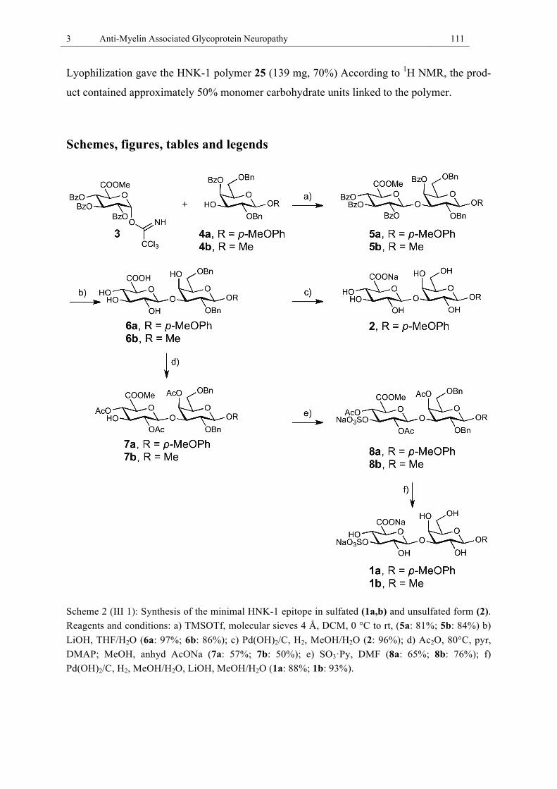
3 Anti-Myelin Associated Glycoprotein Neuropathy 111
Lyophilization gave the HNK-1 polymer 25 (139 mg, 70%) According to 1H NMR, the prod-
uct contained approximately 50% monomer carbohydrate units linked to the polymer.
Schemes, figures, tables and legends
Scheme 2 (III 1): Synthesis of the minimal HNK-1 epitope in sulfated (1a,b) and unsulfated form (2). Reagents and conditions: a) TMSOTf, molecular sieves 4 Å, DCM, 0 °C to rt, (5a: 81%; 5b: 84%) b) LiOH, THF/H2O (6a: 97%; 6b: 86%); c) Pd(OH)2/C, H2, MeOH/H2O (2: 96%); d) Ac2O, 80°C, pyr, DMAP; MeOH, anhyd AcONa (7a: 57%; 7b: 50%); e) SO3·Py, DMF (8a: 65%; 8b: 76%); f) Pd(OH)2/C, H2, MeOH/H2O, LiOH, MeOH/H2O (1a: 88%; 1b: 93%).
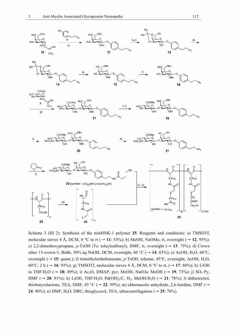
3 Anti-Myelin Associated Glycoprotein Neuropathy 112
Scheme 3 (III 2): Synthesis of the minHNK-1 polymer 25. Reagents and conditions: a) TMSOTf, molecular sieves 4 Å, DCM, 0 °C to rt (→ 11: 53%); b) MeOH, NaOMe, rt, overnight (→ 12, 95%); c) 2,2-dimethoxypropane, p-TsOH (Ts: toluylsulfonyl), DMF, rt, overnight (→ 13: 75%); d) Crown ether 15-crown-5, BnBr, 50% aq NaOH, DCM, overnight, 60 °C (→ 14: 83%); e) AcOH, H2O, 60°C, overnight (→ 15: quant.); f) trimethylorthobenzoate, p-TsOH, toluene, 45°C, overnight; AcOH, H2O, 60°C, 2 h (→ 16: 93%); g) TMSOTf, molecular sieves 4 Å, DCM, 0 °C to rt, (→ 17: 86%); h) LiOH in THF/H2O (→ 18: 89%); i) Ac2O, DMAP, pyr; MeOH, NaOAc MeOH (→ 19, 73%); j) SO3·Py, DMF (→ 20: 91%); k) LiOH, THF/H2O; Pd(OH)2/C, H2, MeOH/H2O (→ 21: 78%); l) dithiotreitol, thiobutyrolactone, TEA, DMF, 85 °C (→ 22: 59%); m) chloroacetic anhydride, 2,6-lutidine, DMF (→ 24: 96%); n) DMF, H2O, DBU; thioglycerol, TEA; ultracentrifugation (→ 25: 70%).

3 Anti-Myelin Associated Glycoprotein Neuropathy 113
Figure 15 (III 1): Representative competitive binding ELISA. (a) Incubation of MAG-coated plates with anti-MAG IgM (patient sera) and minHNK-1 polymer 25 (or ligands 1a,b or 2). (b) Wash step. (c) Incubation with anti-human IgM antibody coupled to horseradish peroxidase. (d) Wash step. (e) Addition of tetramethylbenzidin (TMB) substrate. (f) Addition of acidic stop solution and measure-ment of the optical density.
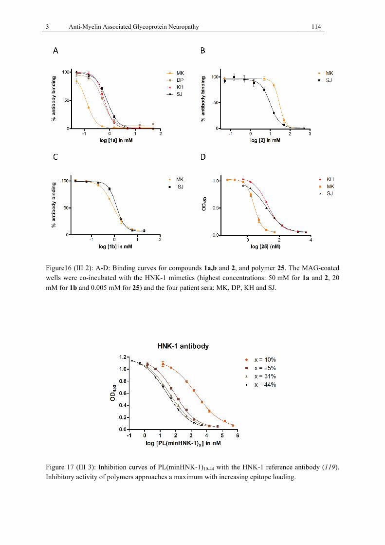
3 Anti-Myelin Associated Glycoprotein Neuropathy 114
Figure16 (III 2): A-D: Binding curves for compounds 1a,b and 2, and polymer 25. The MAG-coated wells were co-incubated with the HNK-1 mimetics (highest concentrations: 50 mM for 1a and 2, 20 mM for 1b and 0.005 mM for 25) and the four patient sera: MK, DP, KH and SJ.
Figure 17 (III 3): Inhibition curves of PL(minHNK-1)10-44 with the HNK-1 reference antibody (119). Inhibitory activity of polymers approaches a maximum with increasing epitope loading.
3 Anti-Myelin Associated Glycoprotein Neuropathy 115
Figure 18 (III 4): Control polymer PL(minHNK-1)0 without minHNK-1 epitope loading does not inhibit IgM antibody binding from patient sera MK, KH, and SJ to MAG.
Table 4 (III 1): IC50 values of compounds 1a, 1b, 2 and the minHNK-1 polymer (25) for the four patient sera MK, DP, KH and SJ.
Serum Compound 1a IC50 (µM)
Compound 1b IC50 (µM)
Compound 2 IC50 (µM)
Polymer (25) IC50 (µM)
MK 124. ± 9.5 860.0 ± 58.2 28967.3 ± 533.0 0.0025 ± 0.0001
DP 536.1 ± 23.5 n.d. n.d. n.d.
KH 614.2 ± 20.1 n.d. n.d. 0.0183 ± 0.0022
SJ 793.1 ± 24.0 1237.0 ± 56.0 9981.1 ± 1002.2 0.0148 ± 0.0006
Table 5 (III 2): IC50 values of PL(minHNK-1)10-44 for patient sera KH, MK and SJ, and the monoclonal HNK-1 antibody (119).
Serum/Antibody PL(minHNK-1)10 IC50 (nM)
PL(minHNK-1)25 IC50 (nM)
PL(minHNK-1)31 IC50 (nM)
PL(minHNK-1)44 IC50 (nM)
MK 10.0 ± 0.7 5.0 ± 0.2 4.0 ± 0.2 4.6 ± 0.1
KH 1936.7 ± 595.9 87.4 ± 2.0 34.0 ± 0.5 24.5 ± 2.5
SJ 27228.7 ± 4066.1 182.7 ± 16.4 47.5 ± 12.6 31.6 ± 6.7
HNK-1 2124.7 ± 443.8 85.6 ± 11.9 39.5 ± 4.0 27.6 ± 2.0
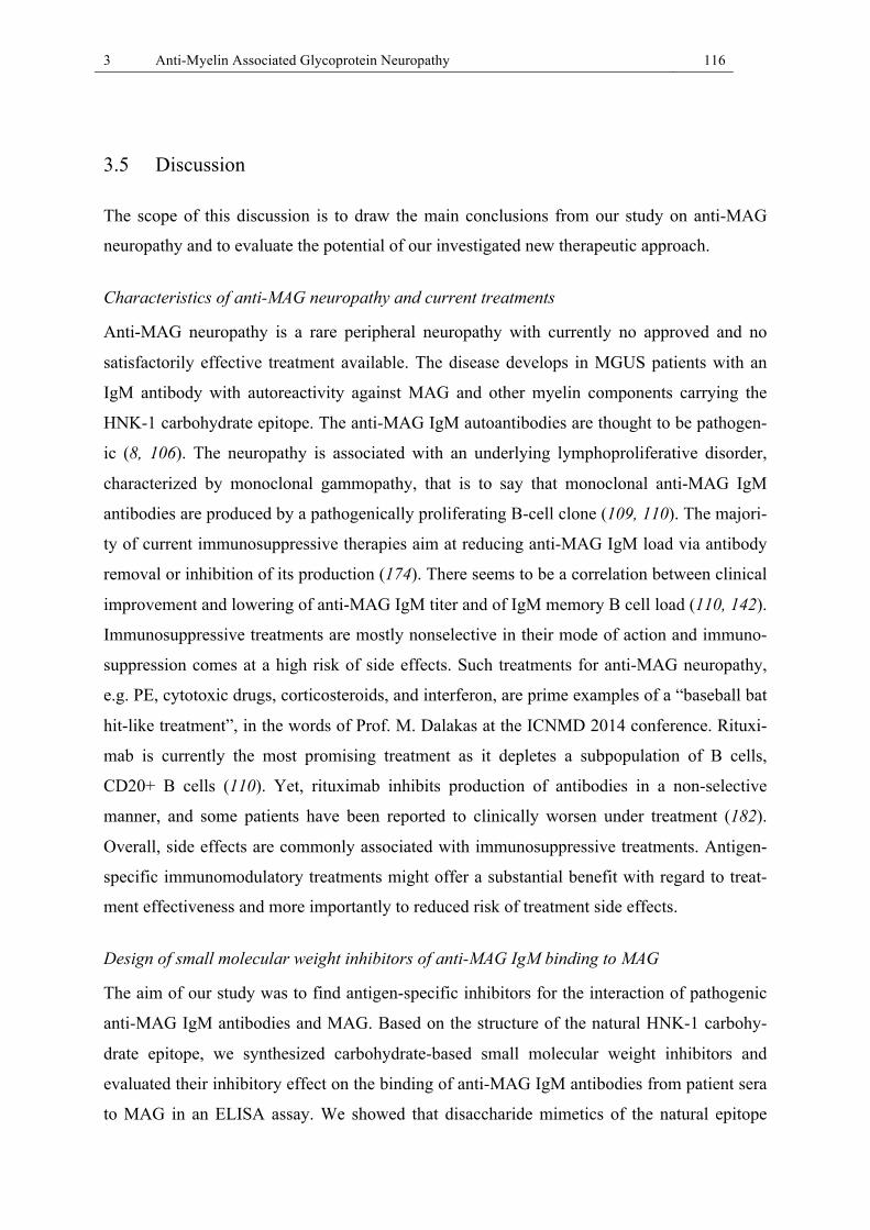
3 Anti-Myelin Associated Glycoprotein Neuropathy 116
3.5 Discussion
The scope of this discussion is to draw the main conclusions from our study on anti-MAG
neuropathy and to evaluate the potential of our investigated new therapeutic approach.
Characteristics of anti-MAG neuropathy and current treatments
Anti-MAG neuropathy is a rare peripheral neuropathy with currently no approved and no
satisfactorily effective treatment available. The disease develops in MGUS patients with an
IgM antibody with autoreactivity against MAG and other myelin components carrying the
HNK-1 carbohydrate epitope. The anti-MAG IgM autoantibodies are thought to be pathogen-
ic (8, 106). The neuropathy is associated with an underlying lymphoproliferative disorder,
characterized by monoclonal gammopathy, that is to say that monoclonal anti-MAG IgM
antibodies are produced by a pathogenically proliferating B-cell clone (109, 110). The majori-
ty of current immunosuppressive therapies aim at reducing anti-MAG IgM load via antibody
removal or inhibition of its production (174). There seems to be a correlation between clinical
improvement and lowering of anti-MAG IgM titer and of IgM memory B cell load (110, 142).
Immunosuppressive treatments are mostly nonselective in their mode of action and immuno-
suppression comes at a high risk of side effects. Such treatments for anti-MAG neuropathy,
e.g. PE, cytotoxic drugs, corticosteroids, and interferon, are prime examples of a “baseball bat
hit-like treatment”, in the words of Prof. M. Dalakas at the ICNMD 2014 conference. Rituxi-
mab is currently the most promising treatment as it depletes a subpopulation of B cells,
CD20+ B cells (110). Yet, rituximab inhibits production of antibodies in a non-selective
manner, and some patients have been reported to clinically worsen under treatment (182).
Overall, side effects are commonly associated with immunosuppressive treatments. Antigen-
specific immunomodulatory treatments might offer a substantial benefit with regard to treat-
ment effectiveness and more importantly to reduced risk of treatment side effects.
Design of small molecular weight inhibitors of anti-MAG IgM binding to MAG
The aim of our study was to find antigen-specific inhibitors for the interaction of pathogenic
anti-MAG IgM antibodies and MAG. Based on the structure of the natural HNK-1 carbohy-
drate epitope, we synthesized carbohydrate-based small molecular weight inhibitors and
evaluated their inhibitory effect on the binding of anti-MAG IgM antibodies from patient sera
to MAG in an ELISA assay. We showed that disaccharide mimetics of the natural epitope

3 Anti-Myelin Associated Glycoprotein Neuropathy 117
retain affinity for the IgM antibodies, supporting previous findings, definding the SO3-3-
GlcA(β1-3)Gal disaccharide as the minimal requirement for binding (130). Monosaccharide
ligands, including only the terminal SO3-3-GlcA (non-reducing end), are not expected to
retain affinity for the IgM antibodies. From three synthesized HNK-1 mimetics the compound
structurally closest to the natural trisaccharide epitope displayed best inhibitory activity on
IgM antibody binding to MAG with micromolar IC50 values. We defined this molecule as the
minimal HNK-1 epitope (minHNK-1). Deletion of the sulfate group in position 3 of GlcA
resulted in decreased inhibitory activity. Deletion of the para-methoxyphenyl group at the
reducing end of our minHNK-1 disaccharide also resulted in decreased inhibitory activity. We
suggest, that the minHNK-1 epitope with the terminal aromatic para-methoxyphenyl group
acts as a pseudo-trisaccharide, mimicking the natural HNK-1 trisaccharide epitope. Involve-
ment of aromatic groups in the binding of carbohydrate-based ligands to lectins has been
described before (194). With respect to the complexity of the synthetic route and scale-up
synthesis, disaccharide ligands are of more value than longer, structurally more complex
oligosaccharides. It is reasonable that the closer HNK-1 mimetics are to the natural epitope in
structure, the higher is also their binding affinity to anti-MAG antibodies. Furthermore, the
structurally closer mimetics retain affinity towards anti-MAG IgM antibodies from various
patients, as the antibodies all share the same epitope. Conventional lead optimization does not
make sense for the HNK-1 mimetics, as the natural epitope serves as the “perfect” template.
Chemical optimization of epitope mimetics for the IgM antibodies of one patient will likely
result in decreased binding affinity for IgM antibodies of another patient. Chemical optimiza-
tion makes more sense when aimed at defining a minimal carbohydrate epitope, which is
synthetically more easily available than the whole epitope. Furthermore, the replacement of
single monosaccharides can be considered, such as with lipophilic cyclohexane or aromatic
groups (e.g. para-methoxyphenyl).
Design of an antigen-specific glycopolymer
Carbohydrate-protein interactions are often of a multivalent nature (195). A multivalent inter-
action most likely happens in anti-MAG neuropathy, whereby patients’ IgM antibody mole-
cules with ten binding sites bind to the HNK-1 epitope, which is densely displayed on MAG
at up to eight glycosylation sites (153). The study by Ogino et al. describes significantly
decreased binding of anti-MAG IgM antibodies to MAG upon either breaking pentameric
IgM into their monomers or by digesting MAG into peptides with monomeric HNK-1 oligo-
saccharides. We therefore hypothesized that a polymer that presents the minHNK-1 epitope in

3 Anti-Myelin Associated Glycoprotein Neuropathy 118
a multivalent manner might display significantly increased IgM antibody binding affinity.
Synthesis of a minHNK-1 polylysine polymer yielded a high molecular weight glycopolymer
with a dramatically increased binding affinity towards the IgM antibodies compared to the
corresponding monomeric molecule. IC50 values were determined for three patient sera al-
ready used in previous assays with the minHNK-1 monomer. Testing of an additional fourth
patient serum confirmed the strong inhibition of IgM antibody binding to MAG in vitro, at
low nanomolar polymer concentrations. Also, the monoclonal HNK-1 mouse IgM antibody
(119) was tested and its binding to MAG was successfully inhibited at low nanomolar glyco-
polymer concentrations. A control polymer with no minHNK-1 epitope loading did not inhibit
patient IgM antibody binding to MAG.
Therapeutic potential of the minHNK-1 glycopolymer
An ideal polymer-based drug candidate could be administered to the patient via infusion or as
an injectable. Therefore, besides good inhibitory activity and selectivity for anti-MAG IgM,
the compound should be readily water-soluble, sufficiently stable in solution, and biode-
gradable. Non-biodegradable polymers will accumulate in the body and are of little therapeu-
tic value. The investigated minHNK-1 epitope polymer investigated in this study displayed
good water solubility and its peptide backbone is biodegradable. As outlined earlier, it has
shown very strong inhibition of anti-MAG IgM antibody binding to MAG in vitro. Through
structural similarity of the polymer warhead (minHNK-1 epitope) with the natural HNK-1
epitope this polymer might act as an antigen specific immunomodulatory agent that blocks
anti-MAG IgM antibody binding to MAG in vivo. Besides blocking circulating anti-MAG
antibodies in the patients, the polymer might also induce a down-regulation of anti-MAG IgM
production via binding to the specific B cells, as indicated by a previous study with a similar
glycopolymer (188). Sustained down-regulation of the anti-MAG IgM titers and concomitant
remyelination of the affected peripheral nerves would be expected if long term therapy is
feasible. Taken together, the described minHNK-1 glycopolymer has significant therapeutic
potential. In addition to therapeutic applications, diagnostic applications are also possible, i.e.
for detection of anti-MAG antibodies in patient sera. In fact, the company Bühlmann AG
successfully incorporated our polymer in a diagnostic ELISA assay kit that detects antibody
reactivity in patient sera against several myelin carbohydrate epitopes. The next steps towards
a therapeutic application for this polymer will be discussed in the outlook section.
3 Anti-Myelin Associated Glycoprotein Neuropathy 119
3.6 Outlook
The most important next step towards a therapeutic application of the minHNK-1 glycopoly-
mer will be its evaluation in an animal model. As outlined earlier, there are a few animal
models described for anti-MAG neuropathy: passive transfer of patients’ IgM antibodies into
healthy experimental animals, including cats (143, 144) and chicks (145); and active immun-
ization of cats with the glycolipid SGPG (147). All of these three animal models display
aspects of human myelin/nerve pathology. Rats and mice do not express the HNK-1 epitope
and are therefore less suitable as a neuropathy model (197). However, to study depletion and
down-regulation of anti-MAG antibodies by the glycopolymer, three mouse models are of
particular interest: (i) passive transfer of the HNK-1 mouse IgM antibody to mice, (ii) injec-
tion of the HNK-1 IgM producing hybridoma clone into the peritoneum of mice, and (iii)
active immunization of mice with SGPG. All of these models should result in circulating anti-
HNK-1 antibodies, and titers could be measured before, during, and after treatment with the
glycopolymer by ELISA. Models (ii) and (iii) mimic constant antibody production, as in the
human disease, and are thus preferable. Model (ii) could be problematic, as produced antibod-
ies might not reach the circulation from the peritoneum.
In addition to the animal model experiments, immunohistochemistry of human nerve biopsies
(skin biopsies) could be used to study whether HNK-1-binding of IgM antibodies from anti-
MAG neuropathy patients is inhibited in the presence of the glycopolymer.
Furthermore, the selectivity for anti-MAG antibodies will have to be investigated. Data from
Bühlmann AG suggest that the minHNK-1 polymer is very selective for anti-MAG IgM
antibodies in the IgM subgroup of immunoglobulins, but also displays a certain degree of
nonspecific IgG binding. This nonspecificity could be chemically optimized.
In order to be able to use a chemically defined polymeric molecule (in terms of molecular
weight, the polylysine polymer is not clearly defined) it might be worth to synthesize polymer
backbones with defined polypeptide lengths.
Eventually, polymers such as the minHNK-1 glycopolmer could be designed for neuropathies
associated with autoantibodies against other carbohydrate epitopes (see section 3.1). The
proposed therapeutic approach might pave the way for the first antigen-specific treatment for
anti-MAG neuropathy, and potentially for other antibody-mediated neuropathies.
4 Closing Words 120
4 Closing Words
This doctoral thesis presents novel therapeutic approaches for two NMDs. In a drug discovery
project for DM1 we identified new potent small molecular weight inhibitors of the CUG78-
MBNL1 complex. The concept of searching for small molecules that displace MBNL1 from
DMPK CUGn RNA to treat DM1 is not new (51). However, our approach and methodology
to identify small molecules were new. The format of the developed CUG78-MBNL1 inhibition
assay might be used for drug screenings in other RNA-mediated neuromuscular diseases, in
particular for other TREDs with pathogenic RNA-protein interactions (e.g. DM2).
With regard to a successful in vivo application of small molecules currently being investigated
in the DM1 field, including ours, particularly issues with target CUGn-RNA selectivity, tox-
icity, and poor pharmacokinetic properties will have to be resolved over the coming years.
Eventually, continued effort might result in an effective small molecule treatment for DM1.
According to comments by Prof. Charles Thornton (ICNMD 2014) a small molecule treat-
ment is the desired DM1 therapy. For the moment, however, the small molecules will have to
leave the stage to antisense oligonucleotides (46, 48), until the small molecules will be suited
for a therapeutic application. As kinase inhibitors are being increasingly investigated for DM1
and show ameliorating effects in DM1 cell models (60, 61, 67), the first small molecular
therapeutic agent reaching the patients might be a kinase inhibitor.
The investigated novel therapeutic approach for the autoimmune disorder anti-MAG neuropa-
thy with the minHNK-1 glycopolymer could pave the way for the first antigen-specific treat-
ment for anti-MAG neuropathy and might provide an example for other antibody-mediated
autoimmune diseases. As a targeted immunomodulatory treatment the new therapeutic ap-
proach might provide substantial benefit over current immunosuppressive therapies (174).
The drug discovery projects for both diseases were made possible by many studies elucidating
the molecular disease mechanisms and therefore clearly owe to these previous basic studies.
Hopefully, both drug discovery projects will contribute to the development of new therapies
for both of these disabling NMDs. Indeed, “it is an exciting time to work in the field of neu-
romuscular disorders”, as mentioned in the preface, where better understanding of neuromus-
cular disease mechanisms fosters the development of new therapies.
References 121
References
1. C. M. McDonald, Clinical Approach to the Diagnostic Evaluation of Hereditary and Acquired Neuromuscular Diseases. Physical Medicine and Rehabilitation Clinics of North America 23, 495-+ (2012).
2. F. Muntoni, M. J. A. Wood, Targeting RNA to treat neuromuscular disease. Nature Reviews Drug Discovery 10, 621-637 (2011).
3. J. T. Caroscio, M. N. Mulvihill, R. Sterling, B. Abrams, AMYOTROPHIC-LATERAL-SCLEROSIS - ITS NATURAL-HISTORY. Neurologic Clinics 5, 1-8 (1987).
4. K. E. Davies, K. J. Nowak, Molecular mechanisms of muscular dystrophies: old and new players. Nature Reviews Molecular Cell Biology 7, 762-773 (2006).
5. H. Azhary, M. U. Farooq, M. Bhanushali, A. Majid, M. Y. Kassab, Peripheral Neuropathy: Differential Diagnosis and Management. American Family Physician 81, 887-892 (2010).
6. F. Rahimov, L. M. Kunkel, Cellular and molecular mechanisms underlying muscular dystrophy. Journal of Cell Biology 201, 499-510 (2013).
7. J. Kinter, M. Sinnreich, Molecular targets to treat muscular dystrophies. Swiss Medical Weekly 144, (2014).
8. A. J. Steck, A. K. Stalder, S. Renaud, Anti-myelin-associated glycoprotein neuropathy. Current Opinion in Neurology 19, 458-463 (2006).
9. J. D. Brook et al., Molecular basis of myotonic dystrophy: expansion of a trinucleotide (CTG) repeat at the 3' end of a transcript encoding a protein kinase family member. Cell 69, 385-385 (1992).
10. C. L. Liquori et al., Myotonic dystrophy type 2 caused by a CCTG expansion in intron 1 of ZNF9. Science 293, 864-867 (2001).
11. L. Machuca-Tzili, D. Brook, D. Hilton-Jones, Clinical and molecular aspects of the myotonic dystrophies: A review. Muscle & Nerve 32, 1-18 (2005).
12. A. Mankodi et al., Myotonic dystrophy in transgenic mice expressing an expanded CUG repeat. Science 289, 1769-1772 (2000).
13. T. M. Wheeler et al., Reversal of RNA Dominance by Displacement of Protein Sequestered on Triplet Repeat RNA. Science 325, 336-339 (2009).
14. M. Mahadevan et al., MYOTONIC-DYSTROPHY MUTATION - AN UNSTABLE CTG REPEAT IN THE 3' UNTRANSLATED REGION OF THE GENE. Science 255, 1253-1255 (1992).
15. A. Hunter et al., THE CORRELATION OF AGE OF ONSET WITH CTG TRINUCLEOTIDE REPEAT AMPLIFICATION IN MYOTONIC-DYSTROPHY. Journal of Medical Genetics 29, 774-779 (1992).
16. E. B. Gharehbaghi-Schnell, J. Finsterer, I. Korschineck, B. Mamoli, B. R. Binder, Genotype-phenotype correlation in myotonic dystrophy. Clinical Genetics 53, 20-26 (1998).
17. D. G. Monckton, L. J. C. Wong, T. Ashizawa, C. T. Caskey, SOMATIC MOSAICISM, GERMLINE EXPANSIONS, GERMLINE REVERSIONS AND INTERGENERATIONAL REDUCTIONS IN MYOTONIC-DYSTROPHY MALES - SMALL POOL PCR ANALYSES. Human Molecular Genetics 4, 1-8 (1995).
18. I. V. Kovtun, C. T. McMurray, Trinucleotide expansion in haploid germ cells by gap repair. Nature Genetics 27, 407-411 (2001).

References 122
19. Y. H. Fu et al., DECREASED EXPRESSION OF MYOTONIN PROTEIN-KINASE MESSENGER-RNA AND PROTEIN IN ADULT FORM OF MYOTONIC-DYSTROPHY. Science 260, 235-238 (1993).
20. S. Reddy et al., Mice lacking the myotonic dystrophy protein kinase develop a late onset progressive myopathy. Nature Genetics 13, 325-335 (1996).
21. G. Jansen et al., Abnormal myotonic dystrophy protein kinase levels produce only mild myopathy in mice. Nature Genetics 13, 316-324 (1996).
22. H. Seznec et al., Mice transgenic for the human myotonic dystrophy region with expanded CTG repeats display muscular and brain abnormalities. Human Molecular Genetics 10, 2717-2726 (2001).
23. M. Gomes-Pereira, T. A. Cooper, G. Gourdon, Myotonic dystrophy mouse models: towards rational therapy development. Trends in Molecular Medicine 17, 506-517 (2011).
24. J. W. Miller et al., Recruitment of human muscleblind proteins to (CUG)(n) expansions associated with myotonic dystrophy. Embo Journal 19, 4439-4448 (2000).
25. Y. Yuan et al., Muscleblind-like 1 interacts with RNA hairpins in splicing target and pathogenic RNAs. Nucleic Acids Research 35, 5474-5486 (2007).
26. B. M. Davis, M. E. McCurrach, K. L. Taneja, R. H. Singer, D. E. Housman, Expansion of a CUG trinucleotide repeat in the 3' untranslated region of myotonic dystrophy protein kinase transcripts results in nuclear retention of transcripts. Proceedings of the National Academy of Sciences of the United States of America 94, 7388-7393 (1997).
27. K. L. Taneja, M. McCurrach, M. Schalling, D. Housman, R. H. Singer, FOCI OF TRINUCLEOTIDE REPEAT TRANSCRIPTS IN NUCLEI OF MYOTONIC-DYSTROPHY CELLS AND TISSUES. Journal of Cell Biology 128, 995-1002 (1995).
28. A. Mankodi et al., Ribonuclear inclusions in skeletal muscle in myotonic dystrophy types 1 and 2. Annals of Neurology 54, 760-768 (2003).
29. A. Mankodi et al., Expanded CUG repeats trigger aberrant splicing of CIC-1 chloride channel pre-mRNA and hyperexcitability of skeletal muscle in myotonic dystrophy. Molecular Cell 10, 35-44 (2002).
30. N. Charlet-B et al., Loss of the muscle-specific chloride channel in type 1 myotonic dystrophy due to misregulated alternative splicing. Molecular Cell 10, 45-53 (2002).
31. Y. Kino et al., MBNL and CELF proteins regulate alternative splicing of the skeletal muscle chloride channel CLCN1. Nucleic Acids Research 37, 6477-6490 (2009).
32. R. S. Savkur, A. V. Philips, T. A. Cooper, Aberrant regulation of insulin receptor alternative splicing is associated with insulin resistance in myotonic dystrophy. Nature Genetics 29, 40-47 (2001).
33. W. Dansithong, S. Paul, L. Comai, S. Reddy, MBNL1 is the primary determinant of focus formation and aberrant insulin receptor splicing in DM1. Journal of Biological Chemistry 280, 5773-5780 (2005).
34. S.-i. Hino et al., Molecular mechanisms responsible for aberrant splicing of SERCA1 in myotonic dystrophy type 1. Human Molecular Genetics 16, 2834-2843 (2007).
35. T. H. Ho et al., Muscleblind proteins regulate alternative splicing. Embo Journal 23, 3103-3112 (2004).
36. J. D. Lueck et al., Chloride channelopathy in myotonic dystrophy resulting from loss of posttranscriptional regulation for CLCN1. American Journal of Physiology-Cell Physiology 292, C1291-C1297 (2007).
37. R. N. Kanadia et al., A muscleblind knockout model for myotonic dystrophy. Science 302, 1978-1980 (2003).
References 123
38. R. N. Kanadia et al., Reversal of RNA missplicing and myotonia after muscleblind overexpression in a mouse poly(CUG) model for myotonic dystrophy. Proceedings of the National Academy of Sciences of the United States of America 103, 11748-11753 (2006).
39. X. Y. Lin et al., Failure of MBNL1-dependent post-natal splicing transitions in myotonic dystrophy. Human Molecular Genetics 15, 2087-2097 (2006).
40. D. L. Black, Mechanisms of alternative pre-messenger RNA splicing. Annual Review of Biochemistry 72, 291-336 (2003).
41. N. M. Kuyumcu-Martinez, G.-S. Wang, T. A. Cooper, Increased steady-state in levels of CUGBP1 in myotonic dystrophy 1 are due to PKC-mediated hyperphosphorylation. Molecular Cell 28, 68-78 (2007).
42. T. Yamamoto et al., Long-term treatment of diabetes mellitus in myotonic dystrophy with pioglitazone. Rinsho shinkeigaku = Clinical neurology 45, 287-292 (2005).
43. M. S. Damian, A. Gerlach, F. Schmidt, E. Lehmann, H. Reichmann, Modafinil for excessive daytime sleepiness in myotonic dystrophy. Neurology 56, 794-796 (2001).
44. D. Furling et al., Viral vector producing antisense RNA restores myotonic dystrophy myoblast functions. Gene Therapy 10, 795-802 (2003).
45. M. A. Langlois et al., Cytoplasmic and nuclear retained DMPK mRNAs are targets for RNA interference in myotonic dystrophy cells. Journal of Biological Chemistry 280, 16949-16954 (2005).
46. A. J. Leger et al., Systemic Delivery of a Peptide-Linked Morpholino Oligonucleotide Neutralizes Mutant RNA Toxicity in a Mouse Model of Myotonic Dystrophy. Molecular Therapy 21, S175-S175 (2013).
47. J. E. Lee, C. F. Bennett, T. A. Cooper, RNase H-mediated degradation of toxic RNA in myotonic dystrophy type 1. Proceedings of the National Academy of Sciences of the United States of America 109, 4221-4226 (2012).
48. T. M. Wheeler et al., Targeting nuclear RNA for in vivo correction of myotonic dystrophy. Nature 488, 111-+ (2012).
49. M. A. Langlois, N. S. Lee, J. J. Rossi, J. Puymirat, Hammerhead ribozyme-mediated destruction of nuclear foci in myotonic dystrophy myoblasts. Molecular Therapy 7, 670-680 (2003).
50. W. Zhang et al., Treatment of Type 1 Myotonic Dystrophy by Engineering Site-specific RNA Endonucleases that Target (CUG)(n) Repeats. Molecular Therapy 22, 312-320 (2014).
51. M. B. Warf, M. Nakamori, C. M. Matthys, C. A. Thornton, J. A. Berglund, Pentamidine reverses the splicing defects associated with myotonic dystrophy. Proceedings of the National Academy of Sciences of the United States of America 106, 18551-18556 (2009).
52. J. F. Arambula, S. R. Ramisetty, A. M. Baranger, S. C. Zimmerman, A simple ligand that selectively targets CUG trinucleotide repeats and inhibits MBNL protein binding. Proceedings of the National Academy of Sciences of the United States of America 106, 16068-16073 (2009).
53. A. Pushechnikov et al., Rational Design of Ligands Targeting Triplet Repeating Transcripts That Cause RNA Dominant Disease: Application to Myotonic Muscular Dystrophy Type 1 and Spinocerebellar Ataxia Type 3. Journal of the American Chemical Society 131, 9767-9779 (2009).
54. J. L. Childs-Disney, J. Hoskins, S. G. Rzuczek, C. A. Thornton, M. D. Disney, Rationally Designed Small Molecules Targeting the RNA That Causes Myotonic Dystrophy Type 1 Are Potently Bioactive. Acs Chemical Biology 7, 856-862 (2012).
55. P. C. Gareiss et al., Dynamic Combinatorial Selection of Molecules Capable of Inhibiting the (CUG) Repeat RNA-MBNL1 Interaction In Vitro: Discovery of Lead
References 124
Compounds Targeting Myotonic Dystrophy (DM1). Journal of the American Chemical Society 130, 16254-16261 (2008).
56. L. O. Ofori, J. Hoskins, M. Nakamori, C. A. Thornton, B. L. Miller, From dynamic combinatorial 'hit' to lead: in vitro and in vivo activity of compounds targeting the pathogenic RNAs that cause myotonic dystrophy. Nucleic Acids Research 40, 6380-6390 (2012).
57. J. W. Hoskins et al., Lomofungin and dilomofungin: inhibitors of MBNL1-CUG RNA binding with distinct cellular effects. Nucleic acids research 42, 6591-6602 (2014).
58. C. Z. Chen et al., Two high-throughput screening assays for aberrant RNA-protein interactions in myotonic dystrophy type 1. Analytical and Bioanalytical Chemistry 402, 1889-1898 (2012).
59. L. A. Coonrod et al., Reducing Levels of Toxic RNA with Small Molecules. Acs Chemical Biology 8, 2528-2537 (2013).
60. A. Ketley et al., High-content screening identifies small molecules that remove nuclear foci, affect MBNL distribution and CELF1 protein levels via a PKC-independent pathway in myotonic dystrophy cell lines. Human Molecular Genetics 23, 1551-1562 (2014).
61. C. Wei, K. Jones, N. A. Timchenko, L. Timchenko, GSK3beta is a new therapeutic target for myotonic dystrophy type 1. Rare diseases (Austin, Tex.) 1, e26555-e26555 (2013).
62. K. Oana et al., Manumycin A corrects aberrant splicing of Clcn1 in myotonic dystrophy type 1 (DM1) mice. Scientific Reports 3, (2013).
63. J. R. Thomas, P. J. Hergenrother, Targeting RNA with small molecules. Chemical Reviews 108, 1171-1224 (2008).
64. A. V. Philips, L. T. Timchenko, T. A. Cooper, Disruption of splicing regulated by a CUG-binding protein in myotonic dystrophy. Science 280, 737-741 (1998).
65. J. Berg, H. Jiang, C. A. Thornton, S. C. Cannon, Truncated ClC-1 mRNA in myotonic dystrophy exerts a dominant-negative effect on the Cl current. Neurology 63, 2371-2375 (2004).
66. W. J. Krzyzosiak et al., Triplet repeat RNA structure and its role as pathogenic agent and therapeutic target. Nucleic Acids Research 40, 11-26 (2012).
67. M. Wojciechowska, K. Taylor, K. Sobczak, M. Napierala, W. J. Krzyzosiak, Small molecule kinase inhibitors alleviate different molecular features of myotonic dystrophy type 1. RNA biology 11, (2014).
68. F. E. Koehn, G. T. Carter, The evolving role of natural products in drug discovery. Nature Reviews Drug Discovery 4, 206-220 (2005).
69. O. Potterat, M. Hamburger, Combined Use of Extract Libraries and HPLC-Based Activity Profiling for Lead Discovery: Potential, Challenges, and Practical Considerations. Planta Med 80, 1171-1181 (2014).
70. R. A. Collins, T. B. Ng, W. P. Fong, C. C. Wan, H. W. Yeung, Removal of polyphenolic compounds from aqueous plant extracts using polyamide minicolumns. Biochemistry and Molecular Biology International 45, 791-796 (1998).
71. S. Chaouch et al., Immortalized Skin Fibroblasts Expressing Conditional MyoD as a Renewable and Reliable Source of Converted Human Muscle Cells to Assess Therapeutic Strategies for Muscular Dystrophies: Validation of an Exon-Skipping Approach to Restore Dystrophin in Duchenne Muscular Dystrophy Cells. Human Gene Therapy 20, 784-790 (2009).
72. A. Mykowska, K. Sobczak, M. Wojciechowska, P. Kozlowski, W. J. Krzyzosiak, CAG repeats mimic CUG repeats in the misregulation of alternative splicing. Nucleic Acids Research 39, 8938-8951 (2011).

References 125
73. D. N. Wilson, Ribosome-targeting antibiotics and mechanisms of bacterial resistance. Nature Reviews Microbiology 12, 35-48 (2014).
74. M. M. Islam, S. R. Chowdhury, G. S. Kumar, Spectroscopic and Calorimetric Studies on the Binding of Alkaloids Berberine, Palmatine and Coralyne to Double Stranded RNA Polynucleotides. Journal of Physical Chemistry B 113, 1210-1224 (2009).
75. G. S. Kumar, RNA targeting by small molecules: Binding of protoberberine, benzophenanthridine and aristolochia alkaloids to various RNA structures. Journal of Biosciences 37, 539-552 (2012).
76. S. Nafisi, Z. M. Malekabady, M. A. Khalilzadeh, Interaction of beta-Carboline Alkaloids with RNA. DNA and Cell Biology 29, 753-761 (2010).
77. O. P. Cetinkol, N. V. Hud, Molecular recognition of poly(A) by small ligands: an alternative method of analysis reveals nanomolar, cooperative and shape-selective binding. Nucleic Acids Research 37, 611-621 (2009).
78. M. B. Warf, J. A. Berglund, MBNL binds similar RNA structures in the CUG repeats of myotonic dystrophy and its pre-mRNA substrate cardiac troponin T. Rna-a Publication of the Rna Society 13, 2238-2251 (2007).
79. Y. B. Guan, E. D. Louis, W. Zheng, Toxicokinetics of tremorogenic natural products, harmane and harmine, in male Sprague-Dawley rats. Journal of Toxicology and Environmental Health-Part A 64, 645-660 (2001).
80. J. Lutes, J. F. Lorden, M. Beales, G. A. Oltmans, TOLERANCE TO THE TREMOROGENIC EFFECTS OF HARMALINE - EVIDENCE FOR ALTERED OLIVO-CEREBELLAR FUNCTION. Neuropharmacology 27, 849-855 (1988).
81. N. Turner et al., Berberine and its more biologically available derivative, dihydroberberine, inhibit mitochondrial respiratory complex I: A mechanism for the action of berberine to activate AMP-Activated protein kinase and improve insulin action. Diabetes 57, 1414-1418 (2008).
82. J.-F. Jiang et al., Novel Effect of Berberine on Thermoregulation in Mice Model Induced by Hot and Cold Environmental Stimulation. Plos One 8, (2013).
83. Y. S. Lee et al., Berberine, a natural plant product, activates AMP-activated protein kinase with beneficial metabolic effects in diabetic and insulin-resistant states. Diabetes 55, 2256-2264 (2006).
84. N. Goeckler et al., Harmine specifically inhibits protein kinase DYRK1A and interferes with neurite formation. Febs Journal 276, 6324-6337 (2009).
85. S. S. Lee, M. Kai, M. K. Lee, Effects of natural isoquinoline alkaloids on monoamine oxidase activity in mouse brain: inhibition by berberine and palmatine. Medical Science Research 27, 749-751 (1999).
86. T. Herraiz, D. Gonzalez, C. Ancin-Azpilicueta, V. J. Aran, H. Guillen, beta-Carboline alkaloids in Peganum harmala and inhibition of human monoamine oxidase (MAO). Food and Chemical Toxicology 48, 839-845 (2010).
87. Y. N. Zhao et al., A new approach to investigate the pharmacokinetics of Traditional Chinese Medicine YL2000. American Journal of Chinese Medicine 32, 921-929 (2004).
88. G. Zetler, G. Back, H. Iven, PHARMACOKINETICS IN RAT OF HALLUCINOGENIC ALKALOIDS HARMINE AND HARMALINE. Naunyn-Schmiedebergs Archives of Pharmacology 285, 273-292 (1974).
89. I. Dimauro, T. Pearson, D. Caporossi, M. J. Jackson, A simple protocol for the subcellular fractionation of skeletal muscle cells and tissue. BMC research notes 5, 513-513 (2012).
90. M. M. Lee et al., Controlling the Specificity of Modularly Assembled Small Molecules for RNA via Ligand Module Spacing: Targeting the RNAs That Cause
References 126
Myotonic Muscular Dystrophy. Journal of the American Chemical Society 131, 17464-17472 (2009).
91. A. Garcia-Lopez, B. Llamusi, M. Orzaez, E. Perez-Paya, R. D. Artero, In vivo discovery of a peptide that prevents CUG-RNA hairpin formation and reverses RNA toxicity in myotonic dystrophy models. Proceedings of the National Academy of Sciences of the United States of America 108, 11866-11871 (2011).
92. R. Parkesh et al., Design of a Bioactive Small Molecule That Targets the Myotonic Dystrophy Type 1 RNA via an RNA Motif-Ligand Database and Chemical Similarity Searching. Journal of the American Chemical Society 134, 4731-4742 (2012).
93. W. D. Wilson, L. Ratmeyer, M. Zhao, L. Strekowski, D. Boykin, THE SEARCH FOR STRUCTURE-SPECIFIC NUCLEIC-ACID INTERACTIVE DRUGS - EFFECTS OF COMPOUND STRUCTURE ON RNA VERSUS DNA INTERACTION STRENGTH. Biochemistry 32, 4098-4104 (1993).
94. D. A. Patrick et al., Anti-Pneumocystic carinii pneumonia activity of dicationic carbazoles. European Journal of Medicinal Chemistry 32, 781-793 (1997).
95. A. Kumar, M. Say, D. W. Boykin, Synthesis of new substituted 2-(trimethylstannyl)indoles. Synthesis-Stuttgart, 707-710 (2008).
96. J. Bezencon et al., pK(a) determination by H-1 NMR spectroscopy - An old methodology revisited. Journal of Pharmaceutical and Biomedical Analysis 93, 147-155 (2014).
97. M. Kansy, F. Senner, K. Gubernator, Physicochemical high throughput screening: Parallel artificial membrane permeation assay in the description of passive absorption processes. Journal of Medicinal Chemistry 41, 1007-1010 (1998).
98. L. Sanderson, M. Dogruel, J. Rodgers, H. P. De Koning, S. A. Thomas, Pentamidine Movement across the Murine Blood-Brain and Blood-Cerebrospinal Fluid Barriers: Effect of Trypanosome Infection, Combination Therapy, P-Glycoprotein, and Multidrug Resistance-Associated Protein. Journal of Pharmacology and Experimental Therapeutics 329, 967-977 (2009).
99. L. R. Guan, M. D. Disney, Recent Advances in Developing Small Molecules Targeting RNA. Acs Chemical Biology 7, 73-86 (2012).
100. M. Zhao et al., SMALL CHANGES IN CATIONIC SUBSTITUENTS OF DIPHENYLFURAN DERIVATIVES HAVE MAJOR EFFECTS ON THE BINDING-AFFINITY AND THE BINDING MODE WITH RNA HELICAL DUPLEXES. Bioorganic & Medicinal Chemistry 3, 785-794 (1995).
101. M. N. C. Soeiro et al., Novel amidines and analogues as promising agents against intracellular parasites: a systematic review. Parasitology 140, 929-951 (2013).
102. L. Pang et al., FimH Antagonists: Structure-Activity and Structure-Property Relationships for Biphenyl a-D-Mannopyranosides. Chemmedchem 7, 1404-1422 (2012).
103. M. W. Pantoliano et al., High-density miniaturized thermal shift assays as a general strategy for drug discovery. Journal of Biomolecular Screening 6, 429-440 (2001).
104. E. S. Goers, J. Purcell, R. B. Voelker, D. P. Gates, J. A. Berglund, MBNL1 binds GC motifs embedded in pyrimidines to regulate alternative splicing. Nucleic Acids Research 38, 2467-2484 (2010).
105. Z. Yong, H. Meirong, Z. Jianping, X. Shaofei, PKSolver: an add-in program for pharmacokinetic and pharmacodynamic data analysis in Microsoft Excel. Computer Methods and Programs in Biomedicine 99, 306-314 (2010).
106. M. C. Dalakas, Pathogenesis and Treatment of Anti-MAG Neuropathy. Current Treatment Options in Neurology 12, 71-83 (2010).
107. V. Chaudhry, M. Swash, Multifocal motor neuropathy - Is conduction block essential? Neurology 67, 558-559 (2006).
References 127
108. E. Nobile-Orazio, Antigenic Determinants in IgM Paraprotein-Related Neuropathies. Clinical Lymphoma & Myeloma 9, 107-109 (2009).
109. J. Kriangkum et al., Clonotypic IgM V/D/J sequence analysis in Waldenstrom macroglobulinemia suggests an unusual B-cell origin and an expansion of polyclonal B cells in peripheral blood. Blood 104, 2134-2142 (2004).
110. M. A. Maurer et al., Rituximab induces sustained reduction of pathogenic B cells in patients with peripheral nervous system autoimmunity. Journal of Clinical Investigation 122, 1393-1402 (2012).
111. A. Steck, N. Yuki, F. Graus, Antibody testing in peripheral nerve disorders. Handbook of clinical neurology 115, 189-212 (2013).
112. M. C. Dalakas, Clinical trials in CIDP and chronic autoimmune demyelinating polyneuropathies. Journal of the Peripheral Nervous System 17, 34-39 (2012).
113. R. A. Kyle, J. P. Garton, THE SPECTRUM OF IGM MONOCLONAL GAMMOPATHY IN 430 CASES. Mayo Clinic Proceedings 62, 719-731 (1987).
114. E. Nobileorazio et al., PERIPHERAL NEUROPATHY IN MONOCLONAL GAMMOPATHY OF UNDETERMINED SIGNIFICANCE - PREVALENCE AND IMMUNOPATHOGENETIC STUDIES. Acta Neurologica Scandinavica 85, 383-390 (1992).
115. R. A. Kyle, S. V. Rajkumar, Monoclonal gammopathies of undetermined significance: a review. Immunological Reviews 194, 112-139 (2003).
116. E. Nobileorazio et al., FREQUENCY AND CLINICAL CORRELATES OF ANTI-NEURAL IGM ANTIBODIES IN NEUROPATHY ASSOCIATED WITH IGM MONOCLONAL GAMMOPATHY. Annals of Neurology 36, 416-424 (1994).
117. P. E. Braun, D. E. Frail, N. Latov, MYELIN-ASSOCIATED GLYCOPROTEIN IS THE ANTIGEN FOR A MONOCLONAL IGM IN POLYNEUROPATHY. Journal of Neurochemistry 39, 1261-1265 (1982).
118. A. J. Steck, N. Murray, C. Meier, N. Page, G. Perruisseau, DEMYELINATING NEUROPATHY AND MONOCLONAL IGM ANTIBODY TO MYELIN-ASSOCIATED GLYCOPROTEIN. Neurology 33, 19-23 (1983).
119. T. Abo, C. M. Balch, A DIFFERENTIATION ANTIGEN OF HUMAN NK-CELLS AND K-CELLS IDENTIFIED BY A MONOCLONAL-ANTIBODY (HNK-1). Journal of Immunology 127, 1024-1029 (1981).
120. R. C. McGarry, S. L. Helfand, R. H. Quarles, J. C. Roder, RECOGNITION OF MYELIN-ASSOCIATED GLYCOPROTEIN BY THE MONOCLONAL-ANTIBODY HNK-1. Nature 306, 376-378 (1983).
121. A. A. Ilyas et al., IGM IN A HUMAN NEUROPATHY RELATED TO PARAPROTEINEMIA BINDS TO A CARBOHYDRATE DETERMINANT IN THE MYELIN-ASSOCIATED GLYCOPROTEIN AND TO A GANGLIOSIDE. Proceedings of the National Academy of Sciences of the United States of America-Biological Sciences 81, 1225-1229 (1984).
122. K. H. Chou, A. A. Ilyas, J. E. Evans, R. H. Quarles, F. B. Jungalwala, STRUCTURE OF A GLYCOLIPID REACTING WITH MONOCLONAL IGM IN NEUROPATHY AND WITH HNK-1. Biochemical and Biophysical Research Communications 128, 383-388 (1985).
123. D. K. H. Chou et al., STRUCTURE OF SULFATED GLUCURONYL GLYCOLIPIDS IN THE NERVOUS-SYSTEM REACTING WITH HNK-1 ANTIBODY AND SOME IGM PARAPROTEINS IN NEUROPATHY. Journal of Biological Chemistry 261, 1717-1725 (1986).
124. T. Ariga et al., CHARACTERIZATION OF SULFATED GLUCURONIC-ACID CONTAINING GLYCOLIPIDS REACTING WITH IGM M-PROTEINS IN
References 128
PATIENTS WITH NEUROPATHY. Journal of Biological Chemistry 262, 848-853 (1987).
125. H. Voshol, W. E. M. vanZuylen, G. Orberger, J. F. G. Vliegenthart, M. Schachner, Structure of the HNK-1 carbohydrate epitope on bovine peripheral myelin glycoprotein P0. Journal of Biological Chemistry 271, 22957-22960 (1996).
126. R. H. Quarles, Myelin-associated glycoprotein (MAG): past, present and beyond. Journal of Neurochemistry 100, 1431-1448 (2007).
127. F. Fluri, F. Ferracin, B. Erne, A. J. Steck, Microheterogeneity of anti-myelin-associated glycoprotein antibodies. Journal of the Neurological Sciences 207, 43-49 (2003).
128. A. A. Ilyas, D. K. H. Chou, F. B. Jungalwala, C. Costello, R. H. Quarles, VARIABILITY IN THE STRUCTURAL REQUIREMENTS FOR BINDING OF HUMAN MONOCLONAL ANTI-MYELIN-ASSOCIATED GLYCOPROTEIN IMMUNOGLOBULIN-M ANTIBODIES AND HNK-1 TO SPHINGOGLYCOLIPID ANTIGENS. Journal of Neurochemistry 55, 594-601 (1990).
129. B. Schmitz, M. Schachner, Y. Ito, T. Nakano, T. Ogawa, DETERMINATION OF STRUCTURAL ELEMENTS OF THE L2/HNK-1 CARBOHYDRATE EPITOPE REQUIRED FOR ITS FUNCTION. Glycoconjugate Journal 11, 345-352 (1994).
130. A. Tokuda et al., On the specificity of anti-sulfoglucuronosyl glycolipid antibodies'. Journal of Carbohydrate Chemistry 17, 535-546 (1998).
131. N. Page, N. Murray, G. Perruisseau, A. J. Steck, A MONOCLONAL ANTI-IDIOTYPIC ANTIBODY AGAINST A HUMAN MONOCLONAL IGM WITH SPECIFICITY FOR MYELIN-ASSOCIATED GLYCOPROTEIN. Journal of Immunology 134, 3094-3099 (1985).
132. R. Lombardi et al., IgM deposits on skin nerves in anti-myelin-associated glycoprotein neuropathy. Annals of Neurology 57, 180-187 (2005).
133. M. Takatsu et al., IMMUNOFLUORESCENCE STUDY OF PATIENTS WITH NEUROPATHY AND IGM M-PROTEINS. Annals of Neurology 18, 173-181 (1985).
134. A. Vital, C. Vital, J. Julien, A. Baquey, A. J. Steck, POLYNEUROPATHY ASSOCIATED WITH IGM MONOCLONAL GAMMOPATHY - IMMUNOLOGICAL AND PATHOLOGICAL-STUDY IN 31 PATIENTS. Acta Neuropathologica 79, 160-167 (1989).
135. J. R. Mendell et al., POLYNEUROPATHY AND IGM MONOCLONAL GAMMOPATHY - STUDIES ON THE PATHOGENETIC ROLE OF ANTI-MYELIN-ASSOCIATED GLYCOPROTEIN ANTIBODY. Annals of Neurology 17, 243-254 (1985).
136. A. P. Hays, S. S. L. Lee, N. Latov, IMMUNE REACTIVE C3D ON THE SURFACE OF MYELIN SHEATHS IN NEUROPATHY. Journal of Neuroimmunology 18, 231-244 (1988).
137. S. Monaco et al., COMPLEMENT-MEDIATED DEMYELINATION IN PATIENTS WITH IGM MONOCLONAL GAMMOPATHY AND POLYNEUROPATHY. New England Journal of Medicine 322, 649-652 (1990).
138. E. Nobileorazio et al., TREATMENT OF PATIENTS WITH NEUROPATHY AND ANTI-MAG IGM M-PROTEINS. Annals of Neurology 24, 93-97 (1988).
139. D. C. Haas, A. H. Tatum, PLASMAPHERESIS ALLEVIATES NEUROPATHY ACCOMPANYING IGM ANTI-MYELIN-ASSOCIATED GLYCOPROTEIN PARAPROTEINEMIA. Annals of Neurology 23, 394-396 (1988).
140. S. Renaud et al., Rituximab in the treatment of polyneuropathy associated with anti-MAG antibodies. Muscle & Nerve 27, 611-615 (2003).
141. A. Pestronk et al., Treatment of IgM antibody associated polyneuropathies using rituximab. Journal of Neurology Neurosurgery and Psychiatry 74, 485-489 (2003).
References 129
142. L. Benedetti et al., Predictors of response to rituximab in patients with neuropathy and anti-myelin associated glycoprotein immunoglobulin M. Journal of the Peripheral Nervous System 12, 102-107 (2007).
143. A. P. Hays, N. Latov, M. Takatsu, W. H. Sherman, EXPERIMENTAL DEMYELINATION OF NERVE INDUCED BY SERUM OF PATIENTS WITH NEUROPATHY AND AN ANTI-MAG IGM M-PROTEIN. Neurology 37, 242-256 (1987).
144. H. J. Willison et al., DEMYELINATION INDUCED BY INTRANEURAL INJECTION OF HUMAN ANTIMYELIN-ASSOCIATED GLYCOPROTEIN ANTIBODIES. Muscle & Nerve 11, 1169-1176 (1988).
145. A. H. Tatum, EXPERIMENTAL PARAPROTEIN NEUROPATHY, DEMYELINATION BY PASSIVE TRANSFER OF HUMAN-IGM ANTI-MYELIN-ASSOCIATED GLYCOPROTEIN. Annals of Neurology 33, 502-506 (1993).
146. W. Trojaborg et al., ELECTROPHYSIOLOGIC STUDY OF EXPERIMENTAL DEMYELINATION INDUCED BY SERUM OF PATIENTS WITH IGM-M PROTEINS AND NEUROPATHY. Neurology 39, 1581-1586 (1989).
147. A. A. Ilyas, Y. Gu, M. C. Dalakas, R. H. Quarles, S. Bhatt, Induction of experimental ataxic sensory neuronopathy in cats by immunization with purified SGPG. Journal of Neuroimmunology 193, 87-93 (2008).
148. S. Kelm et al., SIALOADHESIN, MYELIN-ASSOCIATED GLYCOPROTEIN AND CD22 DEFINE A NEW FAMILY OF SIALIC ACID-DEPENDENT ADHESION MOLECULES OF THE IMMUNOGLOBULIN SUPERFAMILY. Current Biology 4, 965-972 (1994).
149. P. R. Crocker et al., Siglecs: a family of sialic-acid binding lectins. Glycobiology 8, v (1998).
150. R. O. Brady, R. H. Quarles, DEVELOPMENTAL AND PATHOPHYSIOLOGICAL ASPECTS OF THE MYELIN-ASSOCIATED GLYCOPROTEIN. Cellular and Molecular Neurobiology 8, 139-148 (1988).
151. S. Sato et al., CDNA CLONING AND AMINO-ACID SEQUENCE FOR HUMAN MYELIN-ASSOCIATED GLYCOPROTEIN. Biochemical and Biophysical Research Communications 163, 1473-1480 (1989).
152. G. Spagnol et al., MOLECULAR-CLONING OF HUMAN MYELIN-ASSOCIATED GLYCOPROTEIN. Journal of Neuroscience Research 24, 137-142 (1989).
153. M. Ogino, A. H. Tatum, N. Latov, AFFINITY STUDIES OF HUMAN ANTI-MAG ANTIBODIES IN NEUROPATHY. Journal of Neuroimmunology 52, 41-46 (1994).
154. N. H. Sternberger, R. H. Quarles, Y. Itoyama, H. D. Webster, MYELIN-ASSOCIATED GLYCOPROTEIN DEMONSTRATED IMMUNOCYTOCHEMICALLY IN MYELIN AND MYELIN-FORMING CELLS OF DEVELOPING RAT. Proceedings of the National Academy of Sciences of the United States of America 76, 1510-1514 (1979).
155. M. Arquint et al., MOLECULAR-CLONING AND PRIMARY STRUCTURE OF MYELIN-ASSOCIATED GLYCOPROTEIN. Proceedings of the National Academy of Sciences of the United States of America 84, 600-604 (1987).
156. C. Lai et al., 2 FORMS OF 1B236 MYELIN-ASSOCIATED GLYCOPROTEIN, A CELL-ADHESION MOLECULE FOR POSTNATAL NEURAL DEVELOPMENT, ARE PRODUCED BY ALTERNATIVE SPLICING. Proceedings of the National Academy of Sciences of the United States of America 84, 4337-4341 (1987).
157. J. L. Salzer, W. P. Holmes, D. R. Colman, THE AMINO-ACID-SEQUENCES OF THE MYELIN-ASSOCIATED GLYCOPROTEINS - HOMOLOGY TO THE IMMUNOGLOBULIN GENE SUPERFAMILY. Journal of Cell Biology 104, 957-965 (1987).
References 130
158. M. Erb et al., Unraveling the differential expression of the two isoforms of myelin-associated glycoprotein in a mouse expressing GFP-tagged S-MAG specifically regulated and targeted into the different myelin compartments. Molecular and Cellular Neuroscience 31, 613-627 (2006).
159. P. Kursula, V. P. Lehto, A. M. Heape, The small myelin-associated glycoprotein binds to tubulin and microtubules. Brain Res Mol Brain Res 87, 22-30 (2001).
160. B. D. Trapp, S. B. Andrews, A. Wong, M. Oconnell, J. W. Griffin, CO-LOCALIZATION OF THE MYELIN-ASSOCIATED GLYCOPROTEIN AND THE MICROFILAMENT COMPONENTS, F-ACTIN AND SPECTRIN, IN SCHWANN-CELLS OF MYELINATED NERVE-FIBERS. Journal of Neurocytology 18, 47-60 (1989).
161. M. P. T. Lunn, T. O. Crawford, R. A. C. Hughes, J. W. Griffin, K. A. Sheikh, Anti-myelin-associated glycoprotein antibodies alter neurofilament spacing. Brain 125, 904-911 (2002).
162. X. H. Yin et al., Myelin-associated glycoprotein is a myelin signal that modulates the caliber of myelinated axons. Journal of Neuroscience 18, 1953-1962 (1998).
163. M. D. Weiss, C. A. Luciano, R. H. Quarles, Nerve conduction abnormalities in aging mice deficient for myelin-associated glycoprotein. Muscle & Nerve 24, 1380-1387 (2001).
164. M. Schachner, U. Bartsch, Multiple functions of the myelin-associated glycoprotein MAG (siglec-4a) in formation and maintenance of myelin. Glia 29, 154-165 (2000).
165. Z. Cai et al., Focal myelin swellings and tomacula in anti-MAG IgM paraproteinaemic neuropathy: Novel teased nerve fiber studies. Journal of the Peripheral Nervous System 6, 95-101 (2001).
166. J. M. Gabriel et al., Confocal microscopic localization of anti-myelin-associated glycoprotein autoantibodies in a patient with peripheral neuropathy initially lacking a detectable IgM gammopathy. Acta Neuropathologica 95, 540-546 (1998).
167. J. M. Gabriel et al., Selective loss of myelin-associated glycoprotein from myelin correlates with anti-MAG antibody titre in demyelinating paraproteinaemic polyneuropathy. Brain 119, 775-787 (1996).
168. H. C. Wilson, M. P. T. Lunn, S. Schey, R. A. C. Hughes, Successful treatment of IgM paraproteinaemic neuropathy with fludarabine. Journal of Neurology Neurosurgery and Psychiatry 66, 575-580 (1999).
169. A. Ghosh, T. Littlewood, M. Donaghy, Cladribine in the treatment of IgM paraproteinemic polyneuropathy. Neurology 59, 1290-1291 (2002).
170. G. Blume, A. Pestronk, L. T. Goodnough, ANTI-MAG ANTIBODY-ASSOCIATED POLYNEUROPATHIES - IMPROVEMENT FOLLOWING IMMUNOTHERAPY WITH MONTHLY PLASMA-EXCHANGE AND IV-CYCLOPHOSPHAMIDE. Neurology 45, 1577-1580 (1995).
171. M.-A. Hospital et al., Immunotherapy-based regimen in anti-MAG neuropathy: results in 45 patients. Haematologica 98, E155-E157 (2013).
172. S. P. Treon, How I treat Waldenstrom macroglobulinemia. Blood 114, 2375-2385 (2009).
173. E. Nobile-Orazio, N. Meucci, L. Baldini, A. Di Troia, G. Scarlato, Long-term prognosis of neuropathy associated with anti-MAG IgM M-proteins and its relationship to immune therapies. Brain 123, 710-717 (2000).
174. M. P. T. Lunn, E. Nobile-Orazio, Immunotherapy for IgM anti-myelin-associated glycoprotein paraprotein-associated peripheral neuropathies. Cochrane Database of Systematic Reviews, (2012).

References 131
175. G. Comi et al., A randomised controlled trial of intravenous immunoglobulin in IgM paraprotein associated demyelinating neuropathy. Journal of Neurology 249, 1370-1377 (2002).
176. X. Mariette et al., A randomised clinical trial comparing interferon-alpha and intravenous immunoglobulin in polyneuropathy associated with monoclonal IgM. Journal of Neurology Neurosurgery and Psychiatry 63, 28-34 (1997).
177. X. Mariette et al., A randomised double blind trial versus placebo does not confirm the benefit of alpha-interferon in polyneuropathy associated with monoclonal IgM. Journal of Neurology Neurosurgery and Psychiatry 69, 279-280 (2000).
178. M. C. Dalakas et al., Placebo-Controlled Trial of Rituximab in IgM Anti-Myelin-Associated Glycoprotein Antibody Demyelinating Neuropathy. Annals of Neurology 65, 286-293 (2009).
179. J.-M. Leger et al., Placebo-controlled trial of rituximab in IgM anti-myelin-associated glycoprotein neuropathy. Neurology 80, 2217-2225 (2013).
180. L. Broglio, G. Lauria, Worsening after rituximab treatment in anti-MAG neuropathy. Muscle & Nerve 32, 378-379 (2005).
181. A. C. J. Stork, N. C. Notermans, A. F. J. E. Vrancken, D. R. Cornblath, W. L. van der Pol, Rapid worsening of IgM anti-MAG demyelinating polyneuropathy during rituximab treatment. Journal of the Peripheral Nervous System 18, 189-191 (2013).
182. M. D. Weiss, P. Becker, PARADOXICAL WORSENING OF ANTI-MYELIN-ASSOCIATED GLYCOPROTEIN POLYNEUROPATHY FOLLOWING RITUXIMAB. Muscle & Nerve 49, 457-458 (2014).
183. F. Belot, A. Otter, M. Fukuda, O. Hindsgaul, Efficient synthesis of two HNK-1 related pentasaccharides. Synlett, 1315-1318 (2003).
184. C. Coutant, J. C. Jacquinet, 2-DEOXY-2-TRICHLOROACETAMIDO-D-GLUCOPYRANOSE DERIVATIVES IN OLIGOSACCHARIDE SYNTHESIS - FROM HYALURONIC-ACID TO CHONDROITIN 4-SULFATE TRISACCHARIDES. Journal of the Chemical Society-Perkin Transactions 1, 1573-1581 (1995).
185. A. V. Kornilov, A. A. Sherman, L. O. Kononov, A. S. Shashkov, N. E. Nifant'ev, Synthesis of oligosaccharides related to HNK-1 antigen, part 3 - Synthesis of 3-O-sulfoglucuronyl lacto-N-neotetraose 2-aminoethyl glycoside and biotinylated neoglycoconjugates thereof. Carbohydrate Research 329, 717-730 (2000).
186. A. Titz, Z. Radic, O. Schwardt, B. Ernst, A safe and convenient method for the preparation of triflyl azide, and its use in diazo transfer reactions to primary amines. Tetrahedron Letters 47, 2383-2385 (2006).
187. R. Bukowski, L. M. Morris, R. J. Woods, T. Weimar, Synthesis and conformational analysis of the T-antigen disaccharide (beta-D-Gal-(1 -> 3)-alpha-D-GalNAc-OMe). European Journal of Organic Chemistry, 2697-2705 (2001).
188. R. O. Duthaler et al., In vivo Neutralization of Naturally Existing Antibodies against Linear alpha(1,3)-Galactosidic Carbohydrate Epitopes by Multivalent Antigen Presentation: A Solution for the First Hurdle of Pig-to-Human Xenotransplantation. Chimia 64, 23-28 (2010).
189. T. Nakano, Y. Ito, T. Ogawa, SYNTHESIS OF SULFATED GLUCURONYL GLYCOSPHINGOLIPIDS - CARBOHYDRATE EPITOPES OF NEURAL CELL-ADHESION MOLECULES. Carbohydrate Research 243, 43-69 (1993).
190. Y. E. Tsvetkov et al., Synthesis and Molecular Recognition Studies of the HNK-1 Trisaccharide and Related Oligosaccharides. The Specificity of Monoclonal Anti-HNK-1 Antibodies as Assessed by Surface Plasmon Resonance and STD NMR. Journal of the American Chemical Society 134, 426-435 (2012).
References 132
191. A. Bhunia et al., Why Structurally Different Cyclic Peptides Can Be Glycomimetics of the HNK-1 Carbohydrate Antigen. Journal of the American Chemical Society 132, 96-105 (2010).
192. T. D. Nelson et al., Synthesis of a potent hNIK-1 receptor antagonist via an S(N)2 reaction of an enantiornerically pure alpha-alkoxy sulfonate. Organic Letters 7, 55-58 (2005).
193. B. G. Davis, Recent developments in glycoconjugates. Journal of the Chemical Society-Perkin Transactions 1, 3215-3237 (1999).
194. C. T. Oberg, H. Leffler, U. J. Nilsson, Inhibition of Galectins with Small Molecules. Chimia 65, 18-23 (2011).
195. B. Ernst, J. L. Magnani, From carbohydrate leads to glycomimetic drugs. Nature Reviews Drug Discovery 8, 661-677 (2009).
196. G. Thoma et al., Versatile functionalization of polylysine: Synthesis, characterization, and use of neoglycoconjugates. Journal of the American Chemical Society 121, 5919-5929 (1999).
197. G. C. Tucker, M. Delarue, S. Zada, J. C. Boucaut, J. P. Thiery, EXPRESSION OF THE HNK-1/NC-1 EPITOPE IN EARLY VERTEBRATE NEUROGENESIS. Cell and Tissue Research 251, 457-465 (1988).