Edinburgh Research Explorer · Page 2 of 31 Mechanistically, BPL belongs to the family of...
32
Edinburgh Research Explorer Structural and functional studies of the biotin protein ligase from Aquifex aeolicus reveal a critical role for a conserved residue in target specificity Citation for published version: Tron, CM, McNae, IW, Nutley, M, Clarke, DJ, Cooper, A, Walkinshaw, MD, Baxter, RL & Campopiano, DJ 2009, 'Structural and functional studies of the biotin protein ligase from Aquifex aeolicus reveal a critical role for a conserved residue in target specificity' Journal of Molecular Biology, vol. 387, no. 1, pp. 129-46. DOI: 10.1016/j.jmb.2008.12.086 Digital Object Identifier (DOI): 10.1016/j.jmb.2008.12.086 Link: Link to publication record in Edinburgh Research Explorer Document Version: Peer reviewed version Published In: Journal of Molecular Biology Publisher Rights Statement: Copyright © 2009 Elsevier Ltd. All rights reserved. General rights Copyright for the publications made accessible via the Edinburgh Research Explorer is retained by the author(s) and / or other copyright owners and it is a condition of accessing these publications that users recognise and abide by the legal requirements associated with these rights. Take down policy The University of Edinburgh has made every reasonable effort to ensure that Edinburgh Research Explorer content complies with UK legislation. If you believe that the public display of this file breaches copyright please contact [email protected] providing details, and we will remove access to the work immediately and investigate your claim. Download date: 20. Aug. 2018
Transcript of Edinburgh Research Explorer · Page 2 of 31 Mechanistically, BPL belongs to the family of...

Edinburgh Research Explorer
Structural and functional studies of the biotin protein ligase fromAquifex aeolicus reveal a critical role for a conserved residue intarget specificity
Citation for published version:Tron, CM, McNae, IW, Nutley, M, Clarke, DJ, Cooper, A, Walkinshaw, MD, Baxter, RL & Campopiano, DJ2009, 'Structural and functional studies of the biotin protein ligase from Aquifex aeolicus reveal a critical rolefor a conserved residue in target specificity' Journal of Molecular Biology, vol. 387, no. 1, pp. 129-46. DOI:10.1016/j.jmb.2008.12.086
Digital Object Identifier (DOI):10.1016/j.jmb.2008.12.086
Link:Link to publication record in Edinburgh Research Explorer
Document Version:Peer reviewed version
Published In:Journal of Molecular Biology
Publisher Rights Statement:Copyright © 2009 Elsevier Ltd. All rights reserved.
General rightsCopyright for the publications made accessible via the Edinburgh Research Explorer is retained by the author(s)and / or other copyright owners and it is a condition of accessing these publications that users recognise andabide by the legal requirements associated with these rights.
Take down policyThe University of Edinburgh has made every reasonable effort to ensure that Edinburgh Research Explorercontent complies with UK legislation. If you believe that the public display of this file breaches copyright pleasecontact [email protected] providing details, and we will remove access to the work immediately andinvestigate your claim.
Download date: 20. Aug. 2018

Structural and Functional Studies of the Biotin Protein Ligase from
Aquifex aeolicus Reveal a Critical Role for a Conserved Residue in
Target Specificity**
Cecile M. Tron,1,† Iain W. McNae,
2,† Margaret Nutley,
3 David J. Clarke,
1 Alan Cooper,
3
Malcolm D. Walkinshaw,2 Robert L. Baxter
1 and Dominic J. Campopiano
1
[1]EaStCHEM,
School of Chemistry, Joseph Black Building, University of Edinburgh, West Mains Road,
Edinburgh, EH9 3JJ, UK.
[2]Structural Biochemistry, The University of Edinburgh, Michael Swann Building, King's Buildings, Edinburgh,
EH9 3JR, Scotland, UK.
[3]WestChem Department of Chemistry, University of Glasgow, Glasgow G12 8QQ, Scotland, UK.
[*
]Corresponding authors; D.J.C. e-mail: [email protected], tel: 44 131 650 4712,
fax: 44 131 650 4743; and R.L.B. e-mail: [email protected]; tel: 44 131 650 4708,
fax: 44 131 650 4743.
[**
]We are grateful to the UK BBSRC (Biotechnology and Biological Sciences Research Council) for their support
of C. M. T. and the ITC facility in Glasgow. C. M. T. and D. J. C. also thank the University of Edinburgh for post-
graduate funding. Thanks also to Chris Brown for his advice on the early stages of this project.
[†
]C.M.T. and I.W.M. contributed equally to this work.
Supporting information: Supplementary data for this article can be found online at http://dx.doi.org/10.1016/j.jmb.2008.12.086
Keywords: biotin protein ligase; biotinylation; post-translation modification; X-ray crystallography; Isothermal titration
calorimetry
Abbreviations:
BPL, biotin protein ligase; BCCP, biotin carboxyl carrier protein; BCCPΔ67, C-terminal biotinyl domain; HCS,
holocarboxylase synthetase; birA; E. coli biotin protein ligase; ITC, Isothermal titration calorimetry.
This is the peer-reviewed author’s version of a work that was accepted for publication in Journal of
Molecular Biology. Changes resulting from the publishing process, such as editing, corrections,
structural formatting, and other quality control mechanisms may not be reflected in this document.
Changes may have been made to this work since it was submitted for publication. A definitive version is
available at: http://dx.doi.org/10.1016/j.jmb.2008.12.086
Cite as:
Tron, C. M., McNae, I. W., Nutley, M., Clarke, D. J., Cooper, A., Walkinshaw, M. D., Baxter, R. L., &
Campopiano, D. J. (2009). Structural and functional studies of the biotin protein ligase from Aquifex
aeolicus reveal a critical role for a conserved residue in target specificity. Journal of Molecular Biology,
387(1), 129-46.
Manuscript received: 15/09/2008; Accepted: 29/12/2008; Article published: 22/01/2009
Page 1 of 31
Abstract
Biotin protein ligase (BPL, EC: 6.3.4.15) catalyses the formation of biotinyl-5’-AMP from biotin and ATP,
and the succeeding biotinylation of the biotin carboxyl carrier protein (BCCP). We describe the crystal
structures at 2.4 Å resolution of the class I BPL from the hyperthermophilic bacteria Aquifex aeolicus
(AaBPL) in its ligand-free form and in complex with biotin and ATP. The solvent-exposed β- and γ-
phosphates of ATP are located in the inter-subunit cavity formed by the N- and C-terminal domains. The
Arg40 residue from the conserved GXGRXG motif is shown to interact with the carboxyl group of biotin and
to stabilise the α- and β- phosphates of the nucleotide. The structures of the mutant AaBPL R40G in both the
ligand-free and biotin-bound forms reveal that the mutated loop has collapsed, thus hindering ATP binding.
Isothermal titration calorimetry experiments indicate that the presence of biotin is not required for ATP
binding to wildtype AaBPL in the absence of Mg2+
ions and the binding of biotin and ATP has been
determined to occur via a random but cooperative process. The affinity for biotin is relatively unaffected by
the R40G mutation. In contrast, the thermodynamic data indicate that binding of ATP to AaBPL R40G is very
weak in the absence or presence of biotin. The AaBPL R40G mutant remains catalytically active but shows
poor substrate specificity - mass spectrometry and western blot studies reveal that the mutant biotinylates the
target A. aeolicus BCCPΔ67 fragment but also BSA, and is subject to self-biotinylation.
Introduction
The vitamin biotin plays key metabolic roles when covalently bound to essential carboxylases and
decarboxylases involved in lipogenesis, glucogenesis, and amino acid degradation.1 The enzymatic attachment
of the biotin co-factor is catalysed by biotin protein ligase (BPL, EC: 6.3.4.15), also known as
holocarboxylase synthetase (HCS). BPL is exceptionally specific, recognising less than five biotin-dependant
enzymes in most eukaryotes and in bacteria only the multi-subunit protein acetyl-CoA carboxylase (ACC) is
biotinylated.2 In the functional ACC complex, the biotin prosthetic group is linked via an amide linkage to a
conserved lysine residue of the biotin carboxyl carrier protein (BCCP) domain.3 Biotin mediates (through its
N1 carboxylated form) the transfer of CO2 from hydrogen carbonate to acetyl-CoA to afford malonyl-CoA.4
The biotinylation reaction catalysed by BPL is Mg2+
and ATP dependant and occurs in two steps (Scheme 1).
The first step generates the reactive intermediate biotinyl-5’-AMP and in the second step, the Nε of the target
lysine of BCCP reacts with the activated carboxyl group to form biotinylated-BCCP with release of AMP.5

Page 2 of 31
Mechanistically, BPL belongs to the family of adenylating enzymes which includes lipoate protein ligase
(LplA) and aminoacyl-tRNA synthetase (aaRS). Analysis of the crystal structures of E. coli BPL, BirA, and
LplA shows that the two enzymes are close structural homologs.6-8
Structural similarities have also been
previously reported between BirA and class II aminoacyl-tRNA synthetases and these suggest that despite
poor sequence identity, the three enzymes could have arisen from a common ancestor.7,8
The BPL enzymes
have been further classified into four sub-classes, according to their domain structures and their ability to
control biotin metabolism.9 E. coli BirA is a bifunctional class II BPL which has regulatory properties as well
as biotin-transfer activity. BirA normally acts as a ligase after converting biotin into biotinyl-5’-AMP, but in
the absence of substrate apo-BCCP, the BirA:biotinyl-5’-AMP complex dimerizes and represses biotin
biosynthesis by binding of the N-terminal domains to the bidirectional O/P region of the biotin operon (which
encodes bioABCFD).10,11
The biotin sensing/birA regulatory circuit has been used as a paradigm for studies of
the control of metabolism by a repressor protein controlled by a small molecule co-regulator.12
The structure of BirA has been reported in its ligand-free form, in complex with biotin and with the biotinyl-
5’-AMP analogue, biotinol-5’-AMP.13-16
These structures show three distinct domains: a N-terminal DNA-
binding domain, a large central catalytic domain, and a small C-terminal domain for which the exact function
remains unclear. The structure of monomeric, ligand-free BirA reveals four disordered surface loops in the
catalytic domain and shows that disorder-to-order transitions occurring upon biotin binding induce
dimerization. In the structure of BirA in complex with biotin, the biotin binding loop protects the biotin
moiety from the solvent while two more loops located at the dimer interface are also ordered. In the structure
of the BirA:biotinol-5’-AMP complex, the last peptide segment forms the adenylate-binding loop and
completes the dimer packing. In BirA, the binding of the substrates and the formation of biotinyl-5’- AMP
occur via an ordered sequential process with ATP binding after biotin and subsequent dimerisation.17
The crystal structures of Pyroccocus horikoshii OT3 BPL (PhBPL) have been determined in the ligand-free
state and in complex with biotin, ADP, and biotinyl-5’- AMP.18
The class I PhBPL lacks the N-terminal
DNA-binding domain and acts solely as a ligase but the overall fold of PhBPL has high structural similarity
with the catalytic and C-terminal subunits of BirA. The structure of ligand-free PhBPL indicates that only the
biotin binding loop is disordered and that after substrate binding, the intermediate biotinyl-5’-AMP is
stabilised within the active site via hydrogen-bonds with the loop. The formation of a BPL:BCCP complex
appears to be required prior to the transfer of the biotin moiety onto the target lysine to ensure the specificity
of the enzymatic biotinylation reaction. Most recently, the structures of the single and double mutants, PhBPL
R48A and PhBPL R48A K111A, as well as their complexes with the corresponding C-terminal BCCP domain
(PhBCCPΔN76) revealed the residues responsible for complex formation and biotin transfer in this
hypothermophilic species.19
A model of the E. coli complex has also been assembled using the structures of
BirA and the E. coli BCCP87 biotinyl domain, and the complex formed between Aquifex aeolicus BPL and
the C-terminal fragment BCCPΔ67 has been characterised by chemical cross-linking.20,21

Page 3 of 31
The conserved glycine-rich motif, GXGRXG, present in all BPLs (e.g. 45GHGRLN50 in P. horikoshii), is
located on the turn of the biotin binding loop in the structures of E. coli BirA and PhBPL. In BirA this
sequence motif, originally thought to be involved in binding the nucleotide portion of ATP, was shown to be
involved in binding of biotin and the adenylate intermediate.22
In contrast, the corresponding loop has been
shown in PhBPL to interact with biotin, ADP and biotinyl-5’-AMP.18
The two mutants PhBPL R48A and
PhBPL R48A K11A have been shown to catalyse the formation of holo-BCCPΔN76.19
However, while the
structure of the single PhBPL mutant in complex with biotinyl-5’AMP has been determined showing near-
identical interactions with the native enzyme, in the structure of PhBPL R48A K11A, only biotin and the
adenosine moiety are visible. In the BirA structures, the glycine-rich motif plays a role in binding both biotin
and biotinol-5’-AMP.14,16
Kinetic and thermodynamic studies carried out on single-site mutants of the
115GRGRRG120 motif of BirA indicate that mutations within this motif alter ligand affinities and affect the
dimerisation process.22-24
Mutant BirA R118G has been shown to have lost its substrate specificity; in vivo and
in vitro assays have shown that the mutant biotinylates itself as well as non-cognate acceptors.25
While the
mechanism involved in this promiscuous biotinylation is not understood, it has been suggested that it could
arises as a result of dissociation of the BirA R118G:biotinyl-5’-AMP complex to give free biotin adenylate
which reacts with available amino groups. Despite the crystallographic data obtained for the BirA and PhBPL
complexes, the role of the conserved arginine in the normal catalytic process remains unclear. The
mechanistic importance of this residue is underscored by the observation that mutation of the corresponding
arginine in the glycine motif of human holocarboxylase synthetase (R508W), which is known to alter its
affinity for biotin, gives rise to multiple carboxylase deficiency syndrome.26,27
We have investigated the importance of the arginine 40 from the glycine-rich motif in the specificity of the
biotinylation reaction catalysed by the Aquifex aeolicus BPL (AaBPL). The X-ray structures of the class I
AaBPL in the ligand-free state and in complex with biotin and ATP are described and these allow us to
propose a role for the Arg40 residue in substrate binding. Isothermal titration calorimetry experiments have
been carried out with AaBPL in order to investigate the substrate binding processes. Characterisation of the
crystal structure of the mutant AaBPL R40G in complex with biotin and complementary ITC experiments
indicate that the conformation of the active site and ATP binding are strongly affected by the R40G mutation.
Furthermore, streptavidin western blot studies and mass spectrometric analyses carried out with wildtype
AaBPL and mutant R40G have shown the importance of the conserved Arg40 in the catalytic process.
Results and Discussion
1. The Structure of A. aeolicus BPL
In the structure of the class I AaBPL, the first 186 residues of the 26 kDa (233 aa) protein folds into a large N-
terminal catalytic domain which contains seven β-strands and five α-helices (Figure 1a).21
The smaller C-

Page 4 of 31
terminal domain (47 aa) is formed by a mixed β-sheet of five strands. This two-domain topology is also
observed in the structures of E. coli BirA and P. horikoshii BPL.15,18
AaBPL displays linear sequence
similarities with BirA and PhBPL of 20.9 % and 30.6 % sequence identity respectively. Analysis of the
crystal structures of the three different BPLs reveals that similarities are also observed in their overall folds
and secondary structures. Several conformational differences, mainly located on the loops involved in
substrate binding, are observed between the active sites of the three BPLs. In the ligand-free structure of
AaBPL, residues 37 to 47 of the biotin binding loop (loop β2-β3) containing the 37GRGRLG42 motif are
disordered, and the active site formed by the strands β3, β6 and β7 is solvent-exposed. The equivalent
adenylatebinding loop of BirA, which only becomes organised upon ATP binding, is ordered in the ligand-
free structure of AaBPL and the ATP binding site is consequently more preformed.14,16
Additionally, in the
structure of AaBPL, the surface loop connecting the β6 and β7 strands is shifted towards the binding site
(Figure 1b).
(a)

Page 5 of 31
(b)
Figure 1. A cartoon ribbon representation of the apo form of AaBPL and PhBPL (PDB code 2DTO). (a)
Monomer structure of apo form of AaBPL showing secondary structure elements. The α-helices are coloured
red and the β-strands are coloured yellow. Secondary structure elements are numbered according to their
position in the protein sequence. The missing loop is represented by a broken green line. (b) Dimer
comparison of PhBPL and AaBPL. Left-hand, respectively. Right-hand,6 and β7 strands is marked with an
asterisk (⁎) and the colouring is as for individual images.
The class I PhBPL is dimeric in solution and this dimer is also observed in the crystal form.18
The β1-strands
of each PhBPL monomer are hydrogen-bonded and form the dimer interface (Figure 1b). The crystals of
AaBPL belong to either the monoclinic space-group P21 or to the orthorhombic space-group P212121 and, like
the P. horikoshii enzyme, consist of two monomers per asymmetric unit related by a pseudo-two fold axis.
However, in the structure of apo-AaBPL the β1-strands are located in close proximity but do not form an inter-
subunit β-sheet (Figure 1b). The monomeric nature of AaBPL observed in the crystal structures of the ligand-
free and ligand-bound forms is also found in solution - supported by gel filtration experiments carried out in
the absence and presence of substrates (data not shown).
2. The biotin and ATP binding sites in AaBPL
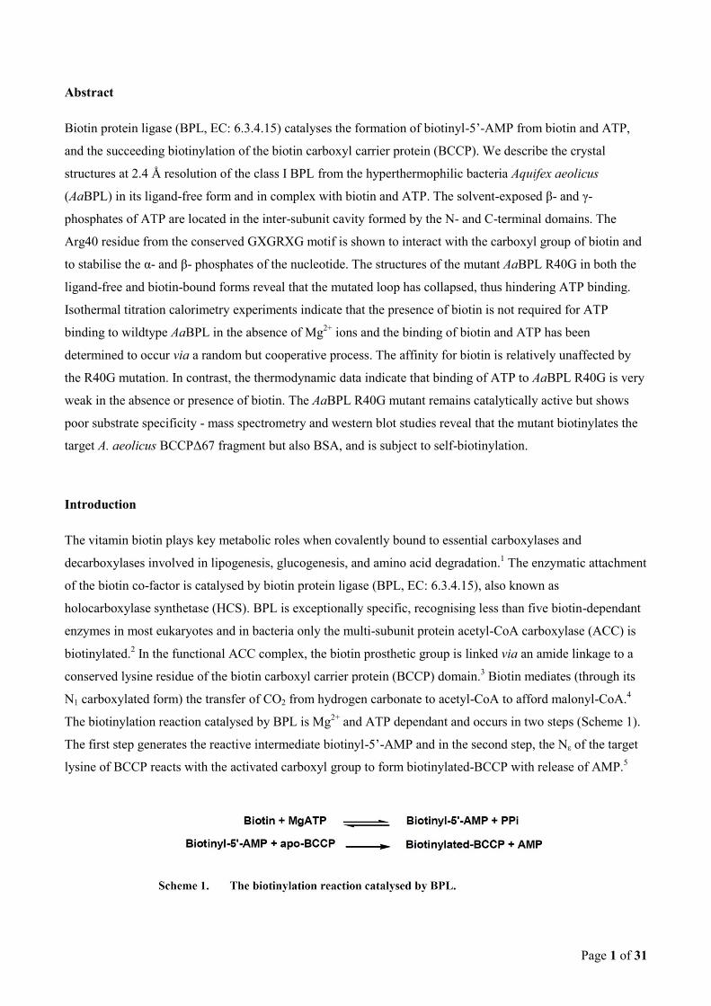
The structure of AaBPL complexed with biotin and ATP (AaBPL:biotin:ATP) was determined at a resolution
of 2.3 Å (Figure 2a). In the complex, residues 37-47, which form the biotin binding loop and act as a lid to the

Page 6 of 31
active site, and the two ligands are fully ordered in the electron density (Figure 2b) The loop stabilises the
biotin and ATP moieties with strong hydrophobic and hydrophilic interactions. The tetrahydrothiophene ring
of the biotin is accommodated in the interior of the binding pocket formed by residues Gly119 and Gly121 of
the β7-strand (Figure 2c) The nitrogen atoms of the biotin ureido group hydrogen-bond with the side chain of
Gln34 and the backbone carbonyl of Arg38 from the biotin binding loop, as well as the Thr14 from the α1-
helix. In addition, the ureido carbonyl forms hydrogen-bonds with the hydroxyl group of Ser13 from the β1- α1
loop and the backbone NH of Arg38. The aliphatic tail of the biotin is held by a sandwich of two hydrophobic
walls formed by the residues Gly106, Val107, and Leu108 from the β6-strand on one side, and on the other
side, the residues Gly37, Gly39, Trp45, and Leu46 from the biotin binding loop.
(a)
(b)

Page 7 of 31
(c)
Figure 2. Stereo views of AaBPL with bound biotin and ATP (AaBPL:biotin:ATP). (a) A cartoon view
of AaBPL monomer, ATP, biotin and Arg40 are represented as sticks. (b) A close-up view of the active site
with electron density around ATP, biotin and Arg40. The final omit |Fo| – |Fc| is contoured at 2.5σ. (Green
chicken wire around ATP and BIOTIN, blue around Arg40) (c) Binding interactions of ATP and biotin within
the AaBPL protomer A active site. ATP and biotin are coloured with green carbons, H-bonds are shown as
broken red lines. Residues (Lys192 and Ser233) from the other protomer are annotated with asterisks (*).
The biotin carboxylate is adjacent to the turn of the biotin binding loop and is strongly stabilised by hydrogen-
bonds with the side chain of the Arg40 from the GRGRLG motif and have weak interactions with the Arg40
backbone NH and the side chain of Lys103 from the β6-strand. The carboxylate group lies toward the outside
of the active site, near the ATP binding pocket. We find that the oxygen atoms of the biotin carboxylate group
and the -phosphate of ATP are located in close proximity (approximately 3.5 Å) (Figures 2b and c).
Therefore, the nucleophilic attack of the carboxylate anion at the α- phosphate of ATP, resulting in the
formation of the phosphoanhydride bond of biotinyl- 5’-AMP, induces only minor conformational changes to
the active site. Comparison of the structures of the AaBPL:biotin:ATP and PhBPL:biotinyl-5’-AMP
complexes shows that the orientations of the adenine ring and sugar moiety of ATP and the biotin within the
active sites are similar in the two complexes.18
We find that polar and electrostatic interactions with residues from both the adenylatebinding loop (Asn123
and Ile133) and the biotin binding loop (Trp45 and Leu46) contribute to the correct positioning of the adenine
ring of ATP in the active site (Figure 2c). The α-phosphate of ATP is stabilised by a network of hydrogen-
bonding interactions with the side chains of Arg40, Arg43 and Lys103. In addition to these interactions, the
crystal packing allows the formation of hydrogen-bonds between the oxygen atoms of the α-phosphate of ATP

Page 8 of 31
and the side chain amino group of Lys192 from the second monomer (monomer B) of the asymmetric unit
(Figure 2c). The structure of the AaBPL:biotin:ATP complex reveals that the β- and γ- phosphates of ATP
protrude from the adenylate binding site pointing into the inter-subunit cavity formed by the N- and C-
terminal domains and are solvent-exposed (Figure 2a). In the crystal, the β-phosphate is stabilised by the
hydrophilic environment formed by Asp96 from the β5-strand and the side chains of Arg40, Lys92 and
Lys103. The γ-phosphate interacts weakly with the hydroxyl group of Tyr98 and the side chain of Lys92 from
the β4- and β5- strands respectively. Interestingly, the structure of the biotin-ATP complex indicates that the
backbone carbonyl of Leu230 and the hydroxyl group of Ser229 from the β12-strand of the C-terminal domain
also form hydrogen-bonds with the oxygen atoms of the γ-phosphate (Figure 2c). The terminal diphosphate, is
thus in a position which allows binding of Mg2+
resulting in the easy elimination of the pyrophosphate leaving
group and the formation of biotinyl-5’-AMP. It is perhaps noteworthy that in this structure the side chain of
Arg231 points towards the phosphate binding cavity. In BirA the equivalent residue (Arg317) has been shown
by mutagenesis to be directly involved in MgATP binding.28
3. Capture of the biotin-ATP intermediate complex of AaBPL
In the AaBPL and PhBPL complexes with biotin and ATP, the biotin binding loops display significant
conformational differences which can be attributed to the sequence differences within their respective glycine
rich motifs - AaBPL 37GRGRLG42 and PhBPL 45GHGRLN50 (Figure 3). In comparison to the
AaBPL:biotin:ATP structure, in the PhBPL complex, the loop is displaced and the side chain of the Asn50 is
solventexposed and interacts weakly with the His46 (Bagautdinov and Kunishima, PDB entry: 2DTO). The
Lys111 was proposed to play a key role in the formation of biotinyl-5’-AMP in PhBPL by stabilising the
pentacoordinate transition state of the α-phosphate before formation of the phosphoanhydride bond.18
In the
AaBPL:biotin:ATP complex the equivalent conserved lysine (Lys103) adopts a similar conformation but
forms long-range hydrogen-bonds with the carboxyl group of the biotin.
Of particular note are the specific orientation and the interactions made by the Arg40 residue in the structure
of the AaBPL complex which differ significantly from those in the PhBPL and BirA substrate
complexes.14,16,18
. Arg40 plays an essential role in stabilising the biotin and ATP ligands within the active site
(Figures 2b and 3). In the AaBPL:biotin:ATP ternary complex, the side chain of Arg40 interacts with the
carboxyl group of the biotin and forms direct hydrogen-bonds with the oxygens of the α- and β- phosphates of
ATP. The backbone NH of the Arg40 residue also interacts with the carboxyl group of the biotin. This
orientation of the conserved arginine side chain has not been observed in other ligand-bound BPL structures.
In the structure of the BirA:biotin complex, the backbone NH of the equivalent arginine (Arg118) forms
hydrogen-bonds with the carboxyl group of the biotin and in the complex with biotinol-5’-AMP, the backbone
NH interacts with the oxygen atoms of the phosphate group of the adenylate.14,16
Thus, in the BirA complexes,

Page 9 of 31
the side chain of the Arg118 appears to play mainly a structural role by closing the active site and sequestering
the ligands via weak hydrophilic interactions. In the PhBPL:biotin:ATP complex the Arg48 strongly stabilises
the α-phosphate of ATP by hydrogen-bonding via its backbone NH and weakly through its sidechain.. While
in the structure of the AaBPL:biotin:ATP complex the side chain of the Arg40 adopts a bent conformation in
order to interact with the biotin and the α- phosphate, in the PhBPL:biotin:ATP complex, the side chain of the
Arg48 points towards the γ-phosphate and has a similar orientation with respect to the triphosphate chain
(Figure 3). This conformation of the Arg48 allows the side chain to form a network of weak hydrogen-bonds
with the oxygen atoms of the α- and β-phosphate groups of ATP (as well as with the phosphate of the biotin in
the PhBPL:biotinyl-5’-AMP complex). In contrast to AaBPL, neither the backbone NH nor the side chain of
the Arg48 interacts with the carboxyl group of the biotin in PhBPL. These combined analyses suggest that
their are subtle differences in the way each BPL isozyme achieves substrate binding and
catalysis.
Figure 3. Comparison of the ligand-binding sites of the ATP and biotin-bound complexes of AaBPL
and PhBPL. The PhBPL PDB file used was 2DTO. The colouring is as follows: AaBPL, red cartoon; ATP,
biotin and Arg40 shown as sticks with purple carbons; PhBPL, blue cartoon; ATP, biotin and Arg48 are
shown as sticks with light blue carbons. The equivalent arginine residues and ATP are shown to be in
significantly different positions.
Page 10 of 31
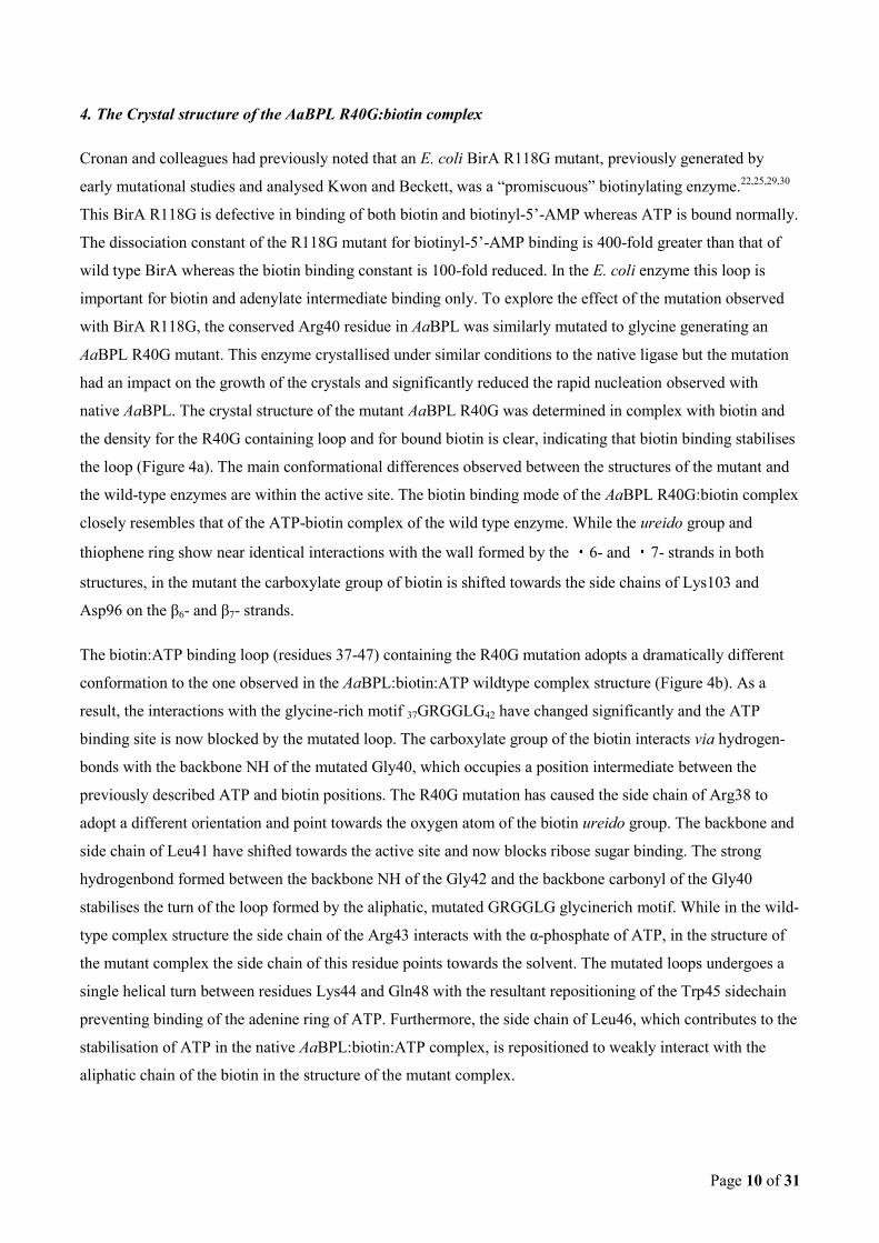
4. The Crystal structure of the AaBPL R40G:biotin complex
Cronan and colleagues had previously noted that an E. coli BirA R118G mutant, previously generated by
early mutational studies and analysed Kwon and Beckett, was a “promiscuous” biotinylating enzyme.22,25,29,30
This BirA R118G is defective in binding of both biotin and biotinyl-5’-AMP whereas ATP is bound normally.
The dissociation constant of the R118G mutant for biotinyl-5’-AMP binding is 400-fold greater than that of
wild type BirA whereas the biotin binding constant is 100-fold reduced. In the E. coli enzyme this loop is
important for biotin and adenylate intermediate binding only. To explore the effect of the mutation observed
with BirA R118G, the conserved Arg40 residue in AaBPL was similarly mutated to glycine generating an
AaBPL R40G mutant. This enzyme crystallised under similar conditions to the native ligase but the mutation
had an impact on the growth of the crystals and significantly reduced the rapid nucleation observed with
native AaBPL. The crystal structure of the mutant AaBPL R40G was determined in complex with biotin and
the density for the R40G containing loop and for bound biotin is clear, indicating that biotin binding stabilises
the loop (Figure 4a). The main conformational differences observed between the structures of the mutant and
the wild-type enzymes are within the active site. The biotin binding mode of the AaBPL R40G:biotin complex
closely resembles that of the ATP-biotin complex of the wild type enzyme. While the ureido group and
thiophene ring show near identical interactions with the wall formed by the 6- and 7- strands in both
structures, in the mutant the carboxylate group of biotin is shifted towards the side chains of Lys103 and
Asp96 on the β6- and β7- strands.
The biotin:ATP binding loop (residues 37-47) containing the R40G mutation adopts a dramatically different
conformation to the one observed in the AaBPL:biotin:ATP wildtype complex structure (Figure 4b). As a
result, the interactions with the glycine-rich motif 37GRGGLG42 have changed significantly and the ATP
binding site is now blocked by the mutated loop. The carboxylate group of the biotin interacts via hydrogen-
bonds with the backbone NH of the mutated Gly40, which occupies a position intermediate between the
previously described ATP and biotin positions. The R40G mutation has caused the side chain of Arg38 to
adopt a different orientation and point towards the oxygen atom of the biotin ureido group. The backbone and
side chain of Leu41 have shifted towards the active site and now blocks ribose sugar binding. The strong
hydrogenbond formed between the backbone NH of the Gly42 and the backbone carbonyl of the Gly40
stabilises the turn of the loop formed by the aliphatic, mutated GRGGLG glycinerich motif. While in the wild-
type complex structure the side chain of the Arg43 interacts with the α-phosphate of ATP, in the structure of
the mutant complex the side chain of this residue points towards the solvent. The mutated loops undergoes a
single helical turn between residues Lys44 and Gln48 with the resultant repositioning of the Trp45 sidechain
preventing binding of the adenine ring of ATP. Furthermore, the side chain of Leu46, which contributes to the
stabilisation of ATP in the native AaBPL:biotin:ATP complex, is repositioned to weakly interact with the
aliphatic chain of the biotin in the structure of the mutant complex.

Page 11 of 31
(a)
(b)
Figure 4. A close-up stereo view of the mutant R40G AaBPL:biotin complex binding site. (a) Electron
density is shown around the R40G containing loop (blue chickenwire) and the biotin ligand (green
chickenwire). The final omit |Fo| – |Fc| map contoured at 3σ is shown. (b) Comparison of the loop position
between AaBPL R40G:biotin complex and wild type AaBPL:biotin:ATP complex. The mutant loop and
bound biotin are coloured green. ATP, biotin and Arg40 are shown as sticks in the native structure with grey
coloured carbons. The mutant loop position can be clearly seen to preclude ATP binding.

Page 12 of 31
5. Binding studies
Isothermal titration calorimetry (ITC) experiments were carried out to determine the thermodynamic
parameters for binding of biotin and ATP with AaBPL, and to investigate if the binding events occur via the
ordered sequential mechanism established for BirA previously. Since the final step of the mechanism, the
formation of biotinyl-5’- AMP, is Mg2+
dependent, the binding of the ligands was studied in the presence of 4
mM EDTA.31
This is a valid simplification since the crystal structure of AaBPL complexed with biotin and
ATP indicates that Mg2+
ions are not required for binding of the substrates. The ITC results indicate that
AaBPL binds both substrates in an exothermic process (Figure 5a and 5b). The thermodynamic parameters
(Table 1) suggest relatively weak protein-ligand interactions for both biotin and ATP binding under these
conditions (KD = 3.5 μM and KD = 7.2 μM respectively), the interaction with biotin being 1000-fold weaker
than that reported for biotin with BirA (KD = 45 nM).22
ATP and biotin binding are enthalpy-driven with a
favourable entropy change. For an ordered mechanism to occur, binding of one ligand would be prerequisite
for binding of the other. However, in contrast to BirA, biotin is not required for ATP binding to AaBPL. This
result is unlikely to be an artefact since it could be shown by streptavidin titration that less than 4% biotin was
present in the AaBPL preparation and thus the amount of biotin initially present in the active site could be
ignored (see supplemental data). The small difference (a factor of 2) between the KD values for binding of
ATP and biotin suggests that ligand binding to AaBPL occurs via a random process.
Table 1. Thermodynamic parameters derived from the titrations of AaBPL and AaBPL R40G with
biotin and ATP before and after formation of the binary complexes formed with the two ligands.
The error in ΔH is ± 5% and is mainly due to the differences in enzyme and ligand concentrations. ΔGo is
calculated from the binding determined by ITC: ΔG° = -RT lnKA = ΔH – T. ΔSo. KA = 1/KD. The parameters
of BirA and BirA R118G binding to biotin are included in the table for comparison.22, 23

Page 13 of 31
Figure 5. Isothermal titration calorimetry measurements of AaBPL with (a) biotin and (b) ATP and AaBPL
R40G with (c) biotin and (d) ATP. The upper panels show the raw data of heat changes upon addition of
ligand. The lower panels show the processed data corresponding to the heat of each injection plotted against
the molar ratio of ligand to enzyme after subtraction of the heat of ligand dilution. All buffers contained
10 mM Hepes pH 7.5, 4 mM EDTA. The derived thermodynamic parameters are given in Table 1.

Page 14 of 31
The cooperativity of the binding events after formation of the binary complexes AaBPL:biotin and
AaBPL:ATP was further investigated by titration with ATP and biotin, respectively. The ITC results revealed
that binding of biotin and ATP was greatly enhanced by the presence of the other substrate. The affinity (KA)
of biotin with the AaBPL:ATP binary complex is five times greater than for AaBPL alone (KD = 0.7 μM
compared with 3.5 μM), while the KA of ATP for AaBPL increases nearly two-fold for the AaBPL:biotin
complex (4.6 μM compared with 7.2 μM, Table 1). The enthalpy and entropy changes measured for the two
possible pathways—i and iii, and ii and iv—leading to the formation of the AaBPL:biotin:ATP ternary complex
correspond within experimental error and indicate cooperativity between biotin and ATP binding (Figure 6).
Despite relatively large variations in apparent enthalpy and entropy changes for the different AaBPL binary
and ternary complexes, the overall changes in binding free energies are much smaller and this may be
attributed to the ubiquitous enthalpy–entropy compensation frequently observed in biomolecular systems.33
Figure 6. Formation of the ternary AaBPL:biotin:ATP complex via the pathways i and iii, and pathways ii
and iv. In AaBPL, both pathways occur. KA is the association constant (1/KD).
To investigate the role of Arg40 in the ligand binding process, ITC experiments were also performed with
theAaBPL R40G enzyme. Titrations of this mutant under the experimental conditions used for the wild type
indicated that the affinity for biotin is only 2.5-fold lower than that observed for the native enzyme (KD 8.3 μ
M compared with 3.5 μM; see Figure 5c and Table 1). This suggests that, in contrast to the E. coli mutant
BirA R118G, which displayed greatly reduced biotin binding, the Arg to Gly mutation has little effect on
biotin binding in AaBPL. Thus, while the affinity of BirA for biotin is approximately 100-fold greater than
that ofAaBPL, the dissociation constants for the biotin complexes of the two mutants BirA R118G and AaBPL

Page 15 of 31
R40G are similar (KD 1.8 μM and 8.3 μM, respectively).22
Both apparent enthalpy and entropy changes are
favourable for biotin binding to the mutant (Table 1). Biotin was shown to bind to AaBPL R40G withKD =
15.4 μM when the mutant was titrated in the presence of ATP. The small difference, a factor of <2, between
the measured dissociation constants for the AaBPL R40G and the AaBPL R40G binary complex suggests that
the presence of ATP does not significantly alter biotin binding.
In contrast to the biotin data, titrations of the mutant enzyme with ATP showed only small heat signals,
principally due to heat of dilution, indicating that binding of ATP to the AaBPL R40G is negligible (KD > 200
μM) under these conditions (Figure 5d). The side chain of Arg40 thus appears to be necessary for ATP
binding in the absence of Mg2+
. The structure of the mutant AaBPL R40G:biotin complex revealed that biotin
binding affects the conformation of the ATP-binding pocket and titration of this complex with ATP also
showed very weak binding (KD > 200 μM).
6. Substrate biotinylation with AaBPL R40G
Previously, we showed that the C-terminal domain of A. aeolicus BCCP (BCCPΔ67) is a substrate for
biotinylation with AaBPL.21
Incubation of apo-BCCPΔ67 with AaBPL or the mutant AaBPL R40G in the
presence of biotin, ATP and MgCl2 for 20 min at 65 °C led to the formation of biotinylated-BCCPΔ67.
While, on the basis of streptavidin Western blots of the reactions, the R40G mutant appears to be a poorer
catalyst than the wild type enzyme (Figure 7a, lanes 9 and 10), mass spectrometry of the biotinylated BCCPΔ
67 from the R40G-catalysed reaction revealed the formation of a singly biotinylated BCCPΔ67 species with a
mass increase equivalent to one biotin moiety (10,740.7 – 10,966.2 Da) (Figure 8c). The mutant substrate
BCCPΔ67 K117L, which lacks the target Lys117, is not biotinylated by AaBPL or by AaBPL R40G,
showing that none of the remaining four lysine residues was derivatised and suggests a significant degree of
target specificity (Figure 7a, lanes 12–14). However, Western blots of these reactions also showed that AaBPL
R40G, in contrast to the wild type enzyme, self-biotinylates (Figure 7a). This was confirmed by mass
spectrometry analysis of the R40G species after incubation; mass increases in the range 200 ∼ 2000 Da (data
not shown) were observed, indicating that the single R40G mutant, which contains lysine residues, undergoes
multiple biotinylation.
The equivalent BirA mutant, BirA R118G, has been shown to biotinylate itself in vivo and in vitro, and to
biotinylate non-cognate acceptors such as bovine serum albumin (BSA) or RNAse A.25
On the basis of the
fact that BirA R118G binds biotinyl-5′-AMP less tightly than wild-type BirA (KD 20 nM and KD 45 pM,
respectively), it has been suggested that this could be a direct consequence of non-specific solution reactions
of free biotinyl-5′-AMP with exposed lysine residues. 22,25
Our suggestion that A. aeolicusBCCPΔ67

Page 16 of 31
K117L is not multiply biotinylated is supported by recent chemical biotinylation studies of E. coliBCCP87
with free biotinyl-5′-AMP, which have demonstrated that only the target lysine is modified.33
This
specificity has been attributed to the intrinsic reactivity of the target lysine and the stabilisation of the
BCCP:biotinyl-5′-AMP complex through interactions of the ureido ring of the biotin moiety with residues
of the “thumb” region present in both E. coli and A. aeolicus BCCP. 21,34
Figure 7. Streptavidin Western blot analysis of biotinylation with wild type AaBPL and mutant AaBPL
R40G, and with BSA of (a) apo-BCCPΔ67 and BCCPΔ67 K117L and (b) BSA. (a) Left-hand panel: SDS-
PAGE stained with Coomassie brilliant blue (lanes 1–7); right-hand panel: Western blot (lanes 8–14). Lanes 1
and 8, apo-BCCPΔ67; lanes 2 and 9, apo-BCCPΔ67 incubated with AaBPL in the presence of biotin, ATP,
MgCl2 at 65 °C for 20 min; lanes 3 and 10, apo-BCCPΔ67 incubated with AaBPL R40G under the same
conditions as wild type AaBPL; lanes 4 and 11, BCCPΔ67 K117L; lanes 5 and 12, BCCPΔ67 K117L
incubated with AaBPL in the presence of biotin, ATP, MgCl2 at 65 °C; lanes 6 and 13, BCCPΔ67 K117L
incubated withAaBPL R40G under the same conditions; lanes 7 and 14, BCCPΔ67 K117L incubated with
AaBPL R40G in the presence of 100 μM biotin, ATP, MgCl2 at 65 °C. The intensity of the AaBPL R40G
spot (in lane 10), given that the protein concentrations of R40G and BCCPΔ67 are 1/10 indicates the multiple
nature of R40G self-biotinylation. This was confirmed by mass spectrometry. (b) BSA (2 μM) incubated in
the presence of biotin, ATP, MgCl2 at 65 °C for 20 min with AaBPL or AaBPL R40G. Lane 1, BSA; lane 2,
BSA with 50 nM AaBPL; lane 3, BSA with 50 nM AaBPL R40G; lane 4, BSA with 200 nM AaBPL; lane 5,
BSA with 200 nM AaBPL R40G; lane 6, BSA with 500 nM AaBPL; lane 7, BSA with 500 nM AaBPL R40G.

Page 17 of 31
To determine whether AaBPL and AaBPL R40G could also carry out non-specific biotinylation, the enzymes
were incubated with biotin, ATP and MgCl2 in the presence of (BSA) at 65 °C. Western blot analysis of the
products revealed that, while biotinylation of BSA did not occur in the presence of substrates alone or at low
concentration of wild-type AaBPL, the mutant AaBPL R40G biotinylated itself and BSA in a manner similar
to that shown for BirA R118G (Figure 7b). At high concentrations of enzyme, the wild-type AaBPL also
carried out self-biotinylation and non-specific biotinylation of BSA, albeit less effectively.
AaBPL R40G appears to self-biotinylate preferentially rather than derivatise the substrates BSA or BCCPΔ67
(Figure 8a and b), which is likely to be a consequence of the elevated temperature of the reaction with the
thermostable enzymes. Since chemical biotinylation with biotinyl-5′-AMP is proximity-dependent and the
lifetime of AMP mixed anhydrides in aqueous solution (typically t1/2 ∼ 1 min at pH 7, 25 °C) will be
significantly shorter at 65 °C, the amount of reagent surviving to modify a non-cognate acceptor will be
significantly less. 25,35
Figure 8. LC-ESI-MS analysis of apo-BCCPΔ67: the 6+ to 10+ charge states and the deconvoluted masses.
(a) apo-BCCPΔ67 incubated in presence of biotin, ATP, MgCl2 at 65 °C for 15 min; (b) apo-BCCPΔ67

Page 18 of 31
incubated with AaBPL in the same conditions; (c) apo-BCCPΔ67 incubated with AaBPL R40G in the same
conditions. The theoretical mass for apo-BCCPΔ67 is 10,739.63 Da and the theoretical mass for holo-BCCP
Δ67 is 10,965.94 Da.
Conclusion
We have determined the crystal structures of the monomeric class I AaBPL in the ligand-free form and in
complex with biotin and ATP. We found that AaBPL displays a high level of structural similarity with the E.
coli BirA and P. horikoshii BPL enzymes with regard to the domain arrangement and overall fold. 16, 18
The
geometry of the novel AaBPL:biotin:ATP complex presented here suggests that it is similar to that of the
transition state that precedes biotinyl-5′-AMP formation. In the structure of the AaBPL:biotin:ATP complex,
the biotin co-factor and the AMP moiety of ATP are buried in the adenylate-binding site, and the biotin
carboxylate and the α-phosphate are located in close proximity to each other at the entrance of the active site.
This suggests that subsequent nucleophilic attack of the carboxylate anion on the α-phosphate leading to the
formation of the phosphoanhydride bond of the product requires only minor changes to the conformation of
the active site and may be facilitated by Mg2+
binding to the β- and γ-phosphates of ATP, which are
solvent-exposed and located in the inter-subunit domain formed by the N- and C-termini. The oxygen atoms
of the ATP γ-phosphate form long-range hydrogen bonds with residues located on the SH3-like barrel C-
terminal domain also characterized in the structural homologue phenylalanine tRNA synthetase.8 This is the
first demonstration that direct interactions of the substrates and the C-terminal domain of a BPL occur. This
has been further highlighted in the recent co-crystal structures of PhBPL R48A and K111A with BCCPΔ
N76, the C-terminal domain shows the largest variations in the different stages of the complex, which
suggests its functional importance in this isoform.19
The bent conformation of the adenylate described in the PhBPL:biotinyl-5′-AMP and BirA:biotinol-5′-
AMP complexes has also been observed in the structural homologue Thermoplasma acidophilum LplA
(TaLplA) bound to lipoyl-AMP. 36,37
However, the orientation of the triphosphate chain of ATP is not
conserved in these enzymes. In the structure of TaLplA bound to MgATP, the ATP moiety adopts a U-shaped
conformation and the phosphate chain points inward towards the bifurcated lipoyl-AMP-binding pocket,
which is accessible through a tunnel-like entrance. The β-phosphate occupies roughly the same position as
the carboxyl group of lipoyl-AMP inside the predominantly hydrophobic cavity. The U-shaped conformation
of ATP is also observed in the structures of class II aminoacyl-tRNA synthetases bound to ATP but here the
β- and γ-phosphates remain solvent-exposed. For example, comparison of the structures of Thermus
thermophilus glycyl-tRNA synthetase bound to ATP and glycyl-AMP show that the ATP phosphate chain,
located at the entrance of the active site, is bent away from the binding site of the glycyl moiety. The resultant

Page 19 of 31
aminoacyl-adenylate adopts an extended conformation.38
Analysis of the structures ofStaphylococcus aureus
threonyl-tRNA synthetase bound to ATP and Zn2+
and to an adenylate analogue (threonyl-AMS) and the
structures of the MgATP and alanine complexes of A. aeolicus alanyl-tRNA synthetase supports this common
binding mode in class II tRNA synthetases.39,40
The structure of the AaBPL:biotin:ATP complex has revealed the importance of Arg40 from the glycine-rich
motif in the stabilisation of the two ligands. The side chain of this conserved arginine plays an important role
in the neutralisation of the negative charges of the triphosphate chain of ATP as well as positioning the biotin
carboxylate in close proximity to the α-phosphate. The side chain interacts with the ATP phosphate chains in
two different ways in the A. aeolicus and P. horoikoshii BPL:biotin:ATP ternary complexes. In the AaBPL
complex, Arg40 satisfies only the negative charges of the α- and β-phosphates, and leads to a requirement
for a metal ion for catalysis. Mutation of the arginine to glycine resulted in the dramatic collapse of the
substrate-binding loop. The crystal structure of AaBPL R40G bound to biotin shows that the mutated binding
loop is ordered but blocks the ATP-binding pocket. In contrast, in the structures of the mutant PhBPL R48A
complexes, the Ala48 side-chain adopts the same orientation as that of the Arg48 of the wild-type PhBPL
complexes and the turn of the loop containing the 45GHGRLN50 motif shows very little conformational change
in any native or mutant structures of PhBPL.19
To investigate the effects of the R40G mutation on substrate binding in solution, comparative ITC
experiments were carried out with wild-type AaBPL and mutant AaBPL R40G. The ITC analysis showed
thatAaBPL binds biotin and ATP randomly, and that the binding events occur via a cooperative process. This
contrasts with the behaviour of E. coli BirA, where binding of the substrates follows a sequential ordered
mechanism in which formation of the BirA:biotin complex is a prerequisite for ATP binding.16
The ordering
of binding exhibited by BirA appears necessary in view of its dual role as a repressor and ligase 41,42
—it makes
sense that recruitment of ATP should be the second step. In contrast, for a monofunctional BPL such as
AaBPL, which lacks a regulatory role, the binding order is not crucial. The BirA adenylate-binding loop,
which has been shown to be involved in ATP binding and dimer packing, is disordered in the structures of the
ligand-free and the biotin-bound BirA complexes. 14,15
In contrast, this loop is organised in the ligand-free
form of AaBPL, resulting in a well defined ATP-binding pocket. The Km values of AaBPL and BirA for ATP
also reflect the greater affinity of AaBPL for the nucleotide (15 μM and 200 μM, respectively) while the
Kmvalues for biotin are similar (440 nM and 300 nM, respectively). 24,28
The order of binding of the substrate
and ATP in the adenylate-synthesising enzymes appears to be peculiarly isozyme-specific, since the
eukaryotic class IV BPL from Saccharomyces cerevisae binds ATP before biotin, while amino acid and ATP
binding is a random process in the class II tRNA synthetases. 43,44
The collapse of the substrate-binding loop observed in the structure of the R40G mutant adversely affects
ATP binding but does not inactivate the enzyme completely (vide infra). We used mass spectrometry and

Page 20 of 31
streptavidin Western blot studies to reveal that AaBPL R40G can catalyse the formation of biotinyl-5′-AMP
in the presence of high concentrations of ATP and promiscuously biotinylate the target lysine of apo-BCCPΔ
67, and residues on BSA and the mutant itself. It can be envisaged that, as a consequence of the changed
conformation of the binding site, the AaBPL R40G:biotinyl-5′-AMP binary complex can dissociate and
release the adenylate in the absence of the acceptor protein. Similar behaviour has been observed with the
BirA mutant R118G and in native E. coli methionyl-tRNA and valyl-tRNA synthetases. 25,45,46
In the structure
of PhBPL R48A K111A co-crystallised with biotin and ATP, the disordered triphosphate chain reduces the
reactivity between the ligands and the authors subsequently used this double mutant (and the PhBPL R48A
single mutant) to allow co-crystallisation with PhBCCPΔN76, the structure of which was reported during the
final stages of preparation of our manuscript.19
It is interesting to note that in the PhBPL R48A
K111A:PhBCCPΔN76 complex, the biotinyl domain is in the holo form and also contains bound biotin and
adenosine, suggesting that the double mutant is catalytically active. Multiple conformations of the proteins
were observed in the crystals obtained by Bagautdinov and colleagues, giving them a view of the enzyme
throughout the catalytic process. They propose a catalytic dyad role for residues Asn103 and Asp104
(conserved as Asn95 and Asp96 in AaBPL), that orientates and deprotonates the target lysine. On the other
hand, the PhBPL BCCP does not contain the conserved thumb found in E. coli and A. aeolicusBCCPs, and it
appears that the biotinylation process in different species has subtle nuances that require detailed individual
study.
Our study sheds light on the effects of mutation within the biotin-binding loop on substrate specificity of the
bacterial BPLs and may provide insight into the molecular basis of human multiple carboxylase deficiency
syndrome (MCD). This disease is linked to mutations in the human holocarboxylase synthetase enzyme
(HCS), a class IV BPL. It has been demonstrated that the minimum functional HCS protein is retained in the
349 C-terminal amino acids, which show highly conserved sequence similarity to the putative biotin-binding
domain of bacterial BPLs.47
Further deletion of the N-terminus of the human enzyme to allow comparison
with the 276 amino acids of AaBPL resulted in 33.3 % sequence similarity and 19.2 % sequence identity
between the human HCS domain and the A. aeolicus enzyme. A commonly recurring HCS mutation leading
to MCD in the 505GKGRGG510 motif is R508W which maps to Arg40 of AaBPL. Patients with this missense
R508W mutation are responsive to biotin and can be treated clinically with pharmaceutical doses of the
vitamin. 48,49
It seems likely that further studies on the bacterial proteins may uncover details of enzyme action
relevant to this area of human biochemistry.50
Page 21 of 31
Materials and Methods
Materials
All chemicals used in the preparation of buffers were at least reagent grade. D-Biotin was purchased from
Sigma. For analysis with Western blot, the blocking solution (SuperBlock® Blocking buffer in PBS) was
obtained from Pierce and the antibody (Streptavidin HRP) from BD Pharmingen™. The BSA used for the in
vitro biotinylation experiment with AaBPL R40G was also obtained from Pierce. For the DNA manipulations,
PCR was done using Ready To Go PCR™ beads (Amersham) and the QIAprep® Spin Miniprep kit was
obtained from Qiagen. Restriction endonucleases were purchased from New England Biolabs.
Expression and purification of AaBPL
AaBPL was expressed and purified essentially as described but with the following modifications.21
The cell
pellet, suspended in 10 mM Hepes pH 7.5 containing one tablet of Complete™ Proteinase Inhibitor Cocktail
(Roche), was lysed by sonication (15 min of 30 s cycle). The cell lysate was clarified twice by centrifugation
in the presence of DNase (Roche). The protein was then purified on a Tricorn H R 10/100 column loaded with
15S beads (Amersham) and eluted with a linear salt gradient (0–1 M NaCl in 10 mM Hepes pH 7.5).
Cloning, expression and purification of mutant AaBPL R40G
Site-directed mutagenesis was carried out with the Stratagene kit. The complementary oligonucleotides
encoding the desired mutation were obtained from Sigma Genosys (AaBPL R40G for: GGA AGG GGA GGA
CTC GGA AGG AAG TGG CTC, AaBPL R40G rev: GAG CCA CTT CCT TCC GAG TCC TCC CCT
TCC).The mutant AaBPL R40G was then expressed and purified as described for wild-type AaBPL.
Crystallisation of apo-AaBPL, AaBPL:biotin:ATP and mutant AaBPL R40G and AaBPL R40G:biotin
The purified protein AaBPL was concentrated to 5 mg/ml and crystallised by the hanging drop, vapour
diffusion method at 17 °C. Each crystallisation drop contained 2 μl of protein solution and 1 μl of the
reservoir solution (0.1 M Mes, 0.2 M ammonium sulphate, 15 % (w/v) PEG 5000 mono-ethyl ether, pH 6.5).
To limit nucleation due to impurities, the protein was centrifuged for 1 h at 13,000 rpm before crystallisation.
The crystals appeared immediately and grew non-reproducibly in a few hours to approximately 0.5 mm in
length. In the case of the AaBPL:biotin:ATP complex, D-biotin (2 mM, 1 M NaOH final concentration) and
ATP (5 mM, pH 7 final concentration) were added to the protein before centrifugation and crystallisation.

Page 22 of 31
AaBPL R40G was crystallised by the hanging drop, vapour diffusion method at 17 °C at a concentration of 6
mg/ml.
Crystal trials of the apo form of the AaBPL R40G mutant protein were done by the hanging drop, vapour
diffusion method at 17 °C. The rapid and extensive nucleation observed with wild-type AaBPL was
significantly reduced and it took 4–14 days for the crystals to grow. Co-crystallization of the AaBPL R40G
mutant (carried out at 4 °C and 17 °C) in the presence of 2 mM biotin and 5 mM ATP afforded a
heterogeneous mixture of low-quality crystals of various shapes and lengths, while crystals of AaBPL R40G
co-crystallised with ATP (5 mM), indicating no electron density for the ligand. We obtained crystals of the
mutant in the presence of biotin alone; each drop contained 2 μl of protein (6 mg/ml) in the presence of 2
mM D-biotin and 1 μl of the reservoir solution (0.1 M Mes, 0.2 M ammonium sulphate, 10 % (w/v) PEG
5000 mono-ethyl ether, pH 6.0) at 17 °C. The rectangular crystals took one week to grow reproducibly and
were ∼0.5 mm in length.
For data collection, the AaBPL and AaBPL R40G crystals were frozen after being transferred into a
cryoprotectant solution containing the reservoir solution supplemented with either 20 % (v/v) glycerol or a
non-drying immersion oil (Cargille Laboratories Inc).
Data collection and structure analysis
Data were collected at station BM14 ESRF Grenoble and station 10.1 SRS Daresbury. Data were processed
using the programs MOSFLM and SCALA as part of the CCP4 suite of programs and the structure was solved
with the program PHASER using the structure from P. horikoshii (PDB code 1WPY). Initial refinement was
done using the program REFMAC and finished using the program phenix.refine as part of the PHENIX
package.53,54
Manual refinement was performed using the program COOT.55
Details of the X-ray data
collection, processing and statistics are presented in Table 2.
Table 2. X-ray data collection, processing and refinement statistics. →

Page 23 of 31

Page 24 of 31
Isothermal titration calorimetry
Isothermal titration calorimetry measurements were performed on a VP-ITC calorimeter (Microcal Inc). All
experiments were carried out at 25 °C in 10 mM Hepes, 4 mM EDTA, pH 7.5. The reactants (15.4 μM
wild-type AaBPL and 18 μM mutant AaBPL R40G) were placed in the 2 ml sample chamber and the
substrates D-biotin and ATP (0.2 mM) in the syringe were added with 29 successive additions of 10 μl for 20
s (with an initial injection of 1 μl). The interval between each injection lasted 180 s. The peaks generated
were corrected for ligand heat of dilution and integrated using the ORIGIN software (Microcal Inc) by
plotting the values in microcalories against the molar ratio of injectant to reactant within the cell. Data were
fitted using the single-site binding model. From the dissociation constant KD and the reaction enthalpy value
ΔH, the change in free Gibbs energy (ΔGo) and entropy change (ΔS
o) can be calculated using the equation
ΔGo=RTln(1/KD)=ΔH−TΔSo where R is the universal gas constant and T is the absolute temperature.
In vitro biotinylation studies of BSA, apo-BCCPΔ67 and BCCPΔ67 K117L by mutant AaBPL R40G
All in vitro biotinylation experiments, unless stated otherwise, were done in 10 mM Hepes, pH 7.5 at 65 °C
for 15 or 20 min and were terminated by addition of SDS sample buffer or 0.1 % (v/v) TFA. In the
biotinylation studies with BSA, 2 μM BSA was incubated with 5 μM d-biotin, 1 mM ATP, 2 mM MgCl2
and 50, 200, or 500 nM wild-type AaBPL and mutant AaBPL R40G. Biotinylation experiments were also
carried out with the apo form of BCCPΔ67 and with the mutant BCCPΔ67 K117L. The substrates apo-
BCCPΔ67 and BCCPΔ67 K117L were expressed and purified as described previously.21
For the streptavidin
Western blot studies, the reaction mixtures contained 2 μM substrate, 5 μM D-biotin, 1 mM ATP, 2 mM
MgCl2 and 200 nM enzyme AaBPL orAaBPL R40G. For the mass spectrometry analysis, 10 μM apo-BCCP
Δ67 was incubated with 100 μM D-biotin, 1 mM ATP, 2 mM MgCl2 and the reaction was initiated by
addition of purified AaBPL R40G to a final concentration of 2 μM.
Streptavidin Western blot analysis
Pure proteins separated by SDS-PAGE were transferred onto a 0.2 μm pore sized Hybond-ECL
nitrocellulose membrane (Amersham) using a Trans blot Semi-Dry Transfer cell. The membrane was
incubated overnight in the biotin-free SuperBlock solution at 4 °C. Excess blocking solution was then
removed by washing with phosphate buffer containing 0.1% (v/v) Tween (Sigma) four times before
incubation in SuperBlock solution containing 1/50,000 Streptavidin HRP for 1 h at room temperature. The
membrane was washed and dried before immunolabelled proteins were detected using the ECL enhanced

Page 25 of 31
chemiluminescence detection system (Amersham) in accordance with the manufacturer's protocol. Proteins
were visualised on BioMax XAR film (Kodak).
Mass spectrometry
Positive electrospray ionisation (ESI) liquid chromatography (LC-ESI-MS) was used for the characterisation
of AaBPL, AaBPL R40G and biotinylated BCCPΔ67. Protein samples were separated using a Phenomenex
C5 reverse phase column on a Waters HPLC 2690. The proteins were eluted from the column with a 5%–95%
(v/v) acetonitrile gradient (containing 0.1 % TFA) and analysed on a MicroMass Platform II quadrupole mass
spectrometer. The molecular mass was determined by the Transform algorithms of the Mass Lynx software
(MicroMass).
Protein Data Bank accession codes
The coordinates have been submitted to the PDB. The apo form of AaBPL has been assigned the PDB
code3FJP, the AaBPL:biotin:ATP complex has PDB code 3EFS and the AaBPL R40G:biotin complex has
PDB code 3EFR.

Page 26 of 31
References
[1] Chapman-Smith, A. & Cronan, J. E., Jr. (1999). The enzymatic biotinylation of proteins: a post-
translational modification of exceptional specificity. Trends Biochem Sci 24, 359-63.
[2] Cronan, J. E., Jr. & Waldrop, G. L. (2002). Multi-subunit acetyl-CoA carboxylases. Prog Lipid Res 41,
407-35.
[3] Nenortas, E. & Beckett, D. (1996). Purification and characterization of intact and truncated forms of the
Escherichia coli biotin carboxyl carrier subunit of acetyl- CoA carboxylase. J Biol Chem 271, 7559-67.
[4] Tong, L. (2005). Acetyl-coenzyme A carboxylase: crucial metabolic enzyme and attractive target for drug
discovery. Cell Mol Life Sci 62, 1784-803.
[5] Lane, M. D., Rominger, K. L., Young, D. L. & Lynen, F. (1964). The Enzymatic Synthesis of
Holotranscarboxylase from Apotranscarboxylase and (+)-Biotin. Ii. Investigation of the Reaction
Mechanism. J Biol Chem 239, 2865-71.
[6] Fujiwara, K., Toma, S., Okamura-Ikeda, K., Motokawa, Y., Nakagawa, A. & Taniguchi, H. (2005).
Crystal structure of lipoate-protein ligase A from Escherichia coli. Determination of the lipoic acid-
binding site. J. Biol. Chem. 239, 2865-2871.
[7] Artymiuk, P. J., Rice, D. W., Poirrette, A. R. & Willet, P. (1994). A tale of two synthetases. Nat. Struct.
Biol. 1, 758-760.
[8] Safro, M. & Mosyak, L. (1995). Structural similarities in the noncatalytic domains of phenylalanyl-tRNA
and biotin synthetases. Protein Sci. 4, 2429-2432.
[9] Mukhopadhyay, B., Purwantini, E., Kreder, C. L. & Wolfe, R. S. (2001). Oxaloacetate synthesis in the
methanarchaeon Methanosarcina barkeri: pyruvate carboxylase genes and a putative Escherichia coli-
type bifunctional biotin protein ligase gene (bpl/birA) exhibit a unique organization. J Bacteriol 183,
3804-10.
[10] Beckett, D. & Matthews, B. W. (1997). Escherichia coli repressor of biotin biosynthesis. Methods
Enzymol. 279, 362-376.
[11] Eisenstein, E. & Beckett, D. (1999). Dimerization of the Escherichia coli biotin repressor: corepressor
function in protein assembly. Biochemistry 38, 13077- 13084.
[12] Beckett, D. (2007). Biotin sensing: universal influence of biotin status on transcription. Ann. Rev. Genet.
41, 443-464.

Page 27 of 31
[13] Brown, P. H., Cronan, J. E., Grotli, M. & Beckett, D. (2004). The biotin repressor: modulation of
allostery by corepressor analogs. J. Mol. Biol. 337, 857-869.
[14] Weaver, L. H., Kwon, K., Beckett, D. & Matthews, B. W. (2001). Corepressorinduced organization and
assembly of the biotin repressor: a model for allosteric activation of a transcriptional regulator. Proc. Natl
Acad. Sci. USA 98, 6045-6050.
[15] Wilson, K. P., Shewchuk, L. M., Brennan, R. G., Otsuka, A. J. & Matthews, B. W. (1992). Escherichia
coli biotin holoenzyme synthetase/bio repressor crystal structure delineates the biotin- and DNA-binding
domains. Proc. Natl Acad. Sci. USA 89, 9257-61.
[16] Wood, Z. A., Weaver, L. H., Brown, P. H., Beckett, D. & Matthews, B. W. (2006). Co-repressor induced
order and biotin repressor dimerization: a case for divergent followed by convergent evolution. J. Mol.
Biol. 357, 509-523.
[17] Xu, Y. & Beckett, D. (1994). Kinetics of biotinyl-5'-adenylate synthesis catalysed by the Escherichia coli
repressor of biotin biosynthesis and the stability of the enzyme-product complex. Biochemistry 33, 7354-
7360.
[18] Bagautdinov, B., Kuroishi, C., Sugahara, M. & Kunishima, N. (2005). Crystal structures of biotin protein
ligase from Pyrococcus horikoshii OT3 and its complexes: structural basis of biotin activation. J. Mol.
Biol. 353, 322-333.
[19] Bagautdinov, B., Matsuura, Y., Bagautdinova, S. & Kunishima, N. (2008). Protein biotinylation
visualized by a complex structure of biotin protein ligase with a substrate. J. Biol. Chem. 283, 14739-
14750.
[20] Weaver, L. H., Kwon, K., Beckett, D. & Matthews, B. W. (2001). Competing protein:protein interactions
are proposed to control the biological switch of the E. coli biotin repressor. Protein Sci. 10, 2618-2622.
[21] Clarke, D. J., Coulson, J., Baillie, R. & Campopiano, D. J. (2003). Biotinylation in the hyperthermophile
Aquifex aeolicus. Eur. J. Biochem. 270, 1277-1287.
[22] Kwon, K. & Beckett, D. (2000). Function of a conserved sequence motif in biotin holoenzyme
synthetases. Protein Sci. 9, 1530-1539.
[23] Kwon, K., Streaker, E. D. & Beckett, D. (2002). Binding specificity and the ligand dissociation process
in the E. coli biotin holoenzyme synthetase. Protein Sci. 11, 558-570.
[24] Kwon, K., Streaker, E. D., Ruparelia, S. & Beckett, D. (2000). Multiple disordered loops function in
corepressor-induced dimerization of the biotin repressor. J. Mol. Biol. 304, 821-833.

Page 28 of 31
[25] Choi-Rhee, E., Schulman, H. & Cronan, J. E. (2004). Promiscuous protein biotinylation by Escherichia
coli biotin protein ligase. Protein Sci. 13, 3043-3050.
[26] Dupuis, L., Leon-Del-Rio, A., Leclerc, D., Campeau, E., Sweetman, L., Saudubray, J. M., et al. (1996).
Clustering of mutations in the biotin-binding region of holocarboxylase synthetase in biotin-responsive
multiple carboxylase deficiency. Hum. Mol. Genet. 5, 1011-1016.
[27] Suzuki, Y., Yang, X., Aoki, Y., Kure, S. & Matsubara, Y. (2005). Mutations in the holocarboxylase
synthetase gene HLCS. Hum. Mutat. 26, 285-290.
[28] Chapman-Smith, A., Mulhern, T. D., Whelan, F., Cronan, J. E. J. & Wallace, J. C. (2001). The C-
terminal domain of biotin protein ligase from E. coli is required for catalytic activity. Protein Sci. 10,
2608-2617.
[29] Barker, D. F. & Campbell, A. M. (1981). Genetic and biochemical characterization of the birA gene and
its product: evidence for a direct role of biotin holoenzyme synthetase in repression of the biotin operon in
Escherichia coli. J. Mol. Biol. 146, 469-492.
[30] Buoncristiani, M. R., Howard, P. K. & Otsuka, A. J. (1986). DNA-binding and enzymatic domains of the
bifunctional biotin operon repressor (BirA) of Escherichia coli. Gene 44, 255-261.
[31] Perozzo, R., Jelesarov, I., Bosshard, H. R., Folkers, G. & Scapozza, L. (2000). Compulsory order of
substrate binding to herpes simplex virus type 1 thymidine kinase. A calorimetric study. J. Biol. Chem.
275, 16139-16145.
[32] Cooper, A., Johnson, C. M., Lakey, J. H. & Nollmann, M. (2001). Heat does not come in different
colours: entropy-enthalpy compensation, free energy windows, quantum confinement, pressure
perturbation calorimetry, solvation and the multiple causes of heat capacity effects in biomolecular
interactions. Biophys. Chem. 93, 215-230.
[33] Streaker, E. D. & Beckett, D. (2006). Nonenzymatic biotinylation of a biotin carboxyl carrier protein:
unusual reactivity of the physiological target lysine. Protein Sci. 15, 1928-1935.
[34] Solbiati, J., Chapman-Smith, A. & Cronan, J. E., Jr. (2002). Stabilization of the biotinoyl domain of
Escherichia coli acetyl-CoA carboxylase by interactions between the attached biotin and the protruding
"thumb" structure. J. Biol. Chem. 277, 21604-21609.
[35] Pavela-Vrancic, M., Dieckmann, R., Dohren, H. V. & Kleinkauf, H. (1999). Editing of non-cognate
aminoacyl adenylates by peptide synthetases. Biochem. J. 342, 715-719.

Page 29 of 31
[36] Kim, D. J., Kim, K. H., Lee, H. H., Lee, S. J., Ha, J. Y., Yoon, H. J. & Suh, S. W. (2005). Crystal
structure of lipoate-protein ligase A bound with the activated intermediate: insights into interaction with
lipoyl domains. J. Biol. Chem. 280, 38081-38089.
[37] McManus, E., Luisi, B. F. & Perham, R. N. (2006). Structure of a putative lipoate protein ligase from
Thermoplasma acidophilum and the mechanism of target selection for post-translational modification. . J.
Mol. Biol. 356, 625-637.
[38] Arnez, J. G., Dock-Bregeon, A. C. & Moras, D. (1999). Glycyl-tRNA synthetase uses a negatively
charged pit for specific recognition and activation of glycine. J. Mol. Biol. 286, 1449-1459.
[39] Swairjo, M. A. & Schimmel, P. R. (2005). Breaking sieve for steric exclusion of a noncognate amino
acid from active site of a tRNA synthetase. Proc. Natl Acad. Sci. USA 102, 988-999.
[40] Torres-Larios, A., Sankaranarayanan, R., Rees, B., Dock-Bregeon, A. C. & Moras, D. (2003).
Conformational movements and cooperativity upon amino acid, ATP and tRNA binding in threonyl-
tRNA synthetase. J. Mol. Biol. 331, 201-211.
[41] Streaker, E. D. & Beckett, D. (2006). The biotin regulatory system: kinetic control of a transcriptional
switch. Biochemistry 45, 6417-6425.
[42] Streaker, E. D., Gupta, A. & Beckett, D. (2002). The biotin repressor: thermodynamic coupling of
corepressor binding, protein assembly, and sequencespecific DNA binding. Biochemistry 41, 14263-
14271.
[43] Pan, F., Lo, K. Y., Pai, S. H. & Lee, H. H. (1982). Kinetic mechanism of threonyltRNA synthetase from
human placenta. Int. J. Pept. Protein Res. 20, 159-166.
[44] Polyak, S. W., Chapman-Smith, A., Brautigan, P. J. & Wallace, J. C. (1999). Biotin protein ligase from
Saccharomyces cerevisiae. The N-terminal domain is required for complete activity. J Biol Chem 274,
32847-54.
[45] Gillet, S., Hountondji, C., Schmitter, J. M. & Blanquet, S. (1997). Covalent methionylation of
Escherichia coli methionyl-tRNA synthethase: identification of the labeled amino acid residues by matrix-
assisted laser desorption-ionization mass spectrometry. Protein Sci 6, 2426-35.
[46] Hountondji, C., Lazennec, C., Beauvallet, C., Dessen, P., Pernollet, J. C., Plateau, P. & Blanquet, S.
(2002). Crucial role of conserved lysine 277 in the fidelity of tRNA aminoacylation by Escherichia coli
valyl-tRNA synthetase. Biochemistry 41, 14856-14865.

Page 30 of 31
[47] Campeau, E. & Gravel, R. A. (2001). Expression in Escherichia coli of N- and Cterminally deleted
human holocarboxylase synthetase. Influence of the N-terminus on biotinylation and identification of a
minimum functional protein. J. Biol. Chem. 276, 12310-12316.
[48] Dupuis, L., Campeau, E., Leclerc, D. & Gravel, R. A. (1999). Mechanism of biotin responsiveness in
biotin-responsive multiple carboxylase deficiency. Mol. Genet. Metab. 66, 80-90.
[49] Morrone, A., Malvagia, S., Donati, M. A., Funghini, S., Ciani, F., Pela, I., et al. (2002). Clinical findings
and biochemical and molecular analysis of four patients with holocarboxylase synthetase deficiency. Am.
J. Med. Genet. 111, 10-18.
[50] Pendini, N. R., Bailey, L. M., Booker, G. W., Wilce, M. C., Wallace, J. C. & Polyak, S. W. (2008).
Microbial biotin protein ligases aid in understanding holocarboxylase synthetase deficiency. Biochim.
Biophys. Acta. 1784, 973-982.
[51] CCP4. (1994). The CCP4 suite: Programs for Protein Crystallography. Acta Crystallographica Section D
50, 760-763.
[52] McCoy, A. J., Grosse-Kunstleve, R. W., Storoni, L. C. & Read, R. J. (2004). Likelihood-enhanced fast
translation functions. Acta Crystallog. sect. D 61, 458-464.
[53] Adams, P. D., Grosse-Kunstleve, R. W., Hung, L. W., Ioerger, T. R., McCoy, A. J., Moriarty, N. W., et
al. (2002). PHENIX: building new software for automated crystallographic structure determination. Acta
Cryst. D Biol. Crystallogr. 58, 1948-1954.
[54] Murshudov, G. N., Vagin, A. A. & Dodson, E. J. (1997). Refinement of macromolecular structures by the
maximum-likelihood method. Acta Crystallog. sect. D 53, 240-255.
[55] Emsley, P. & Cowtan, K. (2004). Coot: model-building tools for molecular graphics. Acta Crystallog.
sect. D 60, 2126-2132.