Edema and uremia from 1827 to 1905: The first faltering steps of renal ... · Edema and uremia from...
12
Kidney International, Vol. 43 (1993), pp. 1385—1396 HISTORICAL ARCHIVES EDITED BY CARL GOrFSCHALK Edema and uremia from 1827 to 1905: The first faltering steps of renal pathophysiology GABRIEL RICHET 76 rue d'Assas, 75006 Paris, France Edema and uremia from 1827 to 1905: The first faltering steps of renal pathophysiology. After Richard Bright's studies, both edema and uremia were thought to be due to a renal retention of urinary substances; but if so why were they so rarely associated with each other? To solve this dilemma, a few clinicians turned to physics and chemistry. In 1897, Koranyi measured the freezing point depression (FPD) of the urine during water restriction. He found that in advanced renal disease it was lower than normal, approaching that of plasma, a phenomenon which he named isothenuria. He introduced the concept of renal insufficiency when, whatever the lesions, urinary excretory function does not adapt to the needs of the body. In the same year Achard and Castaigne found that in uremia, the excretion of methylene blue into the urine was delayed. In contrast, the dye was normally excreted in edematous patients with proteinuria. In 1902 Strauss and Widal, using a new steel needle to obtain blood, each studied the chemistry of plasma and performed water, chloride and nitrogen balances. They revealed that in advanced nephritis without edema there was a retention of nitrogen metabolites but not of chloride, whereas in proteinuric edematous patients the blood urea was normal, and there was a retention of chloride and then of water. Physical chemistry and its objective results had been introduced into renal medicine. Modern renal pathophysiol- ogy was now launched. Soon after Richard Bright's (1789—1858) publication in 1827, attempts to understand the frequent dissociation between ure- mia and edema, two obvious consequences of a renal retention, stimulated much debate. Bright had remained vague whether the nature of the various clinical manifestations of his disease was single or multiple. In 1839, R. Christison (1797—1882) addressed Bright's quandary as follows: "Future pathological research will probably show that there is more than one organic derangement concerned in the question of nomenclature. There seems a decided advantage to consider two diseases, one primary and one idiopathic" [1]. In 1840 P. Rayer (1793—1867) on the basis of urinary biology [21, identified an albuminous nephritis acute or chronic, with transitory or persistent edema, which he distinguished from other forms of nephritis. Rayer's albuminous nephritis was not generally accepted as an entity and Bright's disease with its variable clinical features and pathological findings, continued to be considered as a single entity. In 1853, a young Guy's physician, S. Wilks' (Fig. 1) reported a series of 61 patients with Bright's disease [3], ten of whom with persistent edema had large white kidneys, in contrast to 29 others with clinical uremia who were not edematous and had small red kidneys. Wilks concluded that there were two dis- eases rather than two forms of the same entity. His view was accepted by G. Johnson [4], W. Dickinson (1832—1913) [5] and Grainger Stewart (1837—1900) [6] in Great Britain, A. Kelsch (1841—191 1) [71 and J.M. Charcot (1825—1893) [8] in France, and K. Bartels (1822—1878) [9] in Germany. F.T. v.Frerichs (1819—1885) was a protagonist of the single disease theory [10], as were H. Reinhardt (1816—1892) and R. Virchow (1821—1902) and, in Paris, B. Lecorché (1830—1904) and C. Talamon (1850—1929) [11]. They considered that, when the kidney was abnormal, the various clinical manifestations and pathological findings were all due to a single entity, that an inflammatory process was responsible, and that the apparent clinical and pathological differences were due to the disease being observed at different stages. This lengthy and futile debate is well described by J. Bleker [12]. Around 1890 the dilemma of what was known as dissociated renal impermeability, that is, edema without clinical uremia versus clinical uremia without edema remained unsolved. Con- temporary renal physiologists including C. Ludwig (1816—1893) who focused on the glomerulus and R. Heidenhain (1834-1897) who was more interested in the tubule were unhelpful. Badly disappointed, some clinicians dared to tackle the enigma by means other than those of morbid anatomy. With the tech- niques then available they began to examine the physical chemistry of the urine and the blood. Their studies covered three areas of investigation. The results from two of these appeared in 1897. One concerned the osmotic pressure of the urine and blood as applied to renal function ("funktionel Nierendiagnostik"), and the other covered the use Received for publication August 3, 1992 and in revised form January 29, 1993 Accepted for publication February 1, 1993 © 1993 by the International Society of Nephrology Sir Samuel Wilks (1824—1912), after having been apprenticed to an apothecary, took his M.D. degree from the University of London by thesis in 1850. Appointed full Physician to Guy's in 1856, he remained on the staff of his hospital till his retirement in 1885. Following the tradition of Bright, he was keenly interested in morbid anatomy. Full of honors and reputation he was known as The Nestor of Guy's till his death. 1385 Kidney International, Vol. 43 (1993), pp. 1385—1396 HISTORICAL ARCHIVES EDITED BY CARL GOrFSCHALK Edema and uremia from 1827 to 1905: The first faltering steps of renal pathophysiology GABRIEL RICHET 76 rue d'Assas, 75006 Paris, France Edema and uremia from 1827 to 1905: The first faltering steps of renal pathophysiology. After Richard Bright's studies, both edema and uremia were thought to be due to a renal retention of urinary substances; but if so why were they so rarely associated with each other? To solve this dilemma, a few clinicians turned to physics and chemistry. In 1897, Koranyi measured the freezing point depression (FPD) of the urine during water restriction. He found that in advanced renal disease it was lower than normal, approaching that of plasma, a phenomenon which he named isothenuria. He introduced the concept of renal insufficiency when, whatever the lesions, urinary excretory function does not adapt to the needs of the body. In the same year Achard and Castaigne found that in uremia, the excretion of methylene blue into the urine was delayed. In contrast, the dye was normally excreted in edematous patients with proteinuria. In 1902 Strauss and Widal, using a new steel needle to obtain blood, each studied the chemistry of plasma and performed water, chloride and nitrogen balances. They revealed that in advanced nephritis without edema there was a retention of nitrogen metabolites but not of chloride, whereas in proteinuric edematous patients the blood urea was normal, and there was a retention of chloride and then of water. Physical chemistry and its objective results had been introduced into renal medicine. Modern renal pathophysiol- ogy was now launched. Soon after Richard Bright's (1789—1858) publication in 1827, attempts to understand the frequent dissociation between ure- mia and edema, two obvious consequences of a renal retention, stimulated much debate. Bright had remained vague whether the nature of the various clinical manifestations of his disease was single or multiple. In 1839, R. Christison (1797—1882) addressed Bright's quandary as follows: "Future pathological research will probably show that there is more than one organic derangement concerned in the question of nomenclature. There seems a decided advantage to consider two diseases, one primary and one idiopathic" [1]. In 1840 P. Rayer (1793—1867) on the basis of urinary biology [21, identified an albuminous nephritis acute or chronic, with transitory or persistent edema, which he distinguished from other forms of nephritis. Rayer's albuminous nephritis was not generally accepted as an entity and Bright's disease with its variable clinical features and pathological findings, continued to be considered as a single entity. In 1853, a young Guy's physician, S. Wilks' (Fig. 1) reported a series of 61 patients with Bright's disease [3], ten of whom with persistent edema had large white kidneys, in contrast to 29 others with clinical uremia who were not edematous and had small red kidneys. Wilks concluded that there were two dis- eases rather than two forms of the same entity. His view was accepted by G. Johnson [4], W. Dickinson (1832—1913) [5] and Grainger Stewart (1837—1900) [6] in Great Britain, A. Kelsch (1841—191 1) [71 and J.M. Charcot (1825—1893) [8] in France, and K. Bartels (1822—1878) [9] in Germany. F.T. v.Frerichs (1819—1885) was a protagonist of the single disease theory [10], as were H. Reinhardt (1816—1892) and R. Virchow (1821—1902) and, in Paris, B. Lecorché (1830—1904) and C. Talamon (1850—1929) [11]. They considered that, when the kidney was abnormal, the various clinical manifestations and pathological findings were all due to a single entity, that an inflammatory process was responsible, and that the apparent clinical and pathological differences were due to the disease being observed at different stages. This lengthy and futile debate is well described by J. Bleker [12]. Around 1890 the dilemma of what was known as dissociated renal impermeability, that is, edema without clinical uremia versus clinical uremia without edema remained unsolved. Con- temporary renal physiologists including C. Ludwig (1816—1893) who focused on the glomerulus and R. Heidenhain (1834-1897) who was more interested in the tubule were unhelpful. Badly disappointed, some clinicians dared to tackle the enigma by means other than those of morbid anatomy. With the tech- niques then available they began to examine the physical chemistry of the urine and the blood. Their studies covered three areas of investigation. The results from two of these appeared in 1897. One concerned the osmotic pressure of the urine and blood as applied to renal function ("funktionel Nierendiagnostik"), and the other covered the use Received for publication August 3, 1992 and in revised form January 29, 1993 Accepted for publication February 1, 1993 © 1993 by the International Society of Nephrology Sir Samuel Wilks (1824—1912), after having been apprenticed to an apothecary, took his M.D. degree from the University of London by thesis in 1850. Appointed full Physician to Guy's in 1856, he remained on the staff of his hospital till his retirement in 1885. Following the tradition of Bright, he was keenly interested in morbid anatomy. Full of honors and reputation he was known as The Nestor of Guy's till his death. 1385
Transcript of Edema and uremia from 1827 to 1905: The first faltering steps of renal ... · Edema and uremia from...

Kidney International, Vol. 43 (1993), pp. 1385—1396
HISTORICAL ARCHIVESEDITED BY CARL GOrFSCHALK
Edema and uremia from 1827 to 1905: The first faltering stepsof renal pathophysiology
GABRIEL RICHET
76 rue d'Assas, 75006 Paris, France
Edema and uremia from 1827 to 1905: The first faltering steps of renalpathophysiology. After Richard Bright's studies, both edema and uremiawere thought to be due to a renal retention of urinary substances; but ifso why were they so rarely associated with each other? To solve thisdilemma, a few clinicians turned to physics and chemistry. In 1897,Koranyi measured the freezing point depression (FPD) of the urineduring water restriction. He found that in advanced renal disease it waslower than normal, approaching that of plasma, a phenomenon whichhe named isothenuria. He introduced the concept of renal insufficiencywhen, whatever the lesions, urinary excretory function does not adaptto the needs of the body. In the same year Achard and Castaigne foundthat in uremia, the excretion of methylene blue into the urine wasdelayed. In contrast, the dye was normally excreted in edematouspatients with proteinuria. In 1902 Strauss and Widal, using a new steelneedle to obtain blood, each studied the chemistry of plasma andperformed water, chloride and nitrogen balances. They revealed that inadvanced nephritis without edema there was a retention of nitrogenmetabolites but not of chloride, whereas in proteinuric edematouspatients the blood urea was normal, and there was a retention ofchloride and then of water. Physical chemistry and its objective resultshad been introduced into renal medicine. Modern renal pathophysiol-ogy was now launched.
Soon after Richard Bright's (1789—1858) publication in 1827,attempts to understand the frequent dissociation between ure-mia and edema, two obvious consequences of a renal retention,stimulated much debate. Bright had remained vague whetherthe nature of the various clinical manifestations of his diseasewas single or multiple. In 1839, R. Christison (1797—1882)addressed Bright's quandary as follows: "Future pathologicalresearch will probably show that there is more than one organicderangement concerned in the question of nomenclature. Thereseems a decided advantage to consider two diseases, oneprimary and one idiopathic" [1]. In 1840 P. Rayer (1793—1867)on the basis of urinary biology [21, identified an albuminousnephritis acute or chronic, with transitory or persistent edema,which he distinguished from other forms of nephritis. Rayer'salbuminous nephritis was not generally accepted as an entityand Bright's disease with its variable clinical features and
pathological findings, continued to be considered as a singleentity.
In 1853, a young Guy's physician, S. Wilks' (Fig. 1) reporteda series of 61 patients with Bright's disease [3], ten of whomwith persistent edema had large white kidneys, in contrast to 29others with clinical uremia who were not edematous and hadsmall red kidneys. Wilks concluded that there were two dis-eases rather than two forms of the same entity. His view wasaccepted by G. Johnson [4], W. Dickinson (1832—1913) [5] andGrainger Stewart (1837—1900) [6] in Great Britain, A. Kelsch(1841—191 1) [71 and J.M. Charcot (1825—1893) [8] in France, andK. Bartels (1822—1878) [9] in Germany.
F.T. v.Frerichs (1819—1885) was a protagonist of the singledisease theory [10], as were H. Reinhardt (1816—1892) and R.Virchow (1821—1902) and, in Paris, B. Lecorché (1830—1904)and C. Talamon (1850—1929) [11]. They considered that, whenthe kidney was abnormal, the various clinical manifestationsand pathological findings were all due to a single entity, that aninflammatory process was responsible, and that the apparentclinical and pathological differences were due to the diseasebeing observed at different stages. This lengthy and futiledebate is well described by J. Bleker [12].
Around 1890 the dilemma of what was known as dissociatedrenal impermeability, that is, edema without clinical uremiaversus clinical uremia without edema remained unsolved. Con-temporary renal physiologists including C. Ludwig (1816—1893)who focused on the glomerulus and R. Heidenhain (1834-1897)who was more interested in the tubule were unhelpful. Badlydisappointed, some clinicians dared to tackle the enigma bymeans other than those of morbid anatomy. With the tech-niques then available they began to examine the physicalchemistry of the urine and the blood.
Their studies covered three areas of investigation. The resultsfrom two of these appeared in 1897. One concerned the osmoticpressure of the urine and blood as applied to renal function("funktionel Nierendiagnostik"), and the other covered the use
Received for publication August 3, 1992and in revised form January 29, 1993Accepted for publication February 1, 1993
© 1993 by the International Society of Nephrology
Sir Samuel Wilks (1824—1912), after having been apprenticed to anapothecary, took his M.D. degree from the University of London bythesis in 1850. Appointed full Physician to Guy's in 1856, he remainedon the staff of his hospital till his retirement in 1885. Following thetradition of Bright, he was keenly interested in morbid anatomy. Full ofhonors and reputation he was known as The Nestor of Guy's till hisdeath.
1385
1386 Richet: Edema in contrast to uremia in the 19th century
Fig. 1. Sir Sa,nuei Wilks (1824—1912). Courtesy of Professor St. J.Cameron and Guys Hospital Medical Photographic Departement,
of the renal excretion of methylene blue in uremic and edema-tous patients. The third area of study covered blood chemistrymeasurements and chloride and nitrogen balances, the resultsof which also emerged simultaneously in Germany and Francefrom, 1898 to 1903.
Within ten years the two main syndromes of nephrology hadbeen distinguished physiologically. A new period of clinicalnephrology had opened that of the biological nosology of signs.A step which would provide a rational for symptomatic treat-ment and lay the foundations of modern renal physiology.
Osmosis as applied to renal function
Initially the phenomenon of osmosis was a biological obser-vation. In 1824, Henri Dutrochet (1776—1847) separated thecells of various animal and plant tissues chemically [131. Thisenabled him to discover that the cell is a fundamental physio-logical unit. Using a microscope he noted, in 1826, that a changein the concentration of a solution in which a cell is suspendedcauses a reciprocal change in the size of the cell. He attributedthis phenomenon to a movement of water into or out of the celland named it osmosis [14]. He then placed pure water on oneside of an animal membrane, and a salt solution on the other,and observed a flow of water towards the salt solution. Toquantify his findings Dutrochet created an osmometer, a verti-
cal tube with which the height of the water or mercury con-tained therein at the point of equilibrium provided a measure ofthe pressure exerted by the water crossing the membrane [15](Fig. 2). In this way Dutrochet studied a variety of solutions andmembranes, biological and otherwise. At first he attributed themovement of water to the density gradient between the twomedia; later, however, he rejected this idea. He finally coinedthe term "exosmosis," to designate an opposite movement to"endosmosis," that is the passage of solutes towards the leastconcentrated side of a membrane.
Dutrochet applied his theory of osmosis to the circulation ofsap and lymph. Indeed, in contrast to the prevailing view,Dutrochet was convinced that". . il n'y a pas deux physiolo-gies, l'une animale l'autre vdgétale. . . . La science de la vie estune. ." (There is no difference between the physiology ofanimals and plants. The science of life is one). Thus, herecognized the unity of physiology and that it was controlled bychemical and physical laws. Since urine is an osmotically activeliquid, he proposed that the membranes of the kidney per-formed chemical filtration. As he put it, "un veritable filtrechimique, . . cette activitd est analogue a la sécrdtion de l'uréepar les reins; car on sait par les experiences de MMr Prevost etDumas, que l'urde existe déjà toute formée dans le sang desanimaux" (a genuine chemical filter. . this activity is analogousto the secretion of urea by the kidney since it is known,according to the experiments of Prevost and Dumas, that ureaexists preformed in blood) [14, p. 2151.
Osmosis rapidly made its mark. In 1829 a review of Dutro-chet's research appeared in Edinburgh [16]. There then fol-lowed studies on the effects of osmosis on the shape and size ofred cells [17], its role in the tubular reabsorption of water [18]and in various metabolic processes [19].
Dutrochet (Fig. 3) started life as a soldier. He then studiedmedicine and was inspired by the writings of L. Spallanzani(1729—1799) to become a naturalist. For his discovery of osmo-sis he was elected to the Acaddmie des Sciences in 1831 whenhe was a relatively inactive country doctor in Touraine in theLoire Valley. There is no doubt that Dutrochet's finding ofosmosis diverted attention from his even more fundamentaldiscovery that all tissues are made of individual cells [13]. Acentury later this fact was pointed out with some vigor by A.R.Rich (1893—1968) [20]. Had Dutrochet not been convinced of theexistence of cells, he would not have been able to deduce thatthe swelling and shrinking of these small membranous sacs wasdue to an osmotic phenomenon.
Physicochemical studies into osmosis were made by T.Graham (1805—1869), who coined the term dialysis (diffusionthrough membranes of different permeabilities), by A. Dubrun-faut (1797—1881) who put osmosis to industrial use and M.Traube (1826-4894) who invented artificial membranes. W.Pfeffer (1845—1920) produced a true semipermeable membranewith which he found that osmotic pressure was linked to theconcentration of molecules. In 1885, J.H. Van't Hoff (1854—1911), defined the laws which control the expansion of mole-cules in liquids and gases, establishing the theoretical basis ofosmotic pressure.
Osmosis was reintroduced into biology in 1871 by H. DcVries (1848—1935), then a student in Leyden. He called the

/1
1111
r111
111,
1111
r111
1i11
1 IJ
•IIJ
Richet: Edema in contrast to uremia in the 19th century 1387
NOUVELLES RECHERCHES
SUR L'ENDOSMOSEET L'EXOSMOSE,
'Ut,".
DE L'APPLICATIOI gXPERIMENTALE DE CES ACTIONIPHYSIQUES
A LA SOLUTION DU PROBLEME
DE LIBJUTABILITE VEGETALE,ET A LA DETERMINATION DE LA CAUSE
bE L'ASCENSION DES TIOES ET bE LA DESCENTE DES RACINES.
PAR M. DUTROCHET,Corpoidant .1. I'Ia.tftat l.a. l'Acsdn.l. roile 1.. Sce,.c.., iamb,. aa,oéi d I'M.d.ii.o.Iedi Md.cw., corrUpoid&aI d. I. 5aciid rojal. at cantrale d'Agr.c,.Iturr
ii. I. S.ci borticalto,.t. d Pan., di. Soctd. hortic,.duur.I. ii 4d.co.boi.aiq.de Load,,, • d. Ii Socii4 d'qricutia.. d'lrnfre—,t—Lo,re • etc., ate
—s
A PARIS,CUEZ JR. HAILL1EIIE,
LIRRAIRE DE L'ACADEMIE ROYALE I'E MIDEC1NE,flUE DI L'kCOLE-Dl.-MEDC1NE, N° ii Its;
LOiVTiRES, MME MAtSON,3, U1111'Qltt tTflggT, UEDFOT SUAk!;
1RUXELLES AU DEP>1 LtZ LA LI8RALtuE MEDICALE.
1328.Fig. 2. Dutrochet's Osmometers. On the left is the title page of Dutrochet's third book gathering all his principal studies on osmosis [151. On theright are wood engravings depicting his osmometers. (I) The first and simpler osmometer. The upper reservoir, sealed at its base with a piece ofbladder wall i-i, is filled with the experimental solution. The osmotic pressure is read on the graduated scale p at the highest fluid level reachedin column d. (2) A more sophisticated device. This osmometer is identical to that of 1, except that the lower extremity d is plunging in a thirdreservoir g, which is filled with colored water. As long as t water is transferred from c to a, the dye ascends from d to e. (3) Device used byDutrochet to measure high osmotic pressures. The u tube c is partly filled with mercury, and the experimental solution is poured through theopening b, which is then securely closed. The reservoir is plunging in pure water h. The difference of level of mercury in the two branches of theu tube measures the osmotic pressure.
progressive contraction of the protoplasm of plant cells, im-mersed in saline solutions of increasing concentrations, p/as-molysis [21]. His findings led him to conclude that cells arepermeable to water but relatively impermeable to mineral saltssuch as sodium chloride. By measuring the concentration byweight of various salts he was able to identify the concentrationat which cells just start to shrink. He noted for example that a8.2% solution of MgSO4 had an osmotic pressure identical tothat of a 4% solution of NaCl, leading him to conclude that"these two solutions must be seen as having (more or less) thesame degree of concentration." The adjective molar is missingbut its inference is perfect. He found that all the cells in a plant
preparation immersed in a 27 to 28% solution of cane sugar hadan identical plasmolysis threshold. He was thus able to statethat all plant cells have the same osmotic pressure [21, 22]. Itwas the biological work of De Vries which inspired Van't Hoff.
De Vries created the terms iso-, hypo-, and hypertonicity anddetermined the isotonicity of a large number of saline solutions.Soon after, the finding that, for some salts, identical molarconcentrations had a different plasmolytic threshold was ex-plained by Arrhenius's ionic dissociation of salts. These resultswere confirmed by the red cell hemolysis threshold method ofH. Hamburger (1859—1924) [23] and by the freezing pointdepression method.

4/A .?.
ZI
1388 Richet: Edema in contrast to uremia in the 19th century
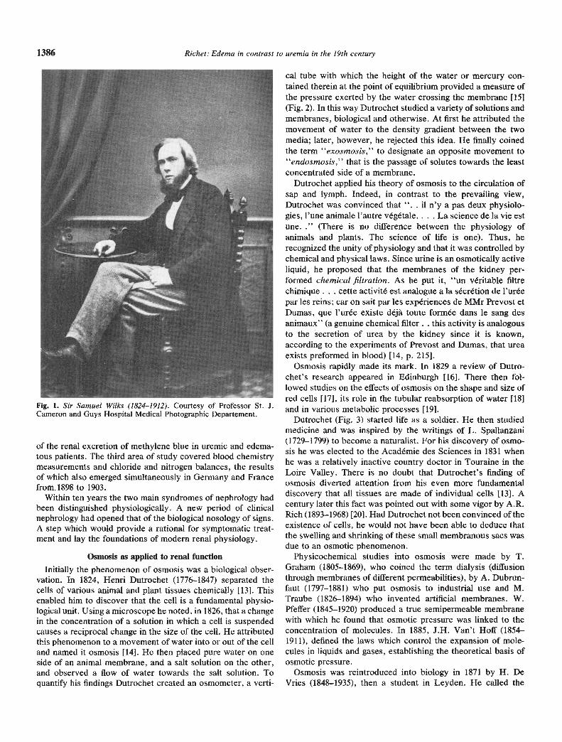
Fig. 3. Henry Dutrochet (1776—1847): bronzemedallion casted in 1842 by Pierre Jean Davidd'Angers (1788—1856). Courtesy of ProfesseurE, Aron, Tours, F.37000.
The estimation of the freezing point depression (FPD) as ameans for measuring total osmotic pressure of a solution was amethodological advance of great value. In 1876, F.M. Raoult(1830—1901) in Grenoble, took up the work of L. de Coppet(1841—1910) of Geneva and found that the extent of FPD was ameasure of the total molecular concentration of a solution.Moreover, he compared the FPD with the rise in boiling pointand the decrease in vapor tension, the colligative properties ofW. Ostwald (1853—1932). As the measurement of FDP requiredonly a small volume of solution this technique was ideally fittedfor use on biological specimens. An investigative tool had beenfound.
From 1890 onwards the measurement of urinary FPD wasintroduced into the study of the physiology of the normal and
abnormal kidneyE. Hoppe Seiler (1825—1892) had put urine and blood from the
same healthy individual on either side of a semi permeablemembrane and observed the transfer of water into the urine. H.Dreser (1860_l925)2 noted this unpublished finding, and in 1892
2 Heinrich Dreser was active in renal physiology and pharmacology,having introduced the therapeutic use of heroin (1898) and aspirin(1899). Besides the research mentioned above, he studied glomerularfunction in frogs according to the Nussbaum technique, the changes inFPD in urine during sugar diuresis, the diuretic action of caffeine and heattributed the excretion of an acid urine to the secretion of H ions by
[24] measured the FPD of blood and urine of men and animalssubjected to various physiological conditions. Dreser was aphysician and a physical chemist. He pointed out that the FPDof blood was 0.56°C and remained constant regardless of thelevel of hydration. In contrast, the FPD of urine varied withwater intake. In humans it ranged from 0.16° to 2° falling to 4°Cin the fluid deprived cat. He considered that the thermodynamicwork of the kidney stemmed from the difference between theFPD of blood and urine. Shortly afterwards, J. Winter in Parisstudied the FPD of serum and serous effusions (which he foundto be the same) and of milk, gastric fluid and urine [25]. TheFPD of urine ranged from 0.45° to 2.40°C thereby confirming thefindings of Dreser.2
The measurement of urinary FPD in renal disease
Between 1895 and 1910 A. v. Korànyi studied the FPD urinefrom normal and abnormal kidneys. In 1907 he summarized hisresults in a famous textbook on medical physical chemistry[26]. He assumed that the kidney regulates the osmolality of theurine so that the osmolality of the blood should remain con-stant. Thus, the kidney controls the milieu intérleur. He there-fore pointed out that (i) a reduced rise in urine FPD to water
the cells of the tubules. Cushny criticized all his work without exceptionin the Secretion of the Urine, 1917.
A
1388 Richet: Edema in contrast to uremia in the 19th century
Fig. 3. Henry Dutrochet (1776—1847): bronzemedallion casted in 1842 by Pierre Jean Davidd'Angers (1788—1856). Courtesy of ProfesseurE, Aron, Tours, F.37000.
The estimation of the freezing point depression (FPD) as ameans for measuring total osmotic pressure of a solution was amethodological advance of great value. In 1876, F.M. Raoult(1830—1901) in Grenoble, took up the work of L. de Coppet(1841—1910) of Geneva and found that the extent of FPD was ameasure of the total molecular concentration of a solution.Moreover, he compared the FPD with the rise in boiling pointand the decrease in vapor tension, the colligative properties ofW. Ostwald (1853—1932). As the measurement of FDP requiredonly a small volume of solution this technique was ideally fittedfor use on biological specimens. An investigative tool had beenfound.
From 1890 onwards the measurement of urinary FPD wasintroduced into the study of the physiology of the normal and
abnormal kidneyE. Hoppe Seiler (1825—1892) had put urine and blood from the
same healthy individual on either side of a semi permeablemembrane and observed the transfer of water into the urine. H.Dreser (1860_l925)2 noted this unpublished finding, and in 1892
2 Heinrich Dreser was active in renal physiology and pharmacology,having introduced the therapeutic use of heroin (1898) and aspirin(1899). Besides the research mentioned above, he studied glomerularfunction in frogs according to the Nussbaum technique, the changes inFPD in urine during sugar diuresis, the diuretic action of caffeine and heattributed the excretion of an acid urine to the secretion of H ions by
[24] measured the FPD of blood and urine of men and animalssubjected to various physiological conditions. Dreser was aphysician and a physical chemist. He pointed out that the FPDof blood was 0.56°C and remained constant regardless of thelevel of hydration. In contrast, the FPD of urine varied withwater intake. In humans it ranged from 0.16° to 2° falling to 4°Cin the fluid deprived cat. He considered that the thermodynamicwork of the kidney stemmed from the difference between theFPD of blood and urine. Shortly afterwards, J. Winter in Parisstudied the FPD of serum and serous effusions (which he foundto be the same) and of milk, gastric fluid and urine [25]. TheFPD of urine ranged from 0.45° to 2.40°C thereby confirming thefindings of Dreser.2
The measurement of urinary FPD in renal disease
Between 1895 and 1910 A. v. Korànyi studied the FPD urinefrom normal and abnormal kidneys. In 1907 he summarized hisresults in a famous textbook on medical physical chemistry[26]. He assumed that the kidney regulates the osmolality of theurine so that the osmolality of the blood should remain con-stant. Thus, the kidney controls the milieu intérleur. He there-fore pointed out that (i) a reduced rise in urine FPD to water
the cells of the tubules. Cushny criticized all his work without exceptionin the Secretion of the Urine, 1917.
Riches: Edema in contrast to uremia in the 19th century 1389
restriction should be an early indication of an impaired excre-tory function, and that (ii) an increase of FDP of the bloodshould mean a severe renal failure.
Koranyi confirmed that normal urine FPD varied from 2.4and 0.08°C, according to the intake of water [27]. In addition henoted that, in patients with terminal uremia, the maximal urineosmolality under water restriction was not modified and ap-proached that of plasma (isosthenuria). Thus the FPD of urinehad a physiological meaning [26, 28]. He established that theurine FPD was dependent on the state of the kidney and not ona systemic metabolic change. Indeed, in unilateral renal diseasewith no detectable abnormalities of the blood, the urine fromthe normal side was normal while the urine from the abnormalside was isosthenuric and little influenced by water intake. Thisfinding was confirmed by Albarran (1860—1912), a Parisianurologist with a passion for investigating renal function [29].Korànyi was aware that he was promoting the idea that thekidney itself could induce a physiological change and thendeveloped the concept of renal insufficiency based upon hypos-thenuria [28]. Though he may not have created the term, hiswork gave it its true meaning, that of an excretory functionwhich fails to meet the needs of the body. He also revealed thata physiological defect of function could be independent of thetype of structural lesion, and that the quantitative measurementof urine FPD during water restriction made it possible to detecta clinically latent stage of uremia before its full symptomaticdevelopment [261.
At this point Korànyi turned his attention to the rapid rise inserum FPD which occurred after bilateral nephrectomy inanimals from 0.57 to 0.65°C or more in 24 hours. However, thewater content of the serum measured by refractometry re-mained unchanged, and Bickel in addition noted that its elec-trical conductivity did not change [30]. Korànyi [26, 28] de-duced that the increase in molecular concentration in the serumdemonstrated by the rise in FPD must therefore be due to thepresence of nitrogen waste products, urea at first. Up to thattime there had been relatively few measurements of serum FPDfrom patients with renal insufficiency. Koranyi collected 170cases, including 10 of his own, whose serum FPD varied from0.60 to 0.70°C. It was particularly high in clinically severecases. His findings in animals thus matched the clinical picture.
Koranyi then tried to link the osmotic pressure of the blood tothe presence of edema as well as to uremia. Such a biophysicalapproach was rare at that time. He considered that the clinicalfindings and the investigations of Achard and Paisseau [311suggested that urea played no part in the accumulation ofedema, but rather that it was due to the retention of some otherdissolved substances. He thought that the possibility it was theretention of sodium chloride in particular had been well dem-onstrated by Widal in Paris and Strauss in Berlin (see below).Korànyi explained the different behavior of urea and sodiumchloride by pointing out that while urea penetrated red cells, asshown by Gryns [32], sodium chloride remained extracellular.There the latter exerted an osmotic pressure which drew waterout of cells which caused a compensatory thirst which led to anincreased intake of water. If edema occurred he thought it wasdue to the inability of the kidney to excrete the extra water.Korànyi had another unprescient hypothesis in that he alsobelieved that retention of sodium chloride and water by thekidney was associated with that of other toxic substances which
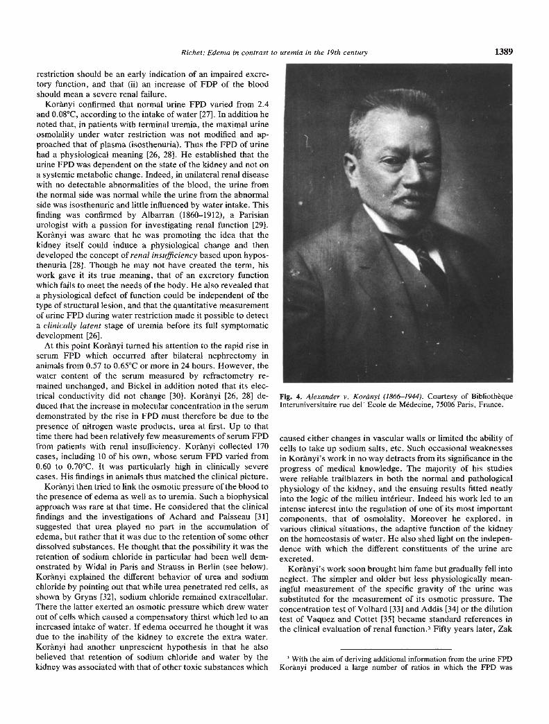
Fig. 4. Alexander v. Kordnyi (1866—1944). Courtesy of BibliothqueInteruniversitaire rue deY Ecole de Mdecine, 75006 Paris, France.
caused either changes in vascular walls or limited the ability ofcells to take up sodium salts, etc. Such occasional weaknessesin Korànyi's work in no way detracts from its significance in theprogress of medical knowledge. The majority of his studieswere reliable trailblazers in both the normal and pathologicalphysiology of the kidney, and the ensuing results fitted neatlyinto the logic of the milieu intérieur. Indeed his work led to anintense interest into the regulation of one of its most importantcomponents, that of osmolality. Moreover he explored, invarious clinical situations, the adaptive function of the kidneyon the homeostasis of water. He also shed light on the indepen-dence with which the different constituents of the urine areexcreted.
Korànyi's work soon brought him fame but gradually fell intoneglect. The simpler and older but less physiologically mean-ingful measurement of the specific gravity of the urine wassubstituted for the measurement of its osmotic pressure. Theconcentration test of Volhard [33] and Addis [34] or the dilutiontest of Vaquez and Cottet [35] became standard references inthe clinical evaluation of renal function. Fifty years later, Zak
With the aim of deriving additional information from the urine FPDKorànyi produced a large number of ratios in which the FPD was
Riches: Edema in contrast to uremia in the 19th century 1389
restriction should be an early indication of an impaired excre-tory function, and that (ii) an increase of FDP of the bloodshould mean a severe renal failure.
Koranyi confirmed that normal urine FPD varied from 2.4and 0.08°C, according to the intake of water [27]. In addition henoted that, in patients with terminal uremia, the maximal urineosmolality under water restriction was not modified and ap-proached that of plasma (isosthenuria). Thus the FPD of urinehad a physiological meaning [26, 28]. He established that theurine FPD was dependent on the state of the kidney and not ona systemic metabolic change. Indeed, in unilateral renal diseasewith no detectable abnormalities of the blood, the urine fromthe normal side was normal while the urine from the abnormalside was isosthenuric and little influenced by water intake. Thisfinding was confirmed by Albarran (1860—1912), a Parisianurologist with a passion for investigating renal function [29].Korànyi was aware that he was promoting the idea that thekidney itself could induce a physiological change and thendeveloped the concept of renal insufficiency based upon hypos-thenuria [28]. Though he may not have created the term, hiswork gave it its true meaning, that of an excretory functionwhich fails to meet the needs of the body. He also revealed thata physiological defect of function could be independent of thetype of structural lesion, and that the quantitative measurementof urine FPD during water restriction made it possible to detecta clinically latent stage of uremia before its full symptomaticdevelopment [261.
At this point Korànyi turned his attention to the rapid rise inserum FPD which occurred after bilateral nephrectomy inanimals from 0.57 to 0.65°C or more in 24 hours. However, thewater content of the serum measured by refractometry re-mained unchanged, and Bickel in addition noted that its elec-trical conductivity did not change [30]. Korànyi [26, 28] de-duced that the increase in molecular concentration in the serumdemonstrated by the rise in FPD must therefore be due to thepresence of nitrogen waste products, urea at first. Up to thattime there had been relatively few measurements of serum FPDfrom patients with renal insufficiency. Koranyi collected 170cases, including 10 of his own, whose serum FPD varied from0.60 to 0.70°C. It was particularly high in clinically severecases. His findings in animals thus matched the clinical picture.
Koranyi then tried to link the osmotic pressure of the blood tothe presence of edema as well as to uremia. Such a biophysicalapproach was rare at that time. He considered that the clinicalfindings and the investigations of Achard and Paisseau [311suggested that urea played no part in the accumulation ofedema, but rather that it was due to the retention of some otherdissolved substances. He thought that the possibility it was theretention of sodium chloride in particular had been well dem-onstrated by Widal in Paris and Strauss in Berlin (see below).Korànyi explained the different behavior of urea and sodiumchloride by pointing out that while urea penetrated red cells, asshown by Gryns [32], sodium chloride remained extracellular.There the latter exerted an osmotic pressure which drew waterout of cells which caused a compensatory thirst which led to anincreased intake of water. If edema occurred he thought it wasdue to the inability of the kidney to excrete the extra water.Korànyi had another unprescient hypothesis in that he alsobelieved that retention of sodium chloride and water by thekidney was associated with that of other toxic substances which
Fig. 4. Alexander v. Kordnyi (1866—1944). Courtesy of BibliothqueInteruniversitaire rue deY Ecole de Mdecine, 75006 Paris, France.
caused either changes in vascular walls or limited the ability ofcells to take up sodium salts, etc. Such occasional weaknessesin Korànyi's work in no way detracts from its significance in theprogress of medical knowledge. The majority of his studieswere reliable trailblazers in both the normal and pathologicalphysiology of the kidney, and the ensuing results fitted neatlyinto the logic of the milieu intérieur. Indeed his work led to anintense interest into the regulation of one of its most importantcomponents, that of osmolality. Moreover he explored, invarious clinical situations, the adaptive function of the kidneyon the homeostasis of water. He also shed light on the indepen-dence with which the different constituents of the urine areexcreted.
Korànyi's work soon brought him fame but gradually fell intoneglect. The simpler and older but less physiologically mean-ingful measurement of the specific gravity of the urine wassubstituted for the measurement of its osmotic pressure. Theconcentration test of Volhard [33] and Addis [34] or the dilutiontest of Vaquez and Cottet [35] became standard references inthe clinical evaluation of renal function. Fifty years later, Zak
With the aim of deriving additional information from the urine FPDKorànyi produced a large number of ratios in which the FPD was

1390 Richet: Edema in contrast to uremia in the 19th century
[361, working with Homer Smith [37], returned to the study ofwater excretion focusing on the filtration reabsorption hypoth-esis, based üpoh a osmolar u/p ratio and the concept of waterclearance.
Alexander v. Korànyi (1866—1944) (Fig. 4) was born inBudapest, the son of a Professor of Medicine who had spentyears under house arrest in a village near Debrecen, for havingtaken part in the Hungarian Revolution of 1848. He graduated inBudapest, then studied with F. Hoppe Seiler in Strasbourgwhere he devoted himself to the physicochemical study of thephysiology of the healthy and diseased kidney [38]. Thereafterhe returned to his native city as Professor where his attentionbecame, to some extent, directed away from the kidney. Hedied in Budapest in 1944.
The renal excretion of methylene blue
Using a similar basic approach to that of Korànyi, C. Achard(1860—1944) and J. Castaigne (1871—1951), between 1897 and1902, studied what they called the permeability of the kidney,by measuring the urinary excretion of methylene blue [39, 40].They stated that "Le besoin se fait sentir d'ajouter a l'étude desorganes lesés, celle des fonctions troubldes et de completerl'investigation anatomique par l'investigation physiologique. IIfaut donc inventer des méthodes spdciales, permettant deverifier non plus simplement le mécanisme des organes a l'étatstatique mais encore observer ces organes en action, a l'étatdynamique. . . . D'observateur, ii (le médecin) se fait expéri-mentateur" (There is a need to study the disturbance offunction of organs in order to supplement anatomical data withphysiological investigation. It is therefore necessary to inventspecial techniques that will allow us to evaluate the functions oforgans, not only under static conditions but also under dynamicconditions . . . The physician should adjust his position fromthat of observer to that of experimenter), These studies on theexcretion of dyes were prompted by Rayer's two clinicalobservations that in advanced nephritis, after eating asparagus,the urine does not smell of mercaptan, and that because of theirdelayed excretion the administration of certain drugs to uremicpatients may produce toxic effects.
The elimination of methylene blue was studied following theinjection of 0.05 g subcutaneously. In normals 50% or more wasexcreted in the urine within the next 24 hours. As expected itwas found that in impending uremia, interstitial nephritis oradvanced Bright's Disease, the excretion of the dye wasdelayed so that less than 50% appeared in the first 24 hours. Thedelay was solely due to the abnormal kidneys for the elimina-tion of the dye continued until the entire dose had beenexcreted, the time required for this to occur being dependent onthe degree of rena/fibrosis. Moreover, it was noted that in thepresence of unilateral disease the diminution in the rate of dyeexcretion was confined to the diseased kidney [41]. In a mannersimilar to the measurement of urine FPD during water restric-tion, the rate of methylene blue excretion also gave a quantita-
Fig. 5. Charles Achard (1860—1944). Courtesy of the Bibliothèque del'AcadCmie Nationale de Médecine.
tive measure of the decline of renal function in what was thencalled the latent stage of chronic nephritis.
Of perhaps even greater interest was the finding that inedematous patients with much proteinuria the excretion ofmethylene blue was normal, L. Bard and L. Bonnet [42]. Thiscondition thus had to be dissociated from that of impendinguremia with little proteinuria and no edema. Having confirmedthis difference L. Bernard [43] in 1900 insisted that there weretwo biological types of nephritis, parenchymatous and intersti-tial, as suggested by Wilks 50 years before purely on clinico-pathological grounds [3]. The elimination of many other dyeswere studied. Rowntree's IV Phenolsulphonaphthalein method[44] replaced all the preceding tests.
In 1902 Ch. Achard (1860-1944) (Fig. 5) published a 435 pagebook 'Les Nouveaux Procédés d'Exp/oration" [45] devoted tothe latest methods with which to investigate functional abnor-malities of various organs including the kidneys. This book andthat of Korànyi and Richter [261, and many others of a similarnature, give an idea of the extent that medicine was permeated,at the beginning of the century, by a scientific approach, theso-called "functional diagnosis." In Europe this fruitful devel-opment was much less evident after World War I. Achard was
compared with the concentration of sodium chloride, or the output ofwater or other urine components over 24 hours. At first, these maneu-vers were enthusiastically followed in Germany and France, but theywere abandoned as they did not appear to contribute to the understand-ing of the observed troubles.
The content of this book depicts the shift towards scientific medi-cine that was taking place. It includes 130 pages devoted to radiology,less than 5 years after the discovery of Röntgen, 120 pages to biohe-matology, 30 pages to clinical cytology, 50 pages to bacteriologicalserology, 30 pages to cryoscopy, 20 pages to the regulation of thecomposition of the blood and 50 pages to methylene blue excretion.

Rjchet: Edema in Contrast to uremia in the 19th century 1391
Fig. 7. H. Strauss's apparatus for rapid Lv. perfusion [50]. At variancewith that appearing on Figure 8, there is no device for gripping theneedle.
Fig. 6. Jules Castaigne (1871—1 951). Courtesy of Professor Alain Cas-taigne, Paris, France.
a leader in biochemical investigation, particularly in the 20's onthe nephrotic syndrome, his steadfast commitment being theregulation of the extracellular spaces [46]. Joseph Castaigne(1871—1951) (Fig. 6), a dedicated internist, left Paris after WorldWar I and became the successful Dean of the Medical School ofClermont-Ferrand.
In summary, therefore, at the end of the 19th century thefunktionel Nierendiagnostik of Korànyi based upon the osmo-lality of the urine and the methylene blue test of Achard andCastaigne had already given a biological scaffold for the twomain clinical syndromes of chronic renal disease: edema anduremia. The two new complementary methodological toolsexplored different aspects of renal function. They had revealedthat the underlying mechanisms responsible for edema anduremia, though still unknown, were different. Although Straussin 1902 considered that these two techniques had produced an"upheaval" [47], the medical community ignored their far-reaching implications.
However, it was not long before the ability to investigate thechemistry of the blood and to perform balance studies con-firmed and extended the suggestions that had been based on theprevious investigative methods.
Clinical blood chemistry, and chloride and nitrogen balances
The Strauss needle: The mandatory too/for blood chemistryThere is good circumstantial evidence that the chemical study
of the blood did not open up until the introduction of thevenesection needle by Strauss between 1898 and 1902. Previ-ously blood chemistry had had a restricted role in clinicalmedicine. Until 1903 clinical papers by Korànyi [28], Bernard[43], Jaksch [48, 49], and Achard and Paisseau [31] clearlystated that blood was obtained by either cutting a superficialvein or making multiple small skin incisions at the site of
cupping. From 1905 on, however, there is no further mentionhow blood is obtained, although by then numerous estimationsof blood substances were being performed by all medicaldisciplines. This change must have been due to the use of theStrauss needle. Nevertheless, it has not been possible to obtainconfirmation of this assumption either from the authorities ofhospitals in Paris or even from some of the original manufac-turers of the needle who are still in business.
H. Strauss was a physician at the "Charitd" in Berlin. Hisoriginal steel needle was 6 cm long with an internal diameter of2 mm. The first illustration of the needle appeared in a paper in1898 [50], which was mainly concerned with the use of apneumatic pressure injecting device (Fig. 7). The needle isbarely visible. It was not until 1902 that Strauss [47] publisheda detailed drawing of the needle including a good view of itsperpendicular "Handgriff" with which it could be grasped (Fig.8).
In 1931, F. Volhard [51] underlined Strauss's highly signifi-cant work on the retention of non-protein nitrogen and urea inthe blood, but he failed to mention Strauss's needle. In theBiographisches Lexicon in 1901 [52] the name of Strauss wasnot associated with nephrology and his needle is ignored. In the1933 edition, however, it is interesting to note that theseomissions are corrected [53].
Strauss and WidalIt was Strauss in Berlin and Widal in Paris, in and around
1900, who overturned the then, almost ritualistic views on renaldisease. Breaking free from the confines of anatomy theystudied the chemical nature of the metabolic troubles afflictingtheir patients. The two men differed considerably in theirapproach and in some of their methods. Strauss (1868—1944)(Fig. 9) opened his chemical net wide by studying all possiblehumoral abnormalities at all stages of chronic nephritis in alarge group of patients. He assumed that the kidney plays acentral role in the metabolic balances of all those substanceswhich are excreted in the urine. He then arranged his resultsaccording to the accepted clinico-anatomical classification ofrenal disease, that is, parenchymatous or interstitial nephritisand transitional forms. Ironically, outside France, Widal (1862—1929) (Fig. 10) is principally remembered today for his bacteri-ological work, typhoid in particular. In his renal work, which hesubsequently took up, he totally ignored any consideration ofrenal pathology. His contribution to nephrology was confined

1392 Richet: Edema in contrast to urenia in the 19th century
Fig. 8. Strauss's steel needle used to drawblood under sterile conditions (Straussche'sNadel zur sterilen Blutentnahme) and histourniquet clamp [45].
Edema
Fig. 9. Hermann Strauss (1866—1944). Courtesy of Herbert Sonnen-feld, Archly, mv. Num, 268/22, Berlin Museum, Abteilung JudischesMuseum.
exclusively to the study of edema and uremia. His method wasto thoroughly study a few patients who were on varying intakesof either sodium chloride or protein. Strauss's and Widal'sfindings and conclusions overlap almost exactly. Their maincontributions were on the origin of edema and uremia.
Strauss noted that in the edema associated with heavyproteinuria and hypoproteinemia, plasma chloride and ureawere normal. Nevertheless, though the concentration of chlo-ride in the plasma remained normal, edema was due to renalretention of chloride, for when the edema spontaneously disap-peared there was an associated brisk increase in the urinaryexcretion of chloride and not of suiphates and phosphates.Moreover, Strauss successfully treated edema with a salt freediet [54, 55]. He concluded that edema was due solely to theurinary retention of sodium chloride. Independently, Widalagreed [56—59]. He and Lemierre [56] found that in four patientswith renal disease but no edema, large changes in salt intakeproduced no change in weight, the urinary excretion of chloridechanging in line with the intake. But in two of three edematousproteinunc patients a sudden large intake of sodium chloridecaused a brisk increase in weight while the extra intake of saltwas not excreted in the urine. In another edematous patient [57]who was studied for six months, it was repeatedly possible toinduce substantial reversible changes in weight and edema bylarge changes in the intake of sodium chloride (Fig. 11). Duringthat six months the blood urea remained constant, eliminatingany lingering possibility that edema was related to proteinintake. An illusion that was nevertheless to persist for another40 years in both Europe and America.
Strauss and Widal's conclusions on the origin of edema inrenal diseases was the culmination of much work and specula-tion by others. Some had confirmed the constancy of plasmachloride in the face of large changes in salt intake (Langlois andRichet) [60]. Others had put forward the idea that in addition tothe retention of sodium chloride edema formation also includeda primary disturbance to water balance. They considered thatthere was some generalized abnormality of the cells whichcaused them to retain water (Koranyi [26], Achard and Loeper[61], and Georgopoulos [62]). This was opposed by Widal whopointed out that the renal retention of sodium chloride, whichdid not penetrate the cells, would on the contrary cause waterto come out of cells. There is a suggestion that Widal did indeedconsider that edema was due to a secondary accumulation ofwater out of cells following the renal retention of sodiumchloride: "La connaissance des lois qul président a l'isotoniedes humeurs de l'organisme, la notion du role fondamentaljoué
0
i4. / /iiI r
p

Richet: Edema in Contrast to uremia in the 19th Century 1393
Fig. 10. Fernand Widal (1862—1929). On theright, the reverse side of the medal coinedafter Widal's death recalling his maincontributions to medicine and the diversity ofhis biomedical interests. Courtesy of theBibliotheque Interuniversitaire Rue de l'Ecolede Médecine.
Fig. 11. Chart of one of Widal's albuminuric and edematous patients who was submitted to various diets. Sodium chloride-free diet induced lossof weight and dispersing of edema. A sodium chloride-rich diet provoked the reappearance of edema. Protein input did not interfere with edema[571.
par le chiorure de sodium dans le maintien de l'équilibreosmotique de ces humeurs, devait tout naturellement conduirea l'hypothèse que la retention de ce sel dans certains tissuspouvait y attirer une partie de l'eau de l'organisme et provoquera leur niveau l'apparition de l'oedème" (Knowledge of the laws
body and the concept of the basic role played by sodiumchloride in maintaining the osmotic equilibrium of this humoralsystem, should quite naturally lead to the hypothesis that theretention of this salt in certain tissues could attract some of thebody's water and cause edema at such sites). Chauffard had
which govern the isotonic nature of the humoral system of the also observed a patient with hepatitis in whom the extent of
Lfi1Htte ,., N eanè. :mumu,unr total..
Richet: Edema in Contrast to uremia in the 19th Century 1393
Fig. 10. Fernand Widal (1862—1929). On theright, the reverse side of the medal coinedafter Widal's death recalling his maincontributions to medicine and the diversity ofhis biomedical interests. Courtesy of theBibliotheque Interuniversitaire Rue de l'Ecolede Médecine.
Fig. 11. Chart of one of Widal's albuminuric and edematous patients who was submitted to various diets. Sodium chloride-free diet induced lossof weight and dispersing of edema. A sodium chloride-rich diet provoked the reappearance of edema. Protein input did not interfere with edema[571.
par le chiorure de sodium dans le maintien de l'équilibreosmotique de ces humeurs, devait tout naturellement conduirea l'hypothèse que la retention de ce sel dans certains tissuspouvait y attirer une partie de l'eau de l'organisme et provoquera leur niveau l'apparition de l'oedème" (Knowledge of the laws
body and the concept of the basic role played by sodiumchloride in maintaining the osmotic equilibrium of this humoralsystem, should quite naturally lead to the hypothesis that theretention of this salt in certain tissues could attract some of thebody's water and cause edema at such sites). Chauffard had
which govern the isotonic nature of the humoral system of the also observed a patient with hepatitis in whom the extent of
"-.5±21? "-m' -
. .. ,. .,. - iThunnaun, Mink.

1394 Richet: Edema in contrast to uremia in the 19th century
edema was linked exactly to the input of sodium chloride [631.Other concepts for explaining edema proliferated. In addition toprimary water retention and an abnormality of protein metab-olism, there was Starling's demonstration [64—66] that the lowoncotic pressure of hypoalbuminuria causes a transfer ofplasma ultrafiltrate towards the tissue spaces.5
Uremia
Strauss's and Widal's contribution to the study of uremia wasto extend and make coherent some interesting isolated findingswhich had appeared in the previous 75 years. There had beenPrevost and Dumas [74], Gmelin [75] and Marchand [76], whobetween 1822 and 1838 had demonstrated that bilateral nephrec-tomy causes a rapid rise in blood urea. As at that time,however, the amount of blood needed to estimate its ureacontent was large, it was not often done in humans, and theresults from the few that were performed were unconvincing(Christison [1], Bostock [77]). In 1850 J.v. Liebig published abetter assay for urea which Picard (1834—1896) adapted for itsestimation in the blood of men and dogs [781. He thus becamethe first to note that the concentration of urea in renal veinblood was lower than in renal arterial blood, and that the bloodurea of patients, before they became symptomatically uremic,rose to what he called intermediate blood urea levels. In otherwords, Picard was the first to show that blood urea might beraised without any clinical evidence of uremia (latent renalinsufficiency). His work was praised by Claude Bernard [79],criticized by v. Recklinghausen [80] and then forgotten. Hisattempts to obtain a university appointment were turned downand he then went into general practice.
In 1880 the hypobromide assay of urea became available,which should have stimulated its wide use in clinical medicine.However, according to Bartels [9], Fleischer [81] and Lecorchéand Talamon [11] it was not performed. R.v. Jaksch's paper onthe measurement of blood urea in various diseases appeared in1902. Subsequently Strauss [47], on a larger number of patients,confirmed that the concentration of blood urea in Bright'sdisease could vary widely over a range of 30 to 500 mg/lOO ml.He pointed out that the clinical signs of uremia were found atthe highest concentration. Symptomatic uremia occurred, how-ever, at a concentration which varied from patient to patient. Inline with Picard, Strauss found that patients with concentra-tions as high as 200 mgIlOO ml could still be free of symptoms.
Characteristically, Widal's primary contribution emergedfrom an intense study of a few patients over a prolonged periodof time. A patient on a constant diet had a blood urea whichremained around 120 mg/lOO ml over a controlled period ofseveral weeks. The protein intake or the oral intake of urea wasthen varied. It caused parallel changes in blood urea over arange of 36 mg to 195 mgIlOO ml [82]. Each change in dietinduced a gradual change in blood urea over a period of fivedays when a new plateau was reached. At the new point of
A large number of publications devoted to the metabolism of NaC1in nephritis with or without edema appeared between 1880 and 1906:C.v. Noorden [67], Bohne [68], Reichel [69], Hofman [70], Lindemann[71], Carrion and Hallion [72], and Claude [73]. However, they oftenused unreliable methods and obtained inconsistent and unreproducibleresults that failed to elucidate the mechanisms of edema. The accom-plishments of Strauss must thus be shared only with Widal.
equilibrium Widal found that the rate of urinary urea excretionhad changed in the same direction as the blood urea and theprotein intake. From these observations he concluded that theconcentration of urea in the blood controlled the rate ofexcretion of urea in the urine, or in other words, that the levelof blood urea was a regulating factor in the maintenance of anitrogen balance.
As a result of Strauss and Widal's work the concentration ofblood urea (or non-protein nitrogen) became as important ashyposthenuria and an impaired excretion of methylene blue forthe detection and assessment of renal failure, whether or notthey were clinical signs of uremia. Strauss and Widal alsoobserved that the blood urea might rise in a variety of diseasesbut that, in the absence of a renal lesion, levels greater than 100mg/lOO ml were exceptional. However, they differed in theirinterpretation of the apparent paradox that most uremic pa-tients do not have edema and can excrete a large additional loadof sodium chloride normally. Strauss' suggestion, which shouldwarm the heart of some contemporary physiologists, was thatthe excretion of sodium was normal because the hypertension,which usually accompanies uremia, forced the sodium throughthe kidney [47]. Widal and Javal were more cautious. Theyproposed that in renal disease a rise in blood urea was due toone unknown mechanism and retention of sodium chloride wasdue to another [83].
Widal later worked on the clinical features of anaphylaxis. Hebecame a leading light of the medical scene in Paris. Strauss in1910 became head of the department of internal medicine at theJewish Hospital in Berlin. In 1944, at the age of 76, on his wayto Auschwitz he became a victim of Hitler's racially besottedtyranny [84].
These are the beginnings of renal pathophysiology. Theirintegration into clinical medicine was extraordinarily slow. Onehas the impression that the few clinical pioneers whose inves-tigation shed light on these problems were working in isolationfrom the main body of their colleagues. On the whole, Straussand Widal's work on edema was ignored until 1940. Neither didthe studies appear to influence their peers in physiology.Cushny, one of the principal pillars of renal physiology at thattime, even appears to have considered that at least one discov-ery made by renal physicians was useless and irrelevant to theproper study of renal function. He denigrated the measurementof urinary FPD: "some years ago the measurement of molecu-lar and ionic concentration of the urine by means of its freezingpoint was introduced as a clinical method of determining theefficiency of the kidney . . . Clinically the results have proveddisappointing and misleading . . . This cryoscopic method wasmuch overvalued and has rightly been abandoned" [85, p. 34].In Cushny's book Korànyi's name does not appear in the 385references. The growing acceptance of the filtration-reabsorp-tion theory made it difficult for renal physiologists to envisagethat a functional lesion of a kidney, such as a leakage of protein,could coexist with an excess of function, such as an increasedabsorption of sodium chloride. Thus for a relatively long timethe early discoveries on the origins of edema and uremia inrenal disease which had emerged from the wards were disre-garded by the physiologists. There is a suggestion that theconcept that an abnormality caused by disease is merely adisturbance of a normal function, had not yet become thor-oughly assimilated.

Richet: Edema in Contrast to uremia in the 19th century 1395
Acknowledgments
The author is deeply indebted to Professor St.Wolf and Dr. Vasmantand their secretaries for translating and preparing the manuscript; toProfessor H. de Wardener for rereading the text; to the Bibliothequeinteruniversitaire de Médecine, the Bibliothque de l'Académie Natio-nate de Médecine and their staffs, especially Mrs. Casseyre, Lenoir,Molitor and Mr. Rivet, Librarians, as well as to Miss M. Chapuis, avery efficient documentalist.
Reprint requests to Gabriel Richet, M.D., 76 rue d'Assas, 75006Paris, France.
References
1. CHRISTISoN R: On the granular degeneration of the kidneys (vol 1).Edinburgh, Adam. and Black, 1839
2. RAYER P: Traité des Maladies des Reins (tome II). Paris, J.B.Baillière, 1840
3. WILKS S: Cases of Bright disease with remarks. Guy's HospitalReport 1:232—3 15, 1853
4. JOHNSON G: On the forms and stages of Bright's disease of thekidneys. Medico Chirurgical Transaction XLII: 153—164, 1859
5. DICKINSON WH: Two essential conditions of kidney giving rise towhat is called Bright's disease. Medico Chir Transaction 43:114,1860
6. GRAINGER STEWART T: Practical Treatise on Bright Disease.London, 1868
7. KELSCH A: Revue critique ci recherches anatomo patholojiques surla maladie de Bright. Arch de Physiol Norm Pathol 1:722—758, 1874
8. CHARCOT JM: Lecons sur les Maladies du Foie et des Reins. Editedby DELAHAYE, Paris, 1877
9. BARTELS K: Les Maladies des Reins. (French Translation). Paris,Germer Bail1ire, 1884
10. FRERICHS FTv: Die Bright'sche Nierenkrankheit und deren Behan-diung. Braunschweig. 1851, 286 p
11. LECORCHE E, TALAMON CH: Traité de l'Albuminurie. Edited byD0IN, Paris, 1888
12. BLEKER J: Die Geschichte der Nierenkrankheiten, BoehringerMannheim, 1972, 147 pp.
13. DUTROCHET H: Recherches Anatomiques et Physiologiques sur laStructure Intime des Animaux et des Végetaux et Sur Leur Motilité.Paris, J.B. Baillière, 1824, 233 pp.
14. DUTROCHET H: L'ageni immédiat du Mouvement Vital DévoiléDans sa Nature et Dans son Mode d'action Chez les Vegetaux etles Animaux. Paris, J.B. Baillière, 1826, 226 pp.
15. DUTROCHET H: Nouvelles Recherches sur l'Endosmose ci l'Exos-mose. Paris, J.B, Baillière, 1828, 106 pp.
16. DUTROCHET H: Critical Analysis, in The Edinburgh Medical andSurgical Journal 31:369—394, 1829
17. REES GO, LANES: On the structure of the blood corpuscule. Guy'sHospital Reports VI:379—391, 1841
18. Luowia C: Nieren und Harnbereitung, in Wagners 1844, II, p.628—640; cite par SCHILLER J Schiller T, Henri Dutrochet, I, vol. A,Paris, Blanchard, 1975, p. 70
19. VIERORDT Ky: Transudation und Endosmose, in Wagners 1846,Bd. III, 631—657
20. RIcH AR: The Place of J.H. Dutrochet in the development of thecell theory. Bull Johns Hopkins Hosp 39:330—365, 1926
21. DE VRIEs H: Sur Ia perméabilitë du protoplasma des betteravesrouges. Revue Néerlandaise des Sciences exactes ci naturellesVI:117—126, 1871
22. DE VRIES H: Sur Ia mort des cellules vegetates par i'effet d'unetemperature élevCe. ibid VI:245—295, 1871
23. HAMBURGER HJ: Sur la determination de Ia tension osmotique desliquides albumineux et particulièrement sur celle qui s'opère aumoyen de l'abaissement du point de congelation. Revue de Méde-cine XV:918—928, 1895
24. DRESER H: Uber Diurese und ihre Beeinflussung durch pharmakol-ogische Mittel. ArchfExperPatho u Pharmak XXIX:303—319, 1892
25. WINTER J: Dc l'équilibre moléculaire des humeurs. Etude de Iaconcentration des urines, ses limites. Arch Physiologic normale etpatholoitique 1896, m,. 114. 287. 296. 529
26. K0RANYI Av: Physikalisch-chemische Methoden und Gesicht-punkte in ihrer Anwendung auf die pathologische Physiologic derNieren, in Physikalische Chemie und Medizin, edited by K01UNYIAv, RICHTER PF, Leipzig, 0. Thieme, 1907, pp. 133—190
27. KORANYI Av: Physiologisch-klinische Untersuchungen Uber denosmotischen Druck tierische Flussigheiten. Zeitsch fKlin Medizin33:1—55, 1897
28. KORANYI Av: Uber den Ham und das Blut bei Nierenkrankheiten.Zeiisch f KIm Medizin 34:1—52, 1898
29. ALBARRAN J: Exploration des Fonciions Renales. Paris, Masson,1905, p. 380
30. BICKEL A: Experimentelle Untersuchungen über den Einfluss derNierenausschaltung auf die elektrische Leitfahigkeit des Blutes.Zeitsch f Klinische Medizin 47:480—489, 1902
31. ACHARD Cu, PAISSEAU G: La rCtention de l'urée dans l'organismemalade. Sern Med 24:209—214, 1904
32. GRYNS G: Uber den Einfluss gelOster Stoffe auf die rothen Blut-zellen, in Verbindung mit den Erscheinungen der Osmose undDiffusion. Pfluger Arch 63:86—119, 1896
33. VOLHARD F: in Mohr and Staehelin's Handbuch der InnerenMedizin (vol III), Berlin, 1918, p. 1197
34. ADDIS T, SCHEVKY MC: A test for the capacity of the kidney toproduce a urine of high specific gravity. Arch mt Med 30:559, 1922
35. VAQUEZ H, COTTET J: Epreuve de Ia diurèse provoquee (polyurieexperimentaie). Valeur sdmiologique du rythme d'élimination uri-naire de l'eau et des chlorures. Presse Médicale 20:993, 1912
36. ZAK GA, BRUN C, SMITH HW: The mechanism of formation ofosmotically concentrated urine during the antidiuretic state. J ClinInvest 33:1064, 1954
37. SMITH HW: Renal excretion of sodium and water. Fed Proc 11:701,1952
38. KORANYI Av: Vorlesungen uber funcktionelle Pathologic undTherapie der Nierenkrankeiien. Berlin, Springer, 1929
39. ACHARD CH, CASTAIGNE J: Diagnostic de Ia permeabilitC rCnale.Bul Mém Soc Med Hptx Paris XIV:637, 831, 1128, 1897, XV:5,1898
40. CASTAIGNE J: Epreuve du bleu de méthylène etperméabiliié rénale.These, Paris, 1900
41. ALBARRAN J, BERNARD L: La perméabilite rénale étudiCe par Icprocédé du bleu de méthylene dans les affections chirurgicales desreins. Ann mal org génito urinaires Avril—Mai, 1899
42. BARD L, BONNET LM: Recherche et considerations cliniques surles differences de perméabilitC rCnale dans les diverses espèces denephrites. Arch Gen Med Chir VIII (l0):129—163, 1898
43. BERNARD L: Les fonctions du rein dans les néphrites chroniques.These, Paris, 1900, 182 pp.
44. ROWNTREE LG, GERAGHTY JT: The Phtalein Test. An experimen-tal and clinical study of Phenolsulfonephtalein in relation to renalfunction in health and disease. Arch Intern Med IX:284, 1912
45. ACHARD CH: Les Nouveaux Procédés d'Exploration. Paris, Mas-son Ed, 1902, 346 pp.
46. ACHARD CH: Le Système Lacunaire, Paris, Masson Ed, 1922, 124pp.
47. STRAUSS H: Die Chronischen Nierenentzdndingen etc. Berlin,Hirschwald, 1902, 158 pp.
48. JAKSCH RUv: Festschrift für Herrn v. Leyden I, 190249. JAKSCH RUv: Weitere Beobachtungen Uber die Mengen des im
Blute des kranken Menschen sich vorfinden Harnstoffes. ZtschHeilkunde 24 (1i):401—423, 1903
50. STRAUSS H: Zur Technik der Aderlasses, der intravenösen undsubcutanen Infusion. ZeitschrfKrankenpflege XX (4):66—67, 1898(Arztliche Polytechnik Sektion)
51. VOLHARD F: Nieren und ableitende Harnweg, 6 Band, 2 Teil, inHandbuch der Inneren Medizin, edited by BERGMANN-STAEHLIN,Berlin, Springer, 1931, pp. 113—115
52. PAGEL J: Biographisches Lexikon. Berlin, Urban Schwarzenberg,1901, p. 1670
53. FISCHER I: Biographishes Lexicon der hervorragende Arzt desletzen funfzig .Jarhre. Berlin, Urban Schwartzenberg, 1933, pp.1527—1528

1396 Richet: Edema in contrast to uremia in the 19th century
54. STRAUSS H: Die Harnkryoscopie in der Diagnostik doppelseitigerNierenerkrankungen. Zeitsc f Kl Medizin 47:337—407, 1902
55. STRAUSS H: Zur Behandlung und Verhutung der Nierenwasser-sucht, in Die Therapie der Gegenwart Mai, 1903, pp. 193—200
56. WIDAL F, LEMIERRE A: Pathogenic de certains oedèmes bright-iques: Action du chiorure de sodium ingére. Bull Mém Soc MedHptx Paris 1903, pp. 678—699; Sem MEd 1903, p. 199
57. WIDAL F: Les regimes déchlorurés. Rapport au Congres Francaisde Médecine, Vaillant Carmanne Liege, Paris, Masson, 1905, pp.181—260
58. WIDAL F, JAVAL A: La cure de ddchloruration; Son action surI'oedème, sur l'hydratation et sur l'albuminurie a certaines périodesde Ia néphrite epithCliale. Bull Mem Soc Med Hptx Paris Séance du26 Juin 1903; Sem Med 1903, p. 219
59. WIDAL F, JAVAL A: La dissociation de Ia perméabilité rénale pourle NaCI et I'urCe dans le Mat de Bright. Cptes Rendus Soc Biol55:1639—1642, 1903
60. LANGLOIS FP, RICHET CH: Proportion des chiorures dans les tissusde l'organisme. J Physiol Pathol GEnEr 2:742—754, 1900
61. ACHARD CH, LOEPER M: Retention des chiorures dans les néph-rites. Bull Mém Soc MEd Hpx Paris 9 Mai, 1902, pp. 429—432
62. GE0RG0PUL05: Experimentelte Beitrage zur Frage der Nierenwass-ersucht. Zeitschr KIm Medizin 60:411—471, 1906
63. CHAUFFARD A: Recherches de physiologic pathologique sur un casd'ictCre infectieux. Sem Med 1900, p. 213, and 1901, p. 233
64. STARLING EH: On the absorption of fluids from the connectivetissue spaces. J Physiol XIX:312, 1895—96
65. STARLING EH: The mechanism of the secretion of urine, in TextBook of Physiology, edited by SCHAEFER EA, Edinburgh, Young J.Pentland, 1898, pp. 639—661
66. STARLING EH: The glomerular function of the kidney. J Physiol24:317—330, 1899
67. NOORDEN Cv: Lehrbuch der Pat hologie des Stoffwechsels, 1893, p.325
68. BOHNE J: Uber die Bedeutung der Retention von Chloriden imOrganismus fur die Einstehung uritmischer und comatöserZustande. Fortschritte der Medizin 15 Fey, 1897 (4), pp. 121—134
69. REICHEL 0: Zur Frage der Oedem's bei Nephritis. Centralblatt furinn Medizin Oct 1898, no. 41
70. H0FMAN A: Uber die Ausscheidung der Chloride bei Nierenent-
zundung und ihren Zusammenhang mit Uramische ZUstanden.Deutsch. Arch fKlin Med LX:603—623, 1898
71. LINDEMANN L: Die Concentration des Harnes und Blutes beiNierenkrankheiten mit einem Beitrag zur Lehre von der Uramie.Deutsch Arch fKlin Med 65:1—80, 1900
72. CARRION H, HALLION L: Contribution expérimentale a Ia pathog-Cnie de l'oedème. CR Soc Biol XI sCrie, Tome I, 1899, pp. 156—157
73. CLAUDE H, MANTE A: La chlorurie alimentaire expCrimentale dansla néphrite. Bul Soc Mém Hpx Paris XIX:(3e sCrie, 2 Mai):42'1—428,1902
74. PREVOST JL, DUMAS JB: Examen du sang et de son action dans lesdivers phdnoménes de Ia vie. Ann Phys Chi 23:90—104, 1823
75. GMELIN L, TIEDEMAN J: Versuche Uber des Blutes. PoggendorfAnn XXXI:303, 1834
76. MARCHAND F: De l'existence de l'urée dans les parties de l'organ-isme animal autres que l'urine (Traduction en Francais). L'ExpEri-ence 11:43—47, 1838
77. BOSTOCK J: in BRIGHT R: Original Papers on Renal Diseases,edited by O5MAN, London, Oxford University Press, 1937
78. PICARD J: De la Presence de l'Uree Dans le Sang. These, Stras-bourg, 1856
79. BERNARD C: Les Liquides de l'Organisme II, Paris, Baillière, 1859,p.31
80. RECKLINGHAUSEN F: Uber Picard Bestimmung's Methode desHarnstoffs im Blut. Virchows Arch XIV:476, 1858
81. FLEISCHER R: Klinische und pathologisch-chemische Beitrage zuLehre von den Nierenkrankheiten. Dtsch Arc f klin Med 29:129—192, 1881
82. WIDAL F, JAVAL A: Le mécanisme régulateur de Ia retention del'urée dans Ic Mal de Bright. CR Soc Biol 56:301—304, 1904;L'indice de retention ureique chez les Brightiques. ibid pp. 304—306
83. WIDAL F, JAVAL A: La retention d'urée dans Ic Mat de BrightcomparCe a la retention des chlorures. Sem Med 1905, p. 313
84. AKADEMIE DER WISSENSCHAFTEN ZU BERLIN: Arbeitsgruppe: Ex-odus der Wissensehaften aus Berlin. Institut für Geschichte derMedizin der FU Berlin, Klingsorsstrasse 119, 1000 Berlin 45 (per-sonal communication)
85. CU5HNY AR: On diuresis and the permeability of the renal cells. JPhysiol 27:1902, 1902 and The Secretion of the Urine, London,Longman, 1917