
Echocardiography diagnosis right endomyocardial · tal myocardial infarction (Feigenbaum, 1972a),...
6
Postgraduate Medical Journal (August 1982) 58, 467-472 Echocardiography in the diagnosis of right ventricular endomyocardial fibrosis B. O. GEORGE F.R.C.P., F.A.C.C., F.W.A.C.P., F.M.C.P. F. E. GABA M.B. Ch.B. A. I. TALABI M.B. Ch.B. D. S. ADENIYI A.S.L.T. Cardiovascular Unit, Department of Medicine, College of Medicine, University of Lagos, P.M.B. 12003, Lagos, Nigeria Summary Twenty-one patients with endomyocardial fibrosis (EMF) and right ventricular involvement were stud- ied by M-mode echocardiography. All 21 patients showed echocardiographic findings consisting of (i) increased right ventricular dimension, (ii) paradoxical septal motion, (lii) increased right ventricular outflow dimension (iv) thickening of the right ventricular anterior wall with increased right ventricular anterior wall motion, and (v) easily recordable tricuspid valve. In addition, some of the patients had posterior pericardial effusion, and fine fluttering of the tricus- pid valve. EMF was diagnosed clinically in all the patients, haemodynamically and angiographically in 15 and confirmed at autopsy in one. None of these echo findings was present in two patients with constrictive pericarditis, and two pa- tients with massive ascites due to portal hyperten- sion. Introduction The clinical features of right-sided endomyocar- dial fibrosis are well recognized (Shillingford and Somers, 1961; Parry, 1976). These features however become obvious only with advanced right ventricular involvement. Problems in diagnosis often arise, when it may be difficult to distinguish at the bedside between endomyocardial fibrosis (EMF), constrictive pericarditis, rheumatic valvular disease, and conges- tive cardiomyopathy (Falase, Kolawole and Lagun- doye, 1976). Congestive cardiomyopathy and rheu- matic valvular disease can be distinguished by their echocardiographic patterns (Millward et al., 1973; Segal, Likoff and Kingsley, 1966), and by cardiac catheterization and angiography. Constrictive peri- carditis and right-sided EMF on the other hand have similar haemodynamic patterns (Somers et al., 1968). If characteristic echocardiographic patterns of right ventricular EMF could be defined, it would enable clinicians to make a bed-side diagnosis, obviating the need for cardiac catheterization and angiography, with their attendant risks. Since the role of surgery in this disease is not yet clearly defined, a method of diagnosis without attendant risks would be most desirable. Furthermore if, it could prove possi- ble with echocardiography to diagnose EMF much earlier, before the classical clinical features present, surgery could perhaps be more beneficial. We here report our observations on M-mode echocardiographic studies on 21 Nigerian patients with right-sided EMF. Subjects and methods The 21 patients (11 females, and 10 males) were referred to the cardiac clinic of the Lagos University Teaching Hospital with diagnoses varying from unexplained cardiomegaly, and EMF, to pericardial effusion. Each patient was examined clinically, fol- lowed by electrocardiographic (ECG) and radiologi- cal examination. Ultrasound examination was car- ried out, using a standard technique, with an SKI Ekoline 20 ultrasonograph. A 2.25 mHz transducer, with a repetition rate of 1000 impulses/sec was placed in the 2nd, 3rd or 4th left parasternal space, with the patient supine. Echocardiograms were ob- tained on Kodak direct print or photographic paper on a Cambridge strip-chart recorder. Fifteen patients had cardiac catheterization and angiocardiography. In addition, 2 patients who had phonocardiograms underwent aortic root angiogra- phy. Autopsy was obtained in one patient. For comparison, two patients with constrictive pericardi- tis, and 2 patients with massive ascites due to portal hypertension were also studied echocardiographi- cally. 0032-5473/82/0800-0467 $02.00 © 1982 The Fellowship of Postgraduate Medicine copyright. on May 31, 2020 by guest. Protected by http://pmj.bmj.com/ Postgrad Med J: first published as 10.1136/pgmj.58.682.467 on 1 August 1982. Downloaded from
Transcript of Echocardiography diagnosis right endomyocardial · tal myocardial infarction (Feigenbaum, 1972a),...
Postgraduate Medical Journal (August 1982) 58, 467-472
Echocardiography in the diagnosis of right ventricular endomyocardial fibrosis
B. O. GEORGEF.R.C.P., F.A.C.C., F.W.A.C.P., F.M.C.P.
F. E. GABAM.B. Ch.B.
A. I. TALABIM.B. Ch.B.
D. S. ADENIYIA.S.L.T.
Cardiovascular Unit, Department of Medicine, College of Medicine, University of Lagos, P.M.B. 12003, Lagos,Nigeria
SummaryTwenty-one patients with endomyocardial fibrosis(EMF) and right ventricular involvement were stud-ied by M-mode echocardiography. All 21 patientsshowed echocardiographic findings consisting of (i)increased right ventricular dimension, (ii) paradoxicalseptal motion, (lii) increased right ventricular outflowdimension (iv) thickening of the right ventricularanterior wall with increased right ventricular anteriorwall motion, and (v) easily recordable tricuspid valve.In addition, some of the patients had posteriorpericardial effusion, and fine fluttering of the tricus-pid valve. EMF was diagnosed clinically in all thepatients, haemodynamically and angiographically in15 and confirmed at autopsy in one.None of these echo findings was present in two
patients with constrictive pericarditis, and two pa-tients with massive ascites due to portal hyperten-sion.
Introduction
The clinical features of right-sided endomyocar-dial fibrosis are well recognized (Shillingford andSomers, 1961; Parry, 1976). These features howeverbecome obvious only with advanced right ventricularinvolvement. Problems in diagnosis often arise, whenit may be difficult to distinguish at the bedsidebetween endomyocardial fibrosis (EMF), constrictivepericarditis, rheumatic valvular disease, and conges-tive cardiomyopathy (Falase, Kolawole and Lagun-doye, 1976). Congestive cardiomyopathy and rheu-matic valvular disease can be distinguished by theirechocardiographic patterns (Millward et al., 1973;Segal, Likoff and Kingsley, 1966), and by cardiaccatheterization and angiography. Constrictive peri-carditis and right-sided EMF on the other hand havesimilar haemodynamic patterns (Somers et al., 1968).
If characteristic echocardiographic patterns of
right ventricular EMF could be defined, it wouldenable clinicians to make a bed-side diagnosis,obviating the need for cardiac catheterization andangiography, with their attendant risks. Since the roleof surgery in this disease is not yet clearly defined, amethod ofdiagnosis without attendant risks would bemost desirable. Furthermore if, it could prove possi-ble with echocardiography to diagnose EMF muchearlier, before the classical clinical features present,surgery could perhaps be more beneficial.We here report our observations on M-mode
echocardiographic studies on 21 Nigerian patientswith right-sided EMF.
Subjects and methodsThe 21 patients (11 females, and 10 males) were
referred to the cardiac clinic of the Lagos UniversityTeaching Hospital with diagnoses varying fromunexplained cardiomegaly, and EMF, to pericardialeffusion. Each patient was examined clinically, fol-lowed by electrocardiographic (ECG) and radiologi-cal examination. Ultrasound examination was car-ried out, using a standard technique, with an SKIEkoline 20 ultrasonograph. A 2.25 mHz transducer,with a repetition rate of 1000 impulses/sec wasplaced in the 2nd, 3rd or 4th left parasternal space,with the patient supine. Echocardiograms were ob-tained on Kodak direct print or photographic paperon a Cambridge strip-chart recorder.
Fifteen patients had cardiac catheterization andangiocardiography. In addition, 2 patients who hadphonocardiograms underwent aortic root angiogra-phy. Autopsy was obtained in one patient. Forcomparison, two patients with constrictive pericardi-tis, and 2 patients with massive ascites due to portalhypertension were also studied echocardiographi-cally.
0032-5473/82/0800-0467 $02.00 © 1982 The Fellowship of Postgraduate Medicine
copyright. on M
ay 31, 2020 by guest. Protected by
http://pmj.bm
j.com/
Postgrad M
ed J: first published as 10.1136/pgmj.58.682.467 on 1 A
ugust 1982. Dow
nloaded from
468 B. 0. George et al.
Results
Nineteen patients presented with the characteristicclinical picture of right ventricular EMF, namelyelevated jugular venous pressure, disproportionatelymassive ascites, and minimal pedal oedema. In 2cases, the patients presented with only mild tomoderate ascites. Fifteen patients had clinical evi-dence of tricuspid regurgitation, 12 had a pansystolicmurmur of mitral regurgitation, and an early dias-tolic murmur was heard in 2 patients. Eighteen werein sinus rhythm, and 3 in atrial fibrillation. There wasno ECG evidence of left bundle branch block. Ofthefifteen who were studied haemodynamically andangiographically, 9 had isolated right ventricularEMF. The remaining 6 had left as well as rightventricular involvement. The 2 patients with anearly diastolic murmur had normal aortic rootangiograms.
Certain echocardiographic features were observedin all 21 patients. Those features, thought to beassociated with right ventricular EMF, were,
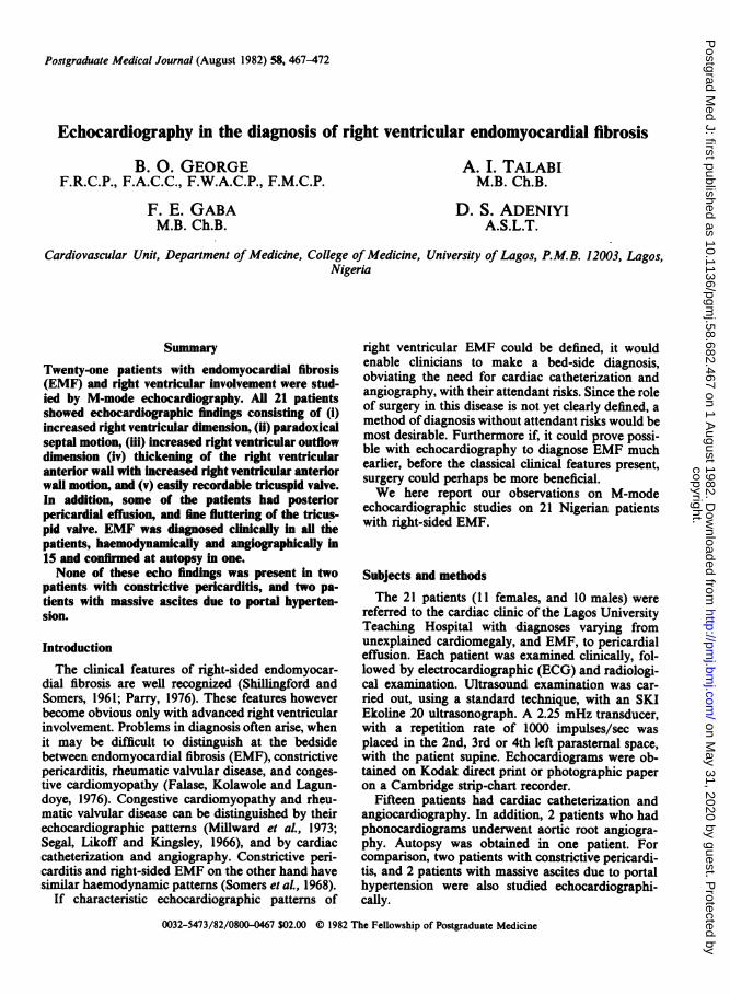
(i) increased right ventricular dimension,(ii) paradoxical septal motion (Fig. 1),(iii) increased right ventricular outflow tract di-
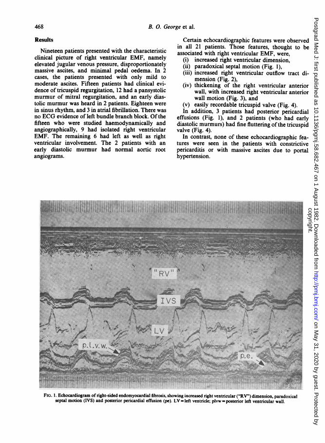
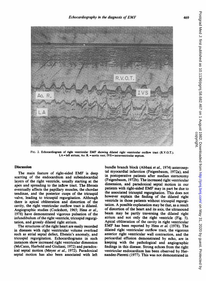
mension (Fig. 2),(iv) thickening of the right ventricular anterior
wall, with increased right ventricular anteriorwall motion (Fig. 3), and
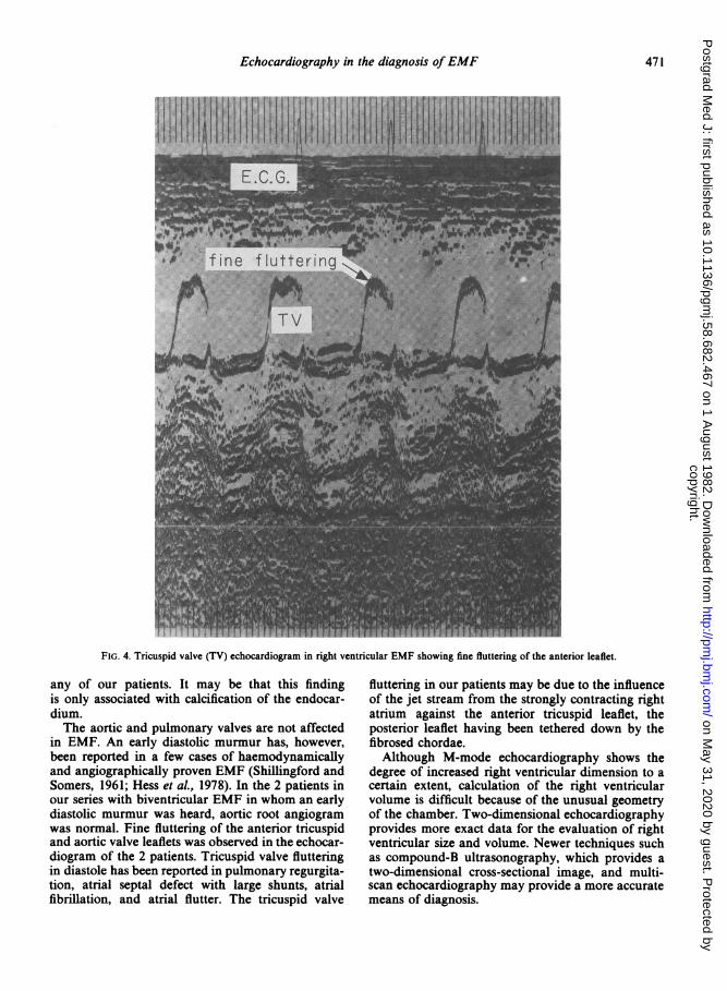
(v) easily recordable tricuspid valve (Fig. 4).In addition, 3 patients had posterior pericardial
effusions (Fig. 1), and 2 patients (who had earlydiastolic murmurs) had fine fluttering ofthe tricuspidvalve (Fig. 4).
In contrast, none of these echocardiographic fea-tures were seen in the patients with constrictivepericarditis or with massive ascites due to portalhypertension.
l~~~~~~~~~~~~~~~~~~~~~~~~~~~~~~~~~~~~~~~~--fa|~~~~~~~~~~~~~~~~~~~~~~~~~~~~~~~~~~~~~~~... .. _
v A!s5--A_ _i__
i 'Sf~~~~~~fLVb
VW
F:~~~~~~~~~~~~~~~~~pe.l vOr'*?ii_-9 = ~ rw St~~~~~~~~~~~~~~~~~~~~~~~~~~~~~~~~~~~ _T # _
FIG. 1. Echocardiogram of right-sided endomyocardial fibrosis, showing increased right ventricular ("RV") dimension, paradoxicalseptal motion (IVS) and posterior pericardial effusion (pe). LV - left ventricle; plvw - posterior left ventricular wall.
copyright. on M
ay 31, 2020 by guest. Protected by
http://pmj.bm
j.com/
Postgrad M
ed J: first published as 10.1136/pgmj.58.682.467 on 1 A
ugust 1982. Dow
nloaded from
Echocardiography in the diagnosis ofEMF 469
.~~~~~~.4~ ~~~1
ji,~~~~~~~v'q
FIG. 2. Echocardiogram of right ventricular EMF showing dilated right ventricular outflow tract (R.V.O.T.).LA - left atrium; Ao. R. - aortic root; IVS - intraventricular septum.
Discussion
The main feature of right-sided EMF is deepscarring of the endocardium and subendocardiallayers of the right ventricle, usually starting at theapex and spreading to the inflow tract. The fibrosiseventually affects the papillary muscles, the chordaetendinae, and the posterior cusps of the tricuspidvalve, leading to tricuspid regurgitation. Althoughthere is apical obliteration and distortion of thecavity, the right ventricular outflow tract is dilated.Angiographic studies (Cockshott, 1965; Hess et al.,1978) have demonstrated vigorous pulsation of theinfundibulum ofthe right ventricle, tricuspid regurgi-tation, and grossly dilated right atrium.The structures ofthe right heart are easily recorded
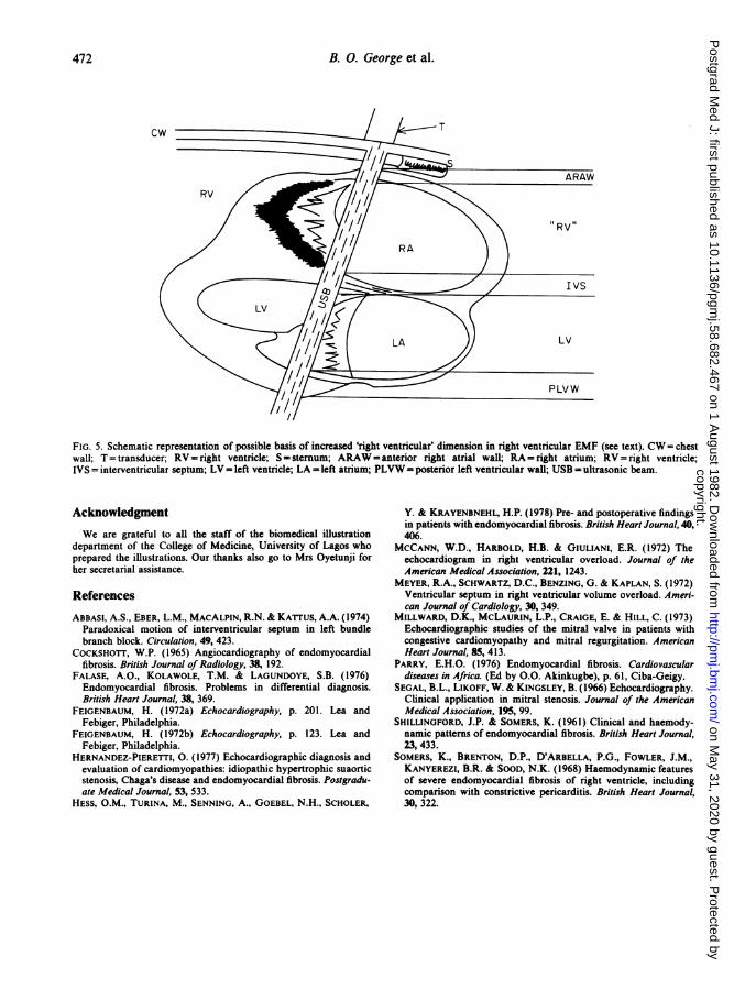
in diseases with right ventricular volume overloadsuch as atrial septal defect, Ebstein's anomaly, andtricuspid regurgitation. Echocardiograms in suchinstances show increased right ventricular dimension(McCann, Harbold and Giuliani, 1972) and paradox-ical septal motion (Meyer et al., 1972). Paradoxicalseptal motion has also been associated with left
bundle branch block (Abbasi et aL, 1974) anterosep-tal myocardial infarction (Feigenbaum, 1972a), andin postoperative patients after median sternotomy(Feigenbaum, 1972b). The increased right ventriculardimension, and paradoxical septal motion in ourpatients with right-sided EMF may in part be due tothe associated tricuspid regurgitation. This does nothowever explain the finding of the dilated rightventricle in those patients without tricuspid regurgi-tation. A possible explanation may be that, as a resultof distortion of the heart and its axis, the ultrasoundbeam may be partly traversing the dilated rightatrium and not only the right ventricle (Fig. 5).Apical obliteration of the cavity in right ventricularEMF has been reported by Hess et aL (1978). Thedilated right ventricular outflow tract, the vigorousanterior right ventricular wall contraction, and thepericardial effusion demonstrated by echo, are inkeeping with the pathological and angiographicfindings in this disease. Strong echoes from the rightventricular endocardium has been observed by Her-nandez-Pieretti (1977). This was not demonstrated in
copyright. on M
ay 31, 2020 by guest. Protected by
http://pmj.bm
j.com/
Postgrad M
ed J: first published as 10.1136/pgmj.58.682.467 on 1 A
ugust 1982. Dow
nloaded from
470 B. 0. George et al.
* | | W W Z RVAW
M.1,I'VIl1I* _
FIG. 3. Echocardiogram of right ventricular EMF showing thickening ofthe right ventricularanterior wall (RVAW), and strong right ventricular anterior wall contraction.
copyright. on M
ay 31, 2020 by guest. Protected by
http://pmj.bm
j.com/
Postgrad M
ed J: first published as 10.1136/pgmj.58.682.467 on 1 A
ugust 1982. Dow
nloaded from
Echocardiography in the diagnosis ofEMF 471
0jli~~l10008k~iS1; A111iSzl I IIk I 1 Hlj11; 11-1i1 11011E.C.G._
as fine f luttering us..'''.,,;,,',',.' '" .. ,.!;.
~~~T Vl
mas*:M:.::~~~~~~~~~~~~~~~~~~~~~~~OA h L
FIG. 4. Tricuspid valve (TV) echocardiogram in right ventricular EMF showing fine fluttering of the anterior leaflet.
any of our patients. It may be that this findingis only associated with calcification of the endocar-dium.The aortic and pulmonary valves are not affected
in EMF. An early diastolic murmur has, however,been reported in a few cases of haemodynamicallyand angiographically proven EMF (Shillingford andSomers, 1961; Hess et al., 1978). In the 2 patients inour series with biventricular EMF in whom an earlydiastolic murmur was heard, aortic root angiogramwas normal. Fine fluttering of the anterior tricuspidand aortic valve leaflets was observed in the echocar-diogram of the 2 patients. Tricuspid valve flutteringin diastole has been reported in pulmonary regurgita-tion, atrial septal defect with large shunts, atrialfibrillation, and atrial flutter. The tricuspid valve
fluttering in our patients may be due to the influenceof the jet stream from the strongly contracting rightatrium against the anterior tricuspid leaflet, theposterior leaflet having been tethered down by thefibrosed chordae.Although M-mode echocardiography shows the
degree of increased right ventricular dimension to acertain extent, calculation of the right ventricularvolume is difficult because of the unusual geometryof the chamber. Two-dimensional echocardiographyprovides more exact data for the evaluation of rightventricular size and volume. Newer techniques suchas compound-B ultrasonography, which provides atwo-dimensional cross-sectional image, and multi-scan echocardiography may provide a more accuratemeans of diagnosis.
copyright. on M
ay 31, 2020 by guest. Protected by
http://pmj.bm
j.com/
Postgrad M
ed J: first published as 10.1136/pgmj.58.682.467 on 1 A
ugust 1982. Dow
nloaded from
472 B. O. George et al.
cw 1T
.' are !)~~~~~~~~~~~~~~/
FIG. 5. Schematic representation of possible basis of increased 'right ventricular' dimension in right ventricular EMF (see text). CW= chestwall; T = transducer; RV - right ventricle; S - sternum; ARAW = anterior right atrial wall; RA = right atrium; RV = right ventricle;IVS = interventricular septum; LV= left ventricle; LA = left atrium; PLVW= posterior left ventricular wall; USB = ultrasonic beam.
AcknowledgmentWe are grateful to all the staff of the biomedical illustration
department of the College of Medicine, University of Lagos whoprepared the illustrations. Our thanks also go to Mrs Oyetunji forher secretarial assistance.
ReferencesABBASI, A.S., EBER, L.M., MACALPIN, R.N. & KArrus, A.A. (1974)
Paradoxical motion of interventricular septum in left bundlebranch block. Circulation, 49, 423.
COCKSHOTT, W.P. (1965) Angiocardiography of endomyocardialfibrosis. British Journal of Radiology, 38, 192.
FALASE, A.O., KOLAWOLE, T.M. & LAGUNDOYE, S.B. (1976)Endomyocardial fibrosis. Problems in differential diagnosis.British Heart Journal, 38, 369.
FEIGENBAUM, H. (1972a) Echocardiography, p. 201. Lea andFebiger, Philadelphia.
FEIGENBAUM, H. (1972b) Echocardiography, p. 123. Lea andFebiger, Philadelphia.
HERNANDEZ-PIERETTI, 0. (1977) Echocardiographic diagnosis andevaluation of cardiomyopathies: idiopathic hypertrophic suaorticstenosis, Chaga's disease and endomyocardial fibrosis. Postgradu-ate Medical Journal, 53, 533.
HESS, O.M., TURINA, M., SENNING, A., GOEBEL, N.H., SCHOLER,
Y. & KRAYENBNEHL, H.P. (1978) Pre- and postoperative findingsin patients with endomyocardial fibrosis. British Heart Journal, 40,406.
MCCANN, W.D., HARBOLD, H.B. & GIULIANI, E.R. (1972) Theechocardiogram in right ventricular overload. Journal of theAmerican Medical Association, 221, 1243.
MEYER, R.A., SCHWARTZ, D.C., BENZING, G. & KAPLAN, S. (1972)Ventricular septum in right ventricular volume overload. Ameri-can Journal of Cardiology, 30, 349.
MILLWARD, D.K., MCLAURIN, L.P., CRAIGE, E. & HILL, C. (1973)Echocardiographic studies of the mitral valve in patients withcongestive cardiomyopathy and mitral regurgitation. AmericanHeart Journal, 85, 413.
PARRY, E.H.O. (1976) Endomyocardial fibrosis. Cardiovasculardiseases in Africa. (Ed by O.O. Akinkugbe), p. 61, Ciba-Geigy.
SEGAL, B.L., LIKOFF, W. & KINGSLEY, B. (1966) Echocardiography.Clinical application in mitral stenosis. Journal of the AmericanMedical Association, 195, 99.
SHILLINGFORD, J.P. & SOMERS, K. (1961) Clinical and haemody-namic patterns of endomyocardial fibrosis. British Heart Journal,23, 433.
SOMERS, K., BRENTON, D.P., D'ARBELLA, P.G., FOWLER, J.M.,KANYEREZI, B.R. & SOOD, N.K. (1968) Haemodynamic featuresof severe endomyocardial fibrosis of right ventricle, includingcomparison with constrictive pericarditis. British Heart Journal,30, 322.
copyright. on M
ay 31, 2020 by guest. Protected by
http://pmj.bm
j.com/
Postgrad M
ed J: first published as 10.1136/pgmj.58.682.467 on 1 A
ugust 1982. Dow
nloaded from