Echocardiography Board Review (500 Multiple Choice Questions with Discussion) || Chapter 16
15
CHAPTER 16 16 Questions 301. The parasternal long-axis image of the mitral valve apparatus shows: A. Mitral annular calcification B. Rheumatic mitral stenosis C. Systolic anterior motion D. Annuloplasty ring Echocardiography Board Review: 500 Multiple Choice Questions with Discussion, Second Edition. Ramdas G. Pai and Padmini Varadarajan. © 2014 John Wiley & Sons, Ltd. Published 2014 by John Wiley & Sons, Ltd. 127
Transcript of Echocardiography Board Review (500 Multiple Choice Questions with Discussion) || Chapter 16

CHAPTER 16
16
Questions
301. The parasternal long-axis image of the mitral valve apparatus shows:
A. Mitral annular calcification
B. Rheumatic mitral stenosis
C. Systolic anterior motion
D. Annuloplasty ring
Echocardiography Board Review: 500 Multiple Choice Questions with Discussion, Second Edition.Ramdas G. Pai and Padmini Varadarajan.© 2014 John Wiley & Sons, Ltd. Published 2014 by John Wiley & Sons, Ltd.
127
128 Echocardiography Board Review
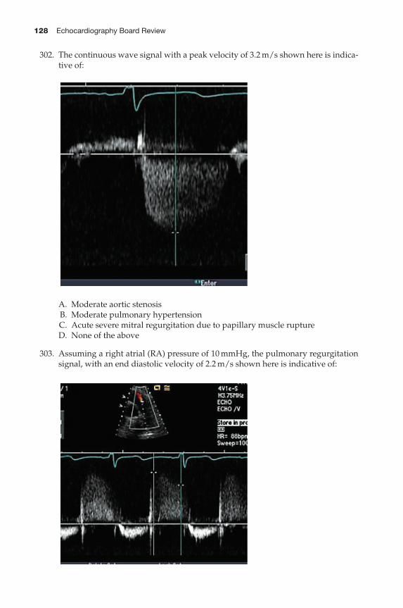
302. The continuous wave signal with a peak velocity of 3.2m/s shown here is indica-
tive of:
A. Moderate aortic stenosis
B. Moderate pulmonary hypertension
C. Acute severe mitral regurgitation due to papillary muscle rupture
D. None of the above
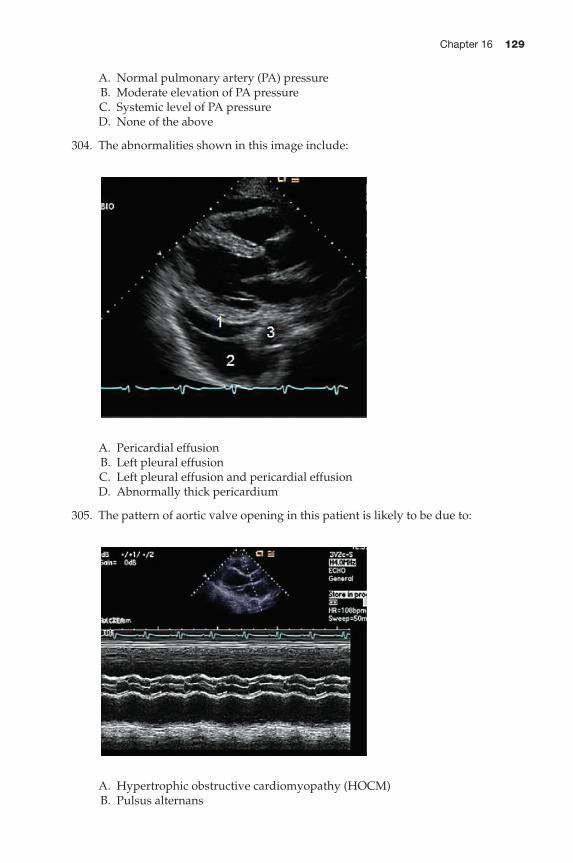
303. Assuming a right atrial (RA) pressure of 10mmHg, the pulmonary regurgitation
signal, with an end diastolic velocity of 2.2m/s shown here is indicative of:
Chapter 16 129
A. Normal pulmonary artery (PA) pressure
B. Moderate elevation of PA pressure
C. Systemic level of PA pressure
D. None of the above
304. The abnormalities shown in this image include:
A. Pericardial effusion
B. Left pleural effusion
C. Left pleural effusion and pericardial effusion
D. Abnormally thick pericardium
305. The pattern of aortic valve opening in this patient is likely to be due to:
A. Hypertrophic obstructive cardiomyopathy (HOCM)
B. Pulsus alternans

130 Echocardiography Board Review
C. Intra-aortic balloon pump (IABP) with 1:3 supportD. Left ventricular assist device (LVAD) with 1:3 support
306. This is an apical four-chamber view of the left ventricle (LV). The structure indi-cated by the arrow in the LV apex is likely to be:
A. LV thrombusB. Rib artifactC. Cannula of LVADD. False tendon in the LV apex
307. The structure indicated by the arrow is:
Chapter 16 131
A. Descending thoracic aorta
B. Coronary sinus
C. Left lower pulmonary vein
D. Left PA
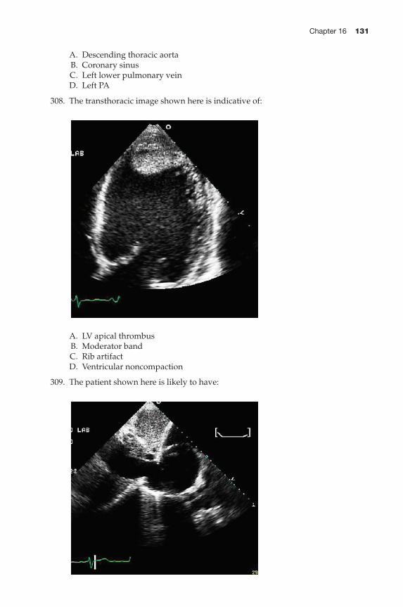
308. The transthoracic image shown here is indicative of:
A. LV apical thrombus
B. Moderator band
C. Rib artifact
D. Ventricular noncompaction
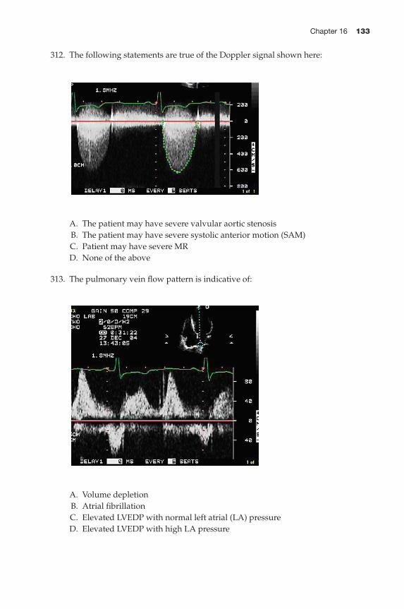
309. The patient shown here is likely to have:

132 Echocardiography Board Review
A. Heart failure
B. Intravascular volume depletion with hypotension
C. Right atrial tumor
D. None of the above
310. The continuous wave Doppler signal shown here is suggestive of:
A. Mixed mitral valve disease with significant mitral stenosis (MS) and mitral
regurgitation (MR)
B. Mixed aortic valve disease with significant aortic stenosis (AS) and aortic
regurgitation (AR)
C. Combination of AR and MR
D. Ventricular septal defect (VSD) with bidirectional flow
311. This patient is likely to have (BP 130/65mmHg):
A. High left ventricular end diastolic pressure (LVEDP)
B. Diastolic MR
C. Premature mitral valve closure
D. All of the above
Chapter 16 133
312. The following statements are true of the Doppler signal shown here:
A. The patient may have severe valvular aortic stenosis
B. The patient may have severe systolic anterior motion (SAM)
C. Patient may have severe MR
D. None of the above
313. The pulmonary vein flow pattern is indicative of:
A. Volume depletion
B. Atrial fibrillation
C. Elevated LVEDP with normal left atrial (LA) pressure
D. Elevated LVEDP with high LA pressure
134 Echocardiography Board Review
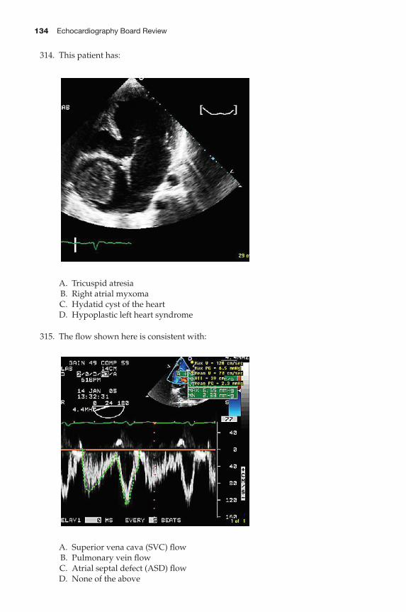
314. This patient has:
A. Tricuspid atresia
B. Right atrial myxoma
C. Hydatid cyst of the heart
D. Hypoplastic left heart syndrome
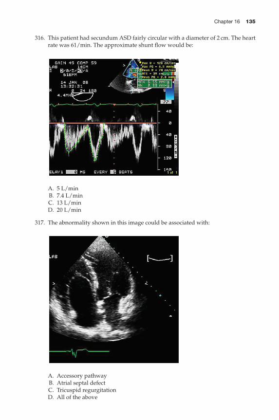
315. The flow shown here is consistent with:
A. Superior vena cava (SVC) flow
B. Pulmonary vein flow
C. Atrial septal defect (ASD) flow
D. None of the above
Chapter 16 135
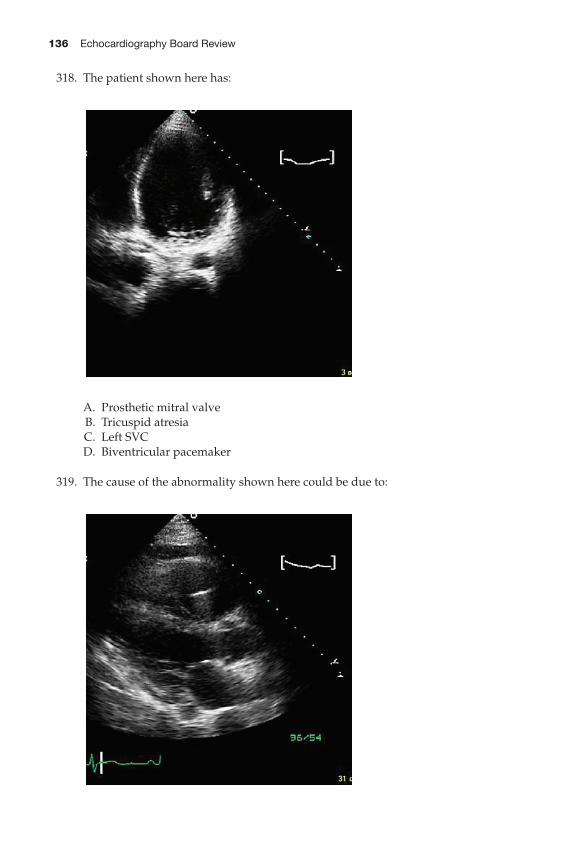
316. This patient had secundum ASD fairly circular with a diameter of 2 cm. The heart
rate was 61/min. The approximate shunt flow would be:
A. 5 L/min
B. 7.4 L/min
C. 13 L/min
D. 20 L/min
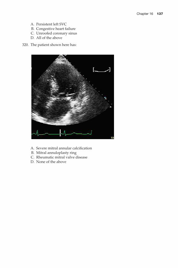
317. The abnormality shown in this image could be associated with:
A. Accessory pathway
B. Atrial septal defect
C. Tricuspid regurgitation
D. All of the above
136 Echocardiography Board Review
318. The patient shown here has:
A. Prosthetic mitral valve
B. Tricuspid atresia
C. Left SVC
D. Biventricular pacemaker
319. The cause of the abnormality shown here could be due to:
Chapter 16 137
A. Persistent left SVC
B. Congestive heart failure
C. Unroofed coronary sinus
D. All of the above
320. The patient shown here has:
A. Severe mitral annular calcification
B. Mitral annuloplasty ring
C. Rheumatic mitral valve disease
D. None of the above
138 Echocardiography Board Review
Answers for chapter 16
301. Answer: D.
Posterior mitral annuloplasty ring in crosssection. This is circular in crosssection
and on the atrial side of the base of the posterior mitral leaflet. Mitral annu-
lar calcification on the contrary will bury the leaflet base inside the calcification
and generally starts from the base of the annulus and extends to the leaflets,
and the shape is not circular in crosssection. There is no restriction of the leaflet
tips to suggest rheumatic involvement and SAM is evaluated in systole. This is a
diastolic frame.
302. Answer: B.This is a signal originating from A–V valve regurgitation as it starts with the QRS
without any isovolumic contraction. Accompanying forward flow velocity is less
than 1/2 m/s suggesting tricuspid origin. Mitral inflow velocity tends to be higher.
The velocity of this signal is 3.2m/s resulting in a transvalvular gradient of about
40mmHg. Assuming an RA pressure of 10mmHg, the RV systolic pressure would
be 50mmHg. Aortic signal is of shorter duration and starts later after the isovo-
lumic contraction period and if mitral inflow is visible the isovolumic relaxation
time could be discerned, that is, the aortic velocity curve will not be continuous
with the mitral inflow velocity curve. In acute severe MR, the gradient could be
low due to hypotension and high LA pressure. In such a situation, a large V-wave
would result in rapid deceleration of the signal soon after finishing acceleration.
This is so-called V-wave “cutoff” sign.
303. Answer: B.End diastolic pulmonary regurgitation velocity is 2m/s, consistent with a PA to
right ventricular (RV) end diastolic gradient of 16mmHg (4 × 22) and assuming
that the RV enddiastolic pressure is close to themeanRApressure, the PAdiastolic
pressure will be 26 mmHg.
304. Answer: C.Number 1 indicates pericardial effusion, 2 indicates pleural effusion, and 3 is the
descending aorta. Pericardial effusion is always anterior to the aorta and pleural
effusion extends posteriorly. The structure separating the two is combined parietal
pericardium and pleura. The combined thickness is <3mm, which is normal.
305. Answer: D.
There is reduced aortic valve openingwith every third beat. This is due to reduced
transaortic flowwith every third beat, which is assisted by the LVAD, and the bulk
of the cardiac output is delivered through the assist device. During the interven-
ing two beats, all the stroke volume is delivered through the aortic valve. With
1:3 IABP support the increase in stroke volume during the IABP deflation occurs
through the aortic valve, hence there is increased opening. In pulsus alternans,
strong and weak beats alternate in a 1:2 fashion. In HOCM, midsystolic closure
occurs with every beat.
306. Answer: C.Echodense walls and echoluscent lumen of the cannula are seen. This LVAD
cannula serves to deliver blood to the assist device. In these patients it is important
to make sure that there is no obstruction to the inlet cannula by surrounding
structures, including the ventricular septum, and no apical thrombi. Thrombus
does not have a central luscency. False tendons are echodense and linear and rib
artifacts are lighter and generally go through the anatomic boundaries.
Chapter 16 139
307. Answer: A.
This vessel is posterior to the left atrium, indicative of descending thoracic aorta.
Coronary sinus is in the posterior A–V groove and is intrapericardial. The left PA
and the left lower pulmonary vein are far away from this location.
308. Answer: A.
An LV apical thrombus. There is a distinctly demarcated thrombus in the apex.
When there is a question, this can be confirmed by obtaining additional views
of the apex, such as two-chamber and short-axis views with color flow imaging
at a low Nyquist limit or using transpulmonary contrast agents such as Definity,
thrombus will be seen as a filling defect. The LV apex is a common place for a
false tendon and may be mistaken for a thrombus. Rib artifact is less dense, goes
beyond the endocardium, and does not move with the heart.
309. Answer: A.
Heart failure. Dilated inferior vena cava (IVC) is suggestive of high RA pressure
if it does not collapse with inspiration. Occasionally, in normal young individuals
one may see a dilated IVC, which readily collapses with inspiration. A general
guideline is that IVC> 2 cm and<10% collapse indicates RApressure> 20mmHg,
>2 cm and 50% collapse indicates RA pressure of 15mmHg, 1.5–2 cm and >50%
collapse indicates RA pressure of 10mmHg, and <1 cm and >50% collapse indi-
cates RA pressure of 5mmHg. However, new ASE guidelines are as follows: IVC
size of≤2.1 cm and>50% collapse with a sniff is suggestive of normal RA pressure
of 3mmHg (range, 0–5mmHg). If IVC size is≥2.1 cmand<50% collapse is sugges-
tive of high RA pressure, 15mmHg (range, 10–20mm hg). In indeterminate cases,
here, the size of IVC and collapse do not fit this paradigm, an intermediate value
of 8mmHg (range, 5–10mm hg) may be used. Alternately, secondary indices of
high RA pressure should be integrated such as tricuspid E/E′>6, diastolic flow
predominance in the hepatic veins. In indeterminate cases, if secondary indices of
elevatedRApressure are not present, RApressure can be downgraded to 3mmHg.
If there is minimal IVC collapse with a sniff (<35%) and secondary indices of high
RA pressure are present, then RA pressure can be upgraded to 15mmHg. If uncer-
tain, leave RA pressure at 8mmHg. In patients who are unable to perform a sniff,
an IVC that collapses <20%with quiet respiration suggests high RA pressure. IVC
collapse does not accurately reflect RA pressure in ventilator-dependent patients.
(J Am Soc Echocardiogr 2010;23:685–713).
310. Answer: B.Mixed aortic valve disease with significant AS andAR. Diastolic signal is diagnos-
tic of AR with a 4m/s early diastolic velocity, which does not occur with MS. The
velocity curve of AR is continuous with the systolic signal, indicating signal ori-
gin at the same valve. TheMR signal would have a longer signal and overlap both
the initial and terminal portions of the AR signal, as MR would occur with both
isovolumic contraction and relaxation phases. Typical VSD signal will have a sys-
tolic component and a presystolic associated with left atrial contraction directed
into the RV. If the patient has Eisenmenger’s syndrome, the flow velocity would
be very low.
311. Answer: D.
All of the above. The AR signal decelerates rapidly with a pressure half-time
of 185ms (less than 250ms indicates very rapid deceleration). The end diastolic
velocity is about 2ms, indicating an end diastolic gradient between the aorta and
LV of 16mmHg, assuming alignment of the ultrasound beam parallel to flow. As
140 Echocardiography Board Review
the patient’s diastolic pressure is 65mmHg, the LVEDP is 49mmHg (65 − 16).
Severe AR, generally acute, or significant AR in the presence of stiff LV may
occur with severe AS or hypertension; high LVEDP resulting from this may cause
diastolic MR and also presystolic closure of the mitral valve.
312. Answer: A.
Severe valvular AS. The timing of the onset slightly after the onset of theQRS com-
plex, suggestive of onset after the LV isovolumic contraction period, is suggestive
of aortic origin. Although the aortic valve area is the best indicator of AS severity,
a mean gradient of >50mmHg is generally consistent with severe AS. In addition,
the signal is mid to late peaking, which has the same significance as the mid to
late peaking of the AS murmur. SAMwould cause a dagger-shaped, late-peaking
signal because of the dynamic nature of the obstruction.
313. Answer: D.
Elevated LVEDP with high LA pressure. The D-wave velocity, which is higher
than the S-wave velocity with a rapid deceleration (time < 170ms), is indicative
of high LA pressure in an adult. Small S and large D could be normal in children
because of very efficient LV relaxation. In the example shown here, the AR wave
duration is markedly increased. Normally, AR wave duration is less than mitral
A-wave duration and is less than 110–120ms. Although A-wave duration is not
shown here, the AR wave duration is grossly abnormal at 200ms, indicating high
LVEDP, causing an increase in the duration of atrial systole because of increased
atrial afterload. In a volume-depleted patient the S-wave will be prominent and
the AR wave would be diminutive; in atrial fibrillation, the AR wave is lost.
314. Answer: A.
Tricuspid atresia. The image shows an absent tricuspid valve, a right atrial mass
that is consistent and likely to be a thrombus due to stasis and a ventricular
septal defect. This patient had cavopulmonary anastomosis with SVC–RPA
and RA–LPA shunt such that IVC blood drained into the LPA through the
RA, causing thrombus formation. The PA was completely banded to facilitate
cavopulmonary flow. This patient has a well-developed left heart and hence does
not have hypoplastic left heart syndrome. Right atrial myxoma is a possibility,
but thrombus is much more likely in this situation.
315. Answer: C.ASD flow. The flow shown is typical of ASD flow with systolic–diastolic wave
and a second wave associated with atrial contraction, all left to right in the same
direction. Both SVC and pulmonary vein flows are triphasic with S, D, and AR
waves, with the AR wave being in an opposite direction to S- and D-waves.
316. Answer: B.7.4 L/min. The shunt flow per heart beat is the time velocity integral (TVI) across
the defect × cross sectional area, that is, 39 × 3.14 × 1 × 1 = 122 cc (TVI of signal is
39 cm). Shunt flow per minute = shunt volume per beat × heart rate = 122 × 61 =7.4 L/min.
317. Answer: D.
The image is diagnostic of Ebstein’s anomaly of the tricuspid valve. This is diag-
nosed when the attachment of the septal leaflet of the tricuspid valve is apically
displaced in relation to the anterior leaflet by>8mm/M2. In this disorder, the sep-
tal leaflet is large, sail like, and could be plastered to the RV wall through the

Chapter 16 141
chordae tendinae. The associations include severe tricuspid regurgitation, atrial
septal defect, and right sided accessory pathway causing PSVT.
318. Answer: D.
Biventricular pacemaker. A coronary sinus lead is clearly seen in this image. This
is imaged from the apical four-chamber view with a posterior transducer tilt to
obtain a tomographic plane through the coronary sinus. Because of this the mitral
valve is not seen. The coronary sinus is not enlarged to support the diagnosis of
left SVC.
319. Answer: D.
All of the above. The coronary sinus is dilated, which could be due to increased
flow or pressure, and any of the conditions listed can potentially result in a dilated
coronary sinus. Note that the coronary sinus is in the A–V groove and intraperi-
cardial versus descending aorta, which is in the posterior mediastinum and is
extrapericardial.
320. Answer: B.Mitral annuloplasty ring. Echocardiographically, this is distinguished frommitral
annular calcification by its rounded shape in crosssection and projection into the
left atrium at the base of the posterior leaflet. On the contrary, mitral annular cal-
cification would incorporate the base of the posterior mitral leaflet into itself.