Echinoderms contain the sea stars, brittle stars, sea...
9
Zoology Exercise #16: Echinoderms Lab Guide Echinoderms contain the sea stars, brittle stars, sea urchins, sand dollars, and sea cucumbers. The name “ echinoderm” is derived from this group’s dermal endoskeleton of calcareous plates and spines. They have a water vascular system that powers a great number of tiny tube feet that are used for locomotion and gathering food. Many members will have pincher like pedicellaria that snap creatures that would settle on them. To breathe, many will rely on numerous dermal branchiae (skin gills) that project through small spaces in their endoskeleton. They lack a definite head and their nervous system/sense organs are poorly developed. Their locomotion is very slow and they lack segmentation. They have pentaradial symmetry. The body parts are most often arranged in multiples of five. But, their larvae are bilaterally symmetrical. Exercise 16A – Class Asteroidea – Sea Stars Sea stars often live along rocky coastlines, but can also be found in heavy surf and at great depths in the ocean. They are strictly bottom dwellers, preying on slow moving prey such as molluscs, crabs, corals, and worms. Adult sea stars have few enemies. External Anatomy The general body plan of sea stars is pentaradial. It typically has 5 arms (rays) surrounding a central disc. It has a body which is flattened. This top is considered the aboral side while the bottom (which contains the mouth) is its oral side. The aboral surface will bear a small, disc shaped madreporite. It is composed of calcium carbonate and allows seawater to seep into the water vascular system which will help the animal in locomotion. The arms on each side of the madreporite are called the bivium. The other 3 arms are the trivium. The anus opens in the center of the central disc, but can be hard to locate. The body is covered with a thin, ciliated epidermis, through which white calcareous spines extend through the endoskeleton. Surrounding the base of each spine is a raised ring of skin that contains tiny, calcareous pincher like pedicellariae. Some might also be found between the spines. These pedicellariae are moved by tiny muscles. Between the spines are soft, transparent, fingerlike projections called dermal branchiae (skin gills). A small eyespot is located at the tip of each arm. The oral surface will show the ambulacral groove of each arm. It contains rows of tiny tube feet (podia). The ambulacral spines will border the groove and these spines are movable, allowing them to lock and protect the delicate tube feet. The tube feet are part of the water vascular system and are hollow. Their tips have suctions discs for attachment. They help in locomotion, but are also very efficient in opening bivalves for food. The central mouth is surrounded by 5 pairs of movable spines. The endoskeleton is formed by calcareous plates (ossicles) and are bound together. The ossicles are often irregular. The outer layer is the epidermis, with the thicker layer connecting the epidermis and peritoneum being called the dermis. The tube feet will extend up between the ambulacral ossicles, emerging on the inside surface of the wall as bulblike ampullae. On the inner surface of each arm there will be an ambulacral ridge. Internal Anatomy The digestive system consists of a pyloric stomach that lies in the central disc. A pyloric duct will extend into each arm, connecting to a large pair of pyloric ceca. A very short intestine leads from the center of the stomach to the anus. Attached to the intestine are two rectal ceca. Below the pyloric stomach is the larger, cardiac stomach. It fills most of the central disc. When a sea star feeds on a bivalve, it folds itself around the animal, attaches its tube feet to the valves, and exerts enough pull to cause the shell to gape slightly. By contracting its body walls to increase pressure, it everts its stomach and inserts it into the partly opened bivalve shell. It will digest the soft parts of the bivalve with digestive juices from the pyloric ceca. When it have finished feeding, it will withdrawal its stomach. Sexes are separate. Their gonads are paired and attached to the sides of the arm (ray) where it joins the central disc. These gonads are much larger during the breeding season. Each will open to the outside on the aboral side by a very small reproductive duct and genital pore. The eggs and sperm are shed into the water where fertilization occurs externally.
Transcript of Echinoderms contain the sea stars, brittle stars, sea...

Zoology Exercise #16: Echinoderms Lab Guide Echinoderms contain the sea stars, brittle stars, sea urchins, sand dollars, and sea cucumbers. The name “echinoderm” is derived from this group’s dermal endoskeleton of calcareous plates and spines. They have a water vascular system that powers a great number of tiny tube feet that are used for locomotion and gathering food. Many members will have pincher like pedicellaria that snap creatures that would settle on them. To breathe, many will rely on numerous dermal branchiae (skin gills) that project through small spaces in their endoskeleton. They lack a definite head and their nervous system/sense organs are poorly developed. Their locomotion is very slow and they lack segmentation. They have pentaradial symmetry. The body parts are most often arranged in multiples of five. But, their larvae are bilaterally symmetrical. Exercise 16A – Class Asteroidea – Sea Stars Sea stars often live along rocky coastlines, but can also be found in heavy surf and at great depths in the ocean. They are strictly bottom dwellers, preying on slow moving prey such as molluscs, crabs, corals, and worms. Adult sea stars have few enemies. External Anatomy The general body plan of sea stars is pentaradial. It typically has 5 arms (rays) surrounding a central disc. It has a body which is flattened. This top is considered the aboral side while the bottom (which contains the mouth) is its oral side. The aboral surface will bear a small, disc shaped madreporite. It is composed of calcium carbonate and allows seawater to seep into the water vascular system which will help the animal in locomotion. The arms on each side of the madreporite are called the bivium. The other 3 arms are the trivium. The anus opens in the center of the central disc, but can be hard to locate. The body is covered with a thin, ciliated epidermis, through which white calcareous spines extend through the endoskeleton. Surrounding the base of each spine is a raised ring of skin that contains tiny, calcareous pincher like pedicellariae. Some might also be found between the spines. These pedicellariae are moved by tiny muscles. Between the spines are soft, transparent, fingerlike projections called dermal branchiae (skin gills). A small eyespot is located at the tip of each arm. The oral surface will show the ambulacral groove of each arm. It contains rows of tiny tube feet (podia). The ambulacral spines will border the groove and these spines are movable, allowing them to lock and protect the delicate tube feet. The tube feet are part of the water vascular system and are hollow. Their tips have suctions discs for attachment. They help in locomotion, but are also very efficient in opening bivalves for food. The central mouth is surrounded by 5 pairs of movable spines. The endoskeleton is formed by calcareous plates (ossicles) and are bound together. The ossicles are often irregular. The outer layer is the epidermis, with the thicker layer connecting the epidermis and peritoneum being called the dermis. The tube feet will extend up between the ambulacral ossicles, emerging on the inside surface of the wall as bulblike ampullae. On the inner surface of each arm there will be an ambulacral ridge. Internal Anatomy The digestive system consists of a pyloric stomach that lies in the central disc. A pyloric duct will extend into each arm, connecting to a large pair of pyloric ceca. A very short intestine leads from the center of the stomach to the anus. Attached to the intestine are two rectal ceca. Below the pyloric stomach is the larger, cardiac stomach. It fills most of the central disc. When a sea star feeds on a bivalve, it folds itself around the animal, attaches its tube feet to the valves, and exerts enough pull to cause the shell to gape slightly. By contracting its body walls to increase pressure, it everts its stomach and inserts it into the partly opened bivalve shell. It will digest the soft parts of the bivalve with digestive juices from the pyloric ceca. When it have finished feeding, it will withdrawal its stomach. Sexes are separate. Their gonads are paired and attached to the sides of the arm (ray) where it joins the central disc. These gonads are much larger during the breeding season. Each will open to the outside on the aboral side by a very small reproductive duct and genital pore. The eggs and sperm are shed into the water where fertilization occurs externally.

The nervous system consists of three systems. The oral system has a nerve ring around the mouth and a radial nerve to each arm running to the eyespot. The aboral system lying near the upper surface of the sea star and a deep system positioned between the oral and aboral systems. There will be an epidermal nerve plexus connected to these three systems. Chemoreceptors and cells sensitive to touch are found all over the surface. Each pigmented eyespot consists of a number of light sensitive ocelli. The water vascular system is found ONLY in echinoderms. They use it for locomotion and in the case of sea stars, for opening bivalves. The madreporite on the aboral surface contains ciliated grooves and pores. There will be a curved stone canal that leads to the ring canal which is found around the outer edge of the central disc. Five radial canals, one in each arm, radiate out from the ring canal, running along the ambulacral groove. Short lateral canals will connect the radial canal with each tube foot. Seawater will enter the madreporite, be passed down the stone canal to the ring canals, from there to the radial canals, and finally through the lateral canals to the ampullae and tube feet. Tube feet have longitudinal muscles while the ampullae have circular muscles. When the tube feet are contracted, most of the water is held in the ampullae. When the ampullae contract, water is forced into the elastic tube feet, which will elongate because of hydrostatic pressure. When the cuplike ends of the tube feet contract on a hard surface, they attach with suction force and then contract, pushing water back into the ampullae and pulling the animal forward. A single foot is not very strong, but hundreds working together can move the animal along slowly and can create a tremendous pull on the shell of a bivalve mollusc. Procedure External
1. Obtain a preserved starfish and rinse off any preservative with water. 2. Place the starfish in the dissecting pan with its aboral (top) surface upward. 3. Observe the starfish and determine its symmetry. ____________________________________ 4. Locate the central disc in the center of the starfish. Count the number of arms or rays the starfish has. _______ Now,
locate the bivium and trivium 5. Locate the small, disc shaped plate called the madreporite on top of the central disc. Water enters through this into
the water vascular system. 6. Feel the upper surface of the starfish for spines. These spines protect the starfish and are part of their internal
skeleton. Locate the pedicellariae that surrounds each spine. Between the spines you might also be able to see the dermal branchia (skin gills).
7. Look at the tip of each arm and attempt to find the eyespot using your magnifying lens. 8. Turn the starfish over to its ventral or oral surface (underside).

9. Locate the mouth in the center of the central disc. Find the ring of oral spines surrounding the mouth. 10. Find the ambulacral groove that extends down the underside of each arm. 11. Feel the numerous, soft tube feet inside each groove. these are part of the water vascular system & aid in movement
and feeding.
SKETCH
Internal
1. With the starfish's aboral surface facing you, cut off the tip of a ray. Cut along lines a, b, and c and then remove this flap of skin. MAKE SURE TO PULL UP WITH YOUR SCISSORS AS YOU CUT!!!
2. Inside each arm, locate two long digestive glands called the pyloric caeca. These make enzymes to digest food in the stomach.
3. Cut a circular flap of skin from the central disc. (You will have to also cut around the madreporite in order to remove this flap.) Be VERY CAREFUL when removing the central disc. Use your blunt probe to remove the pyloric stomach from the underside of the disc. Observe the pyloric stomach. Right below the pyloric stomach is the cardiac stomach. This stomach will be forced out of the animals body as it feeds on molluscs (bivalves).
4. Remove the pyloric caeca from the dissected ray. Find the gonads (testes or ovaries) underneath. These may be small if the starfish is NOT in breeding season. Remove these to see the rest of the water vascular system.
5. Cut off the tip of a ray to observe the parts of the tube feet. Find the zipper-like ambulacral ridge that extends the length of the ray. The tube feet are attached to these.
6. Locate the bulb-like top of a tube foot called the ampulla. This sac works like the top of an eyedropper to create suction. The bottom of the tube foot is a sucker.
7. Embedded in the soft body wall are skeletal plates called ossicles. 8. Running down the center of each arm is a lateral canal to which tube feet are attached. 9. In the central disc the five lateral canals connect to a circular canal called the ring canal. 10. A short, canal called the stone canal leads from the ring canal to the madreporite where water enters.
A
B
C

SKETCH
Exercise 16B – Class Echinoidea – Sea Urchins Sea Urchins lack arms and have a body enclosed in a globose shell (test) made of interlocking plates which have movable spines. They still have the typical pentaradial symmetry and water vascular system of all echinoderms. These animals are herbivores that scrape encrusting algae from rock surfaces, nibble on plants, or eat drifting food. They are called “regular” echinoids because of their symmetry and body shape. Others in this group, such as sand dollars, are called “irregular” echinoids because they have bodies that are bilaterally symmetrical and vary in shape. These animals are strictly marine and are widely distributed in both intertidal areas and the deep sea. Most will graze in turtle grass beds, on coral reefs or bury themselves and feed on microscopic organic matter. External Anatomy Spines are long and movable. They are attached at a ball-and-socket joint by two sets of ring muscles. It’s socket will fit over a rounded tubercle on the test (shell). The tube feet originate from rows of perforations in the five ambulacral regions. The tube feet extend beyond the ends of the spines. The mouth has five converging teeth and a collar like lip. The teeth are part of a complex chewing mechanism called Aristotle’s Lantern. It is operated by several sets of muscles. The membranous peristome surrounds the mouth and is perforated by five pairs of large oral tube feet called buccal tube feet (podia). The peristome also bears small spines. On the peristome there are a number of 3 jawed pedicellariae on the ends of long slender stalks. Smaller but more active pedicellariae are located among the spines. These are there to discourage intruders and help keep the skin clean. The sea urchin will use its spines and tube feet for locomotion. Some are adapted for burrowing into rock or other hard material by using both their spines and chewing mechanisms. Most will have tiny modified spines called sphaeridia to assist in equilibrium. The test, podia, pedicellariae, and spines are covered with ciliated epidermis. The outer edge of the peristome, between the ambulacra, will be five pairs of branching peristomial gills which open into the coelomic cavity. The test (endoskeleton) is composed of calcareous plates (ossicles) symmetrically arranged and interlocked or fused so as to be immovable. The plates are typically arranged into 5 double rows of ambulacral plates alternating with double rows of interambulacral plates. On the aboral surface, which is free of spines, is the periproct. The anus is centrally located, surrounded by four (sometimes five) valve like anal plates. Around the anal plates are five genital plates with each bearing a genital pore. One genital pore is normally larger than the others and has many minute pores. This is the madreporite plate which functions the same as it does in sea stars.
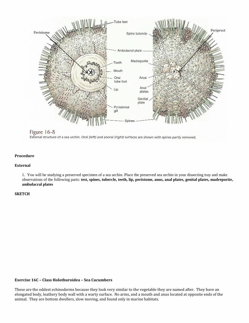
Procedure External
1. You will be studying a preserved specimen of a sea urchin. Place the preserved sea urchin in your dissecting tray and make
observations of the following parts: test, spines, tubercle, teeth, lip, peristome, anus, anal plates, genital plates, madreporite,
ambulacral plates SKETCH Exercise 16C – Class Holothuroidea – Sea Cucumbers These are the oddest echinoderms because they look very similar to the vegetable they are named after. They have an elongated body, leathery body wall with a warty surface. No arms, and a mouth and anus located at opposite ends of the animal. They are bottom dwellers, slow moving, and found only in marine habitats.
Periproct Peristome

External Structure It is orally/aborally elongated with a cylindrical body. Its mouth is encircled with tentacles at one end and the anus at the other. The tentacles, which are modified tube feet, are hollow and a part of the water-vascular system. They are connected internally with radial canals. Most are suspension feeders where they extend their tentacles out and either catch food suspended in the water column or on the substrate. The tentacles become covered with tiny food organisms and is thrust into its mouth where they lick the food off each tentacle. Some will even simply just shovel mud and sand into its mouth, digest out the organic particles, and release the rest. The anus will rhythmically open and close. This is a respiratory movement that is coordinated with the pumping action of the cloaca, which pumps water into and out of the respiratory trees (internal respiratory organs). Internal Structure They have a very large coelomic cavity. Just behind the mouth is the pharynx which is supported by a ring of calcareous plates. It is followed by a short, muscular stomach and a long, convoluted intestine. It is held in place by mesenteries and expands somewhat towards the end to form a cloaca. This cloaca will empty at the anus. Two branched respiratory trees are attached to the cloaca. They serve as both respiratory and excretory organs. They are aerated by rhythmic pumping of the cloaca. Long retractile muscles run from the pharynx to join longitudinal muscles in the body wall. For the water vascular system, a ring canal surrounds the pharynx. One or more rounded or elongated sacs called Polian Vesicles hang from the ring canal into the body cavity. These Polian Vesicles are thought to function as expansion chambers in maintaining pressure within the water vascular system. One or more stone canals also open into the ring canal from the body cavity. In adult sea cucumbers the water vascular system has usually lost contact with the seawater outside. Coelomic fluid, rather than sea water, enters and leaves the system. Five radial canals extend form the ring canal forward along the walls of the pharynx to give off branches to the tentacles, which are actually modified tube feet. From there the radial canals run back along the inner surface of the ambulacra, where each gives off lateral canals to the tube feet and ampullae. Valves in the lateral canals prevent backflow. The reproductive system has gonads that consist of numerous tubules united into one or two tufts on the side of the dorsal mesentery. They become quite large at sexual maturity. A gonadoduct passes anteriorly in the mesentery to the genital pore. The sexes are separate and fertilization is external. The endoskeleton consists largely of tiny, calcareous ossicles scattered in the dermis. These can really only be seen on a prepared slide.

Procedure External 1. You will be studying a preserved specimen of a sea cucumber. Place the preserved sea cucumber in your dissecting tray and make
observations of the following parts: oral end, aboral end, tentacles (tube feet), anus, SKETCH Internal 1. Locate the 5 longitudinal ambulacral areas of a preserved sea cucumber. The sole (ventral side applied to the substrate) has three ambulacra with well-developed podia (tube feet). The dorsal side has two ambulacra with smaller podia (in some species, podia are absent on the dorsal surface). With scissors, open the body by making a longitudinal cut on the ventral (sole) side of the animal, between the central and right ambulacra. Pin down the walls and cover with water. Make observations of the following parts: mouth, pharynx, retractor muscles, stomach, intestine, ring canal, Polian Vesicles, gonads, respiratory tree, longitudinal muscles, cloaca, stone canal, ampullae (if possible) SKETCH Analysis 1. Fill in the following classification scheme for the Sea Star, Sea Urchin, & Sea Cucumber.
Level of Classification
Sea Star Sea Urchin Sea Cucumber
Kingdom Phylum
Class Order Family Genus
Species

2. What does the name “Echinodermata” mean? 3. Go online and find a video which depicts the sequence of action seen in a single tube foot as the animal is moving. Describe this movement. Now, explain what is responsible for this movement. 4. The skeleton of starfish is very different from that seen in arthropods. Compare and explain. 5. Starfish have an aboral and oral side. What is the difference between each and provide a list of structures you would see on each side. 6. The water vascular system of starfish is a significant characteristic used to place this animal into the group identified as Echinoderms. Below, diagram the water vascular system in a starfish. Label the following parts: ring canal, stone canal, Madreporite, one radial canal, lateral canals, and the tube feet/ampullae. 7. What is the function of the system you diagrammed in #6? How is it different in Holothuroideans and Ophiuroideans? 8. Describe the method by which a starfish will feed. 9. Most echinoderms demonstrate a form of radial symmetry. Why would this be an adaption to a sedentary lifestyle? 10. Sea cucumbers have a very unique way of feeding. Describe how they feed and contrast this with how a starfish and sea urchin will feed.

11. How are echinoderms important to humans? 12. The spines in echinoderms are movable. How? 13. Sea urchin anatomy is dominated by gonads and digestive organs. What might this indicate about the ecological role these animals play in the environment? 14. Sea urchins are described as being “regular” echinoderms while sand dollars are “irregular”. What does this mean? 15. Many would think that sea urchins have the ultimate strategy for protecting themselves, but some members of this group have greatly reduced tests. How do these “exceptions” protect themselves? 16. Sea cucumbers have a peculiar power of what appears to be self-mutilation, but actually is a defense mechanism. Explain.