
ECG/EKG. ECG ECG stands for Electrocardiogram Sooo smart students, what do you think it measures?
30
ECG/EKG
-
Upload
brett-hodges -
Category
Documents
-
view
233 -
download
0
Transcript of ECG/EKG. ECG ECG stands for Electrocardiogram Sooo smart students, what do you think it measures?
ECG/EKG

ECG• ECG stands for Electrocardiogram• Sooo smart students, what do you think it
measures?
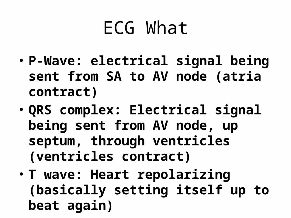
ECG What
• P-Wave: electrical signal being sent from SA to AV node (atria contract)
• QRS complex: Electrical signal being sent from AV node, up septum, through ventricles (ventricles contract)
• T wave: Heart repolarizing (basically setting itself up to beat again)

ECG -- Why1. Check the heart's electrical activity.2. Find the cause of unexplained chest pain
– Pain could be caused by • a heart attack• inflammation of the sac surrounding the heart ( pericarditis)• angina
3. Find the cause of symptoms of heart disease4. Find out if the walls of the heart chambers are too thick
(hypertrophied)5. Check how well medicines are working and whether they are causing
side effects that affect the heart.6. Check how well mechanical devices that are implanted in the heart,
such as pacemakers, are working to control a normal heartbeat.7. Check the health of the heart when other diseases or conditions are
present, such as high blood pressure, high cholesterol, cigarette smoking, diabetes, or a family history of early heart disease.
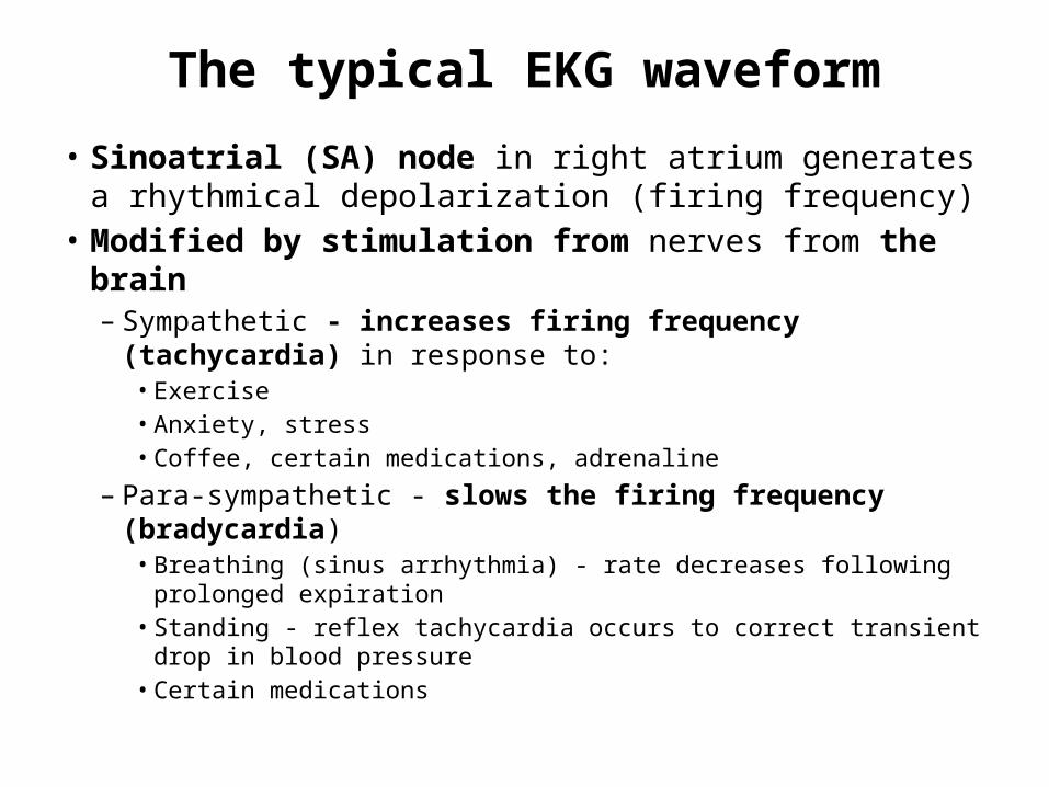
The typical EKG waveform
• Sinoatrial (SA) node in right atrium generates a rhythmical depolarization (firing frequency)
• Modified by stimulation from nerves from the brain– Sympathetic - increases firing frequency (tachycardia) in
response to:• Exercise• Anxiety, stress• Coffee, certain medications, adrenaline
– Para-sympathetic - slows the firing frequency (bradycardia)• Breathing (sinus arrhythmia) - rate decreases following prolonged
expiration • Standing - reflex tachycardia occurs to correct transient drop in
blood pressure• Certain medications
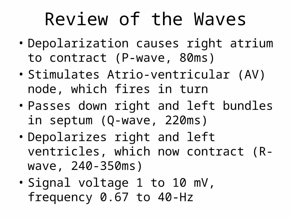
Review of the Waves• Depolarization causes right atrium to contract
(P-wave, 80ms)• Stimulates Atrio-ventricular (AV) node, which
fires in turn• Passes down right and left bundles in septum
(Q-wave, 220ms)• Depolarizes right and left ventricles, which
now contract (R-wave, 240-350ms)• Signal voltage 1 to 10 mV, frequency 0.67 to
40-Hz

ECG Leads• These are what detect the electrical signal of the heart• 6 leads are called Standards Leads and are placed as
follows:
• The additional 6 leads are called Augmented Leads and are placed in different areas according to what is going on with the heart. They make be placed on the arms and legs, or chest.
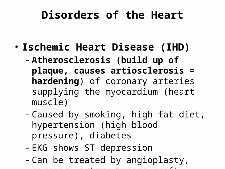
Disorders of the Heart
• Ischemic Heart Disease (IHD) – Atherosclerosis (build up of plaque, causes
artiosclerosis = hardening) of coronary arteries supplying the myocardium (heart muscle)
– Caused by smoking, high fat diet, hypertension (high blood pressure), diabetes
– EKG shows ST depression– Can be treated by angioplasty, coronary artery
bypass graft (CABG)


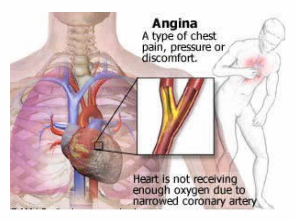
Disorders of the Heart
• · Angina – Reduced blood supply to the heart– Spasm of sclerotic coronary artery– Causes chest pain reversed by nitroglycerine (nitro
spray under tongue)– Unstable (recurring) angina indicates impending
infarction (EWS heart attack)

Disorders of the Heart
• Myocardial Infarction (MI, heart attack) – Thrombosis of coronary artery– Death of heart tissue– Causes chest pain which is not reversed by nitro– Diagnose by EKG (ST elevation, T-wave inversion)– Complications: arrhythmia, cardiac failure– Treatment: morphine, aspirin, monitor for
complications, thrombolysis


Disorders of the Heart
• · Arrhythmia – Irregular heart rhythm, e.g. atrial fibrillation (AF),
heart block, ventricular fibrillation (VF), supraventricular tachycardia (SVT)
– Treatment• digoxin for AF• atropine, pacemaker for bradycardias• VF (may cause sudden death) - treat quickly with
electroshock (defibrillator) and chest compressions


Normal ECG
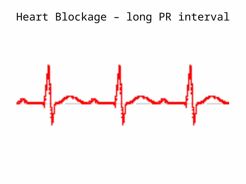
Heart Blockage – long PR interval
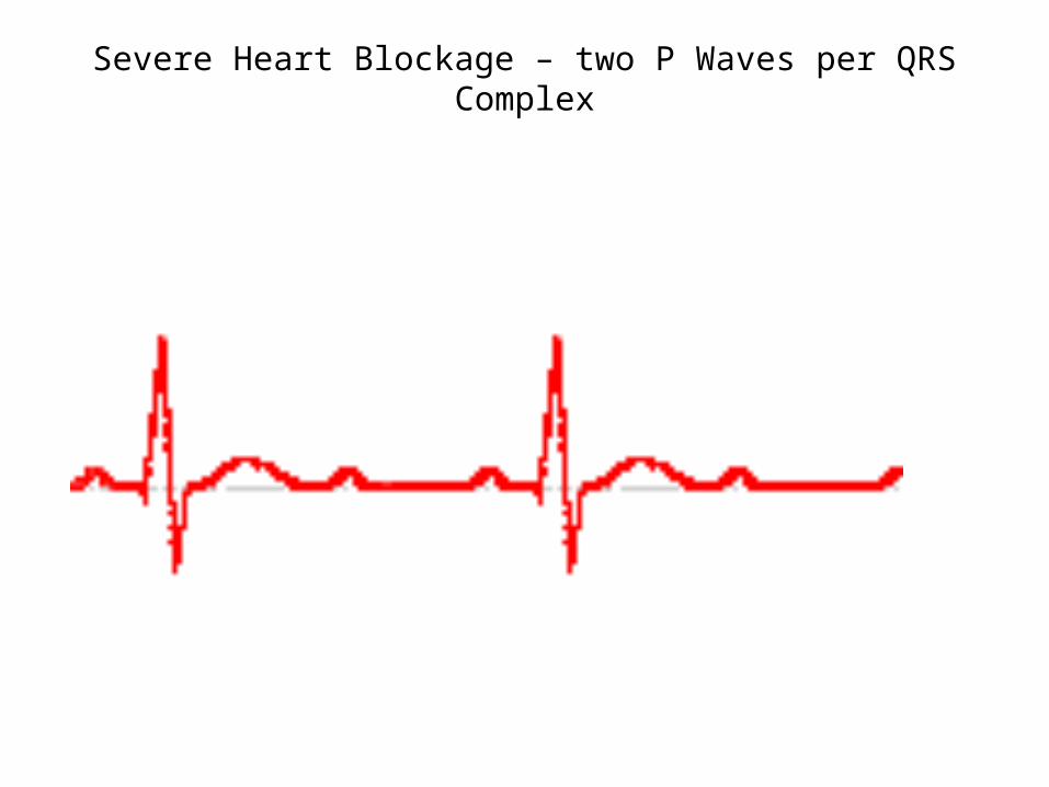
Severe Heart Blockage – two P Waves per QRS Complex

Asystole – flat line
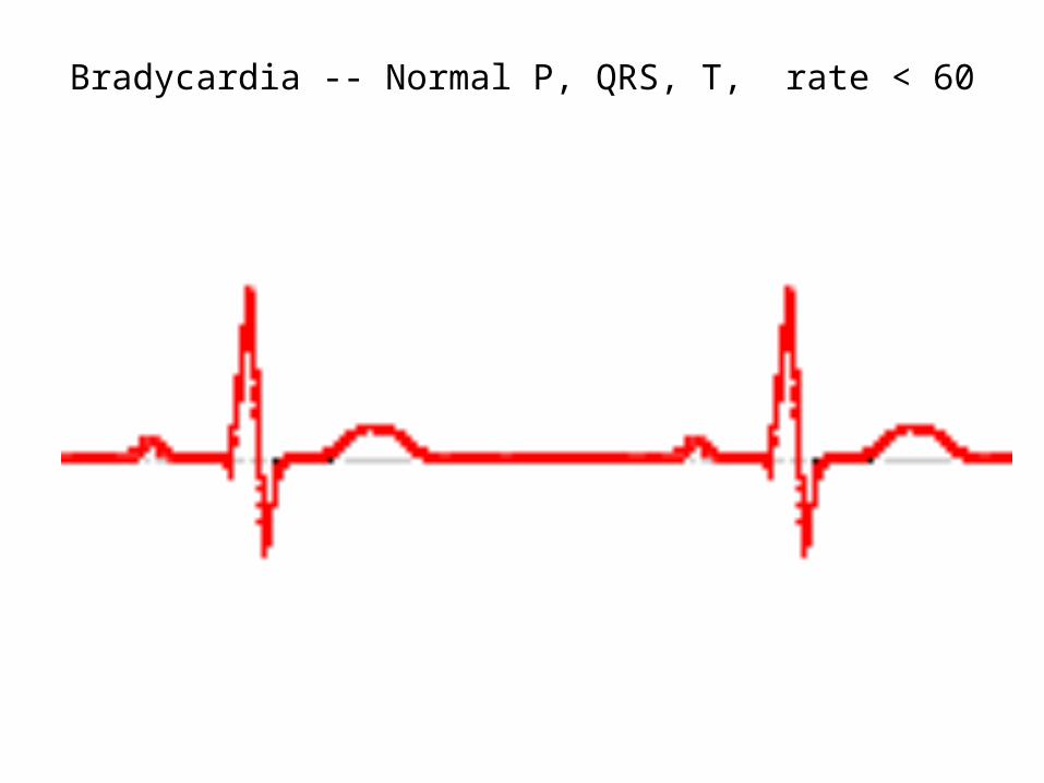
Bradycardia -- Normal P, QRS, T, rate < 60

Hypercalcemia -- Short/absent ST segment
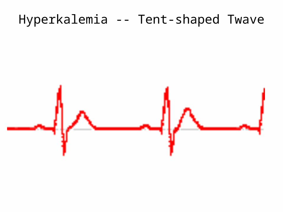
Hyperkalemia -- Tent-shaped Twave
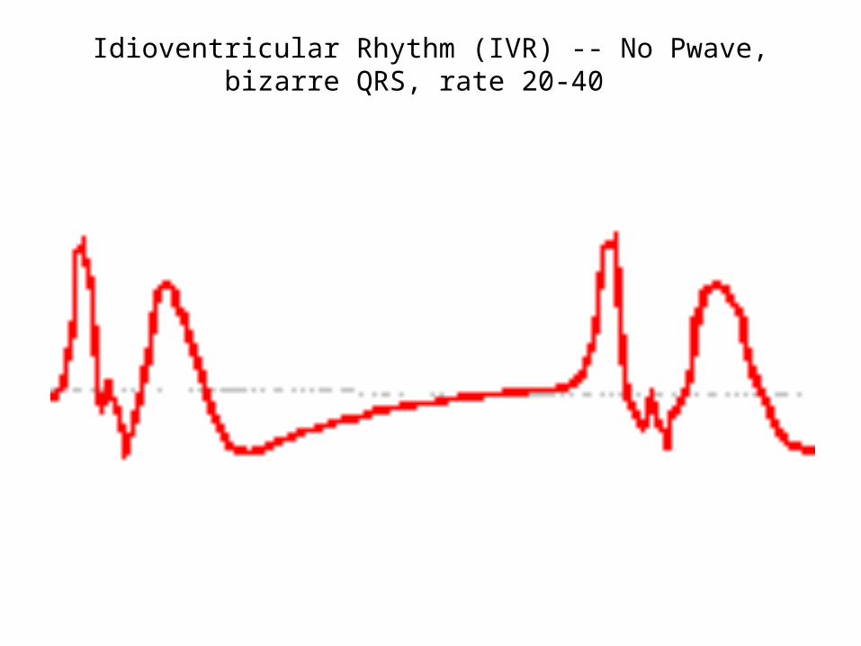
Idioventricular Rhythm (IVR) -- No Pwave, bizarre QRS, rate 20-40
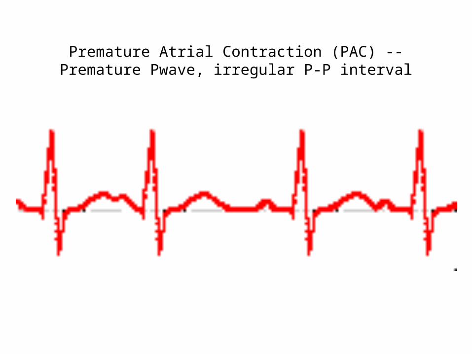
Premature Atrial Contraction (PAC) -- Premature Pwave, irregular P-P interval

Premature Ventricular Contraction (VAC) -- Wide QRS, unrelated
to Pwave
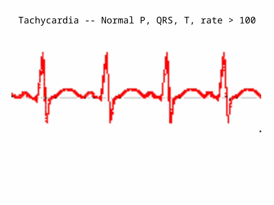
Tachycardia -- Normal P, QRS, T, rate > 100

Ventricular Fibrillation (VFIB) -- Chaotic waves
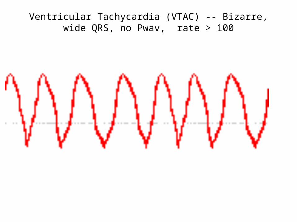
Ventricular Tachycardia (VTAC) -- Bizarre, wide QRS, no Pwav, rate > 100

Now• Choose 10 of the heart irregularities• On white paper: draw the ECGs for each of these 10
heart problems – be sure to leave a blank between each• On notebook paper: write an answer key – include the
following:– Label the waves– Name the disorder– Tell EXACTLY what the ECGs means
• (Example: no P-wave means that there is no electrical signal being sent from the SA to the AV node)
• You will be taking a random person’s quiz tomorrow. Then you will get back the one you wrote to grade according to your answer key


![ECG(ELECTROCARDIOGRAM) [Autosaved] new1.ppt](https://static.fdocuments.in/doc/165x107/577cdafc1a28ab9e78a70e87/ecgelectrocardiogram-autosaved-new1ppt.jpg)


