EBV T cells for EBV-associated Lymphoma Xxx Successfully Generating Multivirus-Specific T cells...
49
Patrick J. Hanley, PhD Director, GMP for Immunotherapy Children’s National Health System The George Washington University School of Medicine and Health Sciences EBV T cells for EBV-associated Lymphoma
Transcript of EBV T cells for EBV-associated Lymphoma Xxx Successfully Generating Multivirus-Specific T cells...

Patrick J. Hanley, PhD Director, GMP for Immunotherapy
Children’s National Health System The George Washington University
School of Medicine and Health Sciences
EBV T cells for EBV-associated Lymphoma

Nothing to disclose
DISCLOSURE

Types of EBV Latency
Type 3 Type 2 Type 1
EBV lymphoma post transplant Lymphoblastoid cell lines
Hodgkin’s Lymphoma Nasopharyngeal carcinoma
Burkitt’s lymphoma
EBNA-1 EBNA-1
LMP 1
LMP 2
EBNA-1
LMP 1
LMP 2
EBNA-3a
EBNA-3b
EBNA-3c
EBNA-2
LP

• Incidence 1-25% following mismatched or unrelated donor BMT
• Predisposing factors:
EBNA-1
EBNA-2
EBNA 3a,3b,3c
LP
LMP2 LMP1
BARF0 E T T
T T
E T T T
T E E E E
E E E
E
EBV Lymphoma After BMT
EBV-Specific CTL Generation EBV-specific CTL Generation
Step 2: T-cell expansion 4-7 weeks
IL-2
PBMC
Step 1: LCL generation 4-6 weeks
EBV
EBV-infected B cells (LCL)
PBMC
Step 3: QA/QC Sterility HLA type Phenotype Cytotoxicity EBV-specific
T cells
EBV-specific T cell Generation
Retroviral vector-neo gene

• Prophylaxis:
• 110 high risk patients
• None developed EBV
lymphoma
• Therapy
• 14 patients treated with active
disease
• 12 attained CR
• No recurrences St Jude Children’s Research Hospital
Baylor College of Medicine Great Ormond St
Marked T cells by in situ PCR at tumor site
Leen et al, Nat Med 2006 and Heslop et al, Blood 2010
Donor-derived EBV-specific T cells for Stem Cell Transplant Recipients

Hematopoietic Stem Cell Transplantation and Virus Infection
• High incidence of viral infections (not just EBV) post-transplant
–Cytomegalovirus (CMV), Epstein-Barr virus (EBV), adenovirus (Ad)
–Highest incidence when donor seronegative (i.e. cord blood)

EBV-Lymphoblastoid cell lines (LCL)
EBNA-1
EBNA-2 EBNA 3a,3b,3c LP
LMP2 LMP1
p 6 5 p
Ad5f35-pp65
Expanding T cells targeting CMV, EBV and adenovirus (Multivirus-specific CTL)

PBMC Multivirus-
specific T cells
(VSTs)
Ad5f35CMVpp65
vector
EBV LCL
IL-2 IL-2
Antigen Stimulation
Generating Multi-Virus-Specific T cells (VSTs) From Peripheral Blood (PB) of Seropositive Donors
Leen et al, Nat Med 2006

The New: Generating Multi-Virus-Specific T cells (VSTs) From Seropositive Donors
V
Dr. Mike Keller, CNMC EXTENDING TO HIV (Lam et al, Mol Ther 2014)
Peptide mixtures CMV IE1 CMVpp65 EBV EBNA1 EBV LMP2 Ad hexon Ad Penton
Gas exchange Gas exchange
GRex
Vera, JIT 2009 Papadopoulou, STM 2014
T cells

Multivirus specificity of VSTs after 10 days
+SEB +Actin
+pp65/IE1 +EBNA1/LMP2 +Hexon/Penton
INF-
ga
mm
a
INF-
ga
mm
a
INF-
ga
mm
a
INF-
ga
mm
a
INF-
ga
mm
a

CBMC Multivirus-specific T cells (VSTs)
Ad5f35CMVpp65 vector
EBV LCL
IL-2 IL-2
Antigen Stimulation
Generating Multivirus-Specific T cells (VSTs) From Cord Blood (CB) Using Same Methodology

Ad5f35CMVpp65 vector
EBV LCL
Antigen Stimulation
IL-7 IL-12 IL-15
IL-15
Virus-specific CB-derived T cells CBMC
CB DCs
Xxx Xxx
Successfully Generating Multivirus-Specific T cells (VSTs) From Cord Blood
Hanley et al, Blood 2009 and Hanley et al, J. Vis. Exp, 2012, Hanley et al, SciTM 2015

Eligibility Criteria
• Prophylaxis and Treatment
• Day +30 post HSCT or CBT
• GvHD <grade III at enrollment
• In addition for Cord Blood:
– 2.5x10⁷ TNC/kg
– Fractionated cord blood unit

Multivirus specific T-cells (VSTs) from Peripheral and Cord Blood
• Peripheral Blood VST Study: • 1x10⁷/m2
• 5x10⁷/m2
• 1x10⁸/m2 • Cord Blood VST Study:
• 5x10⁶/m2
• 1x10⁷/m2
• 1.5x10⁷/m2
• 2.5x10⁷/m2
38 generated from donor peripheral blood (8 with rapid manufacture) 10 generated from cord blood

0%
25%
50%
75%
100%
VSTs Derived from PB and CB Express Effector and Central Memory Markers After Expansion
CD4 CD8 CD45RA- CD62L-
CD45RA- CD62L+
CD3- CD56+
CD3- CD19+
Per
cen
tage
of
Live
Cel
ls
Mean CB VSTs
Mean PB VSTs
CB VSTs
PB VSTs

VSTs Recognize Multiple Viruses
• 33/40 CTL were Ad-specific
• 38/40 CTL Lines were EBV-specific
• 35/40 CTL Lines were CMV-specific
• 8/8 rapid manufacture VSTs – all tri-virus specific
T-cell Line
Adeno EBV CMV Control
Peripheral Blood VST
86 (9-542)
183 (0-351)
648 (28-1278)
14.5 (3-65)
Cord Blood VST
83 (1-504)
117 (4-339)
36 (1-110)
8 (1-28)
IFN-γ ELISPOT: Spots per 1x10⁵ cells

Patient Characteristics
• Most patients received transplant for malignant disease
• Haploidentical donors included (n =14)
• Cord Blood donors (n= 10)
• 10 patients had Adv infection
• 19 patients had CMV infection/reactivation
• 11 patients had EBV reactivation
Group Median Age (range)
Alternative Donors
Campath or ATG in vivo
Median day VST infused
Off Immune Suppression
Peripheral blood VST
10 years (1-62)
84% 90% +84 (35-164)
63%
Cord Blood VST
1.5 years (0.6-5)
100% 0 92 (84-146)
0

Multivirus T cells Protect Against EBV
• 11/58 patients had EBV reactivation
• 11/11 patients had decrease in EBV viral load with coinciding elevation in EBV-specific T cells in PB
• No additional antiviral therapy required
0
50
100
150
200
250
300
350
Pre CTL wk1 wk2 wk4 wk5 wk6 wk8
SF
C p
er
2x1
05
ce
lls
0
10,000
20,000
30,000
40,000
50,000
60,000
70,000
EB
V c
op
ies/u
g D
NA
EBV DNA
EBV T cell
Peripheral blood T cells
0
5
10
15
20
0
500
1000
1500
2000
2500
1 2 3
Sp
ots
pe
r 1
00
,00
0 c
ell
s
EB
V C
op
ies/u
g D
NA
Months Post VST Infusion
EBV LoadEBV-T cells
Cord blood T cells

Rapidly Manufactured Multi-Virus T cell Protect and Treat EBV Lymphoma
Courtesy of Dr. Mike Keller
0
50,000
100,000
150,000
200,000
250,000
0
50
100
150
200
250
day -16 Day -13 day -10 Day -3 Day 0 Day 4 Day 7 Day 14 Day 21
DN
A C
op
ies/
mL
Blo
od
Spo
ts p
er
20
0,0
00
ce
lls
EBV T cells
EBV Viral Load
Multivirus T cells (Targeting LMP2 and EBNA1)
Rituximab

Duration of T cell Persistence Depends on Viral Reactivation
Pre Infusion Month 1 Month 6
% PBMC TCRs
No Reactivation (P3275)
CMV, adeno Reactivation (P2891)
10
0
10 10
10 10 10
0 0
0 0 0
% PBMC TCRs Clones in the patient
Clo
ne
s in
th
e p
rod
uct
Patient 2891 – CMV and Adenovirus Reactivation
Patient P3275 – No Viral Reactivation

Infused VST Clones Persist and Expand in vivo %
of
all T
CR
s
Pre Month 1 Month 3 Month 6 Month 12
4
8
12
16 CASSIKGNNNSP

Minimal Related Toxicity – No GVHD
Study Donor/Recipient Matching
# of Patients Acute GvHD
Peripheral Blood VST
Haplo 14 None
MUD 6 1 Grade I
MRD 10 1 Grade I
Cord Blood VST CBT: 5/6 6 None
CBT: 6/6 4 None
Melenhorst et al, Blood 2010

Donor-derived Multivirus specific T cells After HSCT Improve Outcomes without Toxicity
Leen, Myers et al- Nature Medicine. 2006 Hanley et al – Science Translational Medicine. 2015
Diagnosis of EBV Lymphoma 2 months after
T-cells – CR
Overall response rate = 93% without pharmacotherapy (100% for EBV)

Summary: CB and PB Multivirus-specific T cells are Protective and Efficacious in vivo
• We can now expand multi virus (including EBV)-specific T cells from TWO donor sources: cord blood and peripheral blood
• Safe to infuse to patients (minimal toxicity)
• Persistence of virus-specific T cells in presence of antigen
• Regardless of source of virus-specific T cells (naïve/memory), both populations appear protective

Targeting EBV – Beyond PTLD

Rationale of Immunotherapy for Lymphoma ….Beyond PTLD
• Use of EBV-specific T cells post HSCT/CBT is successful (Rooney and Heslop, Blood 2010 / Doubrovina and O’Reilly, Blood 2012, Leen Nat Med 2006, Hanley Sci. Transl. Med 2015 )
• Significant failure rate of therapy for advanced stage or recurrent disease
• Long-term side effects of chemotherapy and radiation
• EBV antigens expressed by 20-40% of lymphomas are potential targets for T cell immunotherapy

Types of EBV Latency
Type 3 Type 2 Type 1
EBV lymphoma post transplant Lymphoblastoid cell lines
Hodgkin’s Lymphoma Nasopharyngeal carcinoma
Burkitt’s lymphoma
EBNA-1 EBNA-1
LMP 1
LMP 2
EBNA-1
LMP 1
LMP 2
EBNA-3a
EBNA-3b
EBNA-3c
EBNA-2
LP

EBV Specific Cytotoxic T Lymphocytes (CTL) Control EBV Infection In Vivo
EBNA-1
LMP 1
LMP 2
EBV-infected B cells
PBMC
CTL
Lytic EBNA 3a, b, c LMP2 LP LMP1 EBNA 2 EBNA 1
Inhibitory Factors
EBV+ Lymphoma Tumor Cell

EBV-Specific CTL Generation EBV-specific CTL Generation
Step 2: T-cell expansion 4-7 weeks
IL-2
PBMC
Step 1: LCL generation 4-6 weeks
EBV
EBV-infected B cells (LCL)
PBMC
Step 3: QA/QC Sterility HLA type Phenotype Cytotoxicity EBV-specific
T cells
LMP2-CTL
EBV specific T cell Generation

Gene Marked T-cells persisted for 12 months max
EBV-T cells showed small populations of T cells reactive against LMP2
Some expansion of LMP2-specific T cells in PB post infusion.
Anti-tumor effects seen (20% CR/PR)
Marked EBV-T cells by in situ PCR at tumor site
Bollard et al, J Exp Med 2004 Straathof et al, J Immunol 2005
Administration of EBV Specific T-cells to Patients with EBV+ve Hodgkin Lymphoma

• LMP1 and LMP2A are potential T cell targets
Hodgkin R-S Cell/NHL Cell
LMP1 and LMP2A-specific T cells For Hodgkin and non-Hodgkin Lymphoma
EBNA1
LMP1
LMP2A

Making LMP1 and LMP2 Immunodominant Antigens
Bollard et al, JIT 2004 Straathof et al, J Immunol 2005
PBMC LMP-specific T cells
EBV-infected B cells
moDC
GMCSF IL4
IL1b IL6
TNFa PGE2
adherent PBMC
rAd5f35dLMP1-I-LMP2 or Ad5f35LMP2
IL15 IL2 IL2
GMCSF IL4 IL1b IL6 TNFa PGE2
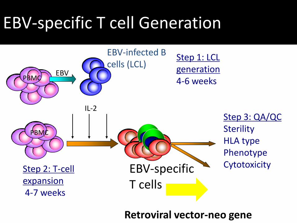
LMP1 & LMP2 Specific Activity in LMP-specific T cells from Patients with EBV+ Lymphoma
0
500
1000
1500
2000
SFC
per
10
5 c
ells
0
300
600
900
1200
SFC
per
10
5 c
ells
LMP2-specific T cells LMP1/2-specific T cells
LMP1 pepmix
LMP2 pepmix
LMP1 pepmix
LMP2 pepmix

Eligibility
• Any age
• EBV+ type III or type II latency lymphoma (EBER and/or LMP1 pos)
• HIV negative
• Either with relapsed disease OR high risk for relapse (e.g. multiply-relapsed patient post chemotherapy or autologous BMT)

CTL Product, Dose, and Administration
• 25 HL and 25 NHL
• Age range 8-79 years
• Autologous LMP-T-cell product
• Dose escalation: 4x10⁷/m2 to 3x10⁸/m2
• Patient received 2 doses (given 2 weeks apart). If stable disease or PR then could receive an additional 6 doses

Clinical Responses Post LMP-specific T cells
• No toxicity
• 11 CR
• 2 PR
• 8 progressive disease
(2-8 weeks)
• Median clinical response: 1.5 years
(range: >6 to >40 months) CR PR no response
Patients with Disease at CTL Infusion (n=21)
Bollard et al, JCO 2014

Clinical Responses Post LMP T cells in Patients with Active Disease 50% Disease Free Survival at 2 Years (n=21)
Bollard et al, JCO 2014 Year
Pro
po
rtio
n d
ise
ase
-fre
e
0 1 2 3
0
0.2
0.4
0.6
0.8
1
AlascerALCI
P=0.882
LMP2 T cell protocol LMP1/2 T cell protocol
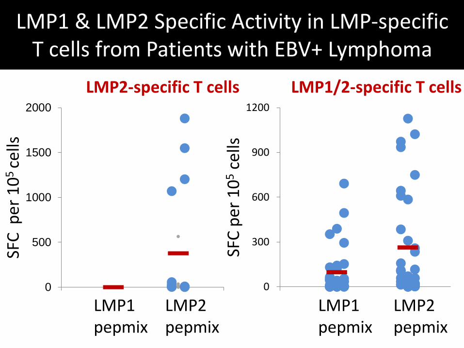
Immune Reconstitution of LMP1 & LMP2- T cells in Patients Treated with Disease
RESPONDERS NON RESPONDERS
LMP1 T Cells LMP1 T Cells
LMP2 T Cells LMP2 T Cells

0
15
30
45
60
SFC
pe
r 2
.5x1
05
ce
lls
Survivin
0
15
30
45
60
SFC
pe
r 2
.5x1
05 c
ells
Survivin
Evidence of Epitope Spreading in Responding Patients Treated with LMP1/2 T cells
LMP2
CD8
0
30
60
90
120
MAGE A4
0
15
30
45
60 PRAME
0
30
60
90
120
MAGEA4
0
15
30
45
60 PRAME
RESPONDERS NON- RESPONDERS
PRE POST PRE POST
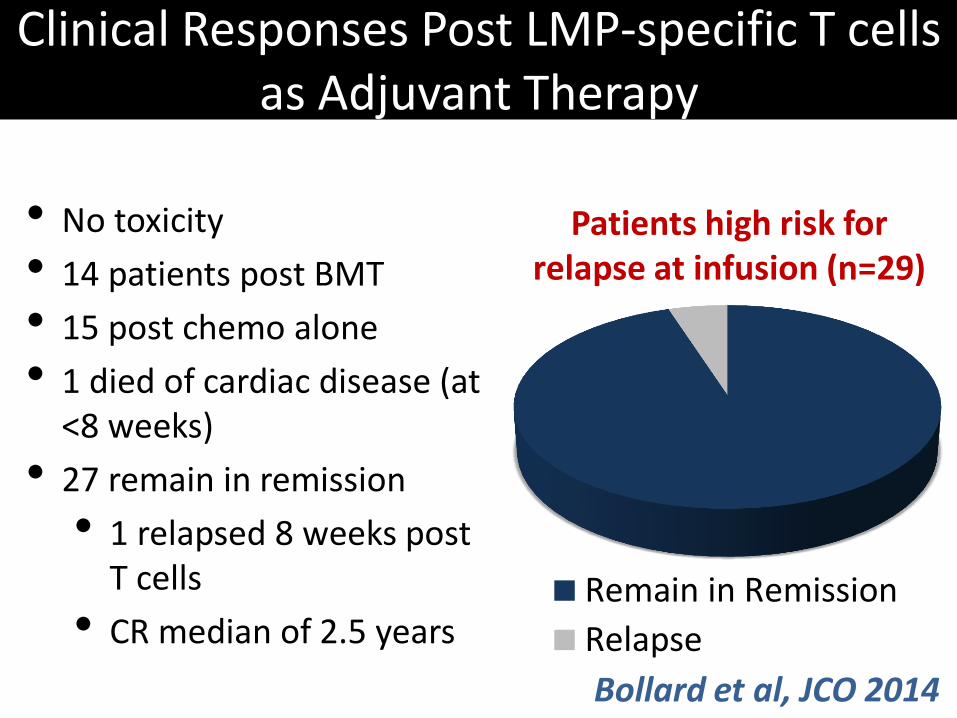
Clinical Responses Post LMP-specific T cells as Adjuvant Therapy
• No toxicity
• 14 patients post BMT
• 15 post chemo alone
• 1 died of cardiac disease (at <8 weeks)
• 27 remain in remission
• 1 relapsed 8 weeks post T cells
• CR median of 2.5 years Remain in Remission
Relapse
Patients high risk for relapse at infusion (n=29)
Bollard et al, JCO 2014
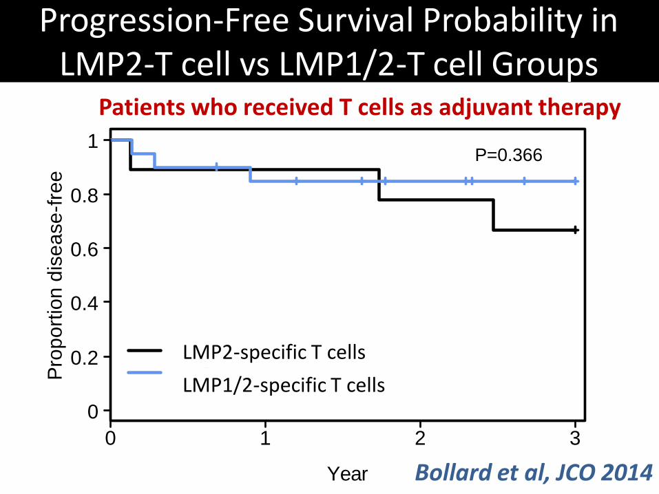
Progression-Free Survival Probability in LMP2-T cell vs LMP1/2-T cell Groups
Patients who received T cells as adjuvant therapy
Bollard et al, JCO 2014 Year
Pro
po
rtio
n d
ise
ase
-fre
e
0 1 2 3
0
0.2
0.4
0.6
0.8
1
Alascer
ALCI
P=0.366
LMP2-specific T cells
LMP1/2-specific T cells

Cause of Death Specific Probability: All Subjects
Patients who received LMP-T cells as
adjuvant therapy
Patients who received LMP-T cells as
treatment XXXX
Cu
mu
lative
In
cid
en
ce
Pro
ba
bili
ty
0 1 2 3 0
0.2
0.4
0.6
0.8
1 Lymphoma
Other
Year
Cu
mu
lative
In
cid
en
ce
Pro
ba
bili
ty
0 1 2 3 0
0.2
0.4
0.6
0.8
1
Year
Year
Cum
ula
tive I
ncid
ence P
robability
Lymphoma
Other
0 1 2 3
0
0.2
0.4
0.6
0.8
1Lymphoma
Other

Deaths from Other Causes
• In adjuvant group, 8/29 patients died
• 1 relapsed, died in CR after allo SCT
• 3 second cancers (2 MDS, 1 sarcoma)
• 3 infection
• 1 cardiac disease
Confirms need for targeted therapies

Conclusions – LMP1/2 Data
• No toxicity
• Accumulation of LMP-specific T cells at disease sites
• Anti-tumor effects seen (13/21 patients PR/CR)
• Next….how to broaden applicability

How Do We Extend Applicability?
• Use bank of allogeneic partially matched CTLs
• Simplify production patient specific product

• Pediatric Lymphoma Cell Therapy Consortium (Funded by St Baldricks Foundation, 7 Centers)
Donor-derived LMP-CTL post allo SCT for HL
Third party LMP-CTL for EBV+ lymphomas
• Multicenter study through Children’s Oncology Group for PTLD post SOT (ANHL1551)
• Industry support (Cellmedica) NK/T cell NHL
LMP-CTL – Moving Beyond Single Center Studies

Proof of Principle Studies
• Learn from the bedside back to the bench
• With an optimal approach the goals are:
- Broaden applicability beyond a few centers
- Multicenter studies are planned
EBV Specific T cell Therapies Broadening Applicability
CAGT Laboratory
A Leen, U Gerdeman B Savoldo, G Dotti S Gottschalk, Carlos Ramos A Gee, B Grilley Malcolm Brenner Cliona Rooney Helen Heslop MDACC EJ Shpall Nina Shah Katy Rezvani NIH John Barrett Jos Melenhorst Barts, London John Gribben
Cath Bollard’s Lab (CAGT-BCM CETI-CNMC) Cath Bollard, Stephanie Ku, Russell Cruz, Sharon Lam, Gerrit Weber, Paul CastilloCaro, Yasmin Hazrat, An Lu, JW Blaney, Francesco Saglio, D Jacobsohn, K Williams, A Abraham, Cecilia Baresce, Kaylor Wright, Fahmida Hoq, M Luo Mike Keller, Renuka Miller, Maria Manso-Martin
T-cell Therapies for EBV+ Lymphomas