EAWAG news 56elibrary.eawag.ch/eawag-publications/EAWAGnews/56E(2003).pdf · examining life...
28
news EAWAG 56e November 2003 Swiss Federal Institute for Environmental Science and Technology (EAWAG), A Research Institute of the ETH-Domain • CH-8600 Dübendorf EAWAG Molecules in Action induced unaltered suppressed Gpxh Defense Genes as Indicators of Toxicity 15 Bacterial Biosensors to Measure Arsenic in Potable Water 12 New Paths in the Analysis of Drinking Water Quality 18 Characterization of Reactive Chemicals Based on their Primary Mode of Action 9
Transcript of EAWAG news 56elibrary.eawag.ch/eawag-publications/EAWAGnews/56E(2003).pdf · examining life...

newsEA
WA
G
56e November 2003Swiss Federal Institute for Environmental Science and Technology (EAWAG), A Research Institute of the ETH-Domain • CH-8600 Dübendorf
EAWAG
Molecules in Action
induced unaltered suppressed
Gpxh
Defense Genes as
Indicators of Toxicity 15
Bacterial Biosensors to Measure
Arsenic in Potable Water 12
New Paths in the Analysis of
Drinking Water Quality 18
Characterization of Reactive Chemicals
Based on their Primary Mode of Action 9

EAWAG news 56 2
EAWAG news 56e • Nov. 2003Information Bulletin of the EAWAG
Publisher Distribution and © by: EAWAG, P.O. Box 611, CH-8600 DübendorfPhone +41-1-823 55 11Fax +41-1-823 53 75http://www.eawag.ch
Editor Martina Bauchrowitz, EAWAG
Translations Norbert Swoboda, USA
Linguistic revision Patricia Colberg, USA; Helen Brügger-Clarke, Zurich
Figures Yvonne Lehnhard, EAWAG; Peter Nadler,Küsnacht
Copyright Reproduction possible on request. Please contact the editor.
Publication Three times yearly in English, German andFrench. Chinese edition in cooperation with INFOTERRAChina National Focal Point.
Cover Photos EAWAG
Design inform, 8005 Zurich
Layout Peter Nadler, 8700 Küsnacht
Printed on original recycled paper
Subscriptions and changes of address New sub-scribers are welcome! Please find the order form in themiddle of this issue.
ISSN 1440-5289
EAWAG
From ecosystem viamolecule to ecosystem
Molecular biology nowadays plays a vital
role in many fields of scientific research. In
medicine, for example, pathogenic proc-
esses are studied on a molecular level. Sub-
sequent to the identification of the under-
lying mechanisms, it becomes possible to
develop specific drugs. These are either
applied as preventive medicine, such as
vaccination, or – once the disease has
broken out – are used as a cure, acting
specifically and with as few side effects
as possible. Less known, however, is the
fact that molecular approaches are also
becoming more and more important in envi-
ronmental research, too.
The EAWAG is committed to the sustainable
use of aquatic ecosystems – rivers, lakes
and groundwater. Aquatic ecosystems are
very complex and offer habitats for a diver-
sity of living creatures, from single cellular
bacteria or algae to multicellular higher
plants and animals. The organisms live in
permanent interaction with each other and
with their environment, which in itself is very
dynamic and subject to continuous change.
Just think of natural changes such as daily
and seasonal fluctuations as an example.
Adding to these natural changes are anthro-
pogenic impacts which are continuously
increasing due to the steadily growing world
population. Problems such as the input of
pollutants into aquatic ecosystems, the
growing pressure on freshwater resources
and the increase of pathogens in surface
waters of developing countries can no
longer be neglected. There is a need for
concepts and approaches which allow the
protection of the complex aquatic environ-
ment for the future by preventive methods
and make it equally possible to deal with
acute problems directly and “without side
effects”. Therefore, the EAWAG is attempt-
ing to analyze processes in ecosystems on
a molecular level, in order to better under-
stand, predict and prevent the effects of
anthropogenic impacts. In this work, we are
well aware that important insights for the
ecosystem can only be obtained if we do
not lose the view of the whole picture of the
ecosystem.
To study processes on molecular level, it is
essential to perform basic molecular re-
search. The EAWAG is studying such differ-
ent aspects as the genetic diversity of
Daphnia in alpine lakes and the mecha-
nisms of action of pollutants on a molecular
level. Furthermore, applied science plays an
important role at the EAWAG. Here as well,
molecular approaches and methods are
used more and more. Examples from this
field are the development of biosensors for
the detection of pollutants, the identification
of a bacterium now being used for the re-
moval of nitrogen in wastewater treatment
plants and the development of a molecular
method for the detection of pathogens in
drinking water.
I invite you to enter the world of the mole-
cules and hopefully become convinced that
molecular biology offers an essential con-
tribution to the sustainable management of
aquatic ecosystems.
Rik Eggen, head of thedepartment “EnvironmentalMicrobiology and MolecularEcotoxicology”
Molecules in Action2 Editorial
Lead Article3 Molecular Strategies in the Environment
– 135 Years of Spell-Binding Research
Research Reports6 Genomic Islands and Horizontal Gene
Transfer Among Bacteria
9 Characterization of Reactive Chemicals
Based on their Primary Mode of Action
12 Bacterial Biosensors to Measure
Arsenic in Potable Water
15 Defense Genes as Indicators of Toxicity
18 New Paths in the Analysis of Drinking
Water Quality
20 The Anammox Process for Nitrogen
Removal in Wastewater Treatment
Plants
22 Genetic Diversity of Daphnia in Alpine
Lakes
In Brief26 Publications (3193–3388)
31 Books
32 In Brief

3 EAWAG news 563 EAWAG news 56
Since the middle of the last century, molecular biology is its own
discipline. It has its roots in microbiology, a discipline traditionally
examining life processes and the role of microorganisms in differ-
ent ecosystems. While today’s applications of molecular biology in
biotechnology and in the medical field are widely known, molecu-
lar approaches to problem-oriented environmental research are
still very much in the background. This is not justified since these
techniques are highly useful in the solution of current problems.
As early as the middle of the 19th century,
scientists began to study the transformation
of specific compounds by microorganisms.
The driving force for most of the studies was
the theory of spontaneous generation which
proposed that life could form repeatedly
and spontaneously. As an unintended con-
sequence, these studies yielded the first
insights into the metabolism of microorgan-
isms. The French scientist Béchamp [1], a
contemporary of Louis Pasteur, was as well
a proponent of the theory of spontaneous
generation. He examined a variety of envi-
ronmental samples for their capacity to
transform specific chemicals, describing for
example the formation of methane from
ethanol. According to his interpretation, the
transformation was accomplished by micro-
organisms that had newly formed in his
flasks; he named these organisms Micro-
zyma cretae. It was Pasteur who refuted
spontaneous generation with his ingenious
experiments. He showed that these obser-
vations were, in reality, the growth and
enrichment of microorganisms that were
already present at the beginning of the
experiments.
In the second half of the 19th century, the
German scientist Felix Hoppe-Seyler con-
tinued the molecular strategy of environ-
mental research. As the first Professor for
Physiological Chemistry at the University
of Strasbourg, he coupled the molecular
understanding of biological processes to
energy considerations. He recognized that
every biochemical transformation yields
energy that can be used by microorganisms
for their growth and metabolism. Remark-
able is that the now well-known classic
theory of thermodynamics was just being
developed (Josiah Gibbs did not introduce
the term free-energy until 1878) [2]. The
original approach used by Hoppe-Seyler
was refined in later years.
Radioactive Isotopes as
Markers for Metabolic Products
The next major breakthrough occurred in
the 1930’s and 1940’s when the chemist
Samual Ruben and the physicist Martin
Kamen discovered the radioactive isotopes11C and 14C [3]. Both scientists immediately
recognized the potential that this discovery
had for science. Experiments with 11C
proved to be difficult since this isotope has
a half-life of only 21 minutes. Only when 14C
with a half-life of ~5700 years was used,
did it became possible to follow the inter-
mediates of biochemical transformations
(even in complex systems) and to ascribe
assimilation products to specific organisms
within an ecosystem. With the use of micro-
autoradiography, organisms that catalyze
certain biochemical transformations in an
ecosystem can be made directly visible
(Fig. 1). Leading the way in this methodol-
ogy were Louise and Thomas Brock during
the 1960’s [4].
Detection of Specific
Microorganisms
During the 1970’s and 1980’s, biologists,
particularly microbial ecologists, concen-
trated their research on the development of
methods for direct identification of microor-
ganisms in complex environmental sam-
ples.
Specific compounds: Some compounds are
found only in very specific groups of organ-
isms. The electron transfer coenzyme F420,
for example, is only found in methanogenic
bacteria, with one exception. This com-
pound is particularly interesting because it
fluoresces, which allows simple detection of
the organism (Fig. 2).
Immunological methods: While the detec-
tion of certain bacteria by immunological
methods was already widely used in med-
ical microbiology by the late 1970’s, this
technique was only starting to be employed
in environmental research. Already at that
time, a wide variety of markers was avail-
able for making antibodies bound to spe-
cific target organisms visible. The options
included radioactive labels, enzymes that
catalyze specific reactions (ELISA tech-
nique), specific heavy metals, or fluorescent
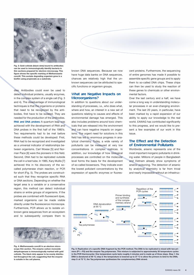
Fig. 1: Autoradiogram of epiphytic bacteria (visible as black colony, lower left) living on marine red algae.The bacteria were fed 14C-glutamate as a carbonsource [from 4].
Fig. 2: F420, an electron transfer coenzyme occurringalmost exclusively in methanogens. UV light causesthis coenzyme to fluoresce. This property allows us todirectly identify methanogens in natural populations,such as Methanobacterium formicicum in this case.
A. Z
ehnd
er, E
AW
AG
Molecular Strategies in theEnvironment
135 Years of Spell-Binding Research
EAWAG news 56 4
dies. Antibodies could even be used to
detect individual proteins, usually enzymes,
in the complex system of a single cell (Fig. 3
and 4). The disadvantage of immunological
techniques is that the organisms or proteins
that need to be recognized by the anti-
bodies, first have to be isolated. They are
needed for the production of the antibodies.
RNA and DNA probes: A quantum leap was
achieved with the development of RNA and
DNA probes in the first half of the 1980’s.
Two requirements had to be met before
these methods could be developed: First,
RNA had to be recognized and investigated
as a universal indicator of relationships be-
tween organisms. Carl Woese [5] and Nor-
man Pace [6] were the pioneers in this field.
Second, DNA had to be replicated outside
the cell in a test tube. In 1985, Kary Mullis [7]
achieved this in his discovery of the so-
called polymerase chain reaction, or PCR
for short (Fig. 5). The probes are construct-
ed such that they recognize specific RNA
or DNA sections. Depending on whether the
target area is a variable or a conservative
region, this method can detect individual
strains or entire groups of organisms. If the
probes are combined with fluorescent dyes,
marked organisms can be made visible
directly under the fluorescence microscope.
Furthermore, PCR allows us to isolate un-
known gene sequences from an ecosystem
and to subsequently compare them to
known DNA sequences. Because we now
have huge data banks on DNA sequences,
chances are relatively high that the un-
known sequences can be attributed to spe-
cific functions or organism groups.
What are Negative Impacts on
Microorganisms?
In addition to questions about our under-
standing of processes, i.e., who does what,
where and how, an interest in a new set of
questions relating to causes and effects of
environmental damage has emerged. This
also includes problems around toxic chem-
icals that are released into the environment
and can have negative impacts on organ-
isms. The urgent need for solutions in this
field has led to enormous progress in ana-
lytical chemistry. Today, a wide variety of
pollutants can be measured at very low
concentrations in complex matrices. In
addition, our knowledge of how biological
processes are controlled on the molecular
level forms the basis for the development
of so-called biosensors. They indicate even
the lowest pollutant concentrations by the
expression of specific enzymes or fluores-
cent proteins. Furthermore, the sequencing
of entire genomes has made it possible to
assemble specific gene groups and to apply
them to so-called DNA chips. These chips
can then be used to study the reaction of
these genes to chemicals or other environ-
mental factors.
Over the last century and a half, we have
come a long way in understanding molecu-
lar processes in an ever-changing environ-
ment. The last 20 years, in particular, have
been marked by a rapid expansion of our
ability to apply our knowledge to the real
world. EAWAG has contributed significantly
to this progress, and we would like to pre-
sent a few examples of our work in this
issue.
The Effect and the Detection
of Environmental Pollutants
Worldwide, arsenic represents one of the
most important inorganic pollutants in drink-
ing water. Millions of people in Bangladesh
and Vietnam already show symptoms of
arsenic poisoning. The detection of arsenic
by analytical chemistry is far from trivial
and nearly impossible without an infrastruc-
Fig. 3: Gold colloids (black dots) bound to antibodiescan be used to immunologically identify bacteria inthin sections prepared for electron microscopy. Thisfigure shows the specific marking of Methanosaetaconcilii. This acetate degrading organism grew in abiofilm using propionate as a substrate.
Fig. 4: Methanosaeta concilii in an electron micro-scope thin section. The enzyme carbon monoxidedehydrogenase is immunologically marked with goldcolloids. The black dots appear to be evenly distribu-ted throughout the cell, suggesting that the enzyme is soluble in the cell plasma.
Pho
togr
aphs
: Wag
enin
gen
Agr
icul
tura
l Uni
vers
ity, N
L
Fig. 5: Replication of a specific DNA fragment by the PCR method. The DNA to be replicated is mixed with two pri-mers (P1 + P2) and the enzyme Taq polymerase. This mixture is subjected to approximately 30 temperature cyclesin the PCR thermocycler. One temperature cycle lasts roughly 4 minutes and is made up of three steps. Step 1: theDNA is denatured at 95 °C; step 2: the temperature is lowered up to 37 °C to allow the primers to bind to the DNA;step 3: at 72 °C, the Taq polymerase synthesizes the complementary DNA strand.
P1
P2
Taq polymerase
DNA denaturationat 95 °C
Primer bindingand synthesisof the comple-mentary strand
Repetition of thePCR cycles

5 EAWAG news 56
ture of analytical laboratories. J.R. van der
Meer and J. Stocker (p. 12) have developed
a bacterial biosensor that is easy to use and
can detect arsenic at the ppb level (one mil-
lionth of a gram per liter). The genetically
engineered bacteria carry a so-called re-
porter gene which is activated in the pres-
ence of arsenic and causes the production
of the corresponding reporter protein. This
enzyme catalyses a reaction that releases
a blue dye. Depending on the arsenic con-
centration, more or less enzyme is pro-
duced, which in turn releases varying
amounts of dye. With a simple paper strip,
even an untrained person can easily test a
well for arsenic contamination.
For many years, ecotoxicology was domi-
nated by a mostly phenomenological ap-
proach. Modern methods of molecular biol-
ogy allow us to detect the expression of
individual genes and, therefore, the pro-
duction of the corresponding proteins in a
very targeted, purposeful manner. When an
organism comes in contact with a pollutant,
various genes are activated, although this is
not externally observable, and the organism
continues its normal biochemical activities.
The products synthesized by the activated
genes aid in the defense against the pol-
lutants inside the cell. In their article on
page 15, B. Fischer and R. Eggen show how
these defense genes can be used to detect
certain pollutants; however, the response
of the cell to an environmental pollutant is
only half the answer. It is equally important
to understand how these pollutants do their
damage, i.e., we need information about
how the pollutants react with biological
structures. B. Escher (p. 9) and her group
have investigated a number of so-called
reactive chemicals for a wide range of toxic
reactions and their primary toxicity mecha-
nism.
Nitrogen Elimination and Clean
Drinking Water
A group of engineers and microbiologists
(see article by C. Fux and colleagues on
p. 20) has examined new ways to remove
nitrogen from waste water. A new method
known as the Anammox process was dis-
covered in the Netherlands and has proved
to be extremely well suited for the treatment
of waste water with high ammonia levels.
In this process, ammonia is transformed
directly to nitrogen using nitrite. A first task
was to identify the microorganism respon-
sible for this transformation. This was ac-
complished using specific gene probes. The
practicality of the method was then tested
in a pilot reactor, where successful opera-
tion cleared the way for the use of the
Anammox process in full-scale reactors in
wastewater treatment plants.
To date, the biological quality of drinking
water is determined by cultivation tech-
niques that are largely based on methods
developed in the 19th century. The main
problem of these cultivation techniques is
the fact that they are rather time-consum-
ing. Results are available after 24 hours at
the earliest, often only after 72 hours. Acute
contamination can, therefore, not be de-
tected quickly. The PCR method has the
potential for significant improvements in
this area. Contamination could be con-
firmed within approximately 4 hours, lead-
ing to much shorter response times. A. Rust
and W. Köster demonstrate in their article
on page 18 how this method can be used in
the monitoring of drinking water quality.
Gene Transfer and
Genetic Diversity
Two other articles deal with evolutionary
aspects. The group of J.R. van der Meer
was able to demonstrate how large sections
of DNA can be exchanged between bac-
teria. These DNA sections are referred to as
“genomic islands” and can account for
more than 10% of the entire genetic ma-
terial. The receptor bacteria can obtain new
capabilities from these transfers, such as
the ability to degrade certain pollutants.
This so-called horizontal gene transfer al-
lows bacteria to accomplish important evo-
lutionary steps with a high success rate and
within very few generations.
M. Winder and P. Spaak (p. 22) examined
the diversity of Daphnia populations in
alpine lakes at varying elevations. The pre-
vailing assumption was that genetic diver-
sity should decrease with increasing eleva-
tion, just as the species diversity decreases
at higher altitudes. Their results, however,
have demonstrated that genetic diversity
does not decrease but remains high even
at high elevations.
“More is Different”
Rapid progress in molecular methods
brings with it the danger of losing the view
for the whole system but trying to explain
the functioning of an entire ecosystem with
a few details. It is important to use molecu-
lar strategies in trying to understand proc-
esses, and to validate this understanding
at the system level. As long ago as 1972,
Philip W. Anderson stated in his article
“More is Different” that the dissection of a
system into individual parts is not enough to
help us understand the functioning of the
entire system [8]. We need both, and
EAWAG is fully aware of this fact.
[1] Béchamp A. (1868): Lettre de M. Béchamp à M. Du-
mas. Annales de Chimie et de Physique 13, 103–111.
[2] Hoppe-Seyler F. (1887): Die Methangährung der
Essigsäure. Hoppe-Seyler’s Zeitschrift der Physiolo-
gischen Chemie 11, 561–568.
[3] Kamen M.D. (1963): Early history of carbon-14.
Science 140, 584–590.
[4] Brock T.D., Brock M.L. (1966): Autoradiography as a
tool in microbial ecology. Nature 209, 734–736.
[5] Woese C.R., Fox G.E. (1977): Phylogenetic structure
of the prokaryotic domain: the primary kingdoms.
Proceedings of the National Academy of Sciences
USA 74, 5088–5090.
[6] Pace N.R., Stahl D.H., Lane D.J., Olson G.J. (1985):
Analyzing of natural populations by RNA sequences.
ASM News 51, 4–12.
[7] Saiki R.K., Gelfand D.H., Stoffel S., Scharf S.J.,
Higuchi R., Horn G.T., Mullis K.B. Ehrlich H.A. (1988):
Primer-directed enzymatic amplification of DNA with
a thermostable DNA-Polymerase. Science 239,
487–491.
[8] Anderson P.W. (1972): More is different. Science 177,
393–396.
Alexander Zehnder, microbiolo-gist, director of EAWAG andprofessor for water protectionand water technology at ETHZurich. His research interestsare environmental microbiologyand the application of microbialprocesses in environmentalbiotechnology. He has recently
also become involved in sustainable development,particularly with respect to water resources.

EAWAG news 56 6
Chromosomes are usually thought to be stable molecules, which
have to be copied carefully for each of the new daughter cells.
Except for a few copying mistakes (“mutations”), not much is
happening to the chromosomal DNA. Or is it? Bacterial chromo-
somes are now known to harbor what is called “genomic islands”,
regions which can cut themselves out of the chromosome, in
some cases travel to other bacterial cells and reinsert into the
recipient’s chromosome. Their function? Very often, they provide
the recipient bacteria with auxiliary capabilities for infecting
eukaryotic hosts or for degrading environmental pollutants.
Almost ten years ago, we started to investi-
gate the process of horizontal gene transfer
in bacteria (see glossary). Our aim was to
estimate how frequent certain types of
genes are transmitted between different
bacteria in the natural environment. As a
model system for our studies, we chose the
bacterium Pseudomonas sp. strain B13,
which was isolated from sewage sludge and
which may use 3-chlorobenzoate as sole
carbon and energy source (Fig. 1). When
this strain was first described in 1974, it
was one of few bacterial strains capable of
degrading chlorinated compounds. This
had attracted considerable attention, since
chlorinated aromatics are often polluting
substances in the environment. Strain B13
became yet more appealing because of
another spectacular feature that we discov-
ered: these bacteria are able to transfer their
genes for the 3-chlorobenzoate metabolism
spontaneously to other bacterial species,
and this even in wastewater treatment re-
actor microcosms [1]. One of the curious
findings, though, was that the rate of hori-
zontal gene transfer seemed to increase in
the presence of 3-chlorobenzoate. At that
time, we interpreted these results such that
3-chlorobenzoate was favoring the growth
of bacteria which had received the genes
for 3-chlorobenzoate degradation. Further-
more, we did not have too much idea on
how these genes were actually distributed
from B13 to other strains.
Genes for 3-Chlorobenzoate
Degradation Combined on a
Genomic Island
Therefore, we looked at the mechanism of
gene transfer in more detail. Roald Ravatn,
who did his PhD thesis on this topic, dis-
covered that the “recipient” bacteria had
actually received a large DNA fragment of
more than 100 000 basepairs from strain
B13. This fragment was called the clc ele-
ment (Fig. 2A) and contains the genes for
3-chlorobenzoate degradation [2]. It had
become inserted into one or two very spe-
cific sites of the recipient chromosome.
Strain B13 itself carries two copies of the
clc element in its chromosome, which didn’t
seem to be lost after transfer to a new bac-
terium (Fig. 2B).
Roald Ravatn also identified the factor re-
sponsible for cutting the clc element out of
the chromosome and for subsequent reinte-
gration. It is an enzyme called “integrase”.
Comparison of the biochemical composi-
tion of the integrase from strain B13 with
other proteins showed that it was related to
integrases from bacterial viruses (bacterio-
phages), which place their genomes into the
chromosomes of the infected cells, and to
integrases from so-called genomic islands
(see glossary) [3]. The gene for the B13 inte-
grase is situated at the right end of the clc
element (Fig. 2A).
Since a few years, the discovery of ge-
nomic islands has accelerated enormously,
mainly because of genome sequencing pro-
jects. Large sequencing laboratories deter-
mined the complete nucleotide sequence of
currently around 100 bacterial genomes.
With the complete nucleotide sequence at
hand, it could be shown that many bacteria
carry genomic islands and even have mul-
tiple different copies. The genomic islands
are characterized by the presence of a gene
for an integrase and a specific site on the
chromosome where they have inserted
(Fig. 2B). Taking together all available infor-
mation, we concluded that the clc element
is a genomic island.
When Do Genomic Islands
Move?
Now that we knew that the genes for the
3-chlorobenzoate degradation lie on a ge-
nomic island, we turned back to our earlier
observation indicating an increased transfer
of the clc element when 3-chlorobenzoate is
present. At this point, Vladimir Sentchilo
started his PhD thesis in 1999 considering
the question of which environmental or
cellular factors regulate the transfer of the
clc element. Because the transfer of the clc
element is always preceded by the activa-
tion of the integrase gene, our assumption
was that we could take the activation of theFig. 1: Specific degradation pathway of the chlorinated aromate 3-chlorobenzoate. The product 3-oxoadipate willbe further degraded in the general cellular metabolism.
3-chlorobenzoate 3-chloropyrocatechol 3-oxoadipate
COOH
Cl
O
O
Cl
OH
OH
Genomic Islands and HorizontalGene Transfer Among Bacteria

7 EAWAG news 56
integrase gene as indicator for the subse-
quent excision and transfer of the clc ele-
ment. Therefore, Vladimir Sentchilo focused
on the gene for the integrase and construct-
ed specific reporter bacteria (similar to the
arsenic biosensor, see p. 12). These report-
er bacteria carried a molecular switch con-
sisting of the integrase gene promoter (see
glossary) coupled to the reporter gene for
the Green Fluorescent Protein (= GFP, see
glossary). Presence of GFP in the cell would
thus signify that the promoter of the inte-
grase gene had been activated and that the
transfer process will subsequently proceed.
ulation. Mainly, however, cells became
fluorescent when they were no longer ac-
tively growing (i.e., starvation conditions).
Strangely enough, though, when cells had
been grown in the presence of 3-chloro-
To our astonishment, we observed that only
very few cells became fluorescent in a
culture of the transgenic strain B13 (Fig. 3),
implying that the transfer mechanism was
only activated in a small subset of the pop-
Fig. 2: The clc element (A) and its hypothetical life on its own (B). After activation of the transfer process, the clc element is cut out of the genome by the integrase and forms a circular molecule (= plasmid) in the bacterial cell. In case this cell comes into contact with another bacterium lacking the clc element, the clc element will be transferred assingle-stranded molecule to the second cell. After replication, the clc element integrates at predetermined insertion sites into the genomes of both cells, a process during whichthe integrase also plays an important role.
Insertion site
A
B
Genes for the degradationof 3-chlorobenzoate
Genes regulating the expressionof the integrase gene
RL
Integrase gen
Conjugation
and transfer
Activtion
of the transferprocess
Genomewith theclc element
Insertion
of the clc element
Replication
of the plasmid
clc element
Donor
Recipient
clc elementas plasmid
Genomewith theclc element
Genomewith theclc element
Glossary
Genomic IslandsUnstable regions on the chromosomes of bacteria, which sometimes transfer themselves fromone bacterium directly into the genome of another one. They increase bacterial fitness and can be divided into several subtypes: e.g., “ecological islands” in environmental bacteria and“pathogenicity islands” in pathogenic bacteria with auxiliary functions in infection, toxin syn-thesis or adhesion [4].
Green Fluorescent Protein or GFPReporter protein; those cells in which GFP is synthesized are fluorescent and can be observedunder the epifluorescence microscope.
Horizontal Gene TransferDNA exchange between bacteria; in contrast to vertical gene transfer signifying the inheritanceof a gene from a progenitor. Bacterial reproduction is usually described as asexual, becausebacteria have no equivalent of the genetic fusion of two different cells that is characteristic ofsexual reproduction in eukaryotes. Nonetheless, bacteria do have the ability to exchange seg-ments of DNA with other bacteria. Because these segments can become fixed in a bacterium’sgenome and confer new traits, gene exchange among bacteria could be considered to be aform of bacterial sex.
PromoterRegulating region of a gene in front of the coding region. Activation of the promoter will lead totranscription of the coding region and to subsequent synthesis of the respective protein.

EAWAG news 56 8
benzoate, the number of fluorescent cells
in starvation conditions was higher than
when other carbon sources were used. This
result confirmed our initial observation and
showed, moreover, that 3-chlorobenzoate
stimulates the transfer of the clc element
at a very early stage, i.e., by activating the
integrase gene expression. However, it is
still unknown why the integrase gene is
activated in some bacteria but not in others.
Vladimir Sentchilo was also able to identify
two proteins which seem to influence the
expression of the integrase gene and may
perhaps interact with cellular or environ-
mental signals. Interestingly, these two pro-
teins are encoded by the genomic island
itself and a database comparison showed
similar proteins in a number of other bacte-
ria. In order to better understand the func-
tion of the genomic island in strain B13 and
its evolutionary relationship to other genom-
ic islands, we are now determining the com-
plete DNA sequence. This is done with the
help of the Institute Pasteur in Paris and the
Genome Center in Bielefeld, Germany. With
this knowledge, we hope to get a better idea
of how the transfer of the B13 element and
other genomic islands is regulated.
Desirable and Undesirable
Implications
If it turns out that certain chemical com-
pounds in the environment, like 3-chloro-
benzoate, really act as a trigger for gene
transfer, this could have profound influence
of the rates of distribution of certain gene
functions among bacterial communities.
From the perspective of degradation of en-
vironmental pollutants, it wouldn’t seem too
problematic if the genes for their degrada-
tion became more widely distributed, since
this would result in a faster degradation of
the pollutant. However, faster distribution
of pathogenicity characteristics providing
other bacteria with auxiliary capabilities for
infecting eukaryotic hosts might not really
be an attractive perspective. It seems that
even the genomes of what we usually con-
sider to be the smallest organisms have
smaller entities, e.g., the genomic islands
with a peculiar life-style of their own.
Fig. 3: The transfer process of the clc element ist activated only in a small number of bacterial cells of a culture of Pseudomonas sp. strain B13. Compare the phase contrast image (left, black on grey) with the same sectionshowing the activated bacteria as bright cells on a black background.
[1] Ravatn R., Zehnder A.J.B., van der Meer J.R. (1998):
Low-frequency horizontal transfer of an element con-
taining the chlorocatechol degradation genes from
Pseudomonas sp. strain B13 to Pseudomonas putida
F1 and to indigenous bacteria in laboratory-scale
activated-sludge microcosms. Applied and Environ-
mental Microbiology 64, 2126–2132.
[2] Ravatn R., Studer S., Springael D., Zehnder A.J.B.,
van der Meer J.R. (1998): Chromosomal integration,
tandem amplification, and deamplification in Pseudo-
monas putida F1 of a 105-kilobase genetic element
containing the chlorocatechol degradative genes from
Pseudomonas sp. strain B13. Journal of Bacteriology
180, 4360–4369.
[3] van der Meer J.R., Ravatn R., Sentchilo V. (2001): The
clc element of Pseudomonas sp. strain B13 and other
mobile degradative elements employing phage-like
integrases. Archives of Microbiology 175, 79–85.
[4] Hacker J., Carniel E. (2001): Ecological fitness, genom-
ic islands and bacterial pathogenicity: A Darwinian
view of the evolution of microbes. EMBO Reports 2,
376–381.
Jan Roelof van der Meer, micro-biologist and head of the group“Molecular Microbiology” in thedepartment “EnvironmentalMicrobiology and MolecularEcotoxikology”. Research areas:Evolution, pollutant degradation,biosensor development andmicrobial ecology.
Coauthors: Vladimir Sentchilo, Muriel Gaillard

9 EAWAG news 56
The ecotoxicological risk of reactive chemi-
cals may be only insufficiently assessed by
classical test methods. The reasons for this
are that reactive chemicals hydrolyze rapid-
ly and that traditional testing methods often
assess only a small part of the broad spec-
trum of effects that reactive chemicals may
cause. Recognizing the mode of action is
crucial, however, particularly for reactive
chemicals, since the reaction mechanism
is one of the major factors determining
the risk potential. For this reason, we are
currently developing a comprehensive bac-
terial test system that will cover a wide
spectrum of reactive chemicals and their
particular modes of action.
Pollutants Damage
Biomolecules
Ultimately, all toxic effects can be traced
back to primary interactions of the pollu-
tants with three groups of biomolecules:
membrane lipids, proteins and genetic ma-
terial (DNA) [1]. Interactions span the entire
range from weak van der Waals forces to
specific interactions, such as the formation
of hydrogen bonds or mutual attraction be-
tween charges, all the way to the formation
of chemical bonds (Fig. 1). Weak interac-
tions typically cause nonspecific, reversible
effects and are only relevant for hydropho-
bic pollutants. Specific interactions are ob-
served, for example, in enzyme inhibition,
where a pollutant competes in the role of
“key fitting” the lock, thus keeping out
the actual substrate. Our special interest,
however, is focused on reactive chemicals
that form covalent – usually irreversible –
bonds with a specific target region of the
affected biomolecule. Among these reactive
chemicals are a large number of com-
pounds with different functional groups
such as the reactive oxygen species (see
article by B. Fischer, p. 15) and the so-called
electrophilic chemicals, which is the focus
of this article.
The Cell Arms Itself against
Electrophilic Compounds
Electrophilic chemicals are molecules that
are electron-poor, due to their electron con-
figuration, and therefore prefer to react with
nucleophilic (= electron rich) groups in pep-
tides, proteins or DNA. Preferred targets
are the thio groups in proteins and peptides
as well as certain oxygen and nitrogen
groups in DNA (Fig. 2). In the extreme case,
proteins can be damaged by electrophilic
compounds to such an extent that they can
no longer perform their functions, while re-
actions between electrophilic compounds
and DNA usually cause instability and muta-
tions of the DNA, leading to cancer at the
worst. Reactions at both targets may lead to
death.
But the cells defend themselves against
such attacks. Glutathione, an intracellular
tripeptide (Fig. 2), intercepts electrophilic
compounds which will subsequently be
transported out of the cell. There are also a
Formation ofcovalent bonds
Nonspecific:v.d. Waals +
H-donor/acceptor
Specific:H-donor/acceptor,ionic interactions,
«steric fit»
Interaction Effect classTarget
Reactive effectse.g., membrane orDNA damage
Nonspecific effectse.g., baseline toxicity
Specific effectse.g., binding to receptors andinhibition of enzymes
Proteins andpeptides
Membranes(lipids)
DNA
Fig. 1: Classification of toxic effects according to the mode of interaction with biomolecules at the target site.
Characterization of ReactiveChemicals Based on their PrimaryMode of Action
Fig. 2: Two examples of primary modes of action ofreactive pollutants. Chemical reactions with proteins(A) or DNA (B) cause toxic effects.
O
NNH2
NHN̈
OH
HOH H
H
H
HO
O
A
2’-DeoxyguanosineGlutathione
Benzylchloride
B
Styreneoxide
OH
HN
O
ONH2
HN
O
OHO
¨SH
CIN
Dead fish washed on the shore “belly up”: who has not seen these
pictures? They illustrate dramatically the fatal effects on aquatic
life that accidents with environmental pollutants can have. How-
ever, our environment is also continuously influenced by chemical
pollutants in low concentrations which unfortunately remain unper-
ceived in most cases. It is important therefore to find out how ex-
actly pollutants react in organisms. Thus, the goal of our research
is to identify and classify the various primary modes of action of
reactive chemicals by means of a bacterial test system.

EAWAG news 56 10
Tab. 1: The 17 pollutants examined in this study and their primary mode of action.
number of repair mechanisms for damage
to the DNA, such as proteins that are able
to recognize and repair errors in the DNA
sequence. However, when pollutants are
present in high concentrations and/or over
long periods of time, defense mechanisms
are overwhelmed and toxic effects begin to
manifest themselves.
Evaluation of Different Mutants
of E. coli and Effect Parameters
Methods showing the activity of the defense
systems are particularly suitable test sys-
tems to identify the primary mode of action
of electrophilic compounds. One has to take
into consideration, however, that the toxi-
city of electrophilic compounds is not only
determined by their chemical reactivity, but
also by their concentration at the intra-
cellular target site. The concentration at the
target site, in turn, depends on how many
electrophilic molecules enter the organism,
how the molecules are distributed within the
organism, and whether the organism is able
to transform the molecule into a nontoxic
form. These processes determine the bio-
availability of the compound in question. In
single-celled organisms, however, one can
assume in case of hydrophilic substances
that the pollutant concentration at the target
site is the same as the extracellular concen-
tration. We therefore have chosen to work
with the bacterium Escherichia coli as our
test organism. An additional advantage of
this organism is that numerous mutants of
E. coli are available.
We evaluated a wide range of E. coli strains
and a total of 17 electrophilic compounds
exhibiting different modes of action (Tab. 1).
We measured the following effect parame-
ters: growth inhibition, intracellular gluta-
thione concentrations as well as the occur-
rence of DNA strand breakage and the
activation of various DNA repair mecha-
nisms [2].
Using Two Pairs of E. coliStrains as Biosensors
Two pairs of E. coli strains have proven to
be especially successful for our test pur-
poses. The strains MJF276 (glutathione+)
and MJF335 (glutathione–) are genetically
identical except for their ability to synthe-
size glutathione. The second pair is also
genetically identical except for the ability to
repair DNA damage: in MV4108 (DNA–),
several genes encoding for DNA repair sys-
tems are mutated, while these genes are
intact in MV1161 (DNA+).
Different concentrations of electrophilic
compounds were added to liquid cultures
of these strain pairs and growth inhibition
Structure Damaged biomolecules
Epoxides
Styrene oxide R = phenyl DNA
2,3-Epoxypropyl benzene R = benzyl DNA
2-(4-Nitrophenyl)-oxirane R = p-nitrophenyl DNA and proteins
1,2-Epoxybutane R = C2H5 DNA
Epichlorohydrin R = CH2Cl DNA and proteins
2-Methyl-2-vinyloxirane DNA and proteins
Reactive organochlorides
Benzyl chloride R = H DNA and proteins
3-Methylbenzyl chloride R = m-CH3 DNA and proteins
4-Nitrobenzyl chloride R = p-NO2 DNA and proteins
2,3-Dichloro-1-propene DNA and proteins
trans-1,4-Dichloro-2-butene DNA and proteins
Compounds with activated double bonds
Acrolein R = H Proteins
Ethyl acrylate R = O-C2H5 Proteins
2-Hydroxyethyl acrylate R = O-C2H4-OH Proteins
Isobutyl acrylate R = HO-sec-C4H9 Proteins
Acrylonitrile Proteins
Acrylamide ProteinsO
H2N
N
O
R
ClCl
ClCl
Cl
R
O
OR

11 EAWAG news 56
Beate Escher, chemist andleader of the work group “Mode-of-action Based Risk Assess-ment of Chemicals” in thedepartment “EnvironmentalMicrobiology and MolecularEcotoxicology”. Lecturer forenvironmental chemistry andecotoxicology at ETH Zurich.
Research interests: Uptake and distribution of pollu-tants by organisms, mechanisms of toxicity, methodsfor hazard and risk assessment.
Coauthors: Angela Harder, Paolo Landini,Christian Niederer, Nicole Tobler
[1] Escher B.I., Hermens J.L.M. (2002): Modes of action
in ecotoxicology: their role in body burdens, species
sensitivity, QSARs, and mixture effects. Environmental
Science & Technology 36, 4201–4217.
[2] Harder A. (2002): Assessment of the risk potential of
reactive chemicals with multiple modes of toxic action.
Dissertation ETH Zurich, 78 p.
Fig. 4: Toxicity data (EC50 values) for pollutants exam-ined in this study. Comparison between EC50 valuesfor E. coli and EC50 values for algae, Daphnia and fish.
A
O
O
HO
100
75
50
25
0
75
50
25
0
-25-2.0 -1.0 0 1.0 -0.5 0 0.5 1.0
C 100
75
50
25
0
log pollutant concentration (mM)
100
75
50
25
0-2.0 -1.5 -0.5 0.5 -2.5
Cl
O
B 100
75
50
25
0
100
75
50
25
0-1.0 -0.5 0 0.5 1.0
-1.0 0 -2.0 -1.5 -1.0 -0.5 0
log pollutant concentration (mM)
-0.5 0 0.5 1.0
2-Hydroxyethylacrylate
Gro
wth
inhi
biti
on (%
)
Benzyl chloride
Styrene oxide
Gro
wth
inhi
biti
on (%
)G
row
th in
hib
ition
(%)
DNA+
DNA–
Glutathione–
Glutathione+
DNA+
DNA–
DNA+
DNA–
Glutathione+
Glutathione+
Glutathione–
Glutathione–
Fig. 3: Growth of the two E. coli strain pairs glutathione+/glutathione– and DNA+/DNA– in the presence of differentpollutant concentrations.A: 2-Hydroxyethyl acrylate as an example of a toxin causing protein damage.B: Styrene oxide as an example of a toxin causing DNA damage. C: Benzyl chloride as an example of a nonspecific reactive chemical, reacting with proteins and DNA.
Algae
Fish
Daphnia
-4
-3
-2
-1
0
1
2
3
log EC50 (mM) E. coli
log
EC
50 (m
M)
aqua
tic o
rgan
ism
s
-2 -1 0 1 2
was measured. Reactive chemicals that
primarily attack at the protein level caused
differences in growth between the gluta-
thione+ and the glutathione– strain, while
the growth of the DNA+ and the DNA–
strains was not affected (Fig. 3A). Six of the
examined compounds, characterized by
activated double bonds, fall into this group
of chemicals (Tab. 1). In contrast, reactive
these compounds obtained in the E. coli
studies are compared to the EC50 values
obtained from experiments on aquatic
organisms. There is a linear correlation
between the different sets of EC50 values
(Fig. 4). The EC50 value is the concentration
of a pollutant where a 50% effect occurs –
in our case growth inhibition for E. coli and
algae as well as lethality for daphnids and
fish.
The study described here is a first step in
assessing reactive chemicals based on their
primary mode of action. The goal of our
further work is to find ways to incorporate
the strain pairs used in this study into eco-
toxicological test batteries and to expand
the concept to other toxicity mechanisms.
Differentiated ecotoxicological risk assess-
ments will only become feasible, however,
when we succeed in construction of the
complete chain of “cause and effect” from
the primary interaction at the molecular
level to the observable effects on the popu-
lation or ecosystem level.
chemicals that primarily target the DNA
clearly cause growth differences in the
DNA+/DNA– strain pair, significantly inhibit-
ing the growth of the DNA– strain (Fig. 3B).
These compounds, which include three
of the examined epoxides (Tab. 1), do not
induce any growth differences between the
glutathione+ and the glutathione– strain. In
addition to these two groups of chemicals,
we identified a third group of compounds,
characterized by non-specific reactivity
since they attack at the protein as well
as at the DNA level (Tab. 1). This group of
compounds causes growth differences in
both strain pairs (Fig. 3C).
Validation and Further Develop-
ment of the Test System
The results from this study are not only
useful in classifying modes of action for
environmental pollutants, but they can also
be used to describe the effects these pol-
lutants have on aquatic organisms. This
becomes evident when EC50 values for

EAWAG news 56 12
Bacterial Biosensors to MeasureArsenic in Potable Water
Worldwide, arsenic is one of the most important inorganic pol-
lutants in drinking water. Particularly alarming is the situation in
Bangladesh where more than one million people are already
suffering from arsenic poisoning. In order to test each one of
the roughly 9 million private drinking water wells, an inexpensive,
reliable and sensitive field method is needed. For this reason,
an EAWAG team has developed a new biosensor for arsenic. The
paper strip test uses genetically modified bacteria that produce
blue coloration even at low arsenic concentrations. EAWAG has
applied for a patent for this sensor.
Inorganic arsenic is a common contaminant
of potable water throughout the world [1–3].
Being usually of geochemical origin, arsen-
ate and arsenite can occur in ground waters
in concentrations up to 1 or 2 mg per liter.
The safety limit for arsenic in drinking water
in most countries is 10 µg or 50 µg per liter.
Chronic exposure to arsenic, even at low
concentrations around 50 µg per liter, leads
to an increased risk for arsenosis and
arsenic-mediated cancers. Therefore, it is
important that arsenic-containing waters
are not used as a drinking water source.
Rather unfortunately, current regions in the
world with the highest exposure to arsenic
in potable water are those with the lowest
scientific infrastructure, such as Bangla-
desh and Vietnam [1, 2]. Furthermore, the
drinking water supply in both countries is
organized very locally, with individual
households each having their own tube
well. Since it has now become clear that
the accurate prediction of the contamina-
tion level of potable water in individual tube
wells is very difficult because of strong local
and seasonal variations in arsenite concen-
trations, accurate analyses of the water
quality do form an important strategy in
arsenic mitigation, as long as no effective
treatment methods for arsenic removal are
available.
Arsenic Measurements
Traditionally, arsenic is measured with col-
orimetric tests, like the mercuric bromide
stain method. However, this method, which
is the basis of several commercial field test
kits, has been shown to be insufficiently
accurate at the level around 70 µg arsenic
per liter and below and, moreover, gives rise
to arsine gas and heavy metal (Zn, Hg, Sn)
contamination. In contrast, arsenic mea-
surement by atomic absorption spectro-
photometry or atomic fluorescence spec-
troscopy is very accurate and reliable, but
requires substantial financial investment.
Therefore, an easy, accurate and inexpen-
sive arsenic test system using genetically
modified bacteria as biosensors has been
developed at EAWAG. How was this made
possible?
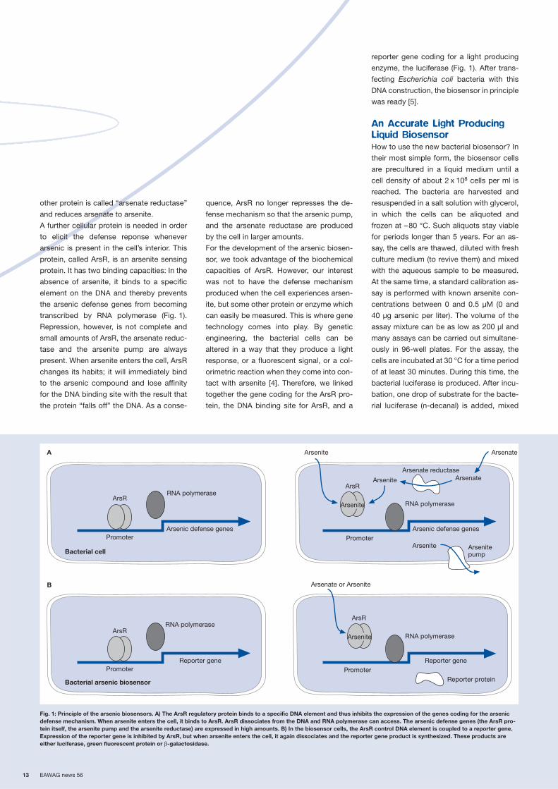
Exploit the Bacterial Defense
Mechanisms Against Arsenic
Arsenic is not only toxic for humans and
animals; arsenic toxicity is even found in
simple organisms like bacteria. They have a
few relatively effective biochemical mecha-
nisms to deal with arsenic ions which have
entered the cell (Fig. 1). Two well known
bacterial proteins deal with the most com-
mon forms of arsenic: arsenite and arsen-
ate. One protein is a pump which is integrat-
ed into the bacterial cell wall and removes
any arsenite from the interior of the cell to
the outside, where it can do no harm. The
Bacterial cells of the light producing biosensor mustalways be kept on ice.
For the assay, the cells are incubated with differentwater samples in a 96-well plate.
The intesity of light emission as a measure of arsenicpollution is analysed by a luminometer.
Pho
togr
aphs
: J.R
. van
der
Mee
r, E
AW
AG
13 EAWAG news 56
other protein is called “arsenate reductase”
and reduces arsenate to arsenite.
A further cellular protein is needed in order
to elicit the defense reponse whenever
arsenic is present in the cell’s interior. This
protein, called ArsR, is an arsenite sensing
protein. It has two binding capacities: In the
absence of arsenite, it binds to a specific
element on the DNA and thereby prevents
the arsenic defense genes from becoming
transcribed by RNA polymerase (Fig. 1).
Repression, however, is not complete and
small amounts of ArsR, the arsenate reduc-
tase and the arsenite pump are always
present. When arsenite enters the cell, ArsR
changes its habits; it will immediately bind
to the arsenic compound and lose affinity
for the DNA binding site with the result that
the protein “falls off” the DNA. As a conse-
quence, ArsR no longer represses the de-
fense mechanism so that the arsenic pump,
and the arsenate reductase are produced
by the cell in larger amounts.
For the development of the arsenic biosen-
sor, we took advantage of the biochemical
capacities of ArsR. However, our interest
was not to have the defense mechanism
produced when the cell experiences arsen-
ite, but some other protein or enzyme which
can easily be measured. This is where gene
technology comes into play. By genetic
engineering, the bacterial cells can be
altered in a way that they produce a light
response, or a fluorescent signal, or a col-
orimetric reaction when they come into con-
tact with arsenite [4]. Therefore, we linked
together the gene coding for the ArsR pro-
tein, the DNA binding site for ArsR, and a
reporter gene coding for a light producing
enzyme, the luciferase (Fig. 1). After trans-
fecting Escherichia coli bacteria with this
DNA construction, the biosensor in principle
was ready [5].
An Accurate Light Producing
Liquid Biosensor
How to use the new bacterial biosensor? In
their most simple form, the biosensor cells
are precultured in a liquid medium until a
cell density of about 2 x 10⁸ cells per ml is
reached. The bacteria are harvested and
resuspended in a salt solution with glycerol,
in which the cells can be aliquoted and
frozen at –80 °C. Such aliquots stay viable
for periods longer than 5 years. For an as-
say, the cells are thawed, diluted with fresh
culture medium (to revive them) and mixed
with the aqueous sample to be measured.
At the same time, a standard calibration as-
say is performed with known arsenite con-
centrations between 0 and 0.5 µM (0 and
40 µg arsenic per liter). The volume of the
assay mixture can be as low as 200 µl and
many assays can be carried out simultane-
ously in 96-well plates. For the assay, the
cells are incubated at 30 °C for a time period
of at least 30 minutes. During this time, the
bacterial luciferase is produced. After incu-
bation, one drop of substrate for the bacte-
rial luciferase (n-decanal) is added, mixed
Fig. 1: Principle of the arsenic biosensors. A) The ArsR regulatory protein binds to a specific DNA element and thus inhibits the expression of the genes coding for the arsenicdefense mechanism. When arsenite enters the cell, it binds to ArsR. ArsR dissociates from the DNA and RNA polymerase can access. The arsenic defense genes (the ArsR pro-tein itself, the arsenite pump and the arsenite reductase) are expressed in high amounts. B) In the biosensor cells, the ArsR control DNA element is coupled to a reporter gene.Expression of the reporter gene is inhibited by ArsR, but when arsenite enters the cell, it again dissociates and the reporter gene product is synthesized. These products areeither luciferase, green fluorescent protein or β-galactosidase.
Bacterial cell
Bacterial arsenic biosensor
Arsenate or Arsenite
Reporter protein
A
B
RNA polymeraseArsR
Arsenic defense genesPromoter
ArsR
Reporter genePromoter
ArseniteArsR
Reporter genePromoter
Arsenate
Arsenitepump
Arsenite
Arsenate reductaseArsenateArsenite
Arsenite
ArsR
RNA polymerase
Arsenic defense genesPromoter
Arsenite
RNA polymerase
RNA polymerase

EAWAG news 56 14
and the light emission measured in a lumi-
nometer. The calibration curve with these
biosensor cells is usually linear within the
range of 0 to 0.5 µM arsenite (Fig. 2). At high
concentrations or for unknown samples,
different dilutions have to be made in order
to perform accurate measurements.
A Simple Paper Strip
Biosensor
For two main reasons, the use of the above
described biosensor is not easy enough and
remains restricted to the laboratory: a rather
expensive luminometer has to be installed
and the handling of liquid bacterial cultures
is too critical in the field. We therefore tried
to develop another biosensor system where
the genetically modified bacterial cells are
immobilized on small paper strips [5]. In-
stead of the luciferase reporter gene, this
second system contains a gene for the
enzyme β-galactosidase that produces a
color reaction in the presence of arsenic.
These biosensor cells are also grown in cul-
ture broth, but mixed after harvesting with a
solution containing various sugars, amino
acids and gelatine. Small amounts of this
mixture are pipetted on paper strips (Fig. 3)
and carefully dried at controlled tempera-
ture and partial vacuum. The cells on the
paper strips remain active for about one
month storage at temperatures between
–20 and 30 °C. For an assay, a paper strip
is placed in a vial with 1 ml of aqueous
sample, incubated for 30 minutes at 30 °C
and taken out. A drop of substrate for the
β-galactosidase enzyme is added to the
paper and – depending on the amount of
β-galactosidase – converted into a blue
product. The color intensity is a qualitative
measure for the amount of arsenite which
the cells have been exposed to. When com-
pared to a standard solution with 10 µg or
50 µg arsenite per liter, one can judge if the
arsenic level is above or below the drinking
water limit (Fig. 3). Thus, the paper strip
biosensor is less accurate and has a short-
er storage life than the liquid biosensor but
seems to be more appropriate for utilization
in the field.
Unsolved Questions
So far laboratory practice. Many important
questions and problems remain before we
can think of using the biosensors routinely
outside the laboratory. For example, how
can the quality of the biosensor cells (i.e.,
their immediate activation potential) be
guaranteed? How good are biosensor mea-
surements when compared to chemical
analyses? Does the chemical composition
of the aqueous sample influence the bio-
sensor response? Would local authorities in
developing countries be sufficiently skilled
to carry out the biosensor test reliably?
What happens with the genetically modified
biosensor bacteria after the test? Solutions
to these questions may only be achieved
by proceeding step-by-step. EAWAG has
applied for a patent for the arsenic bio-
sensor at the European patent office. At
the moment, potential industrial partners
are being traced who might be interested in
licensing the arsenic biosensor technology
and funding additional developments. Fur-
ther improvement and testing of the bio-
sensors will hopefully result in a realistic
comparison of the biosensor test with
chemical methods and its usefulness.
[1] Hug S., Wegelin M., Gechter D., Canonica L. (2000):
Arsenic contamination of ground water: Disastrous
consequences in Bangladesh. EAWAG news 49,
18–20.
[2] Berg M. (2002): Arsenic in drinking water – Vietnam,
new focus of attention. EAWAG news 53, 12–14.
[3] Pfeifer H.-R., Zobrist J. (2002: Arsenic in drinking
water – also a problem in Switzerland? EAWAG news
53, 15–17.
[4] Jaspers M.C.M., Totevova S., Demnerova K., Harms
H., van der Meer J.R. (1999): The use of whole-cell
living biosensors to determine the bioavailability of
pollutants to microorganisms. In: Bavaye P., Block
J.C., Goncharuk V.V. (eds.) Bioavailability of organic
xenobiotics in the environment. Kluwer Academic
Publishers, The Netherlands, 153–158.
[5] Stocker J., Balluch D., Gsell M., Harms H., Feliciano
J., Daunert S., Malik K.A., van der Meer J.R. (2003):
Development of a set of simple bacterial biosensors
for quantitative and rapid field measurements of
arsenite and arsenate in potable water. Environmental
Science & Technology 37, 4743–4750.
R2 = 0.9898
0
0.2·106
0.4·106
0.6·106
0.8·106
1.0·106
0 0.1 0.2 0.3 0.4 0.5
Arsenite (µM)
Rel
ativ
e lig
ht u
nits
(E)
Fig. 2: Example of a calibration curve with the lucifer-ase biosensor. Increasing amounts of arsenite (in arange of 0.05 to 0.5 µM) result in a linear increase inlight production by the sensor cells.
Jan Roelof van der Meer, portrait see page 8.
Coauthor: Judith Stocker
Fig. 3: The arsenic paper strip test: paper strips (4 x 0.5 cm) with spots of dried bacterial cells (arrow). After incuba-tion with arsenite containing water samples, the cells produce β-galactosidase. The activity of this enzyme can bevisualized by conversion of a substrate into a blue molecule (inset). Color intensity depends on the concentration ofarsenite.
0 0.1 0.5 µm

15 EAWAG news 56
As a result of exposure to certain pollutants, various toxic oxygen
derivatives form in the cells of plants and animals, including the
so-called “singlet oxygen”. Fortunately, most cells have specific
defense mechanisms against the toxic effects of these deriva-
tives. Currently, research at EAWAG is examining in detail how the
unicellular green alga Chlamydomonas reinhardtii reacts to the
presence of singlet oxygen. The long-term goal of the work is to
develop a biosensor for the detection of singlet oxygen, which
would give us an indirect indicator of pollutant levels.
Since aerobic respiration provides the ener-
gy required for growth and metabolism in
plants and animals, molecular oxygen (O2)
is required by all higher life forms. Oxygen
may, however, be life-threatening; namely
when reactive, strongly-oxidizing oxygen
species are formed inside the cells (see box
and Fig. 1) [1]. If the cell cannot success-
fully defend against this oxidative stress,
vital components of the cell, such as lipids,
proteins and DNA, can be damaged, and
the cell will die. The formation of reactive
oxygen species is enhanced when organ-
isms are exposed to heavy metals or certain
organic contaminants, such as herbicides
or halogenated organic compounds. This
toxic effect is enhanced by intense solar
radiation. Most cells, however, have devel-
oped specific defense mechanisms against
reactive oxygen species [2].
Molecular Defense Scenarios
Briefly, the molecular defense mechanism
works as follows [3]: the cell possesses sen-
sors that allow it to detect stress situations,
such as the intracellular formation of reac-
tive oxygen species. These sensors, in turn,
activate so-called “stress genes” that are
read by the transcription apparatus, leading
to the production of gene copies (mes-
senger RNA) in quantities proportional to
the strength of the activating stress. Finally,
the corresponding stress proteins are syn-
thesized. These proteins have one of two
functions: (1) they remove the source of
the stress itself (i.e., they convert reactive
oxygen species into non-toxic species), or
(2) they repair cell components that have
already been damaged. The cell produces
general stress proteins that are formed
regardless of the stress source, as well as
specific stress proteins that are able to
target and efficiently remove specific stress
factors [4].
The genetic activation pattern may provide
valuable clues about the nature and inten-
sity of the stress within the cell. Expression
(activation levels) of specific genes has
already been used successfully to detect
and quantify environmental pollutants, such
as in the biosensor for arsenic that was
developed at EAWAG (see article by J.R.
van der Meer, p. 12). The goal of the project
described here is to develop a biosensor for
the detection of water contaminants that
cause the formation of reactive oxygen
species inside cells. Since very little is
known about the defense mechanisms
against singlet oxygen (see box and Fig. 1),
we have decided to focus our work on this
reactive species.
Fig. 1: Formation of reactive oxygen species from molecular oxygen by incomplete reduction or transfer of lightenergy. Oxygen species in parentheses are not stable and immediately transform to their protonated forms.
Reactive Oxygen Species
Due to its electron configuration, naturallyoccurring molecular oxygen is relativelyinert and not harmful to living organisms.It can, however, be activated physically byenergy transfer or chemically by electrontransfer. Such activated oxygen moleculesare called reactive oxygen species. Theyare highly reactive and can be formed inanaerobic organisms even under normalphysiological conditions. Transfer of elec-trons, e.g., from the respiratory chain inthe mitochondria, can lead to the forma-tion of the superoxide anion (O·
2–), hydro-
gen peroxide (H2O2) and the hydroxyl radi-cal (OH·) (Fig. 1) [1]. If light is the energysource causing the electron transfer, sin-glet oxygen is formed (Fig. 1). Singlet oxy-gen is chemically identical to molecularoxygen; only the electronic configurationhas changed. It is, however more reactive,and reacts rapidly with cellular compo-nents forming organic hydroperoxides.Unsaturated fatty acids in cell membranesreact particularly rapidly with singlet oxy-gen, which results in the formation oflipidperoxides and may cause damage tothe membrane. It is, therefore, extremelyimportant for all organisms to have a spe-cific defense mechanism against singletoxygen and the damage it causes.
3O2 O2·– [O·–] [O2–]e–e– e–
O22– and
Molecularoxygen
Lightenergy H+
1O2HO2
· H2O2HO·
H2O
H+ H+ H+
Singletoxygen
Hydroperoxylradical
Hydrogenperoxide
Hydroxylradical
Water
Superoxideanion
Peroxideanion
Defense Genes as Indicatorsof Toxicity
From Basic Research to Practical Application
EAWAG news 56 16
DNA Chips Yield First Clues
Photosynthetic organisms are particularly
affected by oxidative stress [2], since the
photosynthetic apparatus itself is an im-
portant source of reactive oxygen species.
For this reason, we have selected a photo-
synthetic model organism for our studies:
the unicellular, flagellated green alga Chla-
mydomonas reinhardtii. This organism has
the same cellular structure as higher algae
and plants, but is easier to cultivate and
very suitable for the use of molecular tech-
niques [5]. A particular advantage is the fact
that DNA chips with the genetic information
for C. reinhardtii have very recently become
available. These devices allow us to exam-
ine expression patterns for the majority of
the genes of a given organism [3, 6]. In our
case, this will enable us to detect specific
defense genes that are as yet unknown.
The DNA chips we are using contain a total
of 2792 genes of C. reinhardtii. In order to
differentiate between defense genes spe-
cifically responding to singlet oxygen and
nonspecific genes responding to oxidative
stress, the DNA chip experiments were car-
ried out either with singlet oxygen or hydro-
gen peroxide (H2O2), another commonly
occurring reactive oxygen species. A com-
parison of the two expression patterns indi-
cated distinct differences. There were sev-
eral genes that were activated by singlet
oxygen, but not by H2O2 (see image of
DNA chip on the front cover page; each
point represents a Chlamydomonas gene;
induced genes are red, suppressed genes
are green, and genes with unaltered expres-
sion are yellow).
Specific Induction by Singlet
Oxygen
The genes that were most strongly and
specifically induced by the presence of
singlet oxygen were examined further for
their possible expression and function in
other stress situations. One gene was found
to be particularly interesting. The gene in
question is a homologue of glutathione-
peroxidase (Gpxh). Related gene products
in other organisms are responsible for the
degradation of organic hydroperoxides [7].
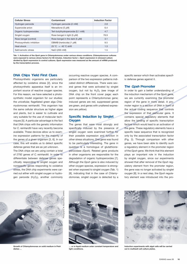
Although the Gpxh gene is also induced by
other oxygen species, expression is strong-
est when exposed to singlet oxygen (Tab. 1)
[8], indicating that in the case of Chlamy-
domonas, singlet oxygen is detected by a
specific sensor which then activates specif-
ic defense genes against it.
The Gpxh Promoter
In order to gain a better understanding of
the induction mechanism of the Gpxh gene,
we are currently examining the promoter
region of the gene in more detail. A pro-
moter region is a section of DNA in front of
the actual coding sequence that controls
the expression of that particular gene. It
contains special regulatory elements that
allow the binding of specific transcription
factors which would lead to an activation of
the gene. These regulatory elements have a
specific base sequence that is recognized
only by the associated transcription factor
(Fig. 2). Through comparison with other
genes, we have been able to identify such
a regulatory element in the promoter region
of the Gpxh gene. We think that this element
plays an important role in the induction
by singlet oxygen, since our experiments
showed that after removal of the Gpxh reg-
ulatory element from the promoter region,
the gene was no longer activated by singlet
oxygen [8]. In a next step, the Gpxh regula-
tory element was introduced into the pro-
Cellular Stress Contaminant Induction Factor
Hydrogen peroxide Hydrogen peroxide (2 mM) 2.8
Superoxide anion Menadione (5 µM) 6.4
Organic hydroperoxides Tert-butylhydroperoxide (0,1 mM) 4.7
Singlet oxygen Rose bengal in light (5 µM) 78.9
Rose bengal (control) Rose bengal in the dark (5 µM) 3.1
Photosynthetic inhibition DBMIB (herbicide) (1 µM) 9.5
Heat shock 25 °C → 40 °C shift 1.3
Salt/osmotic stress NaCl (200 mM) 1.5
Tab. 1: Activation of the Gpxh gene in Chlamydomonas under various stress conditions. Chlamydomonas cultureswere exposed to various stress factors for 60 minutes. Induction factor = Gpxh expression in stressed culturedivided by Gpxh expression in control cultures. Gpxh expression was measured as the amount of mRNA producedby the transcription process.
Growth of Chlamydomonas on solid medium in Petridishes …
… or in liquid medium unter constant temperature andlight conditions.
Induction experiments with algal cells will be carriedout in multiwell cell culture plates.
Pho
togr
aphs
: M. B
auch
row
itz, E
AW
AG

17 EAWAG news 56
moter region of a gene that is not normally
induced by singlet oxygen. We used the
�-tubulin gene of Chlamydomonas, a gene
that encodes the structural protein tubulin
which occurs in the flagella of a number
of microalgae, including Chlamydomonas.
Results confirmed that the transgenic �-
tubulin gene becomes weakly induced by
singlet oxygen after the Gpxh regulatory
element has been inserted into the pro-
moter region of the gene. So far, these ex-
periments suggest that the Gpxh regulatory
element plays a significant role in gene
activation by singlet oxygen. The regulatory
element found in Chlamydomonas has two
closely related and well described homo-
logues in mammals. Despite intensive ef-
forts, it has not yet been correlated with one
of these two mammalian regulatory ele-
ments. It is possible that the sequence was
not completely conserved between mam-
mals and algae or that the Gpxh regulation
element in Chlamydomonas may be a new,
as yet undescribed, regulatory element.
Once the corresponding transcription factor
has been isolated and characterized, it
should be possible to answer these ques-
tions.
Additional research is also needed in order
to determine whether or not there is a spe-
cific mechanism for the induction of Gpxh
by singlet oxygen in Chlamydomonas. If this
were the case, the Gpxh regulatory element
could be used as an element in a molecular
contamination sensor. Such a biosensor
would allow us to detect contaminants that
cause the formation of singlet oxygen in
cells.
[1] Halliwell B., Gutteridge J.M.C. (1999): Free radicals in
biology and medicine. Oxford science publications,
3rd edition. Clarendon Press, Oxford, New York, 968 p.
[2] Mendez-Alvarez S., Leisinger U., Eggen R.I.L. (1999):
Adaptive responses in Chlamydomonas reinhardtii.
International Microbiology 2, 15–22.
[3] Eggen R.I.L. (2001): Biological Tracers in Ecotoxicol-
ogy. EAWG news 52, 8–9.
[3] Gille G., Sigler K. (1995): Oxidative stress and living
cells. Folia Microbiologica 40, 131–52.
[5] Rochaix J.-D., Michel G.-C., Sabeeha M. (1998): The
molecular biology of chloroplasts and mitochondria in
Chlamydomonas. Advances in photosynthesis, Vol. 7.
Kluwer Academic Publishers, Dordrecht, 760 p.
[6] Rockett J.C., Dix D.J. (2000): DNA arrays: technology,
options and toxicological applications. Xenobiotica 30,
155–177.
[7] Leisinger U., Rüfenacht K., Zehnder A.J.B., Eggen
R.I.L. (1999): Structure of a glutathione peroxidase
homologous gene involved in the oxidative stress re-
sponse in Chlamydomonas reinhardtii. Plant Science
149, 139–149.
[8] Leisinger U., Rüfenacht K., Fischer B., Pesaro M.,
Spengler A., Zehnder A.J.B., Eggen R.I.L. (2001):
The glutathione peroxidase homologous gene from
Chlamydomonas reinhardtii is transcriptionally up-
regulated by singlet oxygen. Plant Molecular Biology
46, 395–408.
Fig. 2: Hypothetical activation path of the Gpxh gene by singlet oxygen. In the presence of singlet oxygen in thecell, the inactive form of the Gpxh transcription factor changes to the active form and binds to the Gpxh regulationelement. Only then can the transcription complex bind to the Gpxh gene and read it.
Singlet oxygen
Transcription factor(inactiv)
Transcription factor(activ)
DNA
Bindingsite
Promotor
Gpxh gene
Coding region
Transcriptioncomplex
TGACGCCA
Beat Fischer, molecular biolo-gist and doctoral student in the department “EnvironmentalMicrobiology and MolecularEcotoxicology”.
Coauthor: Rik Eggen
The biflagellated alga Chlamydomonas reinhardtiiunder the microscope (100-times magnified).
B. F
isch
er, E
AW
AG

EAWAG news 56 18
Drinking water is routinely tested for the presence of bacteria by
culturing methods which have the disadvantage of being very time
consuming. It takes at least one day before results are available.
For this reason, EAWAG is currently developing a faster method.
The application of new molecular techniques is promising, although
the development of an actual method is proving to be rather diffi-
cult.
The hygienic quality of drinking water has
been routinely tested for many decades
now by determining several bacterial pa-
rameters. These tests typically screen for
so-called indicator organisms, such as the
intestinal bacterium Escherichia coli [1]. The
assumption is that these harmless bacteria
are secreted along with potential pathogens
and could be introduced into drinking water
supplies. A simple and inexpensive cultur-
ing method for the detection of E. coli has
been used for many decades. However, this
method is rather time consuming: it takes
at least 24 hours before results are avail-
able. There are situations in which the water
supplier would like to be able to determine
more rapidly whether or not the water deliv-
ered to customers is hygienically accept-
able. It is possible, for example, that after
several days of intense precipitation, one
or more of the sources of drinking water
receives poorly filtered water. In a worst-
case scenario, this could lead to fecal
matter-contaminated drinking water. Were
this the case, the drinking water supplier
would have to immediately take measures
to decontaminate the drinking water supply
(disinfection, flushing) and would also have
to advise the population to boil water before
use. When it takes 24 hours before test
results are available, it may be too late for
the appropriate response. For this reason,
EAWAG is developing a faster detection
method that is based on newer molecular
methods of analysis.
The E. coli Reference Method
Drinking water is considered a “food” and
is, therefore, subject to the Swiss Ordinance
on Hygiene [2]. The ordinance specifies that
drinking water quality must be assessed
using the reference methods defined in the
Swiss Food Manual [3]. The use of other
methods is permissible if “they were proven
to lead to the same interpretation as the ref-
erence method” [2]. The reference method
for the detection of E. coli in drinking water
is the culturing method (Fig. 1, top). The
Ordinance on Hygiene sets the tolerance for
E. coli at “not detectable in 100 ml water
sample”. Therefore for the culturing method,
100 ml of water are passed through a filter,
on which E. coli and other organisms are
retained. The filter is then laid on an agar
plate where individual bacterial cells grow to
colonies that can be enumerated. Whether
a particular colony is, in fact, a colony of
E. coli, is confirmed by an enzymatic reac-
tion whereby E. coli colonies turn blue in
color.
The Newly Developed
Alternative Method
In the search for faster alternative methods,
techniques from molecular biology have re-
ceived much interest. In our opinion, the use
of the “Polymerase Chain Reaction” (PCR)
is particularly promising (Fig. 1, bottom). In
this method, short DNA segments charac-
onto an agarplate
Filtrationwater sample 100 ml
Transfer onto aselective agarplate
Ø Filter: 4.7 cm
Transfer of the filter
Ø Filter: 1.3 cm
into an Eppendorf vial
Real-Time-PCR
-1
1
3
5
7
1 10 20 30 40Amount of PCR cycles
Rn
(fluo
resc
ence
)
Colonies
Time 30 min0 min 6 hrs 30 hrs
Precultivation
2–4 hours
Cultivation
18–24 hours
Fig. 1: Individual steps and time requirements for the culturing method (top) and the newly developed PCR method (bottom).
New Paths in the Analysis of Drinking Water Quality
The Tedious Search for a Fast Alternative Method

19 EAWAG news 56
teristic for E. coli are replicated in a temper-
ature cycling process. A fluorescent dye
which stains double stranded DNA makes
the synthesized DNA fragments visible. In
the “real-time PCR method” that we use,
fluorescence is measured in real time and
recorded by a computer. The result is the
number of thermal cycles required to reach
a detectable fluorescence signal. With
respect to a particular water sample, this
means that the more E. coli cells (i.e., the
more E. coli DNA) were initially present in
the sample, the fewer thermal cycles are
needed before the fluorescence reaches
the detection level. A crucial element of our
PCR method is the pretreatment of the sam-
ple with the enzyme DNAse, which removes
any free DNA that might be present due to
dead bacteria, thus preventing false posi-
tive results.
The Difference becomes
Apparent
In order to test the agreement between the
new PCR method and the traditional cultur-
ing method, we inoculated drinking water in
the laboratory with E. coli cells and stored
the water at 4 °C. Over the period of one
month, we withdrew samples and analyzed
them using the two methods. At the begin-
ning of the experiment (t = 0), both methods
detected bacteria (Fig. 2A + B). After that,
the number of bacteria detected by the
culturing method decreased rapidly. We
expected therefore that the number of tem-
perature cycles observed in our PCR
method would increase. However, this was
not the case. Even after one month, the PCR
method still detected the presence of
E. coli. Apparently, the cells were no longer
able to grow on the culture media plates.
Cells that can no longer be cultured but can
still be detected by other methods are often
called “viable but not culturable” (VBNC).
There is a controversy among microbiolo-
gists as to whether the detection of such
VBNC stages is relevant in the case of the
E. coli indicator organism or pathogenic
organisms. Some studies suggest that such
cells can be reactivated or that they can
become infectious, respectively [4]; other
reports indicate the opposite [5, 6]. Based
on our results, we suggest that the cells die
continuously, and that the rate at which the
cells die depends on the “preconditioning”
of the E. coli culture.
A Proven Method is Difficult
to Replace
This simple experiment shows that the two
methods do not lead to the same interpre-
tation and that the PCR method used here
cannot replace the culturing method. The
principles on which the two methods are
based, i.e., the ability to replicate in the cul-
turing method and the detection of specific
DNA fragments in the PCR method, must
be too different from one another. This case
illustrates how strongly the thresholds set
for microbial parameters depend on the
analytical methods employed.
There have been proposals in the literature
to perform a short culturing period of a few
hours before using the PCR method, which
does improve the agreement between the
PCR method and the reference method [7].
In general, there are discussions ongoing
and proposals for standards published or in
progress in order to normalize the validation
of alternative methods in the future [8]. The
PCR method presented here could proba-
bly be established as a “new” method. This
would require, however, that we are able
to determine what specific physiological
stages of E. coli are being detected. For the
time being, our new PCR method appears
to be applicable rather to research than to
the routine monitoring of drinking water.
Acknowledgements
We would like to thank the Bachema Insti-
tute in Schlieren for initiating and supporting
this research project.
[1] Köster W., Egli T., Rust A. (2002) Pathogens in (Drinking) Water? EAWAG news 53, 26–28.
[2] Verordnung des EDI vom 26. Juni 1995 über die hygienischen und mikrobiologischen Anforderungen an Lebensmittel,
Gebrauchsgegenstände, Räume, Einrichtungen und Personal (Hygieneverordnung, HyV), in 817.051, S. 1–16.
[3] Ettel W. (Ed.) (2000): Kapitel 56, Mikrobiologie. In: Schweizerisches Lebensmittelbuch, Bundesamt für Gesundheit, Bern.
[4] Colwell R.R. (2000): Viable but nonculturable bacteria: a survival strategy. Journal of Infection and Chemotherapy 6,
121–125.
[5] Smith R.J., Newton A.T., Harwood C.R., Barer M.R. (2002): Active but nonculturable cells of Salmonella enterica serovar
Typhimurium do not infect or colonize mice. Microbiology 148, 2717–2726.
[6] Bogosian G., Bourneuf E.V. (2001): A matter of bacterial life and death. EMBO reports 21, 770–774.
[7] Frahm E., Obst U. (2003): Application of the fluorogenic probe technique (TaqMan PCR) to the detection of Enterococ-
cus spp. and Escherichia coli in water samples. Journal of Microbiological Methods 52, 123–131.
[8] Hübner P., Gautsch S., Jemmi T. (2002): In house-Validierung mikrobiologischer Prüfverfahren. Mitteilungen aus Lebens-
mitteluntersuchung und Hygiene 93, 118–139.
Fig. 2: Results of E. coli determinations by the cultur-ing method (A) and the PCR method (B) in artificiallycontaminated drinking water.All tests included determinations in non-contaminatedwater; culturing method: 0 colonies; PCR method:detection level of fluorescence not reached after40 temperature cycles.
Annette Rust developed themethod presented here as partof her doctoral research in thedepartment “EnvironmentalMicrobiology and MolecularEcotoxicology”.
Coauthor: Wolfgang Köster
Incubation period (days)
Col
onie
s p
er fi
lter
0
20
40
60
80
100
120A
0 5 10 15 2520 30
Num
ber
of t
emp
erat
ure
cycl
es
Incubation period (days)
B15
20
25
30
350 5 10 15 2520 30

EAWAG news 56 20
As a country bordering the Rhine, Switzerland shares in the
responsibility to reduce nitrogen export to the North Sea. In
wastewater treatment plants, this is currently accomplished
through a costly expansion of the main activated sludge system.
During the 1990s, however, there were several reports of nitrogen
being eliminated rather unexpectedly under certain operating
conditions. It was found that this is due to a recently discovered
group of bacteria, which were also found in Swiss wastewater
treatment plants. Based on this discovery, process engineers have
developed a new process that reliably removes nitrogen.
Nitrogen elimination is commonly regarded
as the conversion of biologically available
nitrogen compounds, such as ammonium
(NH4+), nitrite (NO2
–), or nitrate (NO3–), to ele-
mental nitrogen (N2), which is released into
the atmosphere as a harmless product.
Today, wastewater treatment plants almost
always use a biological nitrification/denitri-
fication process for the elimination of nitro-
gen (see box). Swiss wastewater treatment
plants often omit the denitrification step,
which means that the discharge water still
contains relatively high nitrogen loads,
mostly in the form of nitrate. As a country
that borders the Rhine, Switzerland has
committed itself in the Water Protection Act
of 1998 to reduce nitrogen input to the
Rhine by 2000 tons by the year 2005. Solu-
tions must, therefore, be found in the very
near future. The upgrading of all Swiss
wastewater treatment plants with an ade-
quate nitrification/denitrification stage will
be expensive. In addition, the operation of
this process is energy and resource inten-
sive [1]. Therefore, a new, low-impact proc-
ess is needed.
Tracking Down Unknown
Microorganisms
In the 1980s and 1990s, there were several
indications that nitrification/denitrification
are not the only processes that can remove
ammonium, but that there is an organism
that can oxidize ammonium to nitrogen gas
using nitrite instead of oxygen as its termi-
nal electron acceptor. Dutch and German
scientists first identified these organisms.
They belong to the order of Planctomyce-
tales: Brocadia anammoxidans and Kuene-
nia stuttgartiensis [2, 3]. The phenomenon
of anaerobic ammonium oxidation was also
observed in the Swiss wastewater treat-
ment plant of Kölliken. Our investigations
revealed that a multilayered system of
biofilms had formed in this treatment plant.
Such biofilms create pronounced oxygen
gradients, where the upper-most layer can
be oxygen-rich, while the layer closest to
the carrier material can be anaerobic [4]. We
assumed that the microorganisms we were
looking for were located in the lowest layers
of the biofilms. Using specific gene probes
and the FISH technique (fluorescence in situ
hybridization) [5], we were able to confirm
the presence of a large population of bacte-
ria belonging to the order Planctomycetes
(Fig. 1). Until now, it was not possible to iso-
late a pure culture of these new bacteria
using classic methods of microbiology. We
were able, however, to enrich a sample of
the biofilm, where approximately 90% of
the bacteria were Planctomycetes [6]. The
organism present in the Kölliken wastewater
treatment plant is Kuenenia stuttgartiensis.
Using techniques of molecular biology and
other physiological experiments, we could
show conclusively that K. stuttgartiensis
oxidizes ammonia to nitrogen under anaer-
obic conditions [3, 6]; this process is, there-
fore, called “anaerobic ammonium oxida-
tion” (anammox).
Anaerobic Ammonium Oxida-
tion – a Sustainable Process
for Nitrogen Removal
Process engineers were able to build on this
knowledge. Wastewater treatment plants
with anaerobic sludge digestion produce a
particularly ammonium-rich effluent, which
is nowadays recombined with the influent
to the treatment plant. Consequently the
nitrogen load to be treated is increased by
approximately 15–20%. Instead of “recy-
Fig. 1: Anammox bacteria form dense, globular clus-ters. Individual bacteria are visible as small, opencircles within the clusters. The cell clusters have adiameter of approximately 15 µm. Magnification:1000x.
Nitrification= Aerobic oxidation of ammonia by nitri-fying bacteria.Ammonium (NH4
+) is oxidized by oxygen(O2) via the intermediate nitrite (NO2
–) tonitrate (NO3
–):NH4
+ + 1.5 O2 ⇒ NO2– + H2O + 2 H+
NO2– + 0.5 O2 ⇒ NO3
–
Denitrification= Nitrate respiration by denitrifying bac-teria.Nitrate is reduced under anaerobic condi-tions to elemental nitrogen (N2) with theaddition of organic carbon (e.g., methanol):NO3
– + org. carbon ⇒ N2 + 2 CO2
Anammox= Anaerobic oxidation of ammonium byanammox bacteria.Ammonium is oxidized to elemental nitro-gen by nitrite under anaerobic conditions:NH4
+ + NO2– ⇒ N2 + 2 H2O
Fran
zisk
a B
ossh
ard
, EA
WA
G
The Anammox Process for NitrogenRemoval from Waste Water
The Fruitful Collaboration Between Microbiologists and ProcessEngineers

21 EAWAG news 56
cling” this effluent, it could be treated sepa-
rately using the new anammox process,
requiring minimal input of energy and other
resources. What is required, however, is
nitrite, which is not present in the effluent
but is formed as an intermediate product
in the nitrification process (see box). The
following two-step process (Fig. 2) is a
possible solution: in a first, aerated reactor,
ammonium is partially oxidized to nitrite
(partial nitritation), which is then reduced to
elemental nitrogen in a second oxygen-free
reactor using the remaining ammonium
(anammox). The overall process is, there-
fore, called “partial nitritation/anammox”, or
simply the anammox process. So far, it can
only be used for effluents rich in ammonium.
Thanks to genetic methods, anammox bac-
teria can be detected easily and rapidly,
which is important during the startup phase
and in determining process failure. Com-
pared to traditional nitrification/denitrifica-
tion, the anammox process has several
advantages (Fig. 3):� Oxygen additions can be reduced by
60%. This also reduces the energy required
to introduce the oxygen.� Anammox bacteria do not require organic
carbon. By contrast, organic carbon, such
as methanol, has to be added in the nitrifi-
cation/denitrification process.� Anammox bacteria produce very little bio-
mass, reducing the amount of sludge that
has to be disposed of.
The new anammox process uses not only
less energy and fewer resources, but it is
also less expensive than conventional nitri-
fication/denitrification.
Testing the Anammox Process
In order to test the feasibility of this two-
stage process in practice, EAWAG has built
and operated a pilot plant (4 m3) in cooper-
ation with the wastewater treatment plant
Werdhölzli (Zurich) and other partners [7].
This pilot project has yielded new insights
into the process and has confirmed its
applicability. In addition, the pilot plant has
been used to develop guidelines for the
scale-up and operation of full-scale plants.
Unfortunately, the new process is still met
with caution and hesitation, mostly because
of the slow growth rates of the anammox
bacteria and the lack of experience in oper-
ating the process. Due to the many advan-
tages of the process, however, we antici-
pate the construction of the first large-scale
anammox reactors over the next few years.
Christian Fux, process engineer,completed his doctoral researchon the anammox process in the department “EnvironmentalEngineering” at EAWAG. He is currently a postdoctoralresearcher at the “AdvancedWastewater Management
Centre”, University of Queensland, Australia.
Coauthors: Konrad Egli, Jan Roelof van der Meer,Hansruedi Siegrist
[1] BUWAL (1996): Stickstofffrachten aus Abwasserreini-
gungsanlagen. Schriftenreihe Umwelt Nr. 276, 67 S.
[2] Strous M., Fuerst J.A., Kramer E.H.M., Logemann S.,
Muyzer G., van de Pas-Schoonen K.T., Webb R.,
Kuenen J.G., Jetten M.S.M. (1999): Missing lithotroph
identified as new planctomycete. Nature 400,
446–449.
[3] Schmid M., Twachtmann U., Klein M., Strous M.,
Juretschko S., Jetten M., Metzger J.W., Schleifer K.-H.,
Wagner M. (2000): Molecular evidence for genus-level
diversity of bacteria capable of catalyzing anaerobic
ammonium oxidation. Systematic & Applied Micro-
biology 23, 93–106.
[4] Koch G., Egli K., van der Meer J.R., Siegrist H. (2000):
Mathematical modeling of autotrophic denitrification
in a nitrifying biofilm of a rotating biological contactor.
Water Science and Technology 41, 191–198.
[5] Zepp K. (2001): RNA – A Tracer to Detect Micro-
organisms. EAWAG news 52, 12–13.
[6] Egli K., Fanger U., Alvarez P., Siegrist H., van der Meer
J.R., Zehnder A.J.B. (2001): Enrichment and charac-
terization of a new anammox bacterium from a rotat-
ing biological contactor treating an ammonium-rich
leachate. Archives of Microbiology 175, 198–207.
[7] Fux C., Böhler M., Huber P., Brunner I., Siegrist H.
(2002): Biological treatment of ammonium-rich waste-
water by partial nitritation and subsequent anaerobic
ammonium oxidation (anammox) in a pilot plant.
Journal of Biotechnology 99, 295–306.
2 O2 (100%)
0.75 O2(40%)
Organic carbon(e.g., methanol:3.4 kg/kg N)
Partial nitritation
Anammox
Nitrification
Denitrification
0.5 N2+
lots of biomass
NH4+
NO3–
0.5 N2+
little biomass
NH4–
0.5 NH4+ 0.5 NO2
–
Treated water to thepre-settlement tank
(low concentrations of NH4
+, NO2–, NO3
–)
NH4+ + NO2
– ⇒ N2
NH4+ ⇒ NO2
–
Sludge digester effluent(ammonium-rich)
Aeration (O2)
Mixture ofammonium and nitrite
Elementalnitrogen (N2)
No oxygen
Fig. 2: The anammox process.
Fig. 3: Comparison between the anammox process and conventional nitrogen removal by the nitrification/denitrifi-cation process.

EAWAG news 56 22
The diversity of zoo- and phytoplankton in alpine lakes has been
studied intensively for over 100 years. It is now generally accepted
that the species diversity of plankton communities decreases
with increasing altitude. This study addresses the question as to
whether or not this general pattern also applies at the population
level. Daphnia, a typical member of zooplankton communities, was
chosen as the study organism. Genetic diversity was determined
for 11 Daphnia populations in mountain lakes at varying elevations.
We found that the genetic diversity was very heterogeneous.
The extreme environmental conditions in
alpine regions result in a small selection of
flora and fauna that can adapt to survival in
these regions; therefore, biodiversity (or
more precisely species diversity), decreases
with increasing altitude. It is unclear, how-
ever, whether or not this is also reflected in
another aspect of biodiversity; namely, ge-
netic diversity. In order to answer this ques-
tion, we examined Daphnia populations, a
typical member of zooplankton communi-
ties, in a number of Swiss mountain lakes.
These organisms are particularly interesting
for such a study since they can reproduce
sexually or asexually by parthenogenesis
[1].
Parthenogenesis and
Resting Eggs
Under favorable environmental conditions,
parthenogenesis is the normal reproductive
method in Daphnia; mother organisms form
genetically identical daughters (so-called
clones). This allows Daphnia to multiply
extremely rapidly, thereby making optimal
use of short growth periods in alpine
ecosystems. When environmental condi-
tions worsen, e.g. during cold periods or
when food is sparse, Daphnia produce
males and sexual females. Sexually fertil-
ized eggs are encased in a resistant shell,
the so-called ephippium, which resides in
the breeding cavity and is sloughed with
the next shedding of the skin. Ephippia can
survive for years in the lake sediments. Un-
der favorable conditions, these resting eggs
can produce parthenogenetic females, and
the cycle begins again. If the conditions are
favorable all year, no resting eggs are
formed, so that one parthenogenetic gener-
ation follows another.
Environment, Reproduction
and Genetic Diversity
Our project investigated the relationship
between environmental conditions, repro-
duction method, and genetic diversity in
alpine lakes. We examined 11 mountain
lakes in the Swiss Alps. The lakes varied in
elevation, where environmental conditions
become harsher as elevation increases.
Daphnia populations in these lakes were
sampled in four consecutive years (1997–
2000) during late summer or early fall.
Our experiments were based on the follow-
ing hypotheses:� Alpine lakes should harbor a high propor-
tion of sexual individuals, since Daphnia can
survive cold and food-deprived winters only
in the form of resting eggs.� In Daphnia, genetic diversity depends pri-
marily on the sexual exchange of genetic
material. We postulated that populations
in lakes, where Daphnia reproduce mostly
sexually, would exhibit higher genetic diver-
sity.� Since environmental conditions in alpine
lakes become harsher with increasing ele-
vation, we expected that the genetic diver-
sity in Daphnia populations would increase
with increasing altitude and not decrease
as is the case for species diversity. Alpine
lakes should, therefore, show higher genet-
ic diversity than lakes in the lowlands.
Sexual Individuals in the
Majority
The relative fraction of asexual and sexual
Daphnia in our study lakes varied dramat-
ically and ranged from 2 to 90% (Fig. 1 low-
er panel). In 8 of the 11 mountain lakes
examined, sexual Daphnia outnumbered
asexual individuals. In the two lowest alpine
lakes, the Upper and the Lower Arosasee,
we found almost exclusively asexual fe-
males, while the highest lake, Riffelsee II,
contained almost exclusively sexual individ-
uals. The distribution between sexual and
asexual Daphnia for lakes at intermediate
elevations, however, was heterogeneous;
we could not decipher any clear patterns.
There was, however, one surprising finding:
while males often made up less than 1% of
the Daphnia populations in lowland lakes,
they made up as much as 30–40% of the
The Genetic Examination Method
Allozyme electrophoresis is a proven method for the determination of genetic diversity of popu-lations [2]. It is based on the following principle: the genome of an organism may contain thegenetic information for a certain enzyme once or several times. If the gene is present severaltimes, it is likely that the individual genes have changed over time, usually due to mutations. As a result, the cell will synthesize different enzymes, so-called allozymes, which have slightdifferences in their amino acid sequences, but will exhibit the same functionality as the originalenzyme. In the simplest case, there are two allozymes which differ by only one amino acid. If the new amino acid carries a different charge, the two allozymes will have different migrationvelocities in an electric field and can be separated by gel electrophoresis. Subsequently, theenzyme can be detected through the reaction with its substrate. If the substrate reaction iscoupled with a color reaction, the enzyme can be made directly visible. This method indicateshow many enzyme variations are present within a population and, therefore, provides an indexof genetic diversity.
Genetic Diversity of Daphniain Alpine Lakes

23 EAWAG news 56
populations in the mountain lakes. In addi-
tion, the fraction of sexual male individuals
in alpine lakes was always higher than that
of sexual females.
Genetic Diversity
In order to assess the genetic diversity of
different Daphnia populations, we examined
up to 80 individuals per lake by the allozyme
method (see box). The clonal diversity, i.e.,
the number of different clones within a pop-
ulation, is a first indication of overall diver-
sity. In the 11 mountain lakes examined in
this study, we found between 2 and 42 dif-
ferent clones (Fig. 1 upper panel). Melchsee
and Lago Cadagno showed the highest
clonal diversity.
A better description of genetic diversity is
obtained when the number of individuals for
each of the clones is taken into account in
addition to the number of different clones.
A population with 10 different clones, each
making up 10% of the population, is more
diverse than a population with 10 different
clones, but one single clone accounting for
99% of all the individuals. Therefore, we
calculated the Simpson index (DSim =
1– ∑pi2, pi = fraction of a clone i in the over-
all population) for each of the 11 popula-
tions that were examined (Fig. 1 upper pan-
el). This index describes the probability
that two randomly chosen individuals have
a different genetic structure. High DSim val-
ues (near 1) indicate that all clones occur
with similar frequency, which translates to
high diversity. Low DSim values (near 0) show
that one clone dominates the population,
resulting in low diversity. It was found that
the diversity of Daphnia populations was
relatively high in a number of lakes under
examination, whereas it was low in the
Upper Arosasee, Leisee and Riffelsee II.
The Spatial Genetic Structure of
Daphnia Populations is Complex
The high proportion of sexual individuals in
alpine lakes was surprising and is an indica-
tion that Daphnia survive the harsh winters
only in the form of resting eggs; however,
the fraction of sexual individuals did not
correlate with genetic diversity, neither if
expressed as clonal diversity nor in the
Simpson index. Furthermore, we were not
able to establish a correlation between the
genetic diversity of Daphnia populations
and the elevation of the alpine lakes exam-
ined in this study. We compared our data to
the genetic diversity of Daphnia populations
in two lowland lakes, namely Greifensee
(DSim = 0.96) and Lake Lucerne (DSim = 0.48).
We found that the genetic diversity in moun-
tain lakes can be as low as that of Lake
Lucerne, or almost as high as that of Grei-
fensee.
This study [3] represents a first step in
examining the genetic diversity of a major
zooplankton species in alpine lakes and in
determining those factors that influence
genetic diversity. Apparently, sexual repro-
duction does not necessarily lead to higher
genetic diversity in Daphnia populations.
It can, therefore, be assumed that other
factors, such as the presence of certain
Daphnia species or Daphnia hybrids, have
a stronger influence or that only certain
clones are adapted to the extreme environ-
mental conditions in alpine lakes. High ge-
netic diversity does not appear to be a pre-
requisite for survival of Daphnia in alpine
lakes. Other strategies, such as their flexible
behavior and life cycle, may be more impor-
tant in coping with the harsh conditions in
the alpine environment.
[1] De Meester L. (1996): Local genetic differentiation and
adaptation in freshwater zooplankton populations:
patterns and processes. Ecoscience 3, 385–399.
[2] Monaghan M. (2002): Habitat Fragmentation and
Genetic Diversity. EAWAG news 54, 22–24.
[3] Winder M., Monaghan M.T., Spaak P. (2001): Have
human impacts changed alpine zooplankton diversity
over the past 100 years? Arctic Antarctic and Alpine
Research 33, 467–475.
Monika Winder has recentlyconcluded her doctoral researchon “Zooplankton ecology inhigh-mountain lakes” in theDepartment “Limnology”. She iscurrently a postdoctoral fellowat the University of Washington,Seattle, USA.
Fig. 1: Investigation of Daphnia populations in 11 Swiss alpine lakes at varying elevations. Lower panel: relativeproportions of sexual and asexual individuals; upper panel: genetic diversity, expressed as number of clones and by the Simpson index.
Rel
ativ
e p
ortio
n (%
)
Low
erAr
osas
ee16
91 m U
pper
Aros
asee
1734
m Trüb
see
1764
mM
urgs
ee18
20 m
Mel
chse
e18
91 m
Cad
agno
1923
mPr
ätsc
hsee
1970
mG
otth
ards
ee20
91 m
Grim
sels
ee21
60 m
Leis
ee22
32 m
Schw
arzs
ee25
52 m
Riffe
lsee
II27
45 m
0
20
40
60
80
100
Clo
nes
Sim
pso
n in
dex
�sexual �asexual �
1.0
0.8
0.6
0.4
0.2
0
40
0
10
20
30
50
ClonesSimpson index Piet Spaak, biologist and head
of the section “EvolutionaryEcology” in the department“Limnology”. Lecturer for evolu-tionary ecology at ETH Zurich.Main research interests: Geneticdiversity of Daphnia, alpinelakes and predator-prey inter-actions.

EAWAG news 56 24
Please use the order form in the center fold of EAWAG news to request reprints.
Publications
[3193] Andrade A.P., Witholt B., Hany R., Egli T.,Li Z. (2002): Preparation and Characterization ofEnatiomerically Pure Telechelic Diols from mcl-Poly[(R)-3-hydroxyalkanoates]. Macromolecules35, 684–689.
[3194] Andrade A.P., Neuenschwander P., HanyR., Egli T., Witholt B., Li Z. (2002): Synthesis andCharacterization of Novel Copoly(ester-urethane)Containing Blocks of Poly-[(R)-hydroxyoctanoate]and Poly-[(R)-3-hydroxybutyrate]. Macromole-cules 35, 4946–4950.
[3195] Brun R. (2002): Learning From Data:Parameter Identification in the Context of LargeEnvironmental Simulation Models. Diss. ETHZ No.14 575, Zurich.
[3196] Leisinger U. (2002): Regulation of GeneExpression Upon Oxidative Stress in Chlamydo-monas reinhardtii. Diss. ETHZ No. 14 434, Zurich.
[3197] Monaghan M.T. (2002): Habitat Fragmen-tation in Alpine Sreams: Implications for GeneticStructure and Species Richness of Aquatic In-sects. Diss. ETHZ No. 14 561, Zurich.
[3198] Behringer J. (2002): Legitimität durch Ver-fahren? Bedingungen semi-konventioneller Par-tizipation. Dissertation Universität Stuttgart, S.Roderer Verlag, Regensburg.
[3199] Egli T. (2002): Microbial Degradation ofPollutants at Low Concentrations and in thePresence of Alternative Carbon Substrates:Emerging Patterns. In: “Biotechnology for theEnvironment: Strategy and Fundamentals”, S.N.Agathos, W. Reineke (Eds.). Kluwer AcademicPubl., pp. 131–139.
[3200] Egli T., Witschel M. (2002): Enzymology ofthe Breakdown of Synthetic Chelating Agents. In: “Biotechnology for the Environment: Strategyand Fundamentals”, S.N. Agathos, W. Reineke,(Eds.). Kluwer Academic Publ., pp. 205–217.
[3201] Yang H., Zehnder A.J.B. (2002): WaterScarcity and Food Import: A Case Study forSouthern Mediterranean Countries. World Devel-opment 30 (8), 1413–1430.
[3202] van der Nat D., Tockner K., Edwards P.J.,Ward J.V. (2002): Quantification of Large WoodyDebris in Large Floodplain Rivers: An Area-BasedApproach Using Differential GPS and GIS. Verh.Internat. Verein. Limnol. 28, 332–335.
[3203] Robinson C.T., Uehlinger U., Guidon F.,Schenkel P., Skvare R. (2002): Limitation and Re-tention of Nutrients in Alpine Streams of Switzer-land. Verh. Internat. Verein. Limnol. 28, 263–272.
[3204] Malard F., Hofmann A., Tockner K.,Uehlinger U. (2002): Iron Concentration in the
Water of a Glacial River System. Verh. Internat.Verein. Limnol. 28, 134–139.
[3205] Hieber M., Robinson C.T., Uehlinger U.,Ward J.V. (2002): Are Alpine Lake Outlets LessHarsh Than Other Alpine Streams? Arch. Hydro-biol. 154 (2), 199–223.
[3206] Rechberger H., Graedel T.E. (2002): TheContemporary European Copper Cycle: StatisticalEntropy Analysis. Ecol. Economics 42, 59–72.
[3207] Gerecke A.C., Schärer M., Singer H.P.,Müller S.R., Schwarzenbach R.P., Sägesser M.,Ochsenbein U., Popow G. (2002): Sources ofPesticides in Surface Waters in Switzerland:Pesticide Load Through Waste Water TreatmentPlants – Current Situation and Reduction Poten-tial. Chemosphere 48, 307–315.
[3208] Hug F., Baccini P. (2002): PhysiologicalInteractions Between Highland and LowlandRegions in the Context of Longterm ResourceManagement. Mountain Res. & Develop. 22 (2),168–176.
[3209] Kovar K., Chaloupka V., Egli T. (2002): A Threshold Substrate Concentration is Requiredto Initiate the Degradation of 3-PhenylpropionicAcid in Escherichia coli. Acta Biotechnol. 22 (3–4),285–298.
[3210] Laj C., Kissel C., Mazaud A., Michel E.,Muscheler R., Beer J. (2002): Geomagnetic Fieldintensity, North Atlantic Deep Water Circulationand Atmospheric ∆14C During the Last 50 kyr.Earth and Planetary Sci. Lett. 200, 177–190.
[3211] Mavrocordatos D., Fortin D. (2002):Quantitative Characterization of Biotic Iron Oxidesby Analytical Electron Microscopy. American Min-eralogist 87, 940–946.
[3212] Nguyen H.M., Pham H.V., Giger W., Berg M. (2002): Simultaneous Determination ofPolar and Apolar Organophosphorus Pesticidesand Triazine Herbicides by Solid-phase Microex-traction (SPME) in Aqueous Samples. Anal. Sci. 17(Suppl.), a375–a378.
[3213] Duong H.A., Pham H.V., Gallard H.,Berg M. (2001): Determination of the Breakpoint inChlorine Dosage for Typical Groundwater Sourcesin Hanoi Area. J. Anal. Sci. of the Vietnam Anal.Sci. Soc. 6 (4), 63–66. (in Vietnamese)
[3214] Duong H.A., Hoang M.H., Pham H.V.,Berg M., Giger W. (2001): Evaluation of GC/MS/HS Method for Analysis of Volatile Organic inWater. J. Anal. Sci. of the Vietnam Anal. Sci. Soc.6 (2), 31–36. (in Vietnamese)
[3215] Min N.H., Ha P.N., Viet P.H., Giger W.,Berg M. (2000): Determination of Polar and Non-
polar Pesticides in Aqueous Solutions by SolidPhase Microextraction (SPME) and Gas Chro-matography Mass Spectrometer (GC/MS). Work-shop on Management, Use and Assessment ofEnvironmental Pollution of Pesticides, CEC,Hanoi, Sept. 28–29.
[3216] Boller M., Wagner W. (2001): Urban WaterManagement in Switzerland. Conference-Pro-ceedings EurAqua, Sixth Scientific and TechnicalReview, Laboratorio Nacional de Engenharia Civil,LNEC, Lisbon, Portugal, Lisbon 20–21 October1999.
[3217] Tockner K., Ward J.V., Edwards P.J.,Kollmann J., Gurnell A.M., Petts G.E. (2001): DerTagliamento (Norditalien): Eine Wildflussaue alsModellökosystem für den Alpenraum. Bayer.Akad. für Naturschutz und Landschaftspflege,Laufen/Salzach. Laufener Seminarbeiträge 3 (01),S. 25–34.
[3218] Meyer A., Würsten M., Schmidt A.,Kohler H.-P.E., Witholt B. (2002): Hydroxylationof Indole by Laboratory-Evolved 2-hydroxybi-phenyl 3-monooxygenase. J. Biol. Chem. 277 (37),34161–34167.
[3219] Friedrich J., Dinkel C., Friedl G., Pimen-ov N., Wijsman J., Gomoiu M.-T., Cociasu A.,Popa L., Wehrli B. (2002): Benthic NutrientCycling and Diagenetic Pathways in the North-western Black Sea. Estuarine, Coastal & Shelf Sci.54, 369–383.
[3220] Hügel K., Larsen T., Gujer W. (2002):Ökobilanzen als Massstab für die Ressourcen-Effizienz in der Abwasserentsorgung. Schriften-reihe Wasserforschung 7, 129–141.
[3221] Brun R., Kühni M., Siegrist H., Gujer W.,Reichert P. (2002): Practical Identifiability ofASM2d Parameters – Systematic Selection andTuning of Parameter Subsets. Water Res. 36,4113–4127.
[3222] Granina L., Müller B., Wehrli B., Martin P.(2000): Oxygen, Iron, and Manganese at the Sedi-ment-Water Interface in Lake Baikal. Terra Nostra2000/9, Baikal, First Workshop and Symposium,pp. 87–93.
[3223] Hieber M. (2002): Alpine Streams: Aspectsof Biocomplexity. Diss. ETHZ No. 14 601, Zurich.
[3224] Tockner K., Paetzold A., Karaus U.(2002): Leben in der Flussdynamik zwischen Trok-kenfall und Hochwasser. In: “Rundgespräche derKommission für Ökologie”. Bd. 24. Verlag F. Pfeil,München, S. 37–46.
[3225] Winder M., Spaak P. (2002): Effects ofNatural UV Radiation on the Life History of Alpine

25 EAWAG news 56
[3251] Drescher S., Zurbrügg C. (2002): De-zentrale Kompostierung in Indien. Müllmagazin 3,17–21.
[3252] Schweigert N., Eggen R.I.L., Escher B.I.,Burkhardt-Holm P., Behra R. (2002) Ecotoxico-logical Assessment of Surface Waters: A ModularApproach Integrating in vitro Methods. AltexAltern. Tierexp. 19, 30–37.
[3253] Ammann A.A. (2002): Speciation of HeavyMetals in Environmental Water by Ion Chromatog-raphy Coupled to Icp-Ms. Anal. Bioanal. Chem.372 (3), 448–452.
[3254] Wahli T., Knuesel R., Bernet D., SegnerH., Pugovkin D., Burkhardt-Holm P., Escher M.,Schmidt-Posthaus H. (2002): Proliferative KidneyDisease in Switzerland: Current State of Knowl-edge. J. Fish Dis. 25 (8), 491–500.
[3255] Kuehn K.A., Gessner M.O., Wetzel R.G.K.S. (2000): Standing Litter Decomposition of the Emergent Macrophyte Erianthus giganthus(Plumegrass). Verh. Internat. Verein. Limnol. 27,3846–3847.
[3256] Malard F., Ward J.V. Robinson C.T.(2000): An Expanded Perspecitive of the Hypo-rheic Zone. Verh. Internat. Verein. Limnol. 27,431–337.
[3257] Ackermann G.E., Schwaiger J., NegeleR.D., Fent K. (2002): Effects of Long-Term Nonyl-phenol Exposure on Gonadal Development andBiomarkers of Estrogenicity in Juvenile RainbowTrout (Oncorhynchus mykiss). Aquat. Toxicol. 60(3–4), 203–221.
[3258] Simankova M.V., Parshina S.N., TourovaT.P., Kolganova T.V., Zehnder A.J.B., Nozhev-nikova A.N. (2001): Methanosarcina lacustris SpNov., a New Psychrotolerant Methanogenic Ar-chaeon from Anoxic Lake Sediments. Syst. Appl.Microbiol. 24, (3), 362–367.
[3259] Bossard P. Robarts R. (2001): Editorial.Aquat. Sci. 63, (3), 1–3.
[3260] Beardall J., Berman T., Heraud P., KadiriM.O., Light B.R., Patterson G., Roberts S.,Sulzberger B., Sahan E., Uehlinger U., Wood B.(2001): A Comparison of Methods for Detection ofPhosphate Limitation in Microalgae. Aquat. Sci.63, (1), 107–121.
[3261] Bossard P., Gammeter S., Lehmann C.,Schanz F., Bachofen R., Bürgi H.R., Steiner D.,Zimmermann U. (2001): Limnological Descriptionof the Lakes Zurich, Lucerne, and Cadagno.Aquat. Sci. 63, (3), 225–249.
[3262] Callieri C., Morabito G., Huot Y., NealeP.J., Litchman E. (2001): Photosynthetic Re-sponse of Pico- and Nanoplanktonic Algae toUVB, UVA and PAR in a High Mountain Lake.Aquat. Sci. 63, (3), 286–293.
[3263] Neale P.J., Bossard P., Huot Y. (2001):Incident and in situ Irradiance in Lakes Cadagnoand Lucerne: A Comparison of Methods and Mod-els. Aquat. Sci. 63, (3), 250–264.
[3264] Neale P.J., Litchman E., Sobrino C., Cal-lieri C., Morabito G., Montecino V., Huot Y.,Bossard P., Lehmann, C. Steiner D. (2001):Quantifying the Response of PhytoplanktonPhotosynthesis to Ultraviolet Radiation: BiologicalWeighting Functions Versus in situ Measurementsin Two Swiss Lakes. Aquat. Sci. 63, (3), 265–285.
australis Leaves During Senescence and EarlyAerial Decay. Aquat. Bot. 69, (2-4), 325-339.
[3240] Lotter A.F., Appleby P.G., Bindler R.,Dearing J.A., Grytnes J.-A., Hofmann W., Ka-menik A., Lami A., Livingstone D.M., OhlendorfC., Rose N., Sturm M. (2002): The SedimentRecord of the Past 200 Years in a Swiss High-alpine Lake: Hagelseewli (2339 m a.s.l.). J. Paleo-limnol. 28, 111–127.
[3241] Cameron N.G., Schnell A., Raution M.L.,Lami A., Livingstone D.M., Appleby P.G., Dear-ing J.A., Rose N. (2002): High-resolution Analysesof Recent Sediments from a Norwegian MountainLake and Comparison with Instrumental Recordsof Climate. J. Paleolimnol. 28, 79–93.
[3242] Catalan J., Ventura M., Brancelj A.,Granados I., Thies H., Nickus U., Korhola A.,Lotter A.F., Barbieri A., Stuchlik E., Lien L.,Bitusik P., Buchaca T., Camarero L., GoudsmitG.H., Kopacek J., Lemcke G., Livingstone D.M.,Müller B., Raution M.L., Sisko M., Sorvari S.,Sporka F., Strunecky O., Toro M. (2002): Sea-sonal Ecosystem Variability in Remote MountainLakes: Implications for Detecting Climatic Signalsin Sediment Records. J. Paleolimnol. 28, 25–46.
[3243] Berg M., Hug S., Zobrist J. (2002): Arsen,eine neue Herausforderung für Wasserfachleute.CHemie 10, 3–13.
[3244] Heller Hansraj C. (2000): Trophic Cascad-ing in Lake Lucerne, Switzerland the Influence ofTop-Down Controls on the Pelagic Food Web.Christine Heller Hansraj. Diss. ETHZ Nr. 13 631,Zurich.
[3245] Spaak P., Eggenschwiler L. Bürgi H.(2000): Genetic Variation and Clonal Differentiationin the Daphnia Population of the Greifensee, a Pre-Alpine Swiss Lake. Verh. Internat. Verein. Limnol.27, 1919-1923.
[3246] Wick L.M., Weilenmann H., Egli T. (2002):The Apparent Clock-Like Evolution of Escherichiacoli in Glucose-Limited Chemostats Is Repro-ducible at Large but Not at Small Population Sizesand Can Be Explained with Monod Kinetics.Microbiology (UK) 148, 2889–2902.
[3247] Roth C.M., Goss K.U., SchwarzenbachR.P. (2002): Adsorption of a Diverse Set of Organ-ic Vapors on the Bulk Water Surface. J. ColloidInterface Sci. 252 (1), 21–30.
[3248] Zurbrügg C., Drescher S., Rytz I., SinhaM., Enayetullah I. (2002): Decentralised Com-posting in Dhaka, Bangladesh – Production ofCompost and its Marketing. In: “Appropriate Envi-ronmental and Solid Waste Management andTechnologies for Developing Countries” 2nd Ed.,G. Kocasoy et al., (Eds.). Proceedings Interna-tional Solid Waste Association (ISWA), Istanbul,pp. 1285–1292.
[3249] Zurbrügg C., Drescher S., Sharatchan-dra H.C. (2002): Decentralised Composting inIndia – Lessons Learned. Conf. Proc. 28th WEDCInternat. Conf. on Sustainable EnvironmentalSanitation and Water Services, November 18–22,2002, Calcutta India.
[3250] Ahmed N., Zurbrügg C. (2002): OrganicWaste Management in Karachi, Pakistan. Conf.Proc. 28th WEDC Internat. Conf. on SustainableEnvironmental Sanitation and Water Services, No-vember 18–22, 2002, Calcutta India.
Daphnia. Verh. Internat. Verein. Limnol. 28, 355–359.
[3226] Robinson C.T., Gessner M.O. (2000): LeafBreakdown in an Alpine Spring Brook. Verh. Inter-nat. Verein. Limnol. 27, 744–747.
[3227] Ackermann G.E., Brombacher E., FentK. (2002): Development of a Fish Reporter GeneSystem for the Assessment of Estrogenic Com-pounds and Sewage Treatment Plant Effluents.Environ. Toxicol. Chem. 21 (9), 1864–1875.
[3228] Frutiger A., Meier Bürgisser G.M. (2002):Life History Variability of a Grazing Stream Insect(Liponeura cinerascens minor; Diptera: Blephari-ceridae). Freshwater Biol. 47 (9), 1618–1632.
[3229] Golet E.M., Alder A.C., Giger W. (2002):Environmental Exposure and Risk Assessment ofFluoroquinolone Antibacterial Agents in Waste-water and River Water of the Glatt Valley Water-shed, Switzerland. Environ. Sci. Technol. 36 (17),3645–3651.
[3230] Gessner M.O., van Ryckegem G. (2002):Water Fungi as Decomposers in Freshwater Eco-systems. In: “Encyclopedia of EnvironmentalMicrobiology”, G. Bitton, (Ed.). John Wiley & Sons,pp. 3353–3363.
[3231] Escher B.I., Hermens J.L.M. (2002):Modes of Action in Ecotoxicology: Their Role inBody Burdens, Species Sensitivity, QSARs, andEffects. Environ. Sci. Technol. 36 (20), 4201–4217.
[3232] Gessner M.O., Newell S.Y. (2002): Bio-mass, Growth Rate, and Production of Filamen-tous Fungi in Plant Litter. In: “Manual of Environ-mental Microbiology”, 2nd ed., C.J. Hurst et al.,(Eds.). ASM Press, Washington, D.C., pp. 390–408.
[3233] Ruckstuhl S., Suter M.J.F., Kohler H.P.-E., Giger W. (2002): Leaching and Primary Bio-degradation of Sulfonated Naphthalenes and theirFormaldehyde Condensates from Concrete Su-perplasticizers in Groundwater Affected by TunnelConstruction. Environ. Sci. Technol. 36 (15), 3284–3289.
[3234] Graça M.A.S., Cressa C., Gessner M.O.,Feio M.J., Callies K.A., Barrios C. (2001): FoodQuality, Feeding Preferences, Survival and Growthof Shredders from Temperate and TropicalStreams. Freshwater Biol. 46, 947–957.
[3235] Gessner M.O., Chauvet E. (2002): A Casefor Using Litter Breakdown to Assess FunctionalStream Integrity. Ecol. Appl. 12 (2), 498–510.
[3236] Hieber M., Gessner M.O. (2002): Contri-bution of Stream Detrivores, Fungi, and Bacteriato Leaf Breakdown Based on Biomass Estimates.Ecology 83 (4), 1026–1038.
[3237] Baldy V., Chauvet E., Charcosset J.-Y.,Gessner M.O. (2002): Microbial Dynamics Asso-ciated with Leaves Decomposing in the Mainstemand Floodplain Pond of a Large River. AquaticMicrobial Ecology 28, 25–36.
[3238] Buesing N., Gessner M.O. (2002): Com-parison of Detachment Procedures for DirectCounts of Bacteria Associated with Sediment Par-ticles, Plant Litter and Epiphytic Biofilms. AquaticMicrobial Ecology 27 (1), 29–36.
[3239] Gessner M.O. (2001): Mass Loss, FungalColonisation and Nutrient Dynamics of Phragmites

EAWAG news 56 26
[3265] Kohler J., Schmitt M., Krumbeck H.,Kapfer M., Litchman E., Neale P.J. (2001):Effects of UV on Carbon Assimilation of Phyto-plankton in a Mixed Water Column. Aquat. Sci. 63,(3), 294–309.
[3266] Käch A. (2002): Microbial Degradation ofQuaternary Ammonium Alcohols – HydrolysisProducts of Esterquat Surfactants Used as FabricSofteners. Diss. ETHZ No. 14 575, Zurich.
[3267] Bott M. (2002): Iron Sulfides in Baldeg-gersee During the Last 8000 Years: FormationProcesses, Chemical Speciation and Mineralogi-cal Constraints from EXAFS Spectroscopy. Diss.ETHZ No. 14 767, Zurich.
[3268] Füchslin H.P. (2002): Microbial Competi-tion and Mixed Substrate Utilisation in the Labora-tory: Towards a Better Understanding of MicrobialBehaviour in the Environment. Diss. ETHZ No.14 641, Zurich.
[3269] Golet E.M. (2002): Environmental Expo-sure Assessment of Fluoroquinolone AntibacterialAgents in Sewage, River Water and Soil. Diss.ETHZ Nr. 14 690, Zurich.
[3270] Jaspers M.C.M. (2002): Using ReporterBacteria to Study the Bioavailability of Pollutantsin Aqueous Environments. Diss. ETHZ No. 14 620,Zurich.
[3271] Bloesch J. (2001): The International Asso-ciation for Danube Research (IAD): Its Role inDanube Research in the Perspective of the Future.Verh. Internat. Verein. Limnol. 27, 3939–3941.
[3272] Binder C., Bader H.-P., Scheidegger R.,Baccini P. (2001): Dynamic Models for ManagingDurables Using a Stratified Approach: The Case of Tunja, Colombia. Ecolog. Economics 38 (2),191–207.
[3273] Burkhardt-Holm P. (2002): ProliferativeKidney Disease: Why is it of Interest for the SwissProject “Fishnet”? J. Fish Diseases 25 (8), 441–442.
[3274] Burkhardt-Holm P. (2002): Projekt “Fisch-netz”: Die Spannung steigt. Wasser, Boden, Luft,Umwelttechnik 38 (3), 22–23.
[3275] Hartmann F., Bader H.-P., ScheideggerR., Baccini P. (2001): Water Transport in a BottomAsh Landfill from a Municipial Solid Waste(“MSW”) Incinerator. J. Solid Waste Technol. &Management 27 (2), 76–81.
[3276] Bader H.-P., Real M., Scheidegger R.,Baccini P. (2001): Grossmassstäbliche Einfüh-rung von Solarzellen: Dynamische Modellierungvon Energie und Stoffflüssen. In: “Sustainability inthe Information Society”, Part 2: Methods/Work-shop Papers, L.M. Hilty, P.W. Gilgen (Eds.), Metro-polis Verlag, Marburg, pp. 797–802.
[3277] Real M., Bader H.-P., Scheidegger R.(2001): Minimizing the Environmental Impact ofLarge-scale Rural PV. RenewableENERGYWorld 4(1), 41–54.
[3278] Reichert P., Schervish M., Small M.J.(2002): An Efficient Sampling Technique forBayesian Inference with Computationally De-manding Models. Technometrics 44 (4), 318–327.
[3279] Simoni S.F., Bosma T.N.P., Harms H.,Zehnder A.J.B. (2000): Bivalent Cations IncreaseBoth the Subpopulation of Adhering Bacteria and
their Adhesion Efficiency in Sand Columns. Envi-ron. Sci. Technol. 34 (6), 1011–1017.
[3280] Lorke A., Umlauf L., Jonas T., Wüest A.(2002): Dynamics of Turbulence in Low-speedOscillating Bottom-boundary Layers of StratifiedBasins. Environ. Fluid Mechanics 2, 291–313.
[3281] Holm P. (2001): Das Projekt “NetzwerkFischrückgang Schweiz”: Ziele, Chancen undHindernisse. In: “Fortbildungskurs für Fischerei-aufseher, 30. August bis 1. September 2000 inJongny/Vevey (VD)”. Mitt. zur Fischerei Nr. 68,BUWAL, Bern.
[3282] Kulbe T., Melles M., Verkulich S.R.,Pushina Z.V. (2001): East Antarctic Climate andEnvironmental Variability over the Last 9400 YearsInferred from Marine Sediments of the BungerOasis. Arctic, Antarctic, & Alpine Res. 33 (2), 223–230.
[3283] Müller B. (2002): Biolandbau – eine Lö-sung für das Phosphorproblem der Mittelland-seen? Kommunalmagazin 19 (10), 27–30.
[3284] Tockner K., Peter A. (2002): Totholz spieltim Ökosystem der Gewässer eine wichtige Rolle.Kommunalmagazin 19 (10), 31.
[3285] Sarnthein M., Kennett J.P., Allen J.R.M.,Beer J., Grootes P., Laj C., McManus J.,Ramesh R. (2002): Decadal-to-millennial-scaleClimate Variability – Chronology and Mechanisms:Summary and Recommendations. QuarternarySci. Reviews 21, (10) 1121–1128.
[3286] Thompson L.G., Mosley-Thompson E.,Davis M.E., Henderson K.A., Brecher H.H.,Zagorodnov V.S., Mashiotta T.A., Lin P.-N.,Mikhalenko V.N., Hardy D.R., Beer J. (2002): Kili-manjaro Ice Core Records: Evidence of HoloceneClimate Change in Tropical Africa. Science 298(5593), 589–593.
[3287] Köster W., Egli T. (2002): MolekulareMethoden in der mikrobiellen Trinkwasseranalytik.BIOspektrum 8 (4), 368–372.
[3288] Köster W. (2002): Krankheitserreger imTrinkwasser. Oekoskop Nr. 2, 14–18.
[3289] Kipfer R., Peeters F. (2002): Using Tran-sient Conservative and Environmental Tracers toStudy Water Exchange in Lake Issyk-Kul. In: “LakeIssyk-Kul: Its Natural Environment”, J. Klerkx,B. Imanackunov (Eds.). NATO Science Series. IV.Earth and Environmental Sciences, Vol 13. KluwerAcademic Publ., Dordrechtt NL, pp 89–100.
[3290] Beer J., Muscheler R., Wagner G., KubikP.W. (2001): Past Climate Changes Derived fromIsotope Measurements in Polar Ice Cores. Proc.IAEA Internat. Conf. on “Study of EnvironmentalChange using Isotope Techniques”, Vienna 23.–27.4.2001, C&S Papers Series 13/P, 265–273.
[3291] Uehlinger U., Naegeli M., Fisher S.G.(2002): A Heterotrophic Desert Stream? The Roleof Sediment Stability. Western North Amer. Natu-ralist 62 (4), 466–473.
[3292] Uehlinger U., Tockner K., Burgherr P.(2002): Vielfalt im Gebirgsbach – Resultate ausdem Val-Roseg-Projekt. Hotspot 6, 9.
[3293] Holm P. (2002): Fische in Not – Detektiv-arbeit im Projekt Fischnetz. Hotspot 6, 11.
[3294] Friedl G. (2002): Staudämme stören denNährstoffkreislauf. Hotspot 6, 12.
[3295] Uehlinger U., Robinson C. (2002): Auswir-kungen künstlicher Hochwasser auf die Ökologiedes Spöl. Cratschla H. 2, 20–21.
[3296] Karagounis I., Bundi U. (2002): Ein nach-haltiges Gemeindewerk – was nun? PUSCH –praktischer umweltschutz schweiz: Thema Um-welt H. 2, S. 4–6.
[3297] Bundi U. (2002): Gewässer integral auf-werten. PUSCH – praktischer umweltschutzschweiz: Thema Umwelt H. 3, S. 2–3.
[3298] Bundi U., Karagounis I. (2002): Ziel:Nachhaltige Gemeindewerke. PUSCH – prakti-scher umweltschutz schweiz: Thema Umwelt H. 2,S. 2–3.
[3299] van der Nat D., Schmidt A.P., Tockner K.,Edwards P.J., Ward J.V. (2002): Inundation Dy-namics in Braided Floodplains: Tagliamento River,Northeast Italy. Ecosystems 5, 636–647.
[3300] Tockner K., Stanford J.A. (2002): RiverineFlood Plains: Present State and Future Trends.Environ. Conservation 29 (3), 308–330.
[3301] Baur Keller I. (2002): The Immobilisation ofHeavy Metals and Metalloids in Cement-stabilisedWaters: A Study Focusing on the Selenium Oxy-anions SeO3
2– and SeO42–. Diss. ETHZ No. 14 840,
Zurich.
[3302] Hoehn E. (2002): Hydrogeological Issuesof Riverbank Filtration – A Review. In: “RiverbankFiltration: Understanding Contaminant Biogeo-chemistry and Pathogen Removal”, C. Ray (Ed.).Kluver Academic Publishers, Dordrecht NL,pp. 17–41.
[3303] Golet E.M., Strehler A., Alder A.C., GigerW. (2002): Determination of FluoroquinoloneAntibacterial Agents in Sewage Sludge andSludge-treated Soil Using Accelerated SolventExtraction Followed by Solid-phase Extraction.Anal. Chem. 74 (21), 5455–5462.
[3304] Nesatyy V.J., Ross N.W. (2002): Recoveryof Intact Proteins from Silver Stained Gels. Analyst127, (9) 1180–1187.
[3305] Aldrich A.P., Kistler D., Sigg L. (2002):Speciation of Cu and Zn in Drainage Water fromAgricultural Soils. Environ. Sci. Technol. 36 (22),4824–4830.
[3306] Arscott D.B., Tockner K., Ward J.V.(2001): Thermal Heterogeneity along a BraidedFloodplain River (Tagliamento River, NortheasternItaly). Canad. J. Fish. Aquat. Sci. 58 (12), 2359–2373.
[3307] Tockner K., Ward J.V., Edwards P.J.,Kollmann J. (2002): Riverine Landscapes: an In-troduction. Freshwater Biol. 47 (4), 497–500.
[3308] Ward J.V., Tockner K., Arscott D.B.,Claret C. (2002): Riverine Landscape Diversity.Freshwater Biol. 47 (4), 517–539.
[3309] Schertenleib R. (2002): Globale Wasser-problematik. Umwelt Focus Nr. 4, 45–49.
[3310] Tockner K., Malard F., Uehlinger U.,Ward J.V. (2002): Nutrients and Organic Matter ina Glacial River-floodplain System (Val Roseg,Switzerland). Limnol. Oceanogr. 47 (1), 266–277.
[3311] Volkland H.-P. (2001): From Biocorrosionto Bioprotection a New Approach in CorrosionControl. by Hans-Peter Volkland. Diss. ETHZNr. 14 293, Zurich.

27 EAWAG news 56
Books
Baumann P. (2002): Die Entwicklung des Fisch-nährtier-Bestandes in schweizerischen Fliessge-wässern zwischen 1980 und 2000. Fischnetz-Publ., EAWAG, Dübendorf, 39 S. + Anhänge.
Baumgartner B., Belevi H. (2001): A systematicoverview of urban agriculture in developing coun-tries, Sandec Report, 34 p.
Bloesch J. (2001): “Auf zu neuen Ufern” – For-schungsaktivitäten der IAD. Jubiläumsschrift “25Jahre Österreichisches Nationalkomitee der Inter-nationalen Arbeitsgemeinschaft Donauforschung– Donauforschung neu”. Schriftenreihe Bundes-amt für Wasserwirtschaft, Bd. 12, Wien, S. 91–109. ISBN 3-901605-12-6.
Bohl E., Peter A., Kindle T., Haidvogl G. (2001):Fisch- und Krebsatlas Liechtensteins. Schriften-reihe Amt für Umweltschutz des FürstentumsLiechtenstein, Bd. 2, 83 S.
Bosma T.N.P., Harms H., Zehnder A.J.B. (2001):Biodegradation of xenobiotics in environment andtechnosphere. In: “The handbook of environmen-tal chemistry, Vol. 2, Part K, Biodegradation andpersistence”, B. Beek (Ed.). Springer-Verlag,Berlin, p. 163–202. ISBN 3-540-62576-3.
Bratrich C., Truffer B. (2001): Green electricitycertification for hydropower plants – concept,procedure, criteria. Green Power Publ. Issue 7,EAWAG Kastanienbaum 124 p. ISBN 3-905484-06-4.
Bratrich C., Truffer B. (2001): Ökostrom-Zerti-fizierung von Wasserkraftanlagen – Konzepte, Ver-fahren, Kriterien. – EAWAG, Forschungszentrumfür Limnologie, Kastanienbaum. Ökostrom Publ.Bd. 6, EAWAG Dübendorf, 113 S. ISBN 3-905484-05-6.
Bucher R. (2002): Feinsedimente in schweize-rischen Fliessgewässern; Einfluss auf die Fisch-bestände. Fischnetz-Publ., EAWAG, Dübendorf,87 S.
Dettli R., Markard J. (2001): Kennzeichnung vonElektrizität. Mögliches Vorgehen gemäss Art. 12EMG. Forschungsprogramm Energiewirtschaft-liche Grundlagen, Bundesamt für Energie, Bern,Januar, 80 S.
Escher M. (2002): Zwischenbericht “ProjektSchwarze Forellen”, Fischnetz-Publ., EAWAG,Dübendorf, 7 S.
Frutiger A., Müller R. (2002): Der Rote Sumpf-krebs im Schübelweiher (Gemeinde Küsnacht ZH)– Auswertung der Massnahmen 1998–2001 undErkenntnisse. EAWAG, Dübendorf, 26 S.
Gehrels H., Peters N.E., Hoehn E., Jensen K.,Leibundgut C., Griffioen J., Webb B., Zaadnoor-
dijk W.J. (Eds.) (2001): Impact of Human Activityon Groundwater Dynamics. IAHS-Publ. 269. Inter-nat. Assoc. of Hydrological Sciences, Wallingford,UK, 369 p. ISBN 1-901502-56-2.
Gujer W. (2002): Siedlungswasserwirtschaft, 2.Auflage. Springer-Verlag Berlin, Heidelberg, NewYork, 421 S. ISBN 3-540-43404-6.
Klingel F. (2001): Nam Dinh Urban DevelopmentProject – Septage Management Study. ColencoUrban Development Internat., Nam Dinh City, Viet-nam; EAWAG SANDEC, Dübendorf, 54 p.
Küttel S., Peter A., Wüest A. (2002): Temperatur-präferenzen und -limiten von Fischarten Schwei-zerischer Fliessgewässer. Rhone-Thur-Projekt,EAWAG, Kastanienbaum, Publ. Nr. 1, 36 S.
Markard J. (2001): Fokusgruppen-Erhebung zurKennzeichnung von Elektrizität – Informations-bedürfnisse von Konsumentinnen und Konsumen-ten. Bundesamt für Energie BFE, Ittigen, 40 S.
Markard J., Peter A., Truffer B. (2001): Ökostromaus den Alpen – Die Wasserkraft im liberalisiertenMarkt. Alpenreport, Band 2, CIPRA, Schaan,317–323.
Montangero A., Strauss M. (2001): FaecalSludge Treatment. Lecture Notes, IHE Delft, Feb-ruary 8, 24 p.
Montangero A., Strauss M. (2001): Gestion desboues de vidange. Atelier de planification, CREPA,Ouagadougou, Burkina Faso, 37 p.
Moosmann L., Jorde K., Schneider M., MeierW., Peter A., Wüest A. (2002): Restwasserbemes-sung für Ökostrom mit Beispiel Brenno (Bleniotal,TI). Ökostrom Publ. Bd 9. EAWAG, Kastanien-baum, 121 S. ISBN 3-905484-08-0.
Pianta R., Boller M. (2001): Bericht über quantita-tive und qualitative Eigenschaften von Karstquell-wasser und dessen Aufbereitung zu Trinkwassermittels Membrantechnologie. EAWAG, Düben-dorf.
Reichert P., Borchard D., Henze M., Rauch W.,Shanahan P., Somlyódy L., Vanrolleghem P.(Eds.) (2001): River water quality model No 1. IWASci. & Technol. Report No. 12, London, 144 p.ISBN 1-900222-82-5.
Santiago S., Becker K., Chèvre N., Pardos M.,Benninghoff C., Dumas M., Thybau E., GarrivierF. (2002): Guide pour l’utilisation des tests éco-toxicologiques avec les daphnies, les bactériesluminescentes et les algues vertes, appliqués auxéchantillons de l’environnement. S. Santiago (Ed.).Groupe de travail “Tests écotoxicologiques” de laCommission internat. pour la protection des eauxdu Léman. SOLUVAL SANTIAGO, Couvet, 55 p.
Schager E., Peter A. (2002): Bachforellensöm-merlinge – Phase II. Fischnetz-Publ., EAWAG,Dübendorf, 218 S.
Schälchli U. (2002): Innere Kolmation; Methodenzur Erkennung und Bewertung. Fischnetz-Publ.,EAWAG, Dübendorf, 24 S.
Schwarzenbach R.P., Gschwend P.M., ImbodenD.M. (2002): Environmental Organic Chemistry,2nd Edition. Wiley Interscience, Hoboken, USA,1313 p. ISBN 0-471-35053-2.
Schweigert N., Eggen R.I.L., Escher B.I., HolmP., Behra R. (2001): Methoden zur Untersuchungund Beurteilung der Fliessgewässer in derSchweiz – Vorschläge zur Vorgehensweise imModul Ökotoxikologie. EAWAG Dübendorf, 29 S.
Spreng D., Truffer B., Wüstenhagen R. (2001):Perspektiven für die Wasserkraftwerke in derSchweiz: Die Chancen des Ökostrommarktes.Bundesamt für Energie, Bern, 66 S.
Strehler A. (2002): Immissionsstudie: Beschrei-bung der Immissionsdatenbank. Fischnetz-Publ.EAWAG, Dübendorf, 20 S.
Truffer B., Bloesch J., Bratrich C., Gonser T.,Markard J., Hoehn E., Peter A., Wehrli B., Wü-est A. (2002): Ökostrom aus Wasserkraft – ein transdisziplinäres Forschungsprojekt.Schluss-bericht (1997–2001). EAWAG Kastanienbaum,Ökostrompublikationen, Bd. 10 EAWAG, Kasta-nienbaum. 80 S. ISBN 3-905484-09-9.
Truffer B., Bruppacher S., Behringer J. (2002):Nachfrage nach Ökostrom: Ergebnisse einer Fo-kusgruppenerhebung in den Städten Bern, Zürichund Stuttgart. Ökostrom Publ. Bd. 8 EAWAG,Kastanienbaum, 103 S. ISBN 3-9054884-07-02.
Truffer B., Seiler B. (2001): Umweltzertifizerungvon Kleinwasserkraftanlagen. Grundlagen undKonzepte für ein vereinfachtes Verfahren für kleineWasserkraftwerke. ENET Publikation Nr. 210057.Programm Kleinwasserkraftwerke, Bundesamt fürEnergie, Bern, 130 S.
Ward J.V., Kondratieff B.C., Zuelling R.E. (2002):An Illustrated Guide to the Mountain StreamInsects of Colorado, 2nd Ed. University Press ofColorado. 195 p. ISBN 0870816535.
Wegelin M., Meierhofer R., del Rosario TorresX., Gremion B., Mäusezahl D., Hobbins M.,Indergand-Echeverria S., Grimm B., Aristanti C.(2002): Solar Water Disinfection – a Guide for theApplication of SODIS. SANDEC Report No. 06/02,EAWAG, Dubendorf/Switzerland. 80 p. ISBN 3-906484-24-6.

I N B R I E F
First Graduates from PEAK-Course “Ecotoxicology”The aim of ecotoxicology is to evaluate toxic effects of chemicals and pre-
vent them wherever it is possible. Specialists in industry, administration and
science are responsible for the careful handling of these substances. They
need good basic knowledge of ecotoxicology and have to know the latest
state-of-the-art. The ecotoxicology course “coetox” (collaboration en éco-
toxicologie) – initiated by the EAWAG and the EPF Lausanne in 1994 – serves
the education and further training of these specialists and intends to promote
the exchange of experience. In June 2003, the first graduates were able to
receive their certificates. Presenting their seminar papers was the final step
of their comprehensive training.
The whole course takes three years and contains five modules presented in
German or French. There is a new coetox-course starting in March 2004.
Further information: www.eawag.ch/events/peak
Conclusion of Round Table “Water”In January 2003, the pilot project “Round Table – Science et Cité” on
the topic water came to a close, with a final event at the EAWAG
and the publication of the final report. For three years, citizens and
researchers from EAWAG have taken part in dialogues, have found
ways of mutual understanding and formulated several deductive
principles.
In the panel discussion on the question “Is the dialogue between so-
ciety and science necessary?”, they discussed with Charles Kleiber,
State Secretary for Science and Research. As president, he rep-
resented the interests of the foundation Science et Cité. EAWAG-
director Alexander Zehnder, member of the Round Table, and Cité-
representant Alfred Meier-Jucker reported on their experiences with
the dialogue. Christine Burgener, community president of Thalwil ZH,
explained why politicians are interested in such platforms.
The final report contains several principles showing how similar plat-
forms for a dialogue between society and science can be designed
in a constructive way. It is very important to choose topics which are
closely related both to the scientific work at the research institution
and the problems and interests of the society. Furthermore, such a
dialogue requires time, allowing the vital climate of mutual tolerance
and understanding to develop.
At the EAWAG first steps towards translating the dialogue into action
have already been taken: The citizens of the “Round Table” took part
in the EAWAG budget planning for the period 2004–2007.
The final report (in german only) can be downloaded from:
www.eawag.ch/news/science_et_cite/schlussbericht.pdf
Students in Practice: Case Study ThurModern environmental research cannot be learned in a classroom.
This is why students of the environmental sciences at the ETH Zurich
worked on a real environmental problem between October 2002 and
February 2003, exercising interdisciplinary teamwork. Subject of this
case study was the Thur which had been forced into a tight corset
during 140 years of regulations and is now being revitalised in an
extensive programme started in September 2001. Experiences made
there are not only valuable as landmarks for major projects in the
future but also for the specialists to-be.
Forming six teams, the students analysed – together with scientists
from the ETH Zurich and the EAWAG – a diversity of aspects con-
nected with the revitalisation of the Thur and, for example, thought
about how to evaluate the success of revitalisation projects, how
landowners involved can be convinced of the benefits of the revital-
isation projects, how to align ecology and flood protection, and
whether the climatic change affects the flood situation along the Thur
more than a change of land use does.
The results gained in the teams were presented to the public at a
river inspection in February. They are supposed to be included in the
revitalisation project, which is why the authority for water supply in
Thurgau is now examining the “Case Study Thur” in detail.
Further information: www.eawag.ch/thur