
EASO COURSE ON LUNG CANCER AND … · • Congenial peribronchial ... • Neuroendocrine markers:...
41
Pathology of lung cancer EASO COURSE ON LUNG CANCER AND MESOTHELIOMA DAMASCUS (SYRIA), MAY 3-4, 2007
Transcript of EASO COURSE ON LUNG CANCER AND … · • Congenial peribronchial ... • Neuroendocrine markers:...

Pathology of lung cancer
EASO COURSE ON LUNG CANCER AND MESOTHELIOMADAMASCUS (SYRIA), MAY 3-4, 2007

Gérard ABADJIAN MDPathologist
Pathology Dept. Hôtel-Dieu de France University Hospital, Beirut , LEBANON
Associate Professor, Saint Joseph University

The Pathologist's role in Lung Cancer
• Introduction• Classification• Preinvasive lesions• The biopsies:
– Current presentations– Problem areas– Immunohistochemistry
• Surgical specimens– Staging, check list
• Testing • Rare cases: epathologies.com
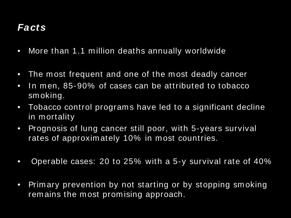
Facts
• More than 1.1 million deaths annually worldwide
• The most frequent and one of the most deadly cancer• In men, 85-90% of cases can be attributed to tobacco
smoking. • Tobacco control programs have led to a significant decline
in mortality• Prognosis of lung cancer still poor, with 5-years survival
rates of approximately 10% in most countries.
• Operable cases: 20 to 25% with a 5-y survival rate of 40%
• Primary prevention by not starting or by stopping smoking remains the most promising approach.

Facts
• No known familial lung cancer• For same cigarette consumption: • Different risks depending of the enzymatic profile
(aryl hydrocarbon hydrolase: increased activity induced by metabolites in tobacco smoke)
• Risk increased from 4 to 10 folds
• Genetics
• The urge to classify….

The Pathologist's role in Lung Cancer
• Introduction• Classification• Preinvasive lesions• The biopsies:
– Current presentations– Problem areas– Immunohistochemistry
• Surgical specimens– Staging, check list
• Testing • Rare cases: epathologies.com

WHO Blue BooksIARC Presshttp://www.iarc.fr/

• Malignant epithelial tumours • Squamous cell carcinoma
– Papillary – Clear cell – Small cell – Basaloid
• Small cell carcinoma – Combined small cell carcinoma
• Adenocarcinoma – Adenocarcinoma, mixed subtype – Acinar adenocarcinoma – Papillary adenocarcinoma – Bronchioloalveolar carcinoma
• Nonmucinous• Mucinous• Mixed nonmucinous and mucinous or
indeterminate
– Solid adenocarcinoma with mucin production • Fetal adenocarcinoma • Mucinous (“colloid”) carcinoma • Mucinous cystadenocarcinoma• Signet ring adenocarcinoma • Clear cell adenocarcinoma
• Large cell carcinoma – Large cell neuroendocrine carcinoma – Combined large cell neuroendocrine
carcinoma – Basaloid carcinoma – Lymphoepithelioma-like carcinoma – Clear cell carcinoma – Large cell carcinoma with rhabdoid phenotype
• Adenosquamous carcinoma • Sarcomatoid carcinoma
– Pleomorphic carcinoma – Spindle cell carcinoma – Giant cell carcinoma – Carcinosarcoma – Pulmonary blastoma
• Carcinoid tumour – Typical carcinoid – Atypical carcinoid
• Salivary gland tumours – Mucoepidermoid carcinoma – Adenoid cystic carcinoma – Epithelial-myoepithelial carcinoma

• Mesenchymal tumours
• Epithelioid haemangioendothelioma• Angiosarcoma• Pleuropulmonary blastoma• Chondroma• Congenial peribronchial
myofibroblastic tumour• Diffuse pulmonary lymphangiomatosis• Inflammatory myofibroblastic tumour• Lymphangioleiomyomatosis• Synovial sarcoma
– Monophasic– Biphasic
• Pulmonary artery sarcoma • Pulmonary vein sarcoma
• Lymphoproliferative tumours• Marginal zone B-cell lymphoma of the
MALT • Diffuse large B-cell lymphoma• Lymphomatoid granulomatosis• Langerhans cell histiocytosis
• Miscellaneous tumours• Harmatoma• Sclerosing hemangioma• Clear cell tumour• Germ cell tumours• Teratoma, mature • Immature • Other germ cell tumours• Intrapulmonary thymoma• Melanoma
• Metastatic tumours

The Pathologist's role in Lung Cancer
• Introduction• Classification• Preinvasive lesions• The biopsies:
– Current presentations– Problem areas– Immunohistochemistry
• Surgical specimens– Staging, check list
• Testing • Rare cases: epathologies.com

Pre-invasive lesions
• Dysplasia and carcinoma insitu – Squamous cell carcinoma (proximal carcinoma)
• Atypical adenomatous hyperplasia– Adenocarcinoma (peripheral carcinoma)
• Diffuse neuro-endocrine hyperplasia– Carcinoid tumours
• No known precursor for Small Cell Carcinoma

Dysplasia and carcinoma insitu Squamous cell carcinoma (proximal carcinoma)
Normal respiratory mucosa
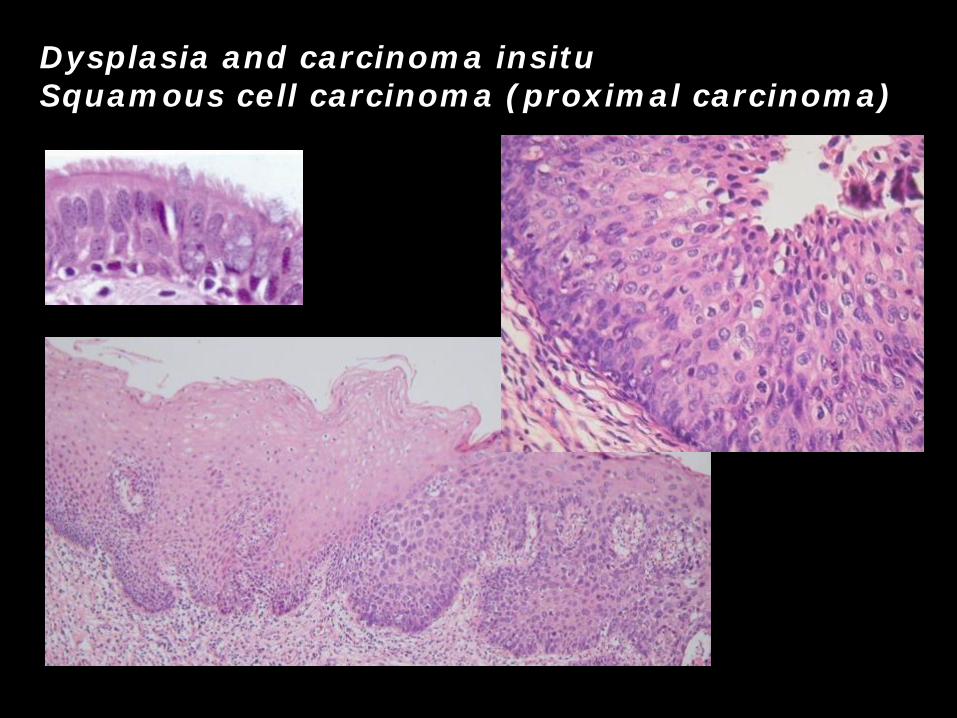
Dysplasia and carcinoma insitu Squamous cell carcinoma (proximal carcinoma)

Dysplasia and carcinoma insitu Squamous cell carcinoma
CIS
Severe Dysplasia
CIS

• Dysplasia and carcinoma insituSquamous cell carcinoma (proximal carcinoma)

Preinvasive lesions. Sequential molecular changes during the multistage pathogenesis of squamous cell lung carcinoma.

Pre-invasive lesions
• Dysplasia and carcinoma insitu – Squamous cell carcinoma (proximal carcinoma)
• Atypical adenomatous hyperplasia– Adenocarcinoma (peripheral carcinoma)
• Diffuse neuro-endocrine hyperplasia– Carcinoid tumours
• No known precursor for Small Cell Carcinoma
Atypical adenomatous hyperplasiaAdenocarcinoma (peripheral carcinoma)

Atypical adenomatous hyperplasiaAdenocarcinoma (peripheral carcinoma)
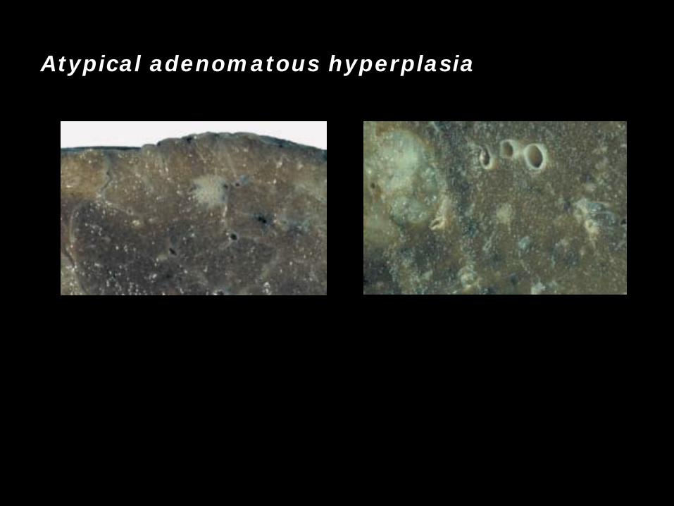
Atypical adenomatous hyperplasia

Pre-invasive lesions
• Dysplasia and carcinoma insitu – Squamous cell carcinoma (proximal carcinoma)
• Atypical adenomatous hyperplasia– Adenocarcinoma (peripheral carcinoma)
• Diffuse neuro-endocrine hyperplasia– Carcinoid tumours
• No known precursor for Small Cell Carcinoma

The Pathologist's role in Lung Cancer
• Introduction• Classification• Preinvasive lesions• The biopsies:
– Current presentations– Problem areas– Immunohistochemistry
• Surgical specimens– Staging, check list
• Testing • Rare cases: epathologies.com

Current pathological presentations: the biopsies
• Squamous Cell Carcinoma
• Adenocarcinoma, NOS
• Small Cell Lung Carcinoma
• Large cell neuroendocrine carcinoma

Tissue collection and interpretation
• Optimal tissue collection, for precise classification (sputum, BALavage, bronchoscopic, thoracoscopic, and needle biopsies)
• Rapid fixation and minimal trauma are important. • Small specimens may not show differentiation when the
tumour is excised; it is, therefore, advisable to limit categorization to SCLC and NSCLC.
• The current classification is largely based on standard H&E sections.
• Some lung carcinomas remain unclassified. They usually fall into the “non-small cell carcinoma” category or are cases where small biopsy or cytology specimens preclude definitive histologic typing.
Ref. : Clinical features and staging, in Pathology and Genetics of Tumours of the Lung..Travis W. and al, WHO, IARC Press 2004

Histologic heterogeneity
• Variation in appearance and differentiation from microscopic field to field and from one histologic section to the next
• Almost 50% of lung carcinomas exhibit more than one of the major histologic types. This fact has important implications for lung tumour classification and must be kept in mind, especially when interpreting small biopsies.
Ref. : Clinical features and staging, in Pathology and Genetics of Tumours of the Lung..Travis W. and al, WHO, IARC Press 2004

Squamous cell carcinoma: M 44 % vs F 25%

Adenocarcinoma : M 28 % vs F 42 %
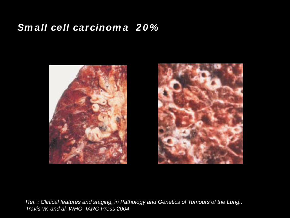
Small cell carcinoma 20%
Ref. : Clinical features and staging, in Pathology and Genetics of Tumours of the Lung..Travis W. and al, WHO, IARC Press 2004

Large cell carcinoma: 9 %
• Large cell neuroendocrine carcinoma– Chromogranin A, or Synaptophysine, or CD56, TTF1 (40%)
• Combined large cell neuroendocrine carcinoma • Basaloid carcinoma
– CK 5/6, 34bE12, NE(-), comedonecrosis, no squamous diff.
• Lymphoepithelioma-like carcinoma – EBV, Lymphoid infiltrate
• Clear cell carcinoma • Large cell carcinoma with rhabdoid phenotype

Neuro-endocrine tumours:
Carcinoid tumour: central and peripheral

Adenosquamous carcinoma

The Pathologist's role in Lung Cancer
• Introduction• Classification• Preinvasive lesions• The biopsies:
– Current presentations– Problem areas– Immunohistochemistry
• Surgical specimens– Staging, check list, and reporting
• Testing • Rare cases: epathologies.com

Immunohistochemistry : the markers
• Epithelial markers:– Cytokeratins– Low Molecular weight
• CK7 CK20
– High Molecular weight• CK 5/6, 34bE12
– Cocktails– Epithelial membrane antigen
• Neuroendocrine markers:– Chromogranin A– Synaptophysin– CD 56
• Specific : – Thyroid Transcription
Factor 1 (TTF1)
• Other markers: – Lymphoid– CD99– Ki67 (MIB-1)– Connective tissue
• Vascular • Adipose• Nervous

The Pathologist's role in Lung Cancer
• Introduction• Classification• Preinvasive lesions• The biopsies:
– Current presentations– Problem areas– Immunohistochemistry
• Surgical specimens (Candidates for surgery: 1/3 cases)
– Staging, check list, and reporting
• Testing • Rare cases: epathologies.com

Surgical specimens: Pathological staging pTNM
• A. Primary Tumor: – pTX Primary tumor cannot be assessed – pT0 No evidence of primary tumor – pTis Carcinoma in situ – pT1 Tumor 3 cm or less in greatest dimension,
surrounded by lung or visceral pleura, not invading the main bronchus
– pT2 Tumor with any of the following features of size or extent
• More than 3 cm in greatest dimension • Invades visceral pleura • Involves main bronchus, 2 cm or more distal to
the carina • Associated with atelectasis or obstructive
pneumonitis which extends to the hilar region but does not involve the entire lung
– pT3 Tumor of any size that directly invades any of the following
• Parietal pleura ,Chest wall (including superior sulcus tumors) ,Diaphragm, Mediastinal pleura, Parietal pericardium
• Tumor in the main stem bronchus less than 2 cm distal to the carina but without involvement of the carina
• Associated atelectasis or obstructive pneumonitisof the entire lung
– pT4 Tumor of any size that invades any of the following • Mediastinum ,heart ,great vessels,trachea
esophagus,vertebral body ,carina • Or tumor with malignant pleural effusion • Or separate tumor nodules in the same lobe.
• B. Regional Lymph Nodes: – pNX Regional lymph nodes cannot
be assessed – pN0 No regional lymph node
metastasis – pN1 Metastasis in ipsilateral
peribronchial and/or hilar lymph nodes, and intrapulmonary nodes, including direct extension.
– pN2 Metastasis in ipsilateral mediastinal and/or subcarinal lymph nodes
– pN3 Metastasis in contralateral mediastinal, contralateral hilar, ipsilateral or contralateral scalene or supraclavicular lymph nodes
• C. Distant Metastasis – pMX Cannot be assessed – pM0 No distant metastasis – pM1 Distant metastasis

The surgical specimens: preparation

The surgical specimens:
• Central tumor, pneumonectomy
• Peripheral tumor, lobectomy
• Peripheral Carcinoid tumor, surgical excision
The Pathologist's role in Lung Cancer
• Introduction• Classification• Preinvasive lesions• The biopsies:
– Current presentations– Problem areas– Immunohistochemistry
• Surgical specimens– Staging, check list, and reporting
• Testing • Rare cases: epathologies.com

The Pathologist's role in Lung Cancer
• Introduction• Classification• Preinvasive lesions• The biopsies:
– Current presentations– Problem areas– Immunohistochemistry
• Surgical specimens– Staging, check list, and reporting
• Testing: Case of Bronchoscopic biopsy • Rare cases: www.epathologies.com

The Pathologist's role in Lung Cancer
• Introduction• Classification• Preinvasive lesions• The biopsies:
– Current presentations– Problem areas– Immunohistochemistry
• Surgical specimens– Staging, check list, and reporting
• Testing: Bronchoscopic biopsy • Rare cases: www.epathologies.com

Thank you for your attention


![Research Article Increased Chromogranin A Cell Density in ...downloads.hindawi.com/journals/grp/2015/823897.pdf · organ in the body [ ]. ese cells project specialized microvilli](https://static.fdocuments.in/doc/165x107/5fd64d49c22ac35b4b7b6b56/research-article-increased-chromogranin-a-cell-density-in-organ-in-the-body.jpg)


