
Early reversal of right ventricular dysfunction in ... · therapy. Our major objective was to...
8
JACC Vol. 10, No.5 November 19H7 :971-8 COOPERATIVE STUDIES 971 Early Reversal of Right Ventricular Dysfunction in Patients With Acute Pulmonary Embolism After Treatment With Intravenous Tissue Plasminogen Activator PATRICIA C. COME, MD, FACC, DUCKSOO KIM, MD, J, ANTHONY PARKER, MD, PHD, SAMUEL Z. GOLDHABER, MD, FACC, * EUGENE BRAUNWALD, MD, FACC, JOHN E. MARKIS, MD and PARTICIPATING INVESTIGATORS Boston, Massachusetts To assess abnormalities of right heart function and their reversal with thrombolysis in pulmonary embolism, se- rial imaging and Doppler echocardiographic studies were performed before and after a 6 hour intravenous infusion of 80 to 90 mg of recombinant tissue-type plasminogen activator (rt-Pa) in seven patients with segmental or lobar acute pulmonary embolism. None of the five men and two women had known prior pulmonary hyperten- sion. Substantial clot lysis and improvement in pulmo- nary blood flow, as determined by serial pulmonary an- giography and perfusion lung scanning, were achieved in all. Coincident with clot lysis, pulmonary artery systolic pressure decreased (from 42 ± 11 to 26 ± 7 mm Hg, p < 0.005), right ventricular diameter decreased (from 3.9 ± 1.0 to 2.0 ± 0.5 em, p < 0.005) and left ven- tricular diameter increased (from 3.7 ± 0.9 to 4.4 ± 0.6 em, p < 0.01). Right ventricular wall movement, initially mildly, moderately or severely hypokinetic in Pulmonary embolism can be a catastrophic event, resulting in early death or serious hemodynamic dysfunction (1-4). Sudden increases in pulmonary artery pressure, especially in patients without prior chronic heart or lung disease re- sulting in progressive right ventricular hypertrophy, may cause right ventricular failure and hypotension (1,2). Pre- vious echocardiographic studies (5-13) have detailed certain A list of Participating Investigators appears in the Appendix. From the Charles A. Dana Research Institute of the Harvard-Thorndike Laboratory, Departments of Medicine and Radiology, Beth Israel Hospital and Harvard Medical School, Boston, Massachusetts and the *Department of Medicine, Brigham and Women's Hospital, Boston, Massachusetts. This study was presented in part at the Annual Meeting of the American College of Car- diology, New Orleans, Louisiana, March 19l57. Manuscript received May II. 1987; revised manuscript received June 17, 19l57, accepted June 26, 1987. Address for reprints: Patricia C. Come, MD, Cardiology Division. Beth Israel Hospital, 330 Brookline Avenue, Boston, Massachusetts 02215. Ii.) 1987 by the American College of Cardiology one, two and four patients, respectively, normalized in five and improved to mild hypokinesia in two. Tricuspid regurgitation was present before lytic therapy in six pa- tients. In five, flow velocity in the tricuspid regurgitant jets indicated a peak systolic right ventricular minus right atrial pressure gradient of 25 to 52 mm Hg. Tri- cuspid regurgitation was detected early after lytic ther- apy in only two patients. Systolic septal flattening was noted before but not after lysis. These findings confirm that pulmonary emboli may result in appreciable right ventricular dysfunction and dilation, resultant tricuspid regurgitation, abnormal septal position and decreased left ventricular size. Their early reversal after rt-PA suggests that this therapy might be efficacious in the management of patients with pulmo- nary embolism presenting with more serious hemody- namic compromise. The potential of rt -PAin this regard merits further investigation. () Am Colt Cardiol 1987;10:971-8) changes that may be seen in pulmonary embolism, namely, thrombi within the right heart chambers or pulmonary artery, right ventricular dilation, right ventricular hypokinesia, ab- normalities of septal movement, reduced left ventricular cavity dimension and dilation of the pulmonary artery. The cited studies generally did not address whether detected abnormalities diminished over time, except to state that detected thrombi often disappeared on follow-up study (5,8,10,12). Nicolosi et al. (7), however, observed de- creases in right ventricular size, increases in left ventricular size and changes in septal curvature in follow-up studies. This study was designed to determine prospectively the presence and magnitude of right ventricular dysfunction, using imaging and Doppler echocardiography, in consec- utive patients with symptomatic, angiographically docu- mented acute pulmonary embolism who met the criteria for inclusion in an ongoing trial of experimental thrombolytic 0735-1097/X7/$3.50
Transcript of Early reversal of right ventricular dysfunction in ... · therapy. Our major objective was to...

JACC Vol. 10, No.5November 19H7:971-8
COOPERATIVE STUDIES
971
Early Reversal of Right Ventricular Dysfunction in Patients WithAcute Pulmonary Embolism After Treatment With Intravenous TissuePlasminogen Activator
PATRICIA C. COME, MD, FACC, DUCKSOO KIM, MD, J, ANTHONY PARKER, MD, PHD,
SAMUEL Z. GOLDHABER, MD, FACC,* EUGENE BRAUNWALD, MD, FACC,
JOHN E. MARKIS, MD and PARTICIPATING INVESTIGATORS
Boston, Massachusetts
To assess abnormalities of right heart function and theirreversal with thrombolysis in pulmonary embolism, serial imaging and Doppler echocardiographic studies wereperformed before and after a 6 hour intravenous infusionof 80 to 90 mg of recombinant tissue-type plasminogenactivator (rt-Pa) in seven patients with segmental orlobar acute pulmonary embolism. None of the five menand two women had known prior pulmonary hypertension. Substantial clot lysis and improvement in pulmonary blood flow, as determined by serial pulmonary angiography and perfusion lung scanning, were achievedin all.
Coincident with clot lysis, pulmonary artery systolicpressure decreased (from 42 ± 11 to 26 ± 7 mm Hg,p < 0.005), right ventricular diameter decreased (from3.9 ± 1.0 to 2.0 ± 0.5 em, p < 0.005) and left ventricular diameter increased (from 3.7 ± 0.9 to 4.4 ±0.6 em, p < 0.01). Right ventricular wall movement,initially mildly, moderately or severely hypokinetic in
Pulmonary embolism can be a catastrophic event, resulting
in early death or serious hemodynamic dysfunction (1-4).
Sudden increases in pulmonary artery pressure, especiallyin patients without prior chronic heart or lung disease resulting in progressive right ventricular hypertrophy, maycause right ventricular failure and hypotension (1,2). Previous echocardiographic studies (5-13) have detailed certain
A list of Participating Investigators appears in the Appendix. From theCharles A. Dana Research Institute of the Harvard-Thorndike Laboratory,Departments of Medicine and Radiology, Beth Israel Hospital and HarvardMedical School, Boston, Massachusetts and the *Department of Medicine,Brigham and Women's Hospital, Boston, Massachusetts. This study waspresented in part at the Annual Meeting of the American College of Cardiology, New Orleans, Louisiana, March 19l57.
Manuscript received May II. 1987; revised manuscript received June17, 19l57, accepted June 26, 1987.
Address for reprints: Patricia C. Come, MD, Cardiology Division.Beth Israel Hospital, 330 Brookline Avenue, Boston, Massachusetts 02215.
Ii.)1987 by the American College of Cardiology
one, two and four patients, respectively, normalized infive and improved to mild hypokinesia in two. Tricuspidregurgitation was present before lytic therapy in six patients. In five, flow velocity in the tricuspid regurgitantjets indicated a peak systolic right ventricular minusright atrial pressure gradient of 25 to 52 mm Hg. Tricuspid regurgitation was detected early after lytic therapy in only two patients. Systolic septal flattening wasnoted before but not after lysis.
These findings confirm that pulmonary emboli mayresult in appreciable right ventricular dysfunction anddilation, resultant tricuspid regurgitation, abnormal septalposition and decreased left ventricular size. Their earlyreversal after rt-PA suggests that this therapy might beefficacious in the management of patients with pulmonary embolism presenting with more serious hemodynamic compromise. The potential of rt -PAin this regardmerits further investigation.
() Am Colt Cardiol 1987;10:971-8)
changes that may be seen in pulmonary embolism, namely,thrombi within the right heart chambers or pulmonary artery,right ventricular dilation, right ventricular hypokinesia, abnormalities of septal movement, reduced left ventricularcavity dimension and dilation of the pulmonary artery. Thecited studies generally did not address whether detectedabnormalities diminished over time, except to state thatdetected thrombi often disappeared on follow-up study(5,8,10,12). Nicolosi et al. (7), however, observed decreases in right ventricular size, increases in left ventricularsize and changes in septal curvature in follow-up studies.
This study was designed to determine prospectively thepresence and magnitude of right ventricular dysfunction,using imaging and Doppler echocardiography, in consecutive patients with symptomatic, angiographically documented acute pulmonary embolism who met the criteria forinclusion in an ongoing trial of experimental thrombolytic
0735-1097/X7/$3.50

972 COME ET AL.VENTRICULAR DYSFUNCTION IN PULMONARY EMBOLISM
lACC Vol. 10, NO.5November 1987:971-8
therapy. Our major objective was to determine whether detected abnormalities of cardiac function are rapidly reversedafter intravenous administration of recombinant tissue-typeplasminogen activator (rt-PA), a relatively fibrin-specificthrombolytic agent that results in significant and markedimprovement in pulmonary artery patency as early as 2 to6 hours after the start of infusion (14).
MethodsStudy patients. Eight consecutive patients undergoing
pulmonary angiography and experimental recombinant tissue-type plasminogen activator (rt-PA) therapy for angiographically confirmed pulmonary embolism were considered for serial echocardiographic evaluation of right ventricularfunction. One patient was excluded because the echocardiographic equipment was not available before administration of lytic therapy. To be included in the study, patientshad to be 18 years of age or older, with angiographic documentation of pulmonary embolism in segmental or moreproximal pulmonary artery branches. Lytic therapy had tobe administered within 5 days of the onset of symptoms,and patients with prior pulmonary embolism were ineligible.Written consent was obtained from all patients, accordingto a protocol approved by the Human Subjects Committeeof the Beth Israel Hospital and by the U.S. Food and DrugAdministration.
Echocardiographic evaluation. M-mode, two-dimensional and pulsed Doppler echocardiographic studies wereperformed using an ATL Mark 600 echocardiograph. Twodimensional scans were obtained in parasternal long- andshort-axis planes, apical two and four chamber planes andthe subcostal four chamber plane. Right ventricular wallmovement was graded as normal or as mildly, moderatelyor severely hypokinetic on the basis of analysis of rightventricular wall movement in the combination of parasternal, apical and subcostal views. M-mode echocardiogramswere generated using a two-dimensionally-directed cursorfrom the left ventricle at the upper chordal level, with thepatient in the supine position. M-mode tracings were takenfrom the parasternal long-axis view (six patients) or fromthe subcostal four chamber view (one patient in whom theparasternal view was technically difficult). Right ventriculardimension was measured from the leading edge of the rightventricular wall to the leading edge of the right septal surfaceat the onset of the QRS complex (15). Right ventricularwall thickness was measured from the leading edge of theright side of the right ventricular wall to the leading edgeof its left side. Left ventricular diastolic diameters at theupper chordal level were measured from the leading edgeof the left septal surface to the leading edge of the posteriorwall at the onset of the QRS complex (15).
Pulsed Doppler echocardiography including, when necessary, high pulsed repetition frequency technology was
used to determine the presence of tricuspid regurgitation andto quantitate its flow velocity. Using short-axis, right ventricular inflow tract and apical four chamber views, thesample volume was positioned on the atrial side of thetricuspid valve. Holosystolic turbulence was considered indicative of tricuspid regurgitation. Maximal flow velocityin the tricuspid regurgitant jet was measured, and the rightventricular (RY) to right atrial (RA) pressure gradient wasdetermined according to the modified Bernoulli equation(16): (RY pressure - RA pressure [in mm Hg]) == 4 v:where Y == maximal flow velocity in the tricuspid regurgitant jet.
The imaging and Doppler echocardiographic studies wereperformed a mean of 10 hours (range 0 to 14) before administration of intravenous rt-PA and a mean of 9 hours (rangeoto 36) after completion of therapy.
Catheterization technique and drug administration.An 8F Grollman catheter was placed, using the Seldingertechnique, through the femoral vein into the right heartchambers and pulmonary artery. Right atrial, right ventricular and pulmonary artery pressures were recorded usingHewlett-Packard transducers (l295A) and a Hewlett-Packard monitoring system (78353A). Systemic arterial pressurewas measured using a cuff sphygmomanometer rather thanan indwelling arterial catheter to avoid a potential site forbleeding with subsequent thrombolytic therapy. Selectivepulmonary angiograms were performed, with the side ofinjection (right or left pulmonary artery) generally chosenaccording to which lung on perfusion scanning had the greaternumber of defects. After diagnosis of thrombus in a segmental or more proximal pulmonary artery branch, 50 mgof rt-PA (Activase, Genentech, Inc.) was infused intravenously for 2 hours at a rate of 25 mg/h. Selective pulmonaryangiography was repeated at the end of the 2 hour infusion.If significant thrombus or thrombi remained, an additional40 mg of rt-PA was infused intravenously over the next 4hours at a rate of 10 mg/hour, unless precluded by bleedingcomplications such as an expanding groin hematoma. Totaldoses of rt-PA given to the seven patients in this study were85 ± 5 mg. At the end of the 6 hour total infusion, selectivepulmonary angiograms were repeated. The total amount ofcontrast medium (diatrizoate meglumine) used was 30 to 40cc for each of the three angiographic evaluations.
Analysis of pulmonary angiograms. The pulmonaryangiograms were coded, scrambled as to whether they werepretreatment, 2 hour or 6 hour films, and read withoutknowledge of patient data by a panel of six investigators.The scoring system used for analysis of the single lungangiogram was similar to the one employed in the UrokinasePulmonary Embolism Trial (17) for assessment of both lungs,and has been described previously (14). The unilateral lungangiograms were graded from 0 (no clot) to 9 (massive clot).Medium-sized emboli were given a score of <3 and generally involved one lobe. Large emboli were given a score
lACC Vol. 10, No 5November 1987:971-8
COME ET AL.VENTRICULAR DYSFUNCTION IN PULMONARY EMBOLISM
973
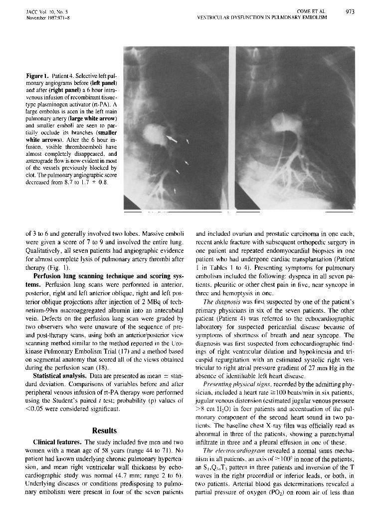
Figure 1. Patient 4. Selective leftpulmonary angiograms before (leftpanel)andafter(right panel) a 6 hourintravenous infusion of recombinant tissuetype plasminogen activator (rt-PA). Alarge embolus is seen in the left mainpulmonary artery (large whitearrow)and smaller emboli are seen to partially occlude its branches (smallerwhite arrows). After the 6 hour infusion, visible thromboemboli havealmost completely disappeared, andanterograde flow isnow evident inmostof the vessels previously blocked byclot. The pulmonary angiographic scoredecreased from 8.7 to 1.7 ± 0.8.
of 3 to 6 and generally involved two lobes. Massive emboliwere given a score of 7 to 9 and involved the entire lung.Qualitatively, all seven patients had angiographic evidencefor almost complete lysis of pulmonary artery thrombi aftertherapy (Fig. I).
Perfusion lung scanning technique and scoring systems. Perfusion lung scans were performed in anterior,posterior, right and left anterior oblique, right and left posterior oblique projections after injection of 2 MBq of technetium-99m macroaggregated albumin into an antecubitalvein. Defects on the perfusion lung scan were graded bytwo observers who were unaware of the sequence of preand post-therapy scans, using both an anterior/posterior viewscanning method similar to the method reported in the Urokinase Pulmonary Embolism Trial (17) and a method basedon segmental anatomy that scored all of the views obtainedduring the perfusion scan (18).
Statistical analysis. Data are presented as mean ± standard deviation. Comparisons of variables before and afterperipheral venous infusion ofrt-PA therapy were performedusing the Student's paired t test; probability (p) values of<0.05 were considered significant.
ResultsClinical features. The study included five men and two
women with a mean age of 58 years (range 44 to 71). Nopatient had known underlying chronic pulmonary hypertension, and mean right ventricular wall thickness by echocardiographic study was normal (4.7 mm; range 2 to 6).Underlying diseases or conditions predisposing to pulmonary embolism were present in four of the seven patients
and included ovarian and prostatic carcinoma in one each,recent ankle fracture with subsequent orthopedic surgery inone patient and repeated endomyocardial biopsies in onepatient who had undergone cardiac transplantation (PatientI in Tables I to 4). Presenting symptoms for pulmonaryembolism included the following: dyspnea in all seven patients, pleuritic or other chest pain in five, near syncope inthree and hemoptysis in one.
The diagnosis was first suspected by one of the patient'sprimary physicians in six of the seven patients. The otherpatient (Patient 4) was referred to the echocardiographiclaboratory for suspected pericardial disease because ofsymptoms of shortness of breath and near syncope. Thediagnosis was first suspected from echocardiographic findings of right ventricular dilation and hypokinesia and tricuspid regurgitation with an estimated systolic right ventricular to right atrial pressure gradient of 27 mm Hg in theabsence of identifiable left heart disease.
Presenting physical signs, recorded by the admitting physician, included a heart rate 2: 100 beats/min in six patients,jugular venous distension (estimated jugular venous pressure>8 em H20 ) in four patients and accentuation of the pulmonary component of the second heart sound in two patients. The baseline chest X-ray film was officially read asabnormal in three of the patients, showing a parenchymalinfiltrate in three and a pleural effusion in one of these.
The electrocardiogram revealed a normal sinus mechanism in all patients, an axis of 2: 100° in none of the patients,an S!,Q3,T3 pattern in three patients and inversion of the Twaves in the right precordial or inferior leads, or both, intwo patients. Arterial blood gas determinations revealed apartial pressure of oxygen (P02) on room air of less than
974 COME ET AL.VENTRICULAR DYSFUNCTION IN PULMONARY EMBOLISM
JACC Vol. 10, No.5November 1987:971-8
Table 1. Hemodynamic Findings in Seven Patients
Heart Rate PA Systolic Pressure RA Pressure Systemic Arterial(beats/min) (mm Hg) (mm Hg) Pressure (mm Hgj PA/SAP
Patient No. Pre Post Pre Post Pre Post Pre Post Pre Post
1 92 97 37 19 4 2 120 120 0.31 0.162 78 80 38 32 7 5 118 130 0.32 0.253 80 84 34 23 2 2 132 114 0.26 0.204 97 82 54 28 19 12 130 160 0.42 0.185 92 72 49 38 10 8 132 114 0.37 0.336 107 95 29 20 2 102 145 0.28 0.147 92 92 56 22 5 2 112 104 0.50 0.21
Mean ±SD 91 ± 10 86 ± 9 42 ± II 26 ± 7* 7 ± 6 5 ± 4t 121 ± II 127 ± 20 0.35 ± 0.08 0.21 ± 0.06*
*p < 0.01, pre vs. post; tp < 0.05, pre vs. post. BP = blood pressure; HR = heart rate; PA = pulmonary artery; PA/SAP = ratio ofpulmonaryartery systolic pressure to systemic arterial systolic pressure; Post and Pre = after and before thrombolytic therapy, respectively; RA = right atrial.
80 torr in five of the six patients whose blood gases wereassessed.
Several patients had minor. easily controlled bleedingfrom venipuncture and prior radial arterial (for blood gasanalysis) sites during or early after rt-PA infusion. The onlymajor complication was hemorrhage into an ovarian carcinoma in Patient 6, occurring several hours after rt-PAinfusion and necessitating exploratory laparotomy and removal of the previously undiagnosed pelvic mass.
Hemodynamic findings (Table 1). Administration ofrecombinant tissue-type plasminogen activator (rt-PA) wasassociated with significant reductions in pulmonary arterysystolic pressure (from 42 ± 11 to 26 ± 7 mm Hg, p <0.01) and right atrial pressure (from 7 ± 6 to 5 ± 4 mmHg, p < 0.05). The ratio of systolic pulmonary artery pressure to systolic systemic arterial pressure decreased from0.35 ± 0.08 to 0.21 ± 0.06 (p < 0.01).
Pulmonary angiographic findings (Table 2, Fig. 1).Significant improvement in pulmonary angiographic scoreswas noted at both 2 hours (p < 0.01) and 6 hours (p <0.001) after the start of rt-PA infusion, and repeat lungperfusion scans at 24 hours demonstrated significant increases in pulmonary blood flow (p < 0.0001 and p <
Table 2. Pulmonary Angiographic Scores in Seven Patients
Patient 2 Hours 6 HoursNo. Pre Post Post
1 7.8 7.5 5.52 8.8 8.4 1.23 8.3 6.8 3.34 8.7 7.5 1.75 5.7 4.0 1.26 8.5 5.8 2.87 8.7 6.0 4.0
Mean 8.1 6.6 2.8± SO 1.1 1.5* 1.6t
*p < 0.01, 2 hours post vs pre; tp < 0.001, 6 hours post vs pre. Postand Pre = after and before thrombolytic therapy, respectively.
0.005 using the segmental and UPET scoring methods, respectively).
Perfusion lung scan findings (Table 3, Fig. 2). Themean time between infusion of thrombolytic therapy andthe post-therapy lung scans was 25.2 ± 3.8 hours. All sevenpatients demonstrated improvement in the perfusion lungscan after rt-PA. The initial score, based on the method ofthe Urokinase Pulmonary Embolism Trial (17), improved65 ± 18% from 0.45 ± 0.21 to 0.16 ± 0.11 (p < 0.005).The initial score, based on the segmental method, improved62 ± 8% from 0.49 ± 0.19 to 0.18 ± 0.08 (p < 0.001).
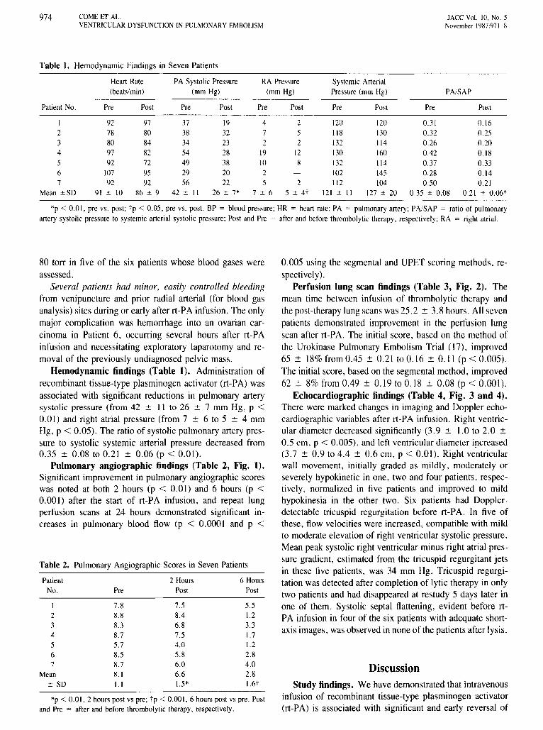
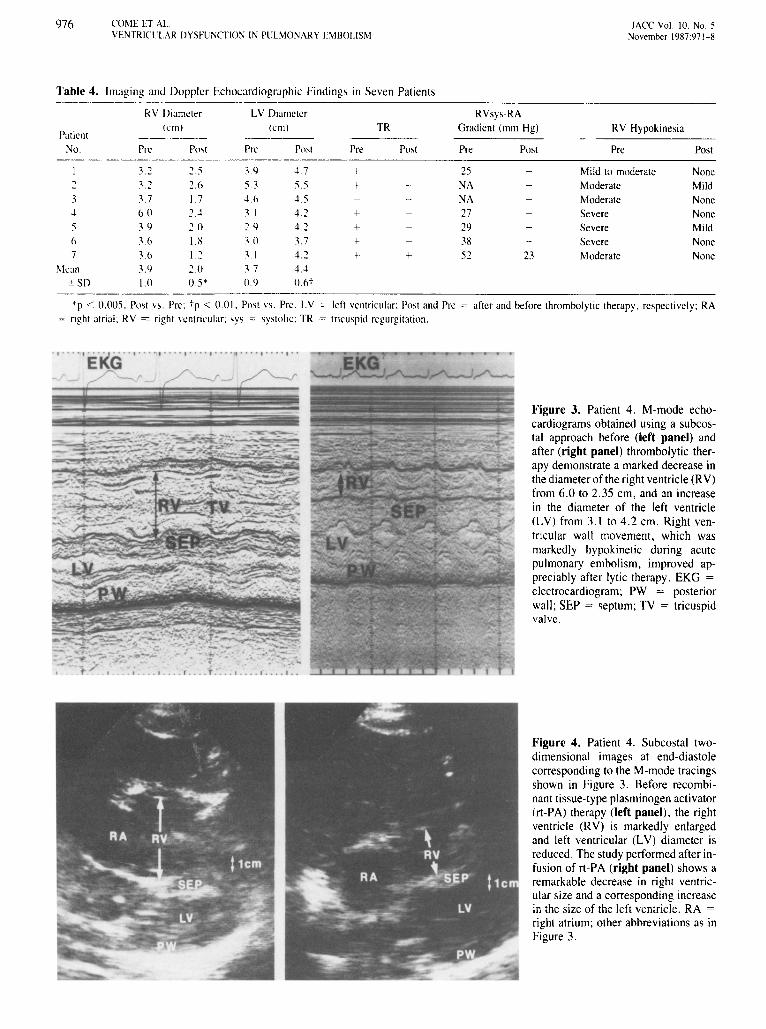
Echocardiographic findings (Table 4, Fig. 3 and 4).There were marked changes in imaging and Doppler echocardiographic variables after rt-PA infusion. Right ventricular diameter decreased significantly (3.9 ± 1.0 to 2.0 ±0.5 ern, p < 0.005), and left ventricular diameter increased(3.7 ± 0.9 to 4.4 ± 0.6 em, p < 0.01). Right ventricularwall movement, initially graded as mildly, moderately orseverely hypokinetic in one, two and four patients, respectively, normalized in five patients and improved to mildhypokinesia in the other two. Six patients had Dopplerdetectable tricuspid regurgitation before rt-PA. In five ofthese, flow velocities were increased, compatible with mildto moderate elevation of right ventricular systolic pressure.Mean peak systolic right ventricular minus right atrial pressure gradient, estimated from the tricuspid regurgitant jetsin these five patients, was 34 mm Hg. Tricuspid regurgitation was detected after completion of lytic therapy in onlytwo patients and had disappeared at restudy 5 days later inone of them. Systolic septal flattening, evident before rtPA infusion in four of the six patients with adequate shortaxis images, was observed in none of the patients after lysis.
DiscussionStudy findings. We have demonstrated that intravenous
infusion of recombinant tissue-type plasminogen activator(rt-PA) is associated with significant and early reversal of

JACC Vol. 10, No.5November 1YH7:971-H
COME ET AI..VENTRICUl.AR DYSFUNCTION IN PUl.MONARY EMBOLISM
Table 3. Lung Scan Scores in Seven Patients
Segmental Method Ul'E'l MethodPatient
No. Pre Post Pre Post
0.40 o 17 0..17 0.192 0.41 0.20 O.4h 0.293 0.31 0.09 o 12 0.044 0.32 0.14 (U7 0.055 0.51 o 13 0.46 0.0.1h O.hX 022 05h 0.257 (l.XI (U2 OXO 0.25
Mean 0.49 o IX 0.45 0.16±SD 0.[9 O.OH' 0.21 0.111"
*p < 0.005, post vs. pre: I"p < 0.001. Post vs. Pre. Post and Pre c; after and hefore thrombolytic therapy,respectively: UPET = Urokinase Pulmonary Embolism Trial.
975
right ventricular dilation and hypokinesia, tricuspid regurgitation and decreased left ventricular cavity dimension.These changes occur with decreases in pulmonary artery
Figure 2. Patient 4, Perfusion lung scans inanterior (ANT), posterior (POST), right anterior oblique (RAO), left anterior oblique(LAO), right posterior oblique (RPO) and left posterior oblique(LPO) projections before (top panels) and after (bottom panels)recombinant tissue-type plasminogen activator (rt-PA) infusion,Multiple segmental areas of absent or reduced perfusion are seenon the first study. The study performed after lytic therapy revealsmuch more homogeneous lung uptake, with only small residualperfusion defects. By the Urokinase Pulmonary Embolism Trial(17) lung scan scoring method, this patient had an 86% improvement inscore from 0.37 to0.05. Bythesegmental scoring system,there was a 56% improvement, with the score decreasing from0,32 before to 0,14 after rt-PA infusion.
pressure and with evidence of reperfusion on angiographicstudy, This study is the first to show reversal of imagingand Doppler echocardiographic abnormalities after thrombolytic therapy. The early reversal of the detected abnormalities suggests that the right ventricle is not "stunned."but rather is capable of recovering quickly in response to adecrease in right ventricular afterload.
Pathophysiology of acute pulmonary embolism. Theright ventricle of an individual without prior heart or lungdisease cannot generate mean pulmonary artery pressureshigher than 30 to 40 mm Hg, presumably because of theabsence of right ventricular hypertrophy (I), With progressive increases in pulmonary vascular resistance. therefore,the right ventricle begins to fail, resulting in elevation ofright atrial pressure and symptoms and signs of right heartfailure (I), There is a significant relation between meanpulmonary artery pressure and mean right atrial pressure( I), Stroke volume and. consequently. cardiac output maybe maintained in some patients on the basis of increasedsympathetic drive and the Frank-Starling mechanism, Inothers, generally those with massive embolism. failure tomaintain stroke volume may result in a decreased cardiacoutput, cardiogenic shock and death, In patients with priorheart or lung disease, or both, right ventricular hypertrophyusually permits generation of higher pulmonary artery pressures (19), In such patients with antecedent pulmonary hypertension, there is no relation between right ventriculardysfunction and the extent of angiographic obstruction (2),Mortality is increased in patients with pulmonary embolismwho have obvious impairment of right ventricular function (2),
Most clinical studies likely have underestimated the prevalence and severity of hemodynamic disturbances resultingfrom symptomatic pulmonary embolism. because they haveincluded patients surviving long enough to be evaluated andenrolled in study protocols. Mortality is very high and occursearly in patients presenting with major hemodynamic compromise. Studies (3,4) have documented that 50. 70 and85% of such patients die within 30 minutes. I hour and 6hours. respectively,
976 COME ET At.VENTRICUl.AR DYSFUNCTlO:-l IN PUl.~lONARY EMBOLISM
lACC Vol. 10. No.5November 1987:971-8
RVsys~RA
Gradient (mm Hg)
Table 4. Imaging and Doppler Echocardiographic Findings in Seven Patients~-------
RV Diameter l.V Diameter(UII) (ern) TR
Patient -_._-- -----No. Pre Pnst Pre Post Pre Post-_.._-- ----~_.-----.__ ._-- ,_._~-------
.L2 2.5 39 47 +2 .1.2 2.0 53 5.5 +3 3.7 1.7 4.0 4.54 60 2.4 3.1 42 +5 39 20 2.9 4.2 +f> 3f> I.K .1.0 3.7 +7 3.6 I 2 .1.1 4.2 + +
Mean 3.9 2.0 37 4.4.~SD 1.0 0.5* 0.9 o.st
----- ._--"_.,._.----_.,--
Pre
25NANA27293X52
Post
23
RV Hypokinesia
Pre
Mild to moderateModerateModerateSevereSevereSevereModerate
Post
NoneMildNoneNoneMildNoneNone
'p < 0.005. Post vs. Pre: tp < 001. Post vs. Pre. LV '" left ventricular; Post and Pre > after and before thrombolytic therapy. respectively; RA'" right atrial: RV = right ventricular; sys '" systolic; TR = tricuspid regurgitation .
• , . •. • I E·KG············,··········,·········,··/' - I' -J -' /"----r
. I .... ' .• . . , . . . . r .... I • • •. I • •• • I ••
-----~-~ ----- - '--- ._--- -. - -r..: -=--- -- - - -
Figure 3. Patient 4. M-mode echocardiograms obtained using a subcostal approach before (left panel) andafter (right panel) thrombolytic therapy demonstrate a marked decrease inthe diameter of the right ventricle (RV)from 6.0 to 2.35 em, and an increasein the diameter of the left ventricle(LV) from 3.1 to 4.2 em. Right ventricular wall movement, which wasmarkedly hypokinetic during acutepulmonary embolism, improved appreciably after lytic therapy. EKG =electrocardiogram; PW = posteriorwall; SEP = septum; TV = tricuspidvalve.
Figure 4. Patient 4. Subcostal twodimensional images at end-diastolecorresponding to the M-mode tracingsshown in Figure 3. Before recombinant tissue-type plasminogen activator(rt-PA) therapy (left panel), the rightventricle (RV) is markedly enlargedand left ventricular (LV) diameter isreduced. The study performed after infusion of rt-PA (right panel) shows aremarkable decrease in right ventricular size and a corresponding increasein the size of the left ventricle. RA =right atrium; other abbreviations as inFigure 3.

lACC Vol. 10, No.5November 1987:971- 8
COME ET AL.VENTRIC ULAR DYSFUNCTION IN PULMONARY EMBOLISM
977
Treatment of acute pulmonary embolism. Conventional therapy for acute pulmonary embolism has been theadministrationof intravenous heparin. Although heparin hasbeen shown experimentally to prevent recurrences of pulmonary emboli (20) and progression of thrombus format ionin areas of pulmonary artery embolism (21), it produces nomajor change in the extent of pulmonary perfusion abnormalities, assessed by lung scan, or in the magnitude ofpulmonary artery pressure, right atrial pressure and totalpulmonary vascular resistance elevation when measurements are repeated 24 hours afterthe start of therapy (17,22).In contrast, thrombolytic therapy with streptokinase or urokinase has been associated with a significant reduction inthe extent of angiographic obstruction, perfusion abnormalities and elevations of pulmonary artery pressure, rightheart pressuresand total pulmonary resistance 24 hoursafterthe start of infusion (17,22,23). Recent reports (14,24) havealso documented the efficacy of rt-PA, a relatively clotspecific fibrinolytic agent. In a study of 30 patients withangiographically proved pulmonary embolism presentingwithin 5 days of the onset of symptoms, Goldhaber et al.(14) found significant angiographic evidence of clot lysisand significant decreases in pulmonary artery pressure 6hours after the start of 2 to 6 hour intravenous infusions ofrt-PA.
The favorable angiographic, perfusion lung scan andechocardiographic changes observed after administration ofthrombolytic agents inour patients suggest that such therapymight be effective in decreasing mortality in that subgroupof patients with massive embolism who survive long enoughto have the diagnosis considered. Certainly, thrombolytictherapy can be instituted more rapidly in hemodynamicallyunstable patients than can open embolectomy on cardiopulmonary bypass, a procedure that has been successful insaving some patients with hemodynamic decompensationbutcarries an operative mortality rate of 24 to 93% (25-27 ).Transvenous catheter embolectomy can be undertaken morequickly than can open surgical embolectomy. Although successful thrombectomy may produce improvement in cardiovascular function in some patients, mortality remainshigh (25) and the procedure requires the availability of acatheterization laboratory. Our study did not address thepotential for rt-PA therapy to improve cardiovascular function or survival in patients with pulmonary embolism withprior cardiac or pulmonary disease or with shock resultingfrom acute pulmonaryembolism. The efficacy of rt-PA therapy in such patients merits investigation.
Most pulmonary emboli probably undergo endogenouslysis over a numberof days, accounting for the observationsin the Urokinase Pulmonary Embolism Trial (17) that meanlung scan improvement by 7 days was equivalent in theheparin-treated and the urokinase/heparin-treated groups. Insome patients, however, pulmonary emboli persist and become organized, and the resultantchanges may be the cause
of a syndrome of unresolved pulmonary emboli characterized by symptoms of dyspnea, persistence of perfusion lungscan abnormalities, mild to moderate pulmonary hypertension and right ventricular hypertrophy (28- 31). It is notknown whether thrombolytic therapy followed by long-termanticoagulation will be preferable to anticoagulation alonein decreasing the incidence of chronic morbidity related topulmonary embolism.
Considerations for future studies. Our favorable shortterm results in selected patients without severe cardiac orrespiratory compromise suggest that randomized trials comparing thrombolytic therapy with anticoagulant therapy aloneand examining different types of thrombolytic therapy shouldbe undertaken to determine their relative risks and efficaciesin decreasing mortality from massive pulmonary embolismand preventing potential syndromes of unresolved pulmonary embolism, chronic hypoxemia and peripheral venousdisease. Such studies should optimally include periodic assessment of right ventricular performance, pulmonary function and the peripheral venous system in addition to evaluation of the acute hemodynamic, angiographic and perfusion lung scan changes after therapy.
AppendixOther participating investigators include
Beth Israel Hospital, Gerald M. Kolodny, MD.Br igham and Women' s Hospital , B. Leonard Holman . MD; Joseph
Loscalzo, MD, PhD; Michael Meyerovitz, MD; Kathleen Reagan. MD;Andrew P. Se lwyn. MD; Sabah Turneh; MD; Douglas Vaughan. MD .
Gene nlech , Inc. , Elliott B. Grossbard, MD .Geor ge Washington Universit y Medical Cenler, Edward Druy, MD:
William Kelly , MD; Craig M. Kessler, MD: K. Siena Kirwin , MD.Veterans Administration Medical Center at Brockton/West Rox
bury, Douglas L. Dawley. MD: Daniel A. Pietro . MD; Arthur Sasahara,MD; G.V.R.K. Sharma, MD.
ReferencesI. Mcint yre KM, Sasahara AA. The hemodynamic response to pulmo
nary embo lism in patients without prior cardiopulmonary disease . AmJ Cardiol 1971;211:2811-93.
2. Mcint yre KM. Sasahara AA. Hemodynamic and ventricular responsesto pulmonary embolism. Prog Cardiovasc Dis 1974;17: 175-88.
3. Gorham LW. A study of pulmonary embolism. Arch Intern Med1961;108:76-90 .
4. Soloff LA. Rodman T. Acute pulmonary embolism. Am Heart J 1967:74:829- 47 .
5. Kasper W. Meinertz T. Henkel B. et al. Echocardiog raphic findingsin patients with proved pulmonary embolism. Am Heart J 1986;112:12114-90.
6. Kasper W. Meinenz T. Kersting F, Lollgen H. Limbourg P. Just H.Echocardiog raphy in asses sing acute pulmonary hyperten sion due topulmonary embolism. Am 1 Cardiol 1980;45:567-72.
7. Nicolosi GC, Dall 'A glio V, Burelli C , Targa S. Zardo F. Zanutt iniD. Studio ecocardiografico Mvmode e bidimensionale nell' embol iapolmonare in fase acuta . G lia l Cardiel 1982;12:17-24.
ll. Armstrong WF, Feigenbaum H, Dillon Jc. Echocardiographic detection of right atrial thromboembolism . Chest 1985;87:801-6.

978 COME ET AL.VENTRICULAR DYSFUNCTION IN PULMONARY EMBOLISM
lACC Vol. 10, NO.5November J9H7:971-H
9. Starkey IR. deBono DP. Echocardiographic identification of rightsided cardiac intracavitary thromboembolus in massive pulmonaryembolism. Circulation 1982;66: 1322-5.
10. Saner HE, Asinger RW, Daniel JA, Elsperger KJ. Two-dimensionalechocardiographic detectionof right-sidedcardiac intracavitary thromboembolus with pulmonary embolism. J Am Coil Cardiol 1984;4:1294-301.
II. Farfel Z, Shechter M, Vered Z, Rath S, Goor D, Gafni J. Review ofechocardiographically diagnosed right heart entrapment of pulmonaryemboli-in-transit with emphasis on management. Am HeartJ 1987;113:171-8.
12. CameronJ, PohlnerPG, Stafford EG, O'Brien MF. BellJHN, MurphyAL. Right heart thrombus: recognition, diagnosis and management.J Am Coil Cardiol 1985;5:1239-43.
13. Bochereau G, Scheuble C, Cereze P. Chauveau P, Juonitzky D,Kleinknecht D. Interet de l'echographie dans Ie diagnostic d'un coeurpulmonaire aigu: a propos de 6 observations. Aggressologie 1980;21:c-112-9.
14. Goldhaber SZ, Vaughan DE, Markis JE, et al. Acute pulmonaryembolism treated with tissue plasminogen activator. Lancet 1986;2:886-9.
15. Sahn DJ, DeMaria A, Kisslo J, Weyman A. Recommendations regarding quantitation in M-modeechocardiography: results of a surveyof echocardiographic measurements. Circulation 1978;58: 1072-82.
16. Hatle L. Angelsen B. Doppler Ultrasound in Cardiology. 2nd ed.Philadelphia; Lea & Febiger, 1982:23.
17. The Urokinase Pulmonary Embolism Trial: a national cooperativestudy. Circulation I973;47(suppl 11):11-1-108.
18. Markis JE, Goldhaber SZ, Kim DS, Palla A, Parker JA, BraunwaldE. Early improved pulmonaryperfusionafter intravenousrecombinanttissue plasminogen activator for acute pulmonary embolism (abstr).Circulation 1986;74(suppl 11):11-127.
19. McIntyre KM. Sasahara AA. Determinants of right ventricular function and hemodynamics after pulmonary embolism. Chest 1974;65:534-42.
20. Wessler S, Morris LE. Studies in intravascular coagulation: the effectof heparin and dicumarol on serum-induced venous thrombosis. Circulation 1955;12:553-6.
21. Carey LC, WilliamsRD. Comparativeeffects ofdicumarol, Tromexanand heparin on thrombus propagation. Ann Surg 1960;152:919-22.
22. HirshJ, Hale GS, McDonaldIG, McCarthy RA, Pitt A. Streptokinasetherapy in acute major pulmonary embolism: effectiveness and problems. Br Med J 1968;4:729-34.
23. Tow DE. Wagner HN. Recovery of pulmonary arterial blood flow inpatients with pulmonaryembolism. N Engl J Med 1967;276:1053-9.
24. Bounameux H, Vermylen 1. Collen D. Thrombolytic treatment withrecombinant tissue-type plasminogen activator in a patient with massive pulmonary embolism. Ann Intern Med 1985;103:64-6.
25. Stewart JR, Greenfield LJ. Transvenous vena cava filtration and pulmonary embolectomy. Surg Clin North Am 1982;62:411-30
26. Mattox KL, Feldtman RW, Beall AC, DeBakey ME. Pulmonary embolectomy for acutemassive pulmonary embolism. Ann Surg 1982; 195:726-30.
27. Glassford DM Jr. Alford WC Jr. Burrus GR, Stoney WS. ThomasCS Jr. Pulmonary embolectomy. Ann Thorac Surg 1981;32:28-32.
28. Hollister LE. Cull VL. The syndrome of chronic thrombosis of themajor pulmonary arteries. Am J Med 1956;21:312-20.
29. Orell SR. The fate and late effects of non-fatal pulmonary emboli.Acta Med Scand 1962:172:473-84.
30. Benotti R, Ockene IS, Alpert S, Dalen JE. The clinical profile ofunresolved pulmonary embolism. Chest 1983;84:669-77.
31. Chitwood WR Jr, Lyerly HK, Sabiston DC Jr. Surgical managementof chronic pulmonary embolism. Ann Surg 1985;20I:11-26.