Early periprosthetic leak after percutaneous closure of a patent foramen ovale - Dr. José A....
12
Early Periprosthetic Leak After Percutaneous Closure of a Patent Foramen Ovale Jose A. G. ÁLVAREZ MD MTSAC FSCAI Head of Interventional Cardiology British Hospital in Buenos Aires
Transcript of Early periprosthetic leak after percutaneous closure of a patent foramen ovale - Dr. José A....
Early Periprosthetic Leak After Percutaneous Closure of a
Patent Foramen Ovale
Jose A. G. ÁLVAREZ MD MTSAC FSCAIHead of Interventional CardiologyBritish Hospital in Buenos Aires

Potential conflicts of interest
Speaker's name: José Amadeo Guillermo Álvarez
I do not have any potential conflict of interest
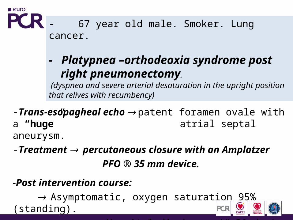
- 67 year old male. Smoker. Lung cancer.
- Platypnea –orthodeoxia syndrome post right pneumonectomy. (dyspnea and severe arterial desaturation in the upright position that relives with recumbency)
-Trans-esopagheal echo patent foramen ovale with a “huge”
atrial septal aneurysm.-Treatment percutaneous closure with an Amplatzer
PFO ® 35 mm device.
-Post intervention course: Asymptomatic, oxygen saturation 95% (standing).
Hospital discharge (# 3º day)
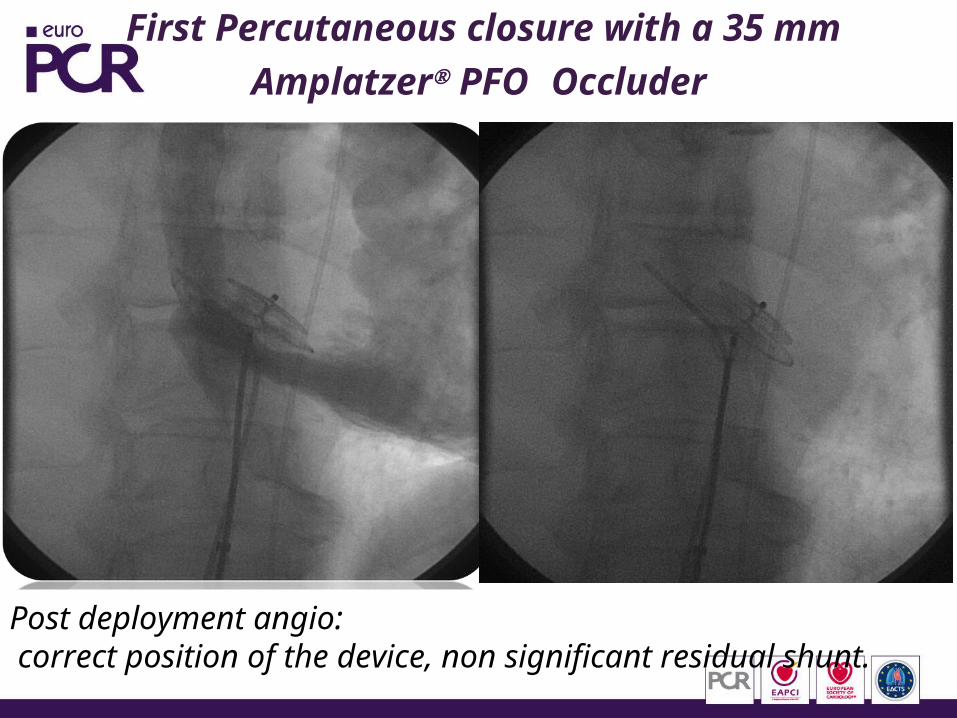
First Percutaneous closure with a 35 mmAmplatzer PFO Occluder
Post deployment angio: correct position of the device, non significant residual shunt.
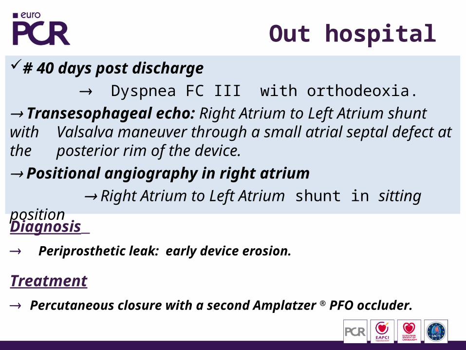
Out hospital course# 40 days post discharge
Dyspnea FC III with orthodeoxia. Transesophageal echo: Right Atrium to Left Atrium shunt with Valsalva maneuver through a small atrial septal defect at the posterior rim of the device. Positional angiography in right atrium
Right Atrium to Left Atrium shunt in sitting position
Diagnosis
Periprosthetic leak: early device erosion.
Treatment
Percutaneous closure with a second Amplatzer ® PFO occluder.
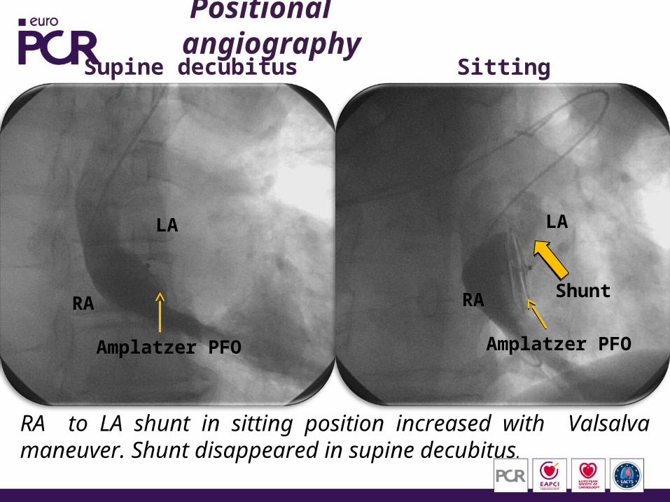
Positional angiographySupine decubitus Sitting
RA to LA shunt in sitting position increased with Valsalva maneuver. Shunt disappeared in supine decubitus.
Amplatzer PFO
LA
RA
Amplatzer PFO
Shunt
LA
RA
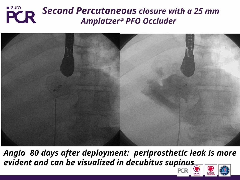
Second Percutaneous closure with a 25 mm Amplatzer PFO Occluder
Angio 80 days after deployment: periprosthetic leak is more evident and can be visualized in decubitus supinus
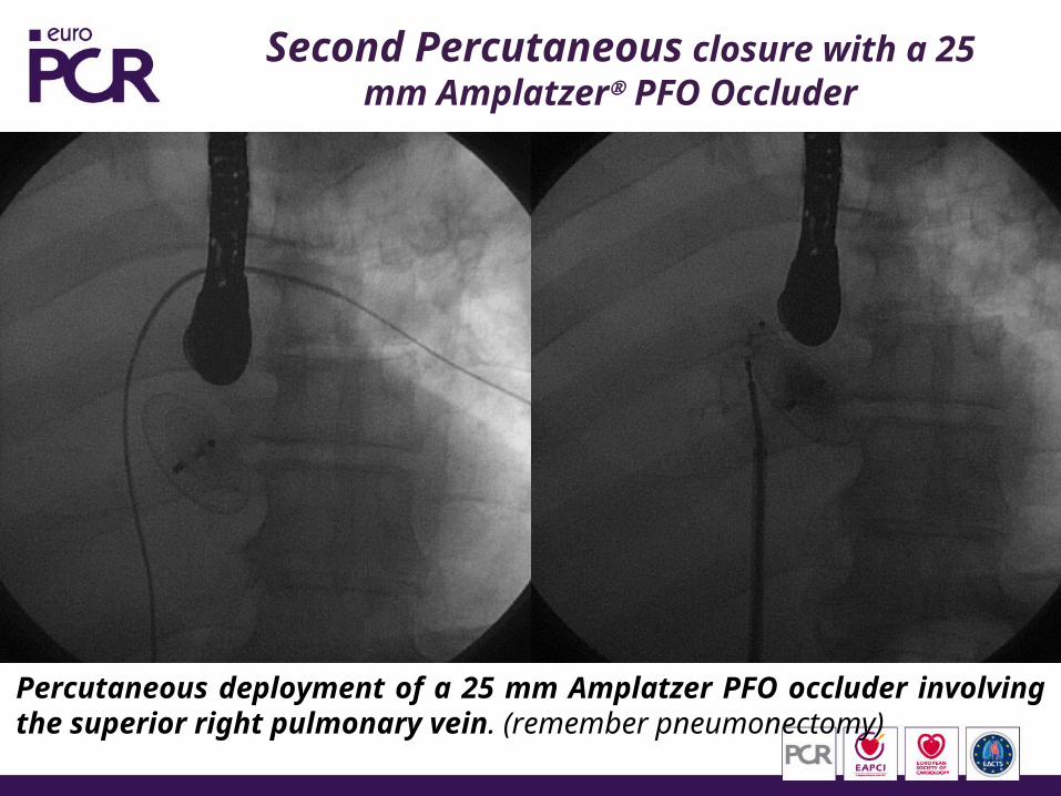
Second Percutaneous closure with a 25 mm Amplatzer PFO Occluder
Percutaneous deployment of a 25 mm Amplatzer PFO occluder involving the superior right pulmonary vein. (remember pneumonectomy)
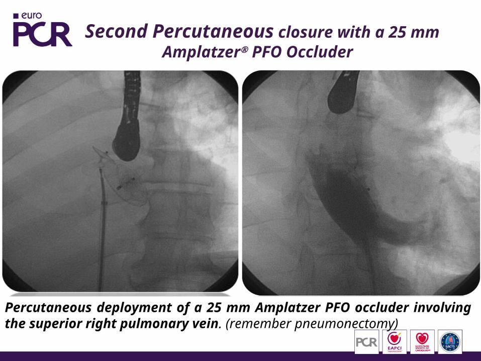
Second Percutaneous closure with a 25 mm Amplatzer PFO Occluder
Percutaneous deployment of a 25 mm Amplatzer PFO occluder involving the superior right pulmonary vein. (remember pneumonectomy)
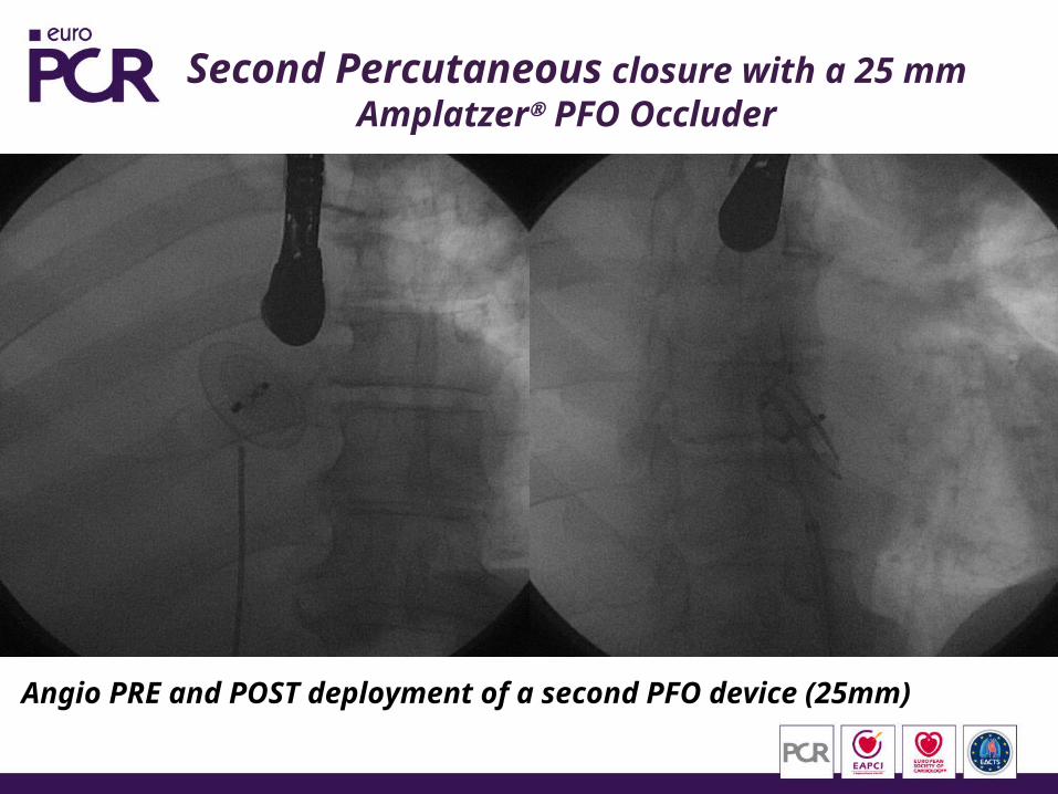
Second Percutaneous closure with a 25 mm Amplatzer PFO Occluder
Angio PRE and POST deployment of a second PFO device (25mm)
Out hospital course
Asymptomatic eversince (3 years of follow up)
In transesophageal echo performed 1 year after the procedure, there was no evidence of residual Right Atrium to Left Atrium shunt
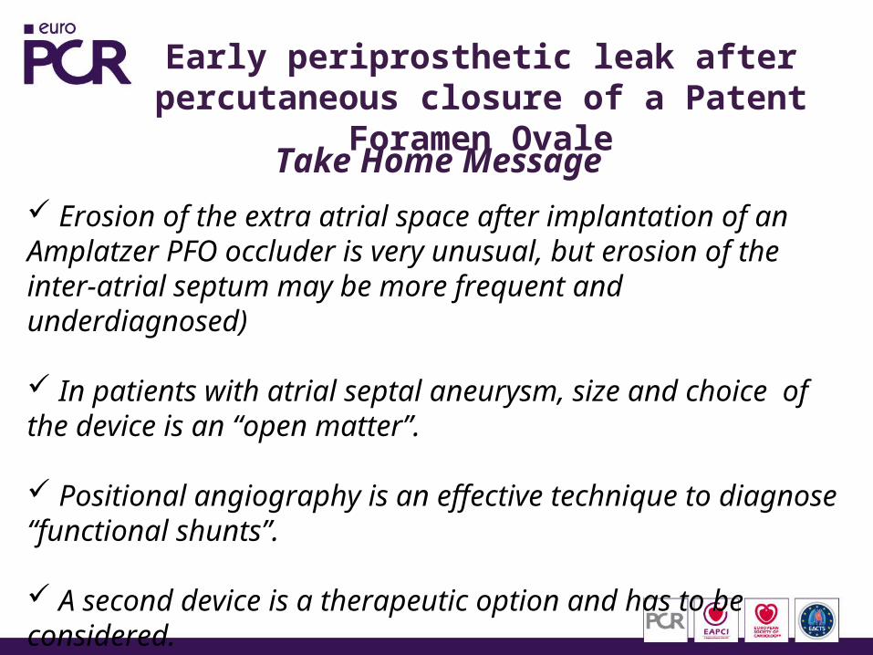
Early periprosthetic leak after percutaneous closure of a Patent Foramen Ovale
Take Home Message Erosion of the extra atrial space after implantation of an Amplatzer PFO occluder is very unusual, but erosion of the inter-atrial septum may be more frequent and underdiagnosed)
In patients with atrial septal aneurysm, size and choice of the device is an “open matter”.
Positional angiography is an effective technique to diagnose “functional shunts”.
A second device is a therapeutic option and has to be considered.