Early neuropathology of somatostatin/NPY GABAergic cells...
15
Neurobiology of Aging 27 (2006) 1658–1672 Early neuropathology of somatostatin/NPY GABAergic cells in the hippocampus of a PS1 × APP transgenic model of Alzheimer’s disease Blanca Ramos a,1 , David Baglietto-Vargas b,1 , Juan Carlos del Rio a,1 , Ines Moreno-Gonzalez b,1 , Consuelo Santa-Maria a , Sebastian Jimenez a , Cristina Caballero a , Juan Felix Lopez-Tellez b , Zafar U. Khan c , Diego Ruano a , Antonia Gutierrez b,∗∗ , Javier Vitorica a,∗ a Department Bioquimica, Bromatologia, Toxicologia y Medicina Legal, Facultad de Farmacia, Universidad de Sevilla, 41012 Sevilla, Spain b Department Biologia Celular, Genetica y Fisiologia, Facultad de Ciencias, Universidad de Malaga, Malaga, Spain c CIMES/Department Medicina, Facultad de Medicina, Universidad de Malaga, Malaga, Spain Received 10 June 2005; received in revised form 26 July 2005; accepted 19 September 2005 Available online 3 November 2005 Abstract At advanced stages, Alzheimer’s disease (AD) is characterized by an extensive neuronal loss. However, the early neurodegenerative deficiencies have not been yet identified. Here we report an extensive, selective and early neurodegeneration of the dendritic inhibitory interneurons (oriens-lacunosum moleculare, O-LM, and hilar perforant path-associated, HIPP, cells) in the hippocampus of a transgenic PS1 × APP AD model. At 6 months of age, from 22 different pre- and postsynaptic mRNA markers tested (including GABAergic, glutamatergic and cholinergic markers), only the expression of somatostatin (SOM) and NPY neuropeptides (O-LM and HIPP markers) displayed a significant decrease. Stereological cell counting demonstrated a profound diminution (50–60%) of SOM-immunopositive neurons, preceding the pyramidal cell loss in this AD model. SOM population co-expressing NPY was the most damaged cell subset. Furthermore, a linear correlation between SOM and/or NPY deficiency and Abeta content was also observed. Though the molecular mechanism of SOM neuronal loss remains to be determined, these findings might represent an early hippocampal neuropathology. Therefore, SOM and NPY neuropeptides could constitute important biomarkers to assess the efficacy of potential early AD treatments. © 2005 Elsevier Inc. All rights reserved. Keywords: Alzheimer’s disease; Neurodegeneration; Transgenic mice; Biomarkers; Hippocampus; Interneurons; Cholinergic; Glutamatergic; GABAergic 1. Introduction Alzheimer’s disease (AD) is the most common neurologi- cal disorder affecting predominantly individuals over age 65. Patients with AD show a loss in memory and a decrease in their cognitive abilities. These changes are due to the progres- sive dysfunction and death of nerve cells that are responsible for the storage and processing of information. The patho- ∗ Corresponding author. Tel.: +34 954 556770; fax: +34 954 233765. ∗∗ Corresponding author. Tel.: +34 952 133344; fax: +34 952 132000. E-mail addresses: [email protected] (A. Gutierrez), [email protected] (J. Vitorica). 1 Authors contributed equally to this work. logical hallmarks of AD are beta amyloid (Abeta) deposits and plaques, hyperphosphorylation of tau and formation of neurofibrillary tangles, degeneration of synapses and loss of neuronal cells [52,53,39,44]. A relatively high number of PS1, APP or PS1 × APP trans- genic (tg) mice has been generated as valuable models of AD [23]. These tg mice models reproduce some but not all aspects of the disease. In this sense, APP or PS1 × APP tg mice displayed Abeta accumulation, neuritic alteration and behav- ioral and cognitive deficiencies but not the massive neuronal loss characteristic of the human disease [23,2]. The appari- tion of Abeta depositions clearly precedes the decrease in the pyramidal cell number [43]. The reasons for these discrep- ancies are actually unknown, however, it is plausible that the 0197-4580/$ – see front matter © 2005 Elsevier Inc. All rights reserved. doi:10.1016/j.neurobiolaging.2005.09.022
Transcript of Early neuropathology of somatostatin/NPY GABAergic cells...
A
diPastclc©
K
1
cPtsf
v
0d
Neurobiology of Aging 27 (2006) 1658–1672
Early neuropathology of somatostatin/NPY GABAergic cells in thehippocampus of a PS1 × APP transgenic model of Alzheimer’s disease
Blanca Ramos a,1, David Baglietto-Vargas b,1, Juan Carlos del Rio a,1,Ines Moreno-Gonzalez b,1, Consuelo Santa-Maria a, Sebastian Jimenez a,
Cristina Caballero a, Juan Felix Lopez-Tellez b, Zafar U. Khan c, Diego Ruano a,Antonia Gutierrez b,∗∗, Javier Vitorica a,∗
a Department Bioquimica, Bromatologia, Toxicologia y Medicina Legal, Facultad de Farmacia, Universidad de Sevilla, 41012 Sevilla, Spainb Department Biologia Celular, Genetica y Fisiologia, Facultad de Ciencias, Universidad de Malaga, Malaga, Spain
c CIMES/Department Medicina, Facultad de Medicina, Universidad de Malaga, Malaga, Spain
Received 10 June 2005; received in revised form 26 July 2005; accepted 19 September 2005Available online 3 November 2005
bstract
At advanced stages, Alzheimer’s disease (AD) is characterized by an extensive neuronal loss. However, the early neurodegenerativeeficiencies have not been yet identified. Here we report an extensive, selective and early neurodegeneration of the dendritic inhibitorynterneurons (oriens-lacunosum moleculare, O-LM, and hilar perforant path-associated, HIPP, cells) in the hippocampus of a transgenicS1 × APP AD model. At 6 months of age, from 22 different pre- and postsynaptic mRNA markers tested (including GABAergic, glutamatergicnd cholinergic markers), only the expression of somatostatin (SOM) and NPY neuropeptides (O-LM and HIPP markers) displayed aignificant decrease. Stereological cell counting demonstrated a profound diminution (50–60%) of SOM-immunopositive neurons, precedinghe pyramidal cell loss in this AD model. SOM population co-expressing NPY was the most damaged cell subset. Furthermore, a linear
orrelation between SOM and/or NPY deficiency and Abeta content was also observed. Though the molecular mechanism of SOM neuronaloss remains to be determined, these findings might represent an early hippocampal neuropathology. Therefore, SOM and NPY neuropeptidesould constitute important biomarkers to assess the efficacy of potential early AD treatments.2005 Elsevier Inc. All rights reserved.
markers
lann
eywords: Alzheimer’s disease; Neurodegeneration; Transgenic mice; Bio
. Introduction
Alzheimer’s disease (AD) is the most common neurologi-al disorder affecting predominantly individuals over age 65.atients with AD show a loss in memory and a decrease in
heir cognitive abilities. These changes are due to the progres-ive dysfunction and death of nerve cells that are responsibleor the storage and processing of information. The patho-
∗ Corresponding author. Tel.: +34 954 556770; fax: +34 954 233765.∗∗ Corresponding author. Tel.: +34 952 133344; fax: +34 952 132000.
E-mail addresses: [email protected] (A. Gutierrez),[email protected] (J. Vitorica).1 Authors contributed equally to this work.
g[odiltpa
197-4580/$ – see front matter © 2005 Elsevier Inc. All rights reserved.oi:10.1016/j.neurobiolaging.2005.09.022
; Hippocampus; Interneurons; Cholinergic; Glutamatergic; GABAergic
ogical hallmarks of AD are beta amyloid (Abeta) depositsnd plaques, hyperphosphorylation of tau and formation ofeurofibrillary tangles, degeneration of synapses and loss ofeuronal cells [52,53,39,44].
A relatively high number of PS1, APP or PS1 × APP trans-enic (tg) mice has been generated as valuable models of AD23]. These tg mice models reproduce some but not all aspectsf the disease. In this sense, APP or PS1 × APP tg miceisplayed Abeta accumulation, neuritic alteration and behav-oral and cognitive deficiencies but not the massive neuronal
oss characteristic of the human disease [23,2]. The appari-ion of Abeta depositions clearly precedes the decrease in theyramidal cell number [43]. The reasons for these discrep-ncies are actually unknown, however, it is plausible that the
gy of A
datra
pesSed
s(plontbbtroidtdbfc(ObIis
GotttsTcpe
ieeqom
mPSA
2
2
aPmrbmAgg
bsfo
2
TiiswarRTatg
opaaa
2
a
B. Ramos et al. / Neurobiolo
ifferent neuronal populations displayed a different vulner-bility to the Abeta depositions. Therefore, the objective ofhis work was to establish whether specific GABAergic neu-onal populations might be particularly vulnerable at earlyges in the hippocampus of tg models.
The possible implications of the GABAergic system in theathological evolution of these tg models have never beenxtensively evaluated, despite that GABAergic neurons con-tituted the major inhibitory system in the Central Nervousystem. In this sense, we have previously observed a prefer-ntial vulnerability of the hippocampal GABAergic systemuring normal aging (see below).
Thus, in this work we have focused on hippocamaplomatostatin (SOM) and NPY expressing interneuronsoriens-lacunosum moleculare, O-LM, and hilar perforantath-associated, HIPP, cells) for four main reasons. First, theoss of SOM and/or NPY in AD patients is a well-reproducedbservation. In this sense, the existence of a reduction in theumber of cortical SOM/NPY immunopositive cells [13,11],he SOM/NPY content in diverse brain areas [14] and in cere-rospinal fluid [37] has been described. In fact, as mentionedy Saito et al. [42], SOM is one of the few transcripts (0.5%)hat consistently reduced in aged human brains (see alsoef. [31]). Second, as mentioned above, we have previouslybserved that the hippocampal GABAergic system [41] and,n particular, the SOM interneurons were primarily affecteduring normal aging [49], the major risk factor in AD. Third,he APP and/or PS1 × APP tg models are hyperactive [48,17],isplaying disturbed activity patterns similar to those inducedy the GABAergic inhibition [4]. Four, at the hippocampalormation, the SOM and NPY neuropeptides were principallyo-expressed by the O-LM (in CA fields) and the HIPP cellsin the dentate gyrus) interneurons [19,36]. Importantly, the-LM and HIPP interneurons constituted a highly vulnera-le cell population to several different insults and conditions.n this sense, these interneurons were preferentially affectedn several models of temporal lobe epilepsy [8,9,12,15], alsoensitive to ischemic conditions [20].
On the other hand, this interneuronal subset is the majorABAergic population innervating the distal dendritic arborf pyramidal and granular neurons [34], fired rhythmically atheta frequency [28,29] and may regulate dendritic integra-ion and plasticity [22]. In this sense, it has been demonstratedhe implication of SOM neuropeptide in the acquisition ofpatial maps and learning induced by novel environments [3].hus, the high cellular vulnerability together with its impli-ation in memory processes makes this particular neuronalopulation a good candidate to be preferentially affected atarly stages of the AD and in AD models.
Therefore, in this work we have evaluated whether thenterneuronal population containing SOM/NPY was prefer-ntially affected in transgenic models of Alzheimer’s dis-
ase. We have thus studied the mRNA expression (usinguantitative RT-PCR) and the neuronal density (using stere-logy) of this interneuronal population in a double tgodel (PS1 × APP mice). This previously characterized tgTHft
ging 27 (2006) 1658–1672 1659
odel [6,43] over-expressed the mutated form of humanS1 (PS1M146L) and human APP (APP751 with both thewedish and London mutations; SL) proteins and developedbeta depositions at early ages (3–4 months).
. Methods
.1. TG mice
The generation and initial characterization of the PS1nd PS1 × APP tg mice has been reported previously [6].S1 tg mice (C57BL/6 background) over-expressed theutated PS1M146L form under the control of the HMGCoA-
eductase promoter. PS1 × APP double tg mice (C57BL/6ackground) were obtained by crossing homozygotic PS1 tgice with heterozygotic Thy1-APP751SL mice (Transgeniclliance). Mice represented F6–F10 offspring of heterozy-ous tg mice. Non-transgenic mice of the same genetic back-round were used as controls.
Anesthetized male mice were killed by decapitation andoth hippocampi were dissected, frozen in liquid N2 andtored at −80 ◦C until use. All animal experiments were per-ormed in accordance with the guidelines of the Committeen Animal Research of the University of Seville.
.2. RNA and protein extraction
Total RNA and proteins were extracted using theripureTM Isolation Reagent (Roche), according to the
nstructions of the manufacturer. This procedure allows thesolation of total RNA, DNA and protein fractions from aingle sample. The contaminating DNA in the RNA samplesas removed by incubation with DNAase (Sigma–Aldrich)
nd confirmed by PCR analysis of total RNA samples prioreverse transcription (RT). After isolation, the integrity of theNA samples was assessed by agarose gel electrophoresis.he yield of total RNA was determined by measuring thebsorbance (260/280 nm) of ethanol precipitated aliquots ofhe samples. The recovery of RNA was comparable in allroups (1.2–1.5 �g/mg of tissue).
In order to analyze the protein fraction, the protein pelletsbtained using the TripureTM Isolation Reagent were resus-ended in 4% SDS and 8 M urea in 40 mM Tris–HCl, pH 7.4nd rotated overnight at room temperature. The total recoverynd integrity of these fractions were determined by Lowry etl. [30] and SDS-PAGE.
.3. RT-competitive PCR and real-time PCR
The retrotranscription (RT) was done using random hex-mers, 3 �g of total RNA as template and either Ready-
o-GoTM You-Prime First-Strand Beads (Amersham) origh-Capacity cDNA Archive Kit (Applied Biosystems)ollowing the manufacturer’s recommendations. After RT,he cDNA was purified using Microcon PCR cartridges
1 gy of A
(2
[iidrptfpot(odcta6
atuticTtatbo
ciuaw
2
[waAaom1aab(
2
(tbhpptcieotdar
2
fpuitpwCasa(mAoatdbprtaSdeop
t
660 B. Ramos et al. / Neurobiolo
Millipore) and quantified by measuring the absorbance at60/280 nm.
Competitive PCR was performed basically as described41,49]. Briefly, 100 ng of cDNA were mixed with increas-ng amounts of internal standards. Each sequence-specificnternal standard was synthesized as described [50] and wasifferent in size respect to their specific PCR product. Theange of internal standard used was previously established inilot experiments using control mice. After PCR (denatura-ion at 94 ◦C for 4 min and 32 cycles of 94 ◦C, 45 s, 60 ◦Cor 45 s and 72 ◦C for 50 s followed by a final elongationeriod of 5 min at 72 ◦C), the PCR products were separatedn a 1.7% agarose gel, photographed under UV illumina-ion and films images were captured by a rotating scannerMustek Scan express 12.000 SP). The optical density (O.D.)f the competitor and target bands in individual lanes wasetermined (PCBAS 2.08) and the log ratio of amplifiedompetitor to target fragments was plotted versus the log ofhe known amount of internal standard. Regression analysisnd calculation of x-intercepts were done with the Sigmaplot.01 program.
For real time RT-PCR, each specific gene product wasmplified using commercial TaqmanTM probes, followinghe instruction of the manufactured (Applied Biosystems),sing an ABI Prism 7000 sequence detector (Applied Biosys-ems). For each assay, a standard curve was constructed usingncreasing amounts of cDNA. In all cases, the slope of theurves indicated adequate PCR conditions (slope 3.2–3.4).he cDNA levels of the different mice were determined using
wo different housekeepers (i.e. GAPDH and beta-actin). Themplification of the housekeepers was done in parallel withhe gene to be analyzed. Similar results were obtained usingoth housekeepers. Thus, the results were normalized usingnly the GAPDH expression.
In order to compare the results between both RT-ompetitive PCR and real time PCR, the data were normal-zed using the same standards and are expressed as relativenits. Identical results were obtained using both competitivend real time RT-PCR (not shown), thus both approachesere used indistinctly.
.4. Western blot
Western blots were performed as described previously1]. Briefly, a total of 2.5 �g of hippocampal proteinsere loaded on a 16% SDS-Tricine and transferred tonitrocellulose (Hybond-C Extra, Amersham, Sweden).
fter blocking, the membranes were incubated overnightt 4 ◦C with the following primary antibodies: (i) mon-clonal 6E10 (Sigma–Aldrich; dilution 1/100) and (ii)onoclonal anti beta-actin (Sigma–Aldrich; dilution
/4000). Then, the membranes were incubated with an
nti-mouse horseradish-peroxidase-conjugated secondaryntibody (Dako, Denmark) at a dilution of 1/8000. Thelots were developed using the ECL-plus detection methodAmersham, Sweden).apwf
ging 27 (2006) 1658–1672
.5. Tissue preparation
After deep anesthesia with sodium pentobarbital60 mg/kg), 6-month-old control (WT), PS1 and PS1 × APPg mice were perfused transcardially with 0.1 M phosphate-uffered saline (PBS), pH 7.4 followed by 4% paraformalde-yde, 75 mM lysine, 10 mM sodium metaperiodate in 0.1 Mhosphate buffer (PB), pH 7.4. Brains were then removed,ost-fixed overnight in the same fixative at 4 ◦C, cryopro-ected in 30% sucrose, sectioned at 40 �m thickness in theoronal plane on a freezing microtome and serially collectedn wells containing cold PBS and 0.02% sodium azide. Eachxperiment was composed of three to six sets of animals (eachne containing one control, one PS1 tg mice and one PS1APPg mice). All animal experiments were carried out in accor-ance with the NIH Guide for the care and use of laboratorynimals and approved by the committee of animal use foresearch at Malaga University.
.6. Immunocytochemistry
Sections that represented 1/7th of the total hippocampusrom control and both tg mice (PS1 and PS1APP) wererocessed in parallel for light microscopy immunostainingsing the same batches of solutions to minimize variability inmmunocytochemical labeling conditions. Free-floating sec-ions were first treated with 3% H2O2/3% methanol in PBS,H 7.4 for 15 min to inhibit endogenous peroxidases, andith avidin-biotin Blocking Kit (Vector Labs, Burlingame,A, USA) for 30 min to block endogenous avidin, biotinnd biotin-binding proteins. For single immunolabelingections were immunoreacted with one of the primaryntibodies: anti-somatostatin (SOM) policlonal antibody1:1000 dilution; Santa Cruz Biotechnology); anti-GAD67onoclonal antibody (1:2500 dilution; Chemicon) or anti-beta monoclonal antibody 6E10 (1:1500 dilution; Sigma)vernight at room temperature. The tissue-bound primaryntibody was then detected by incubating for 1 h withhe corresponding biotinylated secondary antibody (1:500ilution, Vector Laboratories), and then followed by incu-ating for 90 min with streptavidin-conjugated horseradisheroxidase (Sigma–Aldrich) diluted 1:2000. The peroxidaseeaction was visualized with 0.05% 3-3′-diaminobenzidineetrahydrochloride (DAB, Sigma–Aldrich), 0.03% nickelmmonium sulphate and 0.01% hydrogen peroxide in PBS.ections were mounted on gelatin-coated slides, air dried,ehydrated in graded ethanols, cleared in xylene and cov-rslipped with DPX (BDH) mounting medium. Specificityf the immune reactions was controlled by omitting therimary antiserum.
For double immunoperoxidase SOM-NeuN labeling, sec-ions were first immunostained for somatostatin as described
bove. After the DAB-nickel incubation (black reaction endroduct), sections were then washed and incubated overnightith anti-NeuN monoclonal antibody (1:1000 dilution),ollowed by 1 h incubation with anti-mouse biotinilated
gy of A
I9pi(oemc
swa(aA
stm
2
ce1osapfiCmattpBTtm(ttvlaeoibSST
stwsawswtfp�
l
ttCa
2
aie
V
w‘tcst[amf
2
l(mtsa
2
B. Ramos et al. / Neurobiolo
gG antibody (1:500 dilution; Vector Laboratories), and0 min incubation with streptavidin-conjugated horseradisheroxidase (Sigma–Aldrich) diluted 1:2000. This secondmmunoperoxidase reaction was developed with DAB onlybrown reaction end product). Sections were finally mountedn gelatin-coated slides, air dried, dehydrated in gradedthanol, cleared in xylene and coverslipped with DPX (BDHounting medium). Specificity of the immune reactions was
ontrolled by omitting the primary antisera.For double SOM/NPY immunofluorescence, labeling,
ections were incubated with goat anti-SOM (1:1000) andith rabbit anti-NPY (1:5000; Sigma–Aldrich). Immunore-
ction was visualized with Alexa 568 donkey anti-goat1:1000 dilution, Molecular Probes) and biotinylated donkeynti-rabbit (1:200; Amersham) and streptavidin-conjugatedlexa 488 (1:2000 dilution; Molecular Probes).Sections were mounted onto gelatin-coated slides, cover-
lipped with 0.01 M PBS containing 50% glycerin and 2.5%riethylenediamine and then examined under a confocal laser
icroscope (Leica TCS-NT).
.7. Stereological analysis
Immunopositive cells for SOM, GAD67 or NeuN, andresyl-violet stained principal cells belonging to the differ-nt animal groups (WT, PS1 and PS1 × APP) (n = 3–6/group;0–15 sections per animal) were quantified according to theptical fractionator method, using an Olympus BX51 micro-cope (Olympus, Denmark) interfaced with a computer andcolor JVC digital videocamera. The CAST-Grid software
ackage (Olympus, Glostrup, Denmark) generated samplingrames with a known area (aframe) and directed the motor-zed X–Y stage (Prior proscan, Prior Scientific Instruments,amba, UK), and a microcator (MT12, Heidenheim, Ger-any), which monitored the movements in the Z-axis withresolution of 0.5 �m. The number of neurons was quan-
ified in every seventh section (with a distance of 280 �m)hrough the entire antero-posterior extent of the hippocam-us (between −0.94 mm anterior and 3.64 mm posterior toregman according to the atlas of Paxinos and Watson).his selection criteria prevented counting neurons from con-
iguous sections. An average of 10–15 serial sections waseasured in each animal. One side of the entire hippocampus
CA1, CA2–3 and DG) was defined using a 4× objective andhe number of neurons was counted using a 100×/1.35 objec-ive. The number of counting frames (each one 1874.2 �m2)aried with the hippocampal region or subfield layer ana-yzed. We used the optical 3 �m from de upper surfacess look-up, and those 3–13 �m from the surfaces as ref-rence sections. The numerical density (ND; cells/mm3)f immunopositive cells was estimated using the follow-ng formula: ND = N/(A × 10 �m/SV), where N is the num-
er of dissector-counted somatic profiles, A the area andV is the volumetric shrinkage factor of the sample (theV was determined in the same way as described) [25,26].he area shrinkage factor (0.76–0.91) and the thicknessbdg
ging 27 (2006) 1658–1672 1661
hrinkage factor (0.45–0.56) were calculated according tohe type of animal and age. For the principal cells, the soft-are calculated the estimated total number of cresyl-violet
tained nuclei in the CA1 region utilizing the optical fraction-tor formula, N = 1/bsf × 1/ssf × 1/asf × 1/hsf × �Q− [16],here bsf is the block sampling fraction, ssf represents the
ection sampling fraction, asf the area sampling fraction,hich is calculated by dividing the area sampled with the
otal area of the layer, hsf stands for the height samplingraction, which is calculated by dividing the height sam-led (10 �m in this study) with the section thickness, andQ− is the total count of nuclei sampled for the entire
ayer.The precision of the individual estimations is expressed by
he coefficient of error (CE) [7] and here we have estimatedhe total CE (CE group value) that was calculated using theEs in each individual animal. The CEs ranged between 0.01nd 0.03.
.8. Hippocampal volume
We estimated the hippocampal volume of wildtype (n = 6)nd PS1 × APP mice (n = 6) applying the Cavaliers principlen combination with point counting, a method which providesfficient and unbiased volume estimation:
= a(p) · d̄ ·n∑
i=1
Pi
here ‘a(p)’ is the area associated with each sampling point,d̄’ the mean distance between two consecutively studied sec-ions and ‘
∑ni=1Pi’ is the sum of points hitting. From the
omplete rostrocaudal set of sections in each animal a 1:7eries was selected for analysis. An average of 10–12 sec-ions was measured in each animal. The CE of the volume7] ranged between 0.03 and 0.07. Data demonstrated thebsence of differences between WT, PS1 and PS1 × APPice (10.50 ± 0.84, 9.98 ± 0.93 or 9.93 ± 1.52 mm3, n = 6,
or WT, PS1 and PS1 × APP, respectively).
.9. Lacunosum moleculare volume
The volume of the SOM-positive lacunosum moleculareayer in the CA1 subfield of wildtype (n = 3) and PS1 × APPn = 3) mice was estimated according to the following for-ula: V = A × h, where ‘A’ is the area of the layer and ‘h’ is
he height of each section. From the complete rostrocaudalet of sections in each animal a 1:7 series was selected fornalysis.
.10. Statistical analysis
Data was expressed as mean ± S.D. The comparisonetween two mice groups (WT and PS1APP tg mice) wasone by two-tailed t-test. For comparison between severalroups (WT, PS1, APP and PS1APP mice) and ages, we
1 gy of A
uS3
3
3n
mmaPWmn
coaaNtasroSot(
FaTnamle
662 B. Ramos et al. / Neurobiolo
sed one-way ANOVA or multifactor ANOVA, followed bycheffe post hoc multiple comparisons test (Statgraphics plus.1). The significance was set at 95% of confidence.
. Results
.1. Early reduction of hippocampal SOM and NPYeuropeptide mRNAs in PS1 × APP tg mice
We first determined the expression of SOM and NPYRNAs in hippocampus from a large mice population (25–30ice per group) and in a wide age range (2-, 4-, 6-, 12-
nd 18-month-old mice) of non-tg mice (WT), PS1 and
S1 × APP double tg mice. As compared with age-matchedT or PS1 mice (Fig. 1A and B), in PS1 × APP double tgice the expression of both neuropeptides decreased sig-ificantly even at very early ages. At 4 months, a signifi-
t
So
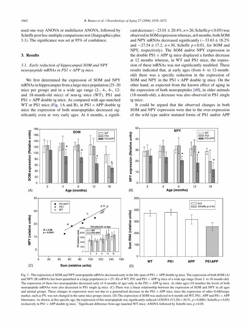
ig. 1. The expression of SOM and NPY neuropeptide mRNAs decreased early in thnd NPY (B) mRNAs has been quantified in a large population (n = 25–30) of WT, Phe expression of these two neuropeptides decreased early (4–6 months of age) oneuropeptide mRNAs were also decreased in PS1 single tg mice. (C) There was and animal groups. These changes in expression were not due to a generalized darker, such as PV, was not changed in the same mice groups (inset). (D) The expre
ittermates. As shown, at this specific age, the expression of this neuropeptide was sixclusively in PS1 × APP double tg mice. *Significant difference from age matche
ging 27 (2006) 1658–1672
ant decrease (−23.01 ± 20.4%, n = 26; Scheffe p < 0.05) wasbserved in SOM expression whereas, at 6 months, both SOMnd NPY mRNAs decreased significantly (−33.63 ± 18.2%nd −27.54 ± 17.2, n = 30, Scheffe p < 0.01, for SOM andPY, respectively). The SOM and/or NPY expression in
he double PS1 × APP tg mice displayed a further decreaset 12 months whereas, in WT and PS1 mice, the expres-ion of these mRNAs was not significantly modified. Theseesults indicated that, at early ages (from 4- to 12-month-ld) there was a specific reduction in the expression ofOM and NPY in the PS1 × APP double tg mice. On thether hand, as expected from the known effect of aging inhe expression of both neuropeptides [49], in older animals18-month-old), a decrease was also observed in PS1 single
g mice.It could be argued that the observed changes in bothOM and NPY expression were due to the over-expressionf the wild type and/or mutated forms of PS1 and/or APP
e life-span of PS1 × APP double tg mice. The expression of both SOM (A)S1 and PS1 × APP tg mice of a wide age range (from 2- to 18-month-old).ly in the PS1 × APP tg mice. At older ages (18 months) the levels of bothlinear relationship between the expression of SOM and NPY in all ages
ecrease in the PS1 × APP mice since the expression of other GABAergicssion of SOM was analyzed in 6-month-old WT, PS1, APP and PS1 × APPgnificantly reduced (ANOVA F(3,20) = 20.51, p = 0.0001; Scheffe p < 0.05)d WT mice; ANOVA followed by Scheffe test, p < 0.05.
gy of A
tsSPe
tftwIsmTcigs
sgTwad(soeanetSst
3sh
moiSt
ttSoIlcC
hmCine(
iPriose56ebbo
iitnm1r
pmdioFt
3mh
atPbdcso
B. Ramos et al. / Neurobiolo
ransgenes. Although we have not directly tested this pos-ibility, it seemed unlikely since: (i) no differences in theOM and NPY expression were observed in 2-month-oldS1 × APP tg mice, compared to WT mice and (ii) no differ-nces were observed between PS1 and WT until 18 months.
On the other hand, it has been proposed that most ofhe NPY positive interneurons also express SOM [19,36]. Inact (Fig. 1C), there was a highly significant linear correla-ion between both parameters. A reduced expression in SOMas accompanied by a parallel decrease in NPY expression.
nterestingly, the age-dependent modifications on the expres-ion of both neuropeptides, determined in WT and PS1 tgice, also fit well with those observed in PS1 × APP mice.his parallel decrease in SOM and NPY expression indi-ated that the same neuronal population should be affectedn the PS1 × APP tg mice. The expression of other GABAer-ic marker, such as parvalbumin, was not modified in theame PS1 × APP tg mice (Fig. 1C inset).
To further determine if the reduction in the SOM expres-ion was specific of the PS1 × APP double tg mice, hemizy-ous PS1 mice were crossed with hemizygous APP mice.hus, all four genotypes (WT, PS1, APP and PS1 × APP)ere obtained in the first generation. At 6 months of
ge, as compared with WT or PS1 mice (Fig. 1D), theouble PS1 × APP tg mice showed a significant decrease−30.9 ± 9.1%, n = 6, Scheffe p < 0.05) in the SOM expres-ion, similar to that showed previously. No differences werebserved between PS1 tg mice and WT littermates. Thexpression of SOM mRNA in APP single tg mice displayedn intermediate reduction (−14.8 ± 10.8%, n = 6), althoughon significantly as compared with WT mice (Fig. 1D). Inter-stingly, the Abeta deposition was delayed in this single APPg mice, as compared to double tg [6]. Thus, the decrease inOM and/or NPY expression seems to be correlated with theeverity of the pathology (Abeta depositions, see below) inhese mice models.
.2. The number of SOM immunoreactive cells isignificantly decreased in PS1 × APP tg miceippocampus at 6 months of age
We next tested if the observed reduction in the SOMRNA was also accompanied by a decrease in the number
f SOM expressing cells. We have thus combined specificmmunohistochemical detection of interneurons, expressingOM, with an unbiased stereological cell counting method in
he hippocampus of PS1 × APP transgenic at 6 months of age.The immunohistochemical study showed that the distribu-
ion of SOM-positive neurons in the hippocampus of WT andg animals (Fig. 2) was comparable to previous reports. MostOM-positive neurons were found in the stratum oriens (SO)f the CA1–3 subfields and in the hilus of dentate gyrus [36].
n addition, few SOM-positive cells were also found in statumucidum of CA3 region. Occasionally, some immunoreactiveells were also seen in strata pyramidale and radiatum ofA1–3 regions.bdTh
ging 27 (2006) 1658–1672 1663
A reduced number of SOM-positive cells throughout theippocampus of PS1 × APP mice, when compared to the age-atched WT or PS1 mice, was clearly observed (see Fig. 2,1–C2 versus A1–A2). In addition, the morphological exam-
nation showed the presence of dystrophic SOM-positiveeurons or neurites in the double tg mice. However, no differ-nces were detected between PS1 tg mice and control groupsee Fig. 2, B1–B2 versus A1–A2).
The stereological quantification of SOM-positive cellsn CA1–3 subfield and dentate gyrus of WT, PS1 andS1 × APP mice (n = 6, 10–15 sections per animal; Fig. 2E)evealed a significant and pronounced reduction in the numer-cal density (cells/mm3) of SOM-positive cells in all regionsf the hippocampus of PS1 × APP tg mice, in compari-on with WT or PS1 mice. The number of SOM cellsxhibited a 63.9 ± 4.6% decrease in CA1 (Scheffe p < 0.05);2.5 ± 10.8% in CA2–3 regions (Scheffe p < 0.05) and3.1 ± 5.9% in dentate gyrus hilus (Scheffe p < 0.05). How-ver, the number of SOM interneurons remained unchangedetween PS1 tg and WT mice. Furthermore, no differencesetween PS1 × APP tg mice and either WT or PS1 mice werebserved at younger ages (2-month-old, not shown).
To determine the selective vulnerability of hippocampalnterneurons containing SOM, we have counted striatalnterneurons that express SOM using the same immunos-ained brain sections. The stereological analysis showedo significant differences between PS1 × APP and WTice (1182 ± 69 cells/mm3 versus 945 ± 104 cells/mm3,
0–12 sections per mouse, n = 3; for WT and PS1 × APP,espectively).
On the other hand, stereological measurement of totalyramidal cell number, in hippocampal CA1 subfield of 6-onth-old PS1 × APP tg mice, did not reveal any significant
ifference when compared to WT or PS1 mice (see Fig. 2Enset). Furthermore, no significant differences in the densityf perisomatic inhibitory PV positive cells (not shown but seeig. 2D1–3) were detected between the double tg and wild
ype mice.
.3. SOM cells co-expressing NPY are the interneuronsost severely affected in 6-month-old PS1 × APP miceippocampus
As suggested by the parallel reduction in the SOMnd NPY mRNA expression, the SOM and NPY posi-ive GABAergic cells could be preferentially affected inS1 × APP tg mice. Thus, we have analyzed the vulnera-ility of SOM/NPY subpopulation in PS1 × APP mice byouble immunofluorescence labeling. Representative confo-al images (from five different independent experiments) arehown in Fig. 3. Though we have not quantified the extentf this co-localization, numerous interneurons co-expressed
oth neuropeptides in stratum oriens of CA1 and hilus ofentate gyrus of WT (Fig. 3A3) and PS1 (Fig. 3B3) mice.he number of interneurons immunolabeled for NPY wasighly reduced in PS1 × APP mice (Fig. 3C1) as compared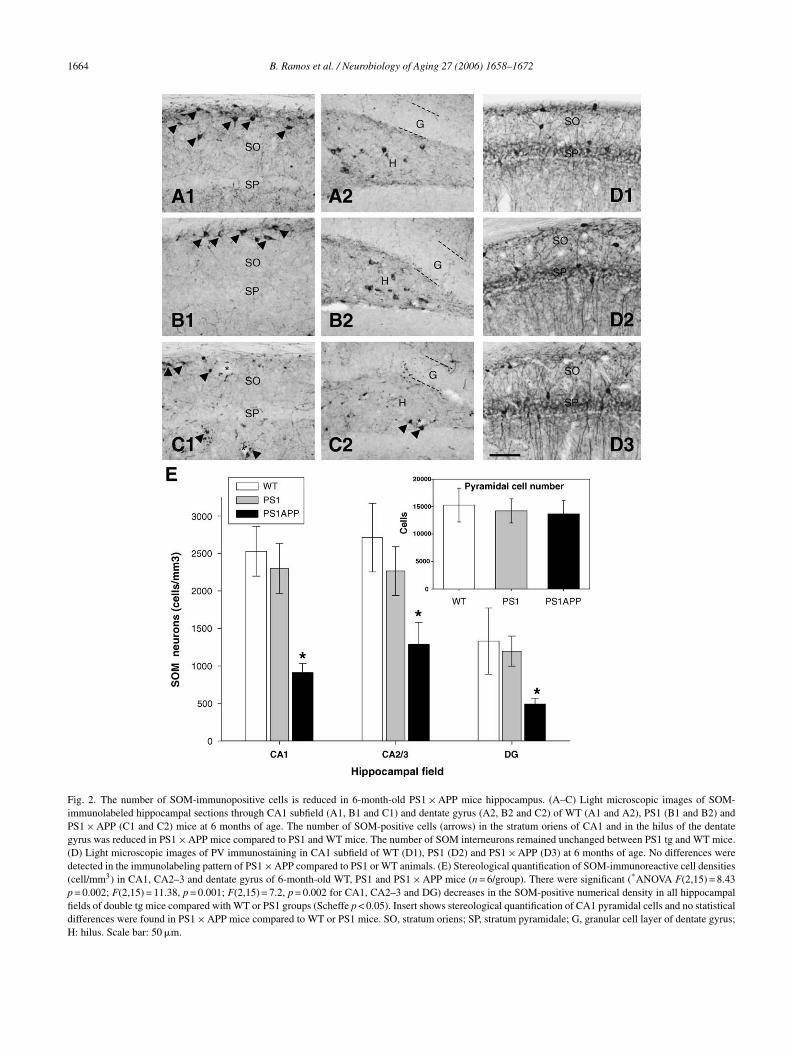
1664 B. Ramos et al. / Neurobiology of Aging 27 (2006) 1658–1672
Fig. 2. The number of SOM-immunopositive cells is reduced in 6-month-old PS1 × APP mice hippocampus. (A–C) Light microscopic images of SOM-immunolabeled hippocampal sections through CA1 subfield (A1, B1 and C1) and dentate gyrus (A2, B2 and C2) of WT (A1 and A2), PS1 (B1 and B2) andPS1 × APP (C1 and C2) mice at 6 months of age. The number of SOM-positive cells (arrows) in the stratum oriens of CA1 and in the hilus of the dentategyrus was reduced in PS1 × APP mice compared to PS1 and WT mice. The number of SOM interneurons remained unchanged between PS1 tg and WT mice.(D) Light microscopic images of PV immunostaining in CA1 subfield of WT (D1), PS1 (D2) and PS1 × APP (D3) at 6 months of age. No differences weredetected in the immunolabeling pattern of PS1 × APP compared to PS1 or WT animals. (E) Stereological quantification of SOM-immunoreactive cell densities(cell/mm3) in CA1, CA2–3 and dentate gyrus of 6-month-old WT, PS1 and PS1 × APP mice (n = 6/group). There were significant (*ANOVA F(2,15) = 8.43p = 0.002; F(2,15) = 11.38, p = 0.001; F(2,15) = 7.2, p = 0.002 for CA1, CA2–3 and DG) decreases in the SOM-positive numerical density in all hippocampalfields of double tg mice compared with WT or PS1 groups (Scheffe p < 0.05). Insert shows stereological quantification of CA1 pyramidal cells and no statisticaldifferences were found in PS1 × APP mice compared to WT or PS1 mice. SO, stratum oriens; SP, stratum pyramidale; G, granular cell layer of dentate gyrus;H: hilus. Scale bar: 50 �m.

B. Ramos et al. / Neurobiology of Aging 27 (2006) 1658–1672 1665
Fig. 3. Loss of SOM/NPY-containing interneurons in 6-month-old PS1 × APP tg mice hippocampus. (A–C) Immunofluorescent double-labeled confocal laserscanning images, representative of five different mice per group, for neuropeptides NPY (A1, B1 and C1) and SOM (A2, B2 and C2) in the dentate gyrus of6-month-old WT (A1–3), PS1 (B1–3) and PS1 × APP (C1–3) mice. The corresponding merged images showed that the density of hilar interneurons containingboth neuropeptides (arrows) is clearly reduced in PS1 × APP mice (C3) compared with PS1 (B3) and WT (A3). No apparent differences in the number ofd ages shoh
wNc
3hP
sipmcsttap
mtsocSarTwdpuG[
ouble labeled somata were found between WT and PS1 mice. Merged imilus; g, granular cell layer. Scale bar: 50 �m.
ith WT (Fig. 3A1) and PS1 groups (Fig. 3B1). The loss ofPY-positive cells correlates with the loss of SOM-positive
ells as shown in the merged image (Fig. 3C3).
.4. The decrease of SOM and NPY neuropeptides isighly selective at early ages in the hippocampus ofS1 × APP tg mice
The reduction in the expression of SOM and NPY could bepecific or, on the other hand, reflecting a generalized mod-fication in the GABAergic system. In order to analyze thisoint, using a new population of 6-month-old PS1 × APP tgice (n = 18), we have quantified the expression of mRNAs
onsidered as presynaptic and postsynaptic markers of thisystem (i.e. GAD65 enzyme, the neuropeptides cholecys-
okinine (CCK) and VIP; and the calcium binding pro-eins PV and Calbindin (Cb))[36]. The expression of SOMnd NPY neuropeptides was included for comparative pur-oses. In parallel, we also quantified the expression of theqca
wed that most of the interneurons co-localize SOM and NPY peptides. h,
ajor hippocampal alpha subunits of the GABAA recep-or (alpha1, alpha2 and alpha5 subunits). The results arehown in Fig. 4A. Concerning the interneuronal markers,nly the expression of SOM and NPY displayed a signifi-ant reduction (−31.3 ± 17.9% or −34.7 ± 16.9%, n = 18, forOM and NPY, respectively; two-tailed t = 5.96, p < 0.0001nd t = 4.64, p < 0.0001) as compared with WT mice. Thiseduction was similar to that observed in Fig. 1A, B and D.he absence of changes in the Cb expression was in contrastith the results reported by Palop et al. [38]. This apparentiscrepancy could be due to differences in the onset of theathological modifications between the different tg modelssed. On the other hand, at this age, the expression of theABAA receptor subunits was not modified (see also ref.
40]).
In addition, using the same mRNA samples, we have alsouantified the expression of synaptophysin and several spe-ific markers of glutamatergic (AMPA receptor subunits)nd cholinergic (muscarinic and nicotinic receptor subunits)

1666 B. Ramos et al. / Neurobiology of A
Fig. 4. The SOM and NPY expression was specifically altered in 6-month-old PS1 × APP double tg mice. The expression of multiple GABAergic (A),glutamatergic and cholinergic (B) neurotransmission markers was assayed ina new population of 6-month-old WT and PS1 × APP tg mice (n = 18). Froma total of 22 different mRNAs quantified, only the expression of SOM andNWS
sielro
3ci
piwogSraob
almicauTisbbNfiip
3lP
(smpatrc(t((twIov−o
3rt
fStie
PY mRNAs was significantly decreased, as compared with the age matchedT mice (two-tailed t-test, t = 5.96, p < 0.0001 and t = 4.64, p < 0.0001 for
OM and NPY, respectively).
ystems (Fig. 4B). As shown, no differences were observedn any of the mRNAs quantified. The expression of the ChATnzyme, determined in the basal forebrain, was also simi-ar between control and PS1 × APP mice (Fig. 4B). Theseesults emphasized the specificity and reproducibility in thebserved SOM and NPY modifications at this age.
.5. The decrease of SOM/NPY positive cells is aonsequence of an early neurodegeneration of inhibitorynterneurons in the hippocampus of PS1 × APP tg mice
To ascertain whether the reduction in the number of SOM-ositive neurons in PS1 × APP mice reflects a diminutionn the absolute number of interneurons or hypofunctionalityith altered phenotype, we have performed parallel stere-logical counts of SOM-positive cells and total GABAer-ic population in the stratum oriens of CA1 using doubleOM-NeuN immunostained sections. NeuN is a specific neu-
onal marker and, in the stratum oriens, most of the neuronsre GABAergic [19,36]. A comparison of the distributionf SOM/NeuN interneurons in the stratum oriens of CA1etween WT and PS1 × APP tg mice is shown in Fig. 5Ac
u4
ging 27 (2006) 1658–1672
nd B. The density of SOM labeled cells as well as NeuNabeled cells was significantly reduced in PS1 × APP tg
ice. The results of stereological quantification are shownn Fig. 5C. There was a marked reduction of SOM-positiveells (−61.8 ± 19.4%, n = 4; 10–15 sections per mouse)ccompanied by a decline in the total NeuN positive pop-lation (−41.4 ± 7.9%, n = 4; 10–15 sections per mouse).his decrease was similar to that observed using anti-GAD67
mmunolabeled sections (−46.3 ± 2.8%, n = 3, results nothown). Importantly, the loss in the SOM-positive cell num-er (5114 ± 695 cells/mm3; calculated from the differenceetween WT and PS1 × APP) was similar to the loss ofeuN labeled cells (5893 ± 222 cells/mm3; see Fig. 5C). Thisnding demonstrated a significant early loss of inhibitory
nterneurons that mostly correspond to the SOM-containingopulation in the hippocampus of PS1 × APP tg mice.
.6. The SOM-immunolabeled axonal field in stratumacunosum moleculare is reduced in 6-month-oldS1APP mice hippocampus
The SOM-immunostained stratum lacunosum molecularethe axon terminal field of O-LM cells) of PS1 × APP micehowed an apparent shrinkage, when compared to WT ani-als (Fig. 6A and B). To investigate whether the loss of SOM-
ositive cells in the stratum oriens produces a diminishedxonal field, we have measured the volume of this layer inhe CA1 region of PS1 × APP and control mice (Fig. 6C). Theesults revealed a generalized reduction in rostral, medial andaudal levels being statistically significant at the caudal level−32.54 ± 6.91%, n = 3; 10–15 sections per mouse; two-ailed t = 3.11, p = 0.04). As a consequence, the total volumerostral + medial + caudal) also showed a significant decrease−28.02 ± 4.9%, n = 3; two-tailed t = 3.87, p = 0.03). Thus,he decrease in the SOM-positive cell number in PS1 × APPas also paralleled by a decrease in the innervated area.
t is noteworthy that the decrease in the numerical densityf SOM cells exceeded the reduction of the total axonalolume immunopositive for SOM (−63.9 ± 4.6% versus28.02 ± 4.9%). This difference could indicate the existence
f axonal sprouting of the remaining SOM-positive fibers.
.7. The decrease in SOM and/or NPY neuropeptides iselated to the Abeta content in 6-month-old PS1 × APPg mice
As shown (Fig. 7C), the expression of the mRNA encodingor SOM displayed variability in the PS1 × APP tg mice. TheOM expression in some PS1 × APP individuals was iden-
ical to controls whereas a profound decrease was observedn other cases. Similar results were obtained for NPY mRNAxpression (not shown). Thus, we have determined the Abeta
ontent from 23 different 6-month-old PS1 × APP tg mice.The total Abeta content was determined by Western blot,sing the mAb 6E10. As shown in Fig. 7A, a major band of.5 kDa was detected using this approach. This 4.5 kDa band

B. Ramos et al. / Neurobiology of Aging 27 (2006) 1658–1672 1667
Fig. 5. Comparison of the density of SOM-NeuN immunopositive interneurons in the CA1 subfield between 6-month-old wildtype and PS1 × APP mice.(A and B) Light microscopic images of double immunolabeling with SOM (blue) and NeuN (brown) in CA1 subfield of WT (A) and PS1 × APP tg (B)mice. A reduced density of stratum oriens interneurons (NeuN-positive cells) as well as SOM-immunoreactive cells (double labeled cells; arrows) was foundin PS1 × APP mice compared with WT. (C) Stereological quantification of NeuN-positive cells and SOM-positive cells in CA1 stratum oriens of WT, PS1a 15; F(2i 4) andt P) wasp
c[sl(Ha
tdhtsanprs
npAmAn
iioTcEa
nd PS1 × APP demonstrated a significant (ANOVA F(2,10) = 8.1, p = 0.0nterneurons, respectively, in PS1 × APP mice (n = 4) compared to WT (n =he difference in the SOM-positive cell number between WT and PS1 × APyramidale. Scale bar: 25 �m.
orresponded to the monomeric form of the Abeta peptide51]. This band was also detected using the mAb WO2 (nothown) [24]. Other immunostained bands of higher molecu-ar weight could be also observed at higher exposition timesdimeric and/or trimeric forms of Abeta, not shown) [51].owever, this approach did not resolve between Abeta 1–40
nd 1–42 (not shown).Fig. 7A showed a representative Western blot. As shown,
he amount of 4.5 kDa band varied drastically between theifferent mice tested, from very low levels (animal #15) toigh levels (#233 or #200). This high variability was not dueo differences in the original protein loading (Fig. 7B). Ashown (Fig. 7C), the expression of Abeta displayed high vari-bility within the PS1 × APP population (similar to the SOM
europeptide in the same mice population, Fig. 7C). We nextlotted the mRNA levels of SOM and NPY versus the cor-esponding Abeta value, determined in the same animals. Ashown in Fig. 7D, for both neuropeptides, there was a sig-ioAh
,10) = 6.70, p = 0.019) decrease of total interneurons and SOM-containingPS1 (n = 4) mice. The loss in the SOM-positive cell number (calculated bysimilar to the loss of NeuN labeled cells. SO, stratum oriens; SP, stratum
ificant inverse linear correlation with the amount of Abetaeptide. Those PS1 × APP mice displaying low amount ofbeta content also showed high levels of both SOM and NPYRNAs whereas those PS1 × APP mice with high content ofbeta peptides displayed low levels of both SOM and NPYeuropeptides.
In order to determine the intracellular/extracellular local-zation of Abeta in PS1 × APP tg model, we have performedmmunostaining with 6E10 antibody in hippocampal sectionsf 2-, 4- and 6-month-old mice (Fig. 7E–G, respectively).he results showed intense immunolabeling of pyramidalell bodies and proximal dendrites at all three ages tested.xtracellular deposition was first observed at 4 months oldnd increased with age. Abeta deposits were mainly located
n stratum oriens of CA subfields (Fig. 7F and G) and hilusf dentate gyrus (not shown). Interestingly, no intracellularbeta expression was detected in the stratum oriens or in theilus.
1668 B. Ramos et al. / Neurobiology of Aging 27 (2006) 1658–1672
Fig. 6. The SOM-immunolabeled axonal field in stratum lacunosum moleculare is reduced in 6-month-old PS1 × APP mice hippocampus. (A and B) Lightmicroscopic images of SOM-immunolabeled hippocampal CA1 subfield of 6-month-old WT (A) and PS1 × APP tg (B) mice showing the immunoreactive axonalfield of SOM cells in the stratum lacunosum moleculare. The thickness of the SOM-immunoreactive axonal field was reduced in PS1 × APP mice comparedwith WT animals. (C) The volume (mm3) of the SOM-immunolabeled axonal field in CA1 stratum lacunosum moleculare was significantly (*two-tailed t-test,s to WTl oriensm
4
crfp
itb
ee text) reduced only at caudal levels in PS1 × APP mice (n = 3) comparedevels) also showed significant decrease in the double tg mice. SO, stratum
oleculare. Scale bar: 50 �m.
. Discussion
We have recently demonstrated the existence of a signifi-
ant deficit in the SOM/NPY GABAergic population in agedat hippocampus [49]. Since aging is the main risk factoror AD, we investigated in this work whether this hippocam-al interneuronal population was also preferentially affectedadso
(n = 3). The total volume measurement (including rostral + middle + caudal; SP, stratum pyramidale; SR, stratum radiatum; SLM, stratum lacunosum
n AD transgenic models. Our results clearly demonstratedhe existence of a profound decrease in the expression ofoth SOM and NPY mRNAs in PS1 × APP mice, as early
s 4–6 months of age, progressing towards lower valuesuring the aging process. At this early period in the lifepan of the tg mice, no modifications were detected in anyf the other GABAergic, glutamatergic or even cholinergic
B. Ramos et al. / Neurobiology of Aging 27 (2006) 1658–1672 1669
Fig. 7. The decrease in SOM and NPY expression was related to the relative abundance of Abeta peptides in 6-month-old PS1 × APP hippocampus. Theproduction of Abeta peptides was quantified, by Western blots, in the protein fraction obtained from 23 samples of 6-month-old PS1 × APP hippocampi. (A)Representative Western blot of seven different age matched PS1 × APP mice. Equal amount of protein (2.5 �g/lane) was run in parallel and developed using themAb 6E10. These experiments were repeated three times using different animal combinations. As shown, the amount of Abeta peptides (4.5 kDa band) variedwithin the different PS1 × APP mice tested. This high variation was not due to the amount of protein loaded (B). (C) Quantitative representation of the differentWestern blots (the Abeta signal was normalized by beta-actin). As expected, there is a large heterogeneity in the amount of Abeta peptides (closed squares).This heterogeneity was similar to that observed in the expression of SOM mRNAs (gray circles) from the same mice. The expression of SOM in WT (opencircles) was included for comparative purposes. (D) Relationship between the Abeta content and the expression of SOM (gray circles) or NPY (closed squares)mRNAs from 6-month-old PS1 × APP mice. The WT values (triangles) were included for comparison. Note the difference in scales between SOM and NPYmRNAs. (E–G) Light microscopic images showing CA1 subfield of PS1APP mice hippocampus at 2 (E), 4 (F) and 6 (G) months of age immunostained withanti-beta-specific antibody 6E10. Pyramidal cell layer showed intense intracellular immunolabeling at all these ages. However, GABAergic interneurons didn detectabo SP, stra
ma(cc[iaat
wwair
ot appear immunolabeled. Extracellular Abeta deposits (arrows) were firstf extracelullar amyloid deposition increased with age. SO, stratum oriens;
arkers tested. Furthermore, this change was observed inbsence of modifications in the expression synaptophysina presynaptic marker) or ChAT in the basal forebrain (alassical AD marker) [32]. Since SOM neuropeptide is impli-ated in both memory acquisition and Abeta degradation3,42], this selective loss could also represent one of the
nitial steps in the pathologic evolution of AD. Addition-lly, the expression of both neuropeptides could constitutevaluable tool to determine the efficacy of therapeuticreatments.
(com
le at 4 months of age and mostly located in the stratum oriens. The amounttum pyramidale; SR, stratum radiatum. Scale bar: 100 �m.
The decrease in the SOM and/or NPY content in AD casesas already known (see Section 1 and ref. [7]). However, heree demonstrate that this specific and early neurochemical
lteration is associated to a reduced number of SOM-NPYnterneurons in our PS1 × APP tg mice. In this respect, ouresults clearly probed the existence of a marked reduction
50–60%) in the numerical density of SOM immunopositiveells in CA1–3 stratum oriens and dentate gyrus of 6-month-ld PS1 × APP tg mice. Furthermore, this reduction was noterely reflecting a modification in the phenotypic proper-
1 gy of A
tciodtdinbteoNIitmsctIpmvP
d(sLsSasliTireitenGp[to
Hitmg
PtrmietnopiiPalctbrtiitiTctat
tduLcugnw
StinIppc[tis
670 B. Ramos et al. / Neurobiolo
ies of this particular neuronal population (hypofunctionalells)[46]. The presence of SOM-positive dystrophic neuritesndicated the existence of a neurodegenerative deteriorationf this interneuronal population. Moreover, the SOM-NeuNouble immunolabeling experiments clearly established thathe decrease in the SOM immunopositive cell number wasue to cell death. The SOM-positive cells were concentratedn the stratus oriens in the CA fields [36] and, in this layer, alleurons are GABAergic [19,36]. Thus, the direct comparisonetween NeuN and SOM density in stratum oriens allowed uso discriminate between hypofunctionality and neurodegen-ration. Importantly, our results demonstrated the existencef a similar, if not identical, decrease in both SOM andeuN immunopositive cells in PS1 × APP tg mice (Fig. 5).
n consequence, we conclude that the decrease in the SOMmmunopositive cells was due to the neurodegeneration ofhis particular interneuronal population in the PS1 × APP tg
ice. Importantly, in parallel experiments, the numerical den-ity of either SOM in striatum, or hippocampal PV positiveells (not shown) and CA1 pyramidal cells remained unal-ered (as compared with age matched WT or PS1 tg mice).t should be noted that, in this tg model, the decrease in theyramidal cell number was observed in 17-month-old ani-als [43]. Thus, these data demonstrated the specific an early
ulnerability of SOM expressing-population in the doubleS1 × APP tg model.
On the other hand, in the hippocampus, at least threeifferent subsets of SOM interneurons have been identifiedi.e. apical projecting cells, O-LM and HIPP cells; medialeptal projecting cells and bistratified cells). Most of the O-M and HIPP cells coexpressed SOM and NPY whereas theeptal projecting cells and the bistratified cells coexpressedOM/Cb or SOM/PV, respectively [33,19]. Our confocalnd double labeling experiments suggested that SOM/NPYubpopulation decreased extensively. In fact, the molecu-ar characterization also demonstrated a parallel reductionn both SOM and NPY mRNA expression (see Fig. 1).aken together, the molecular and immunohistological exper-
ments clearly demonstrated that the SOM-NPY interneu-onal subset (mostly the O-LM and HIPP cells) was prefer-ntially affected at early steps of the pathological evolutionn PS1 × APP models. To the best of our knowledge, this ishe first time that an early and neuron specific neurodegen-rative process has been described in tg AD models. It isoteworthy that the O-LM and HIPP cells represent a majorABAergic population in the hippocampal formation com-rising, approximately, 40–60% of the total inhibitory cells27]. Thus, the decrease in the SOM cell density, observed inhis work (50–60%), should constitute a substantial declinef the total GABAergic population in this brain area.
The reasons that determined this cell loss are unknown.owever, our results also demonstrated the existence of an
nverse correlation between the SOM/NPY expression andhe Abeta content in the different PS1 × APP mice at 6
onths of age. Further, this effect was also consistent with theradual deficiency in the SOM expression observed between
pllm
ging 27 (2006) 1658–1672
S1, APP and PS1 × APP littermates (Fig. 1D). In this sense,he implication of Abeta is well established as one of theesponsible factors for the pathological events that deter-ined the neuronal death observed in AD [35]. However,
t is unclear whether the toxic effects are due to intra- orxtra-cellular Abeta (see ref. [54] for review). In this respect,he existence of an extensive pyramidal cell loss and the cog-itive deterioration have been associated with the expressionf intracellular Abeta peptides [10,5]. We have also tested theossible expression of intracellular Abeta by the interneuronsn the stratum oriens (see Fig. 6), yet, no immunostainingn these cells was observed in either 2-, 4- or 6-month-oldS1 × APP mice (even after formic acid treatment), whereasprominent expression was detected in the pyramidal cell
ayer. Thus, in this particular case, the SOM expressingells may be affected by extracellular, secreted Abeta pep-ides. The present results do not allow us to discriminateetween a direct Abeta effect on the SOM/NPY interneu-ons or, on the contrary, an indirect Abeta cytotoxic effect onhis neuronal population. Concerning to this latter possibil-ty, the 4–6-month-old PS1 × APP mice develop an extensivenflammatory response with a substantial microglial activa-ion and induction of, among other factors, TNF-alpha andNOS expression (manuscript in preparation and ref. [6]).hese factors could be implicated in the degenerative pro-esses observed in the AD models (ref. [18] and referencesherein). These two alternatives are not mutually exclusivend we cannot exclude a synergic effect of both processes inhe degeneration induced by the Abeta deposition.
On the other hand, our results also demonstrated thathe reduction in SOM cell number was accompanied by aecrease in the corresponding immunoreactive axonal vol-me in the lacunosum moleculare field (see Fig. 6). The O-M and HIPP neurons belong to the distal dendritic inhibitoryells, e.g. oriens/alveus interneurons with lacunosum molec-lare axon arborization [45]. Therefore, this decrease sug-ested that, not only the number of SOM cells, but also theumber of inhibitory synapses at the distal apical dendritesere reduced in the PS1 × APP tg model.The physiological consequences of the reduction in the
OM content, the total number and the innervated area ofhe SOM cells are actually unknown. However, the distalnhibitory cells played an essential role in the synaptic sig-al integration and input plasticity of principal cells [47,21].n fact, their axonal terminations were co-aligned with theerforant path input from the entorhinal cortex and the SOM-ositive O-LM cells fired rhythmically at theta frequencies,ontributing to the hyperpolarization of the principal cells28,29]. This rhythmic hyperpolarization could contribute tohe de-inactivation of voltage-gated ion channels, facilitat-ng somatodendritic back-propagation action potentials andynaptic plasticity of the principal cells [28,29]. Thus, the
rofound (50–60%) and specific loss of SOM cells and theiracunosum moleculare innervation could be implicated, ateast in part, in the memory impairment observed in AD tgodels. On the other hand, the specific degeneration of the O-
gy of A
Ladti
osSpbHSnpaa
sfsicrditaahieccotcicmm
A
ttgFDfwTIt
R
[
[
[
[
[
[
[
[
B. Ramos et al. / Neurobiolo
M and HIPP cells in the PS1 × APP tg mice might producen imbalance between excitation and inhibition on the distalendritic tree, increasing the pyramidal excitability, similaro that observed in temporal lobe epilepsy models [12] and,n consequence, increasing the vulnerability of these cells,
Finally, it has recently been demonstrated the implicationf SOM in the regulation of neprilysin activity at the neuronalurface and the degradation of Abeta peptides [42]. In fact,OM upregulated the neprilysin activity transported to thelasmatic membrane and, in consequence, reducing the solu-le Abeta deposition. Thus, the degeneration of the O-LM andIPP cells, that determines the decrease in the expression ofOM neuropeptide, could also produce a diminution in theeprilysin activity. In consequence, this neurodegenerativerocess could contribute to the progressive age-dependentccumulation of Abeta peptides observed in both AD casesnd tg models.
In sum, our findings demonstrate, for the first time, a sub-tantial early loss of SOM and/or NPY GABAergic neuronsrom the hippocampus of double PS1 × APP tg mice. Thiselective modification was observed in absence of changesn other neuronal markers of GABAergic, glutamatergic andholinergic systems or the principal cell number. This neu-odegenerative process provides a cellular explanation to theecrease in the SOM neuropeptide expression (also observedn AD cases). Since SOM neuropeptide is implicated inhe control of neprilysin activity, the loss of SOM neuronslso explains the observed decrease in neprilysin activitynd Abeta accumulation in the hippocampus. On the otherand, the SOM-NPY GABAergic neurons are implicatedn the coordination of pyramidal cells activity, then, thearly neurodegeneration of these interneurons could impli-ate a reduction in the synaptic plasticity of the principalells and determine, at least in part, the deficits in mem-ry/learning process observed in early phases of AD. Finally,he expression of both SOM and NPY mRNAs could beonsidered as specific and early molecular markers of ADn the PS1 × APP models. Thus, these neuropeptides couldonstitute important molecular tools to improve the assess-ent of the efficacy of potential AD treatments in this miceodel.
cknowledgments
We would like to thanks to Dr. J.A. Aguirre for his assis-ance with the stereological analysis and Aventis-Pharma forhe transgenic mice models. This work was supported byrants SAF2002-03448 (to J.V.), FIS PI030214 (to A.G.),IS PI030177 (to D.R.) and Aventis Grants (to J.V. and A.G.)..R. and Z.U.K. were supported by a Ramon y Cajal program
rom Ministerio de Ciencia y Tecnologia. B.R. and J.F.L.-T.
ere recipients of fellowships from Ministerio de Ciencia yecnologia, and I.M.-G. and J.C.R., D.B.-V. from Fondo denvestigaciones Sanitarias and Junta de Andalucia, respec-ively.[
ging 27 (2006) 1658–1672 1671
eferences
[1] Araujo F, Tan S, Ruano D, Schoemaker H, Benavides J, VitoricaJ. Molecular and pharmacological characterization of native corticalgamma-aminobutyric acid(A) receptors containing both alpha(1) andalpha(3) subunits. J Biol Chem 1996;271:27902–11.
[2] Auld DS, Kornecook TJ, Bastianetto S, Quirion R. Alzheimer’s dis-ease and the basal forebrain cholinergic system: relations to [beta]-amyloid peptides, cognition, and treatment strategies. Prog Neurobiol2002;68:209–45.
[3] Baraban SC, Tallent MK. Interneuron diversity series: interneuronalneuropeptides—endogenous regulators of neuronal excitability. TrendsNeurosci 2004;27:135–42.
[4] Bast T, Zhang W-N, Fledon J. Hyperactivity, decreased startle reactiv-ity, and disrupted prepulse inhibition following disinhibition of the ratventral hippocampus by the GABAA receptor antagonist picrotoxin.Psychopharmacology 2001;156:225–33.
[5] Billings LM, Oddo S, Green KN, McGaugh JL, LaFerla FM. Intra-neuronal A[beta] causes the onset of early Alzheimer’s disease-relatedcognitive deficits in transgenic mice. Neuron 2005;45:675–88.
[6] Blanchard V, Moussaoui S, Czech C, Touchet N, Bonici B, PlancheM, et al. Time sequence of maturation of dystrophic neurites associ-ated with A[beta] deposits in APP/PS1 transgenic mice. Exp Neurol2003;184:247–63.
[7] Blasco B, Avendano C, Cavada C. A stereological analysis of the lateralgeniculate nucleus in adult Macaca nemestrina monkeys. Vis Neurosci1999;16:933–41.
[8] Bouilleret V, Loup F, Kiener T, Marescaux C, Fritschy JM. Early lossof interneurons and delayed subunit-specific changes in GABA(A)-receptor expression in a mouse model of mesial temporal lobe epilepsy.Hippocampus 2000;10:305–24.
[9] Bouilleret V, Schwaller B, Schurmans S, Celio MR, Fritschy JM.Neurodegenerative and morphogenic changes in a mouse model oftemporal lobe epilepsy do not depend on the expression of the calcium-binding proteins parvalbumin, calbindin, or calretinin. Neuroscience2000;97:47–58.
10] Casas C, Sergeant N, Itier JM, Blanchard V, Wirths O, van der Kolk N,et al. Massive CA1/2 neuronal loss with intraneuronal and N-terminaltruncated A{beta}42 accumulation in a novel Alzheimer transgenicmodel. Am J Pathol 2004;165:1289–300.
11] Chan-Palay V, Lang W, Allen YS, Haesler U, Polak JM. Corti-cal neurons immunoreactive with antisera against neuropeptide Yare altered in Alzheimer’s-type dementia. J Comp Neurol 1985;238:390–400.
12] Cossart R, Dinocourt C, Hirsch JC, Merchan PA, De Felipe J, Ben AriY, et al. Dendritic but not somatic GABAergic inhibition is decreasedin experimental epilepsy. Nat Neurosci 2001;4:52–62.
13] Davies P, Katzman R, Terry RD. Reduced somatostatin-like immunore-activity in cerebral cortex from cases of Alzheimer disease andAlzheimer senile dementa. Nature 1980;288:279–80.
14] Davis KL, Mohs RC, Marin DB, Purohit DP, Perl DP, Lantz M, et al.Neuropeptide abnormalities in patients with early Alzheimer disease.Arch Gen Psychiatry 1999;56:981–7.
15] Dinocourt C, Petanjek Z, Freund TF, Ben Ari Y, Esclapez M. Lossof interneurons innervating pyramidal cell dendrites and axon initialsegments in the CA1 region of the hippocampus following pilocarpine-induced seizures. J Comp Neurol 2003;459:407–25.
16] Dorph-Petersen KA, Nyengaard JR, Gundersen HJG. Tissue shrinkageand unbiased stereological estimation of particle number and size. JMicrosc 2001;204:232–46.
17] Dumont M, Strazielle C, Staufenbiel M, Lalonde R. Spatial learn-ing and exploration of environmental stimuli in 24-month-old female
APP23 transgenic mice with the Swedish mutation. Brain Res2004;1024:113–21.18] Floden AM, Li S, Combs CK. {beta}-Amyloid-stimulated microgliainduce neuron death via synergistic stimulation of tumor necrosis factor{alpha} and NMDA receptors. J Neurosci 2005;25:2566–75.

1 gy of A
[
[
[
[
[
[
[
[
[
[
[
[
[
[
[
[
[
[
[
[
[
[
[
[
[
[
[
[
[
[
[
[
[
[
[
672 B. Ramos et al. / Neurobiolo
19] Freund TF, Buzsaki G. Interneurons of the hippocampus. Hippocampus1996;6:347–470.
20] Georg M, Arne M, Flemming J. Stereological cell counts of GABAergicneurons in rat dentate hilus following transient cerebral ischemia. ExpBrain Res 2001;141:380–8.
21] Gulledge AT, Stuart GJ. Excitatory actions of GABA in the cortex.Neuron 2003;37:299–309.
22] Harney SC, Jones MV. Pre- and postsynaptic properties of somaticand dendritic inhibition in dentate gyrus. Neuropharmacology2002;43:584–94.
23] Hock BJ, Lamb BT. Transgenic mouse models of Alzheimer’s disease.Trends Genet 2001;17:S7–12.
24] Ida N, Hartmann T, Pantel J, Schroder J, Zerfass R, Forstl H, et al.Analysis of heterogeneous beta A4 peptides in human cerebrospinalfluid and blood by a newly developed sensitive Western blot assay. JBiol Chem 1996;271:22908–14.
25] Jinno S, Aika Y, Fukuda T, Kosaka T. Quantitative analysis of GABAer-gic neurons in the mouse hippocampus, with optical disector usingconfocal laser scanning microscope. Brain Res 1998;814:55–70.
26] Jinno S, Kosaka T. Patterns of colocalization of neuronal nitric oxidesynthase and somatostatin-like immunoreactivity in the mouse hip-pocampus: quantitative analysis with optical disector. Neuroscience2004;124:797–808.
27] Jinno S, Aika Y, Fukuda T, Kosaka T. Quantitative analysis of GABAer-gic neurons in the mouse hippocampus, with optical disector usingconfocal laser scanning microscope. Brain Res 1998;814:55–70.
28] Klausberger T, Magill PJ, Marton LF, Roberts JD, Cobden PM, BuzsakiG, et al. Brain-state- and cell-type-specific firing of hippocampalinterneurons in vivo. Nature 2003;421:844–8.
29] Klausberger T, Marton LF, Baude A, Roberts JD, Magill PJ, Som-ogyi P. Spike timing of dendrite-targeting bistratified cells duringhippocampal network oscillations in vivo. Nat Neurosci 2004;7:41–7.
30] Lowry OH, Rosebrough NJ, Farr AL, Randall RJ. Protein mea-surement with the Folin phenol reagent. J Biol Chem 1951;193:265–75.
31] Lu T, Pan Y, Kao SY, Li C, Kohane I, Chan J, et al. Gene regulation andDNA damage in the ageing human brain. Nature 2004;429:883–91.
32] Lyness SA, Zarow C, Chui HC. Neuron loss in key cholinergic andaminergic nuclei in Alzheimer disease: a meta-analysis. NeurobiolAging 2003;24:1–23.
33] Maccaferri G, Roberts JDB, Szucs P, Cottingham CA, Somogyi P.Cell surface domain specific postsynaptic currents evoked by identi-fied GABAergic neurones in rat hippocampus in vitro. J Physiol London2000;524:91–116.
34] Maccaferri G, Lacaille JC. Interneuron diversity series: hippocampalinterneuron classifications—making things as simple as possible, notsimpler. Trends Neurosci 2003;26:564–71.
35] Mattson MP. Pathways towards and away from Alzheimer’s disease.Nature 2004;430:631–9.
36] Matyas F, Freund TF, Gulyas AI. Immunocytochemically definedinterneuron populations in the hippocampus of mouse strains used in
transgenic technology. Hippocampus 2004;14:460–81.37] Nilsson CL, Brinkmalm A, Minthon L, Blennow K, Ekman R. Process-ing of neuropeptide Y, galanin, and somatostatin in the cerebrospinalfluid of patients with Alzheimer’s disease and frontotemporal dementia.Peptides 2001;22:2105–12.
[
ging 27 (2006) 1658–1672
38] Palop JJ, Jones B, Kekonius L, Chin J, Yu GQ, Raber J, et al. Neuronaldepletion of calcium-dependent proteins in the dentate gyrus is tightlylinked to Alzheimer’s disease-related cognitive deficits. Proc Natl AcadSci USA 2003;100:9572–7.
39] Price DL, Tanzi RE, Borchelt DR, Sisodia SS. Alzheimer’s dis-ease: genetic studies and transgenic models. Ann Rev Genet1998;32:461–93.
40] Rissman RA, Mishizen-Eberz AJ, Carter TL, Wolfe BB, De BlasAL, Miralles CP, et al. Biochemical analysis of GABAA recep-tor subunits [alpha]1, [alpha]5, [beta]1, [beta]2 in the hippocampusof patients with Alzheimer’s disease neuropathology. Neuroscience2003;120:695–704.
41] Ruano D, Araujo F, Revilla E, Vela J, Bergis O, Vitorica J. GABA(A)and alpha-amino-3-hydroxy-5-methylsoxazole-4-propionate receptorsare differentially affected by aging in the rat hippocampus. J Biol Chem2000;275:19585–93.
42] Saito T, Iwata N, Tsubuki S, Takaki Y, Takano J, Huang SM, et al.Somatostatin regulates brain amyloid [beta] peptide A[beta]42 throughmodulation of proteolytic degradation. Nat Med 2005;11:434–9.
43] Schmitz C, Rutten BPF, Pielen A, Schafer S, Wirths O, Tremp G, et al.Hippocampal neuron loss exceeds amyloid plaque load in a transgenicmouse model of Alzheimer’s disease. Am J Pathol 2004;164:1495–502.
44] Selkoe DJ. Alzheimer’s disease is a synaptic failure. Science2002;298:789–91.
45] Sik A, Penttonen M, Ylinen A, Buzsaki G. Hippocampal CA1interneurons: an in vivo intracellular labeling study. J Neurosci1995;15:6651–65.
46] Sloviter RS, Zappone CA, Harvey BD, Bumanglag AV, Bender RA,Frotscher M. Dormant basket cell hypothesis revisited: relative vul-nerabilities of dentate gyrus mossy cells and inhibitory interneu-rons after hippocampal status epilepticus in the rat. J Comp Neurol2003;459:44–76.
47] Tamas G, Szabadics J, Somogyi P. Cell type- and subcellular position-dependent summation of unitary postsynaptic potentials in neocorticalneurons. J Neurosci 2002;22:740–7.
48] Van Dam D, D’Hooge R, Staufenbiel M, Van Ginneken C, Van MeirF, De Deyn PP. Age-dependent cognitive decline in the APP23 modelprecedes amyloid deposition. Eur J Neurosci 2003;17:388–96.
49] Vela J, Gutierrez A, Vitorica J, Ruano D. Rat hippocampal GABAergicmolecular markers are differentially affected by ageing. J Neurochem2003;85:368–77.
50] Vela J, Vitorica J, Ruano D. Rapid PCR-mediated synthesis of competi-tor molecules for accurate quantification of beta(2) GABA(A) receptorsubunit mRNA. Brain Res Brain Res Protoc 2001;8:184–90.
51] Walsh DM, Klyubin I, Fadeeva JV, Cullen WK, Anwyl R, WolfeMS, et al. Naturally secreted oligomers of amyloid [beta] proteinpotently inhibit hippocampal long-term potentiation in vivo. Nature2002;416:535–9.
52] West MJ, Coleman PD, Flood DG, Troncoso JC. Differences in thepattern of hippocampal neuronal loss in normal ageing and Alzheimer’sdisease. Lancet 1994;344:769–72.
53] West MJ, Kawas CH, Stewart WF, Rudow GL, Troncoso JC. Hip-
pocampal neurons in pre-clinical Alzheimer’s disease. Neurobiol Aging2004;25:1205–12.54] Wirths O, Multhaup G, Bayer TA. A modified “beta”-amyloid hypoth-esis: intraneuronal accumulation of the “beta”-amyloid peptide—thefirst step of a fatal cascade. J Neurochem 2004;91:513–20.