Early functional and transcriptomic changes in the ... · These pathways merit further study to...
17
Zurich Open Repository and Archive University of Zurich Main Library Strickhofstrasse 39 CH-8057 Zurich www.zora.uzh.ch Year: 2013 Early functional and transcriptomic changes in the myocardium predict outcome in a long-term rat model of sepsis Rudiger, Alain; Dyson, Alex; Felsmann, Karen; Carré, Jane E; Taylor, Valerie; Hughes, Sian; Clatworthy, Innes; Protti, Alessandro; Pellerin, Denis; Lemm, Jana; Claus, Ralf A; Bauer, Michael; Singer, Mervyn Abstract: Myocardial function is depressed in sepsis and is an important prognosticator in the human condition. Using echocardiography in a long-term fluid-resuscitated Wistar rat model of faecal peritonitis we investigated whether depressed myocardial function could be detected at an early stage of sepsis and, if so, whether the degree of depression could predict eventual outcome. At 6 h post-insult, a stroke vol- ume <0.17 ml prognosticated 3-day mortality with positive and negative predictive values of 93 and 80%, respectively. Subsequent fluid loading studies demonstrated intrinsic myocardial depression with poor- prognosis animals tolerating less fluid than either good-prognosis or sham-operated animals. Cardiac gene expression analysis at 6 h detected 527 transcripts significantly up- or down-regulated by the septic process, including genes related to inflammatory and cell cycle pathways. Predicted mortality was asso- ciated with significant differences in transcripts of genes expressing proteins related to the TLR2/MyD88 (Toll-like receptor 2/myeloid differentiation factor 88) and JAK/STAT (Janus kinase/signal transducer and activator of transcription) inflammatory pathways, -adrenergic signalling and intracellular calcium cycling. Our findings highlight the presence of myocardial depression in early sepsis and its prognostic significance. Transcriptomic analysis in heart tissue identified changes in signalling pathways that cor- related with clinical dysfunction. These pathways merit further study to both better understand and potentially modify the disease process. DOI: https://doi.org/10.1042/CS20120334 Posted at the Zurich Open Repository and Archive, University of Zurich ZORA URL: https://doi.org/10.5167/uzh-69561 Journal Article Published Version Originally published at: Rudiger, Alain; Dyson, Alex; Felsmann, Karen; Carré, Jane E; Taylor, Valerie; Hughes, Sian; Clatworthy, Innes; Protti, Alessandro; Pellerin, Denis; Lemm, Jana; Claus, Ralf A; Bauer, Michael; Singer, Mervyn (2013). Early functional and transcriptomic changes in the myocardium predict outcome in a long-term rat model of sepsis. Clinical Science, 124(6):391-401. DOI: https://doi.org/10.1042/CS20120334
Transcript of Early functional and transcriptomic changes in the ... · These pathways merit further study to...

Zurich Open Repository andArchiveUniversity of ZurichMain LibraryStrickhofstrasse 39CH-8057 Zurichwww.zora.uzh.ch
Year: 2013
Early functional and transcriptomic changes in the myocardium predictoutcome in a long-term rat model of sepsis
Rudiger, Alain; Dyson, Alex; Felsmann, Karen; Carré, Jane E; Taylor, Valerie; Hughes, Sian;Clatworthy, Innes; Protti, Alessandro; Pellerin, Denis; Lemm, Jana; Claus, Ralf A; Bauer, Michael;
Singer, Mervyn
Abstract: Myocardial function is depressed in sepsis and is an important prognosticator in the humancondition. Using echocardiography in a long-term fluid-resuscitated Wistar rat model of faecal peritonitiswe investigated whether depressed myocardial function could be detected at an early stage of sepsis and,if so, whether the degree of depression could predict eventual outcome. At 6 h post-insult, a stroke vol-ume <0.17 ml prognosticated 3-day mortality with positive and negative predictive values of 93 and 80%,respectively. Subsequent fluid loading studies demonstrated intrinsic myocardial depression with poor-prognosis animals tolerating less fluid than either good-prognosis or sham-operated animals. Cardiacgene expression analysis at 6 h detected 527 transcripts significantly up- or down-regulated by the septicprocess, including genes related to inflammatory and cell cycle pathways. Predicted mortality was asso-ciated with significant differences in transcripts of genes expressing proteins related to the TLR2/MyD88(Toll-like receptor 2/myeloid differentiation factor 88) and JAK/STAT (Janus kinase/signal transducerand activator of transcription) inflammatory pathways, �-adrenergic signalling and intracellular calciumcycling. Our findings highlight the presence of myocardial depression in early sepsis and its prognosticsignificance. Transcriptomic analysis in heart tissue identified changes in signalling pathways that cor-related with clinical dysfunction. These pathways merit further study to both better understand andpotentially modify the disease process.
DOI: https://doi.org/10.1042/CS20120334
Posted at the Zurich Open Repository and Archive, University of ZurichZORA URL: https://doi.org/10.5167/uzh-69561Journal ArticlePublished Version
Originally published at:Rudiger, Alain; Dyson, Alex; Felsmann, Karen; Carré, Jane E; Taylor, Valerie; Hughes, Sian; Clatworthy,Innes; Protti, Alessandro; Pellerin, Denis; Lemm, Jana; Claus, Ralf A; Bauer, Michael; Singer, Mervyn(2013). Early functional and transcriptomic changes in the myocardium predict outcome in a long-termrat model of sepsis. Clinical Science, 124(6):391-401.DOI: https://doi.org/10.1042/CS20120334

Clinical Science (2013) 124, 391–401 (Printed in Great Britain) doi: 10.1042/CS20120334
Early functional and transcriptomic changes inthe myocardium predict outcome in a long-termrat model of sepsisAlain RUDIGER*, Alex DYSON*, Karen FELSMANN†, Jane E. CARRE*, Valerie TAYLOR*, Sian HUGHES‡,Innes CLATWORTHY§, Alessandro PROTTI*, Denis PELLERIN‖, Jana LEMM¶, Ralf A. CLAUS¶,Michael BAUER¶1 and Mervyn SINGER*
*Bloomsbury Institute of Intensive Care Medicine, Division of Medicine, University College London, Gower Street, London WC1E 6BT, U.K.†SIRS-Lab GmbH, Otto-Schott-Strasse 15, D-07745 Jena, Germany‡Department of Histopathology, Royal Free and University College Medical School, University College London, Rockefeller Building, University Street,London WC1E 6JJ, U.K.§Imaging Facility, UCL Institute of Opthalmology, 11–43 Bath Street, London EC1V 9EL, U.K.‖The Heart Hospital, UCL Hospitals NHS Foundation Trust, 16-18 Westmoreland Street, London W1G 8PH, U.K.¶Integrated Research and Treatment Center–Center for Sepsis Control and Care, Jena University Hospital, Allee 101, D-07747 Jena, Germany
AbstractMyocardial function is depressed in sepsis and is an important prognosticator in the human condition. Usingechocardiography in a long-term fluid-resuscitated Wistar rat model of faecal peritonitis we investigated whetherdepressed myocardial function could be detected at an early stage of sepsis and, if so, whether the degreeof depression could predict eventual outcome. At 6 h post-insult, a stroke volume <0.17 ml prognosticated 3-daymortality with positive and negative predictive values of 93 and 80%, respectively. Subsequent fluid loading studiesdemonstrated intrinsic myocardial depression with poor-prognosis animals tolerating less fluid than eithergood-prognosis or sham-operated animals. Cardiac gene expression analysis at 6 h detected 527 transcriptssignificantly up- or down-regulated by the septic process, including genes related to inflammatory and cell cyclepathways. Predicted mortality was associated with significant differences in transcripts of genes expressingproteins related to the TLR2/MyD88 (Toll-like receptor 2/myeloid differentiation factor 88) and JAK/STAT (Januskinase/signal transducer and activator of transcription) inflammatory pathways, β -adrenergic signalling andintracellular calcium cycling. Our findings highlight the presence of myocardial depression in early sepsis and itsprognostic significance. Transcriptomic analysis in heart tissue identified changes in signalling pathways thatcorrelated with clinical dysfunction. These pathways merit further study to both better understand and potentiallymodify the disease process.
Key words: animal model, echocardiography, faecal peritonitis, gene transcript, heart failure, sepsis
INTRODUCTION
Sepsis, the systemic inflammatory response syndrome to infec-tion, is the leading cause of death in the critically ill. This occursprimarily through the development of multiple organ failure [1],in which cardiac dysfunction is a well-recognized manifestation[2,3]. Identified mechanisms for myocardial depression include
Abbreviations: ARDB2 adrenergic receptor β2; ATP2A3, ubiquitous calcium-transporting ATPase; CDKN1A, cyclin-dependent kinase inhibitor 1A; CI, confidence interval; COX2,cyclo-oxygenase 2; EM, electron microscopy; H&E, haematoxylin and eosin; HMBS, hydroxymethylbilane synthase; HR, heart rate; IL, interleukin; i.p., intraperitoneal; i.v., intravenous;JAK, Janus kinase; LBP, lipopolysaccharide-binding protein; LV, left ventricular; MyD88, myeloid differentiation factor 88; PDE10A, phosphodiesterase 10A; PKA, protein kinase A;PLA2G4A, phospholipase A2 group IVA; PP1, protein phosphatase 1; PPP1CB, protein phosphatase 1 catalytic subunit β -isoform; PRKACA, protein kinase cAMP-dependent catalytic α;PRKAG2, protein kinase AMP-activated γ 2; PTGS2, prostaglandin-endoperoxidase synthase 2, ROC, receiver operator characteristic; SERCA, sarcoplasmic/endoplasmic reticulumCa2 + -ATPase; SR, sarcoplasmic reticulum; STAT, signal transducer and activator of transcription; TLR, Toll-like receptor.1 A member of the scientific advisory board of SIRS-Lab GmbH, Jena, Germany, where the microarray experiments were performed.
Correspondence: Professor Mervyn Singer (email [email protected]).
alterations in adrenergic signalling, intracellular calcium cycling,impaired electromechanical coupling and mitochondrial dysfunc-tion [4]. The hierarchy of these mechanisms, however, remainsuncertain and individual variations in severity inadequately ex-plained.
A clinically relevant sepsis model achieving a 50 % mortal-ity rate raises the important but hitherto unaddressed question
www.clinsci.org 391
Clin
ical
Sci
ence
ww
w.c
linsc
i.org

A. Rudiger and others
as to why some animals survive while the remainder die des-pite similar, if not, identical characteristics in terms of genotype,gender, age, weight and environment, and the septic insult re-ceived. Though these factors differ considerably in humans withsepsis, resulting in variable degrees and combinations of mul-tiple organ dysfunction, a wide selection of inflammatory [5],hormonal [6,7], circulatory [8], bioenergetic [9] and organ dys-function biomarkers [7,10] can prognosticate as early as the firstfew days of intensive care admission. Notably, survivors and non-survivors are often clinically indistinguishable at this early timepoint. This implies that mortality may be determined to a largeextent by a pattern of molecular genetic and functional responsesthat differentiate survivors from non-survivors at an early stage.
Although the degree of myocardial depression has beenreported in several clinical studies to be an indicator of pooroutcome [11,12], others suggest this may have a protectiverole [13]. This disparity may relate to timing of the studyrelative to the duration of critical illness. However, the roleand prognostic significance of early myocardial dysfunctionhas received relatively scant attention. We hypothesized thatearly cardiac performance could predict eventual outcome, andperformed transcriptomic analysis to probe putative pathwaysthat could reconcile the physiological and molecular mechanismsof sepsis-induced cardiac dysfunction.
MATERIALS AND METHODS
Animal modelAll experiments were performed according to local ethics com-mittee (University College London) and Home Office (U.K.)guidelines under the 1986 Scientific Procedures Act. Under 2 %isoflurane anaesthesia, male Wistar rats (Charles River) had PVCtubes (inner diameter, 0.58 mm; outer diameter, 0.96 mm) inser-ted into the right jugular vein and left carotid artery, and tunnelledsubcutaneously to emerge at the nape of the neck. These lineswere subsequently mounted on to a swivel-tether system allowingthe rat, on recovery from anaesthesia, to have unimpeded move-ment in its cage and free access to food and water. After 24 h,sepsis was induced by i.p. (intraperitoneal) injection of faecalslurry (1.8 ml of a 40 % preparation obtained from the bowel con-tent of a rat from the same batch). Fluid resuscitation, consistingof a 1:1 solution of 6 % hetastarch (Elohaes®; Fresenius-Kabi)and 5 % glucose, was commenced 2 h later through the centralvenous line. Glucose supplementation prevented hypoglycaemiain the animals with sepsis due to decreased appetite and intake.The infusion rate was maintained at 10 ml/kg of body weight perh between 2 h and 24 h, halving thereafter at 24 h intervals. An-imals received additional 25 ml/kg of body weight boluses of 6 %hetastarch at 6 h and 10 ml/kg of body weight at 24 h to optimizefluid resuscitation (results from pilot studies). Sham animals ofsimilar age, gender and weight underwent identical instrumenta-tion and fluid administration, but did not receive the i.p. injectionof faecal slurry. At 6, 24, 48 and 72 h post-sepsis induction, andin sham-operated controls, clinical scoring was performed. This
included an assessment of appearance, alertness and movement,as previously described [14].
EchocardiographyEchocardiography studies were carried out using a 14 MHzlinear-array transducer connected to a digital ultrasound system(Vivid 7; GE Healthcare). Images were acquired in left paras-ternal long and short axis views at the papillary muscle level.Radius (r) and length (l) of the left ventricle were measured atend-diastole by M-mode and two-dimensional mode respectively.LV (left ventricular) end-diastolic volumes were calculated usinga prolate spheroid formula (4/3·π ·r2·l). Stroke volume was meas-ured in the ascending aorta (radius 1.3 mm) from the velocity–time integral using pulsed-wave Doppler. The vessel diameter atthis level remains stable despite intravascular volume variations[15].
Study 1: long-term outcome studyA total of 20 rats with sepsis and eight sham-operated rats (bodyweight, 334 +− 4.5 g) were monitored for 72 h, and time of deathwas recorded. At baseline, 3, 6, 24, 48 and 72 h, spontaneouslybreathing animals were removed from their cage, anaesthetizedwith isoflurane and placed in a supine position on a warming matfor echocardiography. The concentration of isoflurane was keptbetween 1.0 and 1.5 %, so that the animals tolerated the study butcould react to a leg pinch with a withdrawal response.
Study 2: fluid loading studyIn 14 rats with sepsis and six sham rats (body weight, 305 +− 6.0 g)i.v. (intravenous) fluid resuscitation was started 2 h after the in-sult with echocardiography performed at 6 h, as described above.Following echocardiography, animals received a 2.5 ml bolus (ap-proximately 8 ml/kg of body weight) of 6 % hetastarch intraven-ously over 2 min, and this was repeated at approximately 5 minintervals. Echocardiography was performed immediately aftereach fluid bolus. Bolus fluid administration was repeated untildeath occurred.
Study 3: histologyTwo sham animals and 11 animals with sepsis were prepared asabove. At 6 h post-insult, echocardiography was performed todistinguish predicted sepsis survivors from non-survivors. An-imals were killed at 24 h. Cardiac samples for histology andEM (electron microscopy) were taken from the LV free wallat the papillary muscle level and were analysed by blindedinvestigators.
Study 4: network-based gene expression analysisThe model was prepared as described above. At 6 h post-insult,clinical severity was recorded [14] and echocardiography per-formed to distinguish predicted survivors from non-survivors.Freshly culled (naıve) and sham-operated animals served ascontrols. Hearts (four per group) were promptly removed andstored in liquid nitrogen. Heart muscle samples (30–50 mg each)were transferred into 900 μl of lysis solution (RNeasy Mini Kit;Qiagen) including mercaptoethanol, homogenized in a tissuelyser at 30 Hz for 4 min at room temperature (Qiagen), then
392 C© The Authors Journal compilation C© 2013 Biochemical Society

Cardiac dysfunction during early sepsis
Table 1 Differences at 6 h between sham animals, sepsis survivors and sepsis non-survivorsParameters were recorded at 6 h, before administration of the fluid bolus. Values represent means (+−S.E.M.). Ejection fraction(EF) was calculated as (stroke volume/end-diastolic volume)×100. *P < 0.05 compared with sepsis survivors; †P < 0.05compared with sepsis non-survivors; ‡P < 0.05 compared with sham controls. LV, EDV, LV end-diastolic volume; IVC, inferiorvena cava; MAP, mean arterial pressure.
Parameter Sham (n = 8) Sepsis survivors (n = 5) Sepsis non-survivors (n = 15)
Core temperature (◦C) 36.9 (+−0.21) 37.2 (+−0.36) 38.0 (+−0.32)
Respiratory rate (breaths/min) 76 (+−2.8) 78 (+−5.9) 86 (+−3.4)
MAP (mmHg) 129 (+−9.2) 127 (+−5.0) 133 (+−4.1)
HR (beats/min) 411 (+−14) 404 (+−20)† 491 (+−10)‡
LV EDV (ml) 0.52 (+−0.02)* 0.37 (+−0.02) 0.30 (+−0.02)‡
Stroke volume (ml) 0.27 (+−0.02) 0.21 (+−0.03)† 0.12 (+−0.01)‡
Cardiac output (ml/min) 111 (+−3.5) 86 (+−14) 60 (+−4.7)‡
LV EF (%) 53 (+−2.4) 55 (+−7.0) 43 (+−3.5)
IVC diameter (mm) 0.30 (+−0.02) 0.24 (+−0.03) 0.20 (+−0.01)‡
centrifuged and processed further, according to manufacturer’sinstructions. RNA integrity and purity were analysed (Agilent2100; Agilent), and 300 ng of total RNA was reverse-transcribedand amplified (Illumina RNA amplification Kit IL1791; Am-bion). All cRNA samples were quantified with a NanoDropspectrophotometer before proceeding to sample hybridization.A portion (0.75μg of cRNA) of each sample was hybridized toSentrix Rat Ref-Seq-12 Beadchips (BD-27-302; Illumina) with>24 000 gene-specific oligonucleotide probes per array, targetingall known genes and splice variants. The hybridized arrays werestained with Streptavidin-Cy3 (FluoroLinkTM Cy3TM; Amer-sham Biosciences), diluted to 1 mg/ml or 1 μg/μl with RNase-free water, washed, dried and scanned immediately on the Il-lumina Bead Station. Data were normalized using a variancestabilizing transformation [16]. For the heatmap, the expressionvalues of selected genes were Z-score standardized (S.D. = 1,mean = 0). Data were analysed using Ingenuity Pathway Ana-lysis (Ingenuity Systems) for functional analyses of the datasetto identify significant biological functions and diseases connec-ted with the expression profile. The microarray experiment de-scription file according to the MIAME checklist is available atftp://rudiger2012:[email protected]/
To confirm changes in mRNA transcript levels, ATP2A3(ubiquitous calcium transporting ATPase), PRKAG2 (proteinkinase AMP-activated protein kinase γ 2 non-catalytic subunit),PRKACA (protein kinase cAMP-dependent catalytic α) andPPP1CB (protein phosphatase 1 catalytic subunit β-isoform)were quantified by real-time PCR. Results were computed withspecialized software (qBase plus; Biogazelle) and given as theratio between the gene of interest and the housekeeping geneHMBS (hydroxymethylbilane synthase).
StatisticsResults are expressed as means +− S.E.M. or percentages. Con-tinuous variables were compared with ANOVA. If appropriate,post-hoc testing was performed using Bonferroni corrections. χ2
testing compared the categorical variables. The quality of strokevolume as a diagnostic test was described by the area underthe ROC (receiver operator characteristic) curve. Relationshipsbetween gene array and PCR values are described by the square
of the sample correlation coefficient. All testing was two-tailed;P values <0.05 were considered significant.
RESULTS
Study 1: long-term outcome studyAnimals with sepsis developed clinical manifestations of illnessincluding hunched appearance, piloerection, bloating and leth-argy, reaching a nadir between 16–48 h. A spectrum of clinicalseverity was observed, even between animals from the same lit-ter, which received the same batch of faecal slurry. Animals withsepsis showed early signs of sepsis at 6 h after i.p. slurry injection;their clinical severity score of 2.6 +− 0.4 contrasted with 0 +− 0 forsham animals (P < 0.001). However, the clinical severity wassimilar between animals predicted to survive or not. Survivalrates at 6, 24, 48 and 72 h were 95, 55, 30 and 25 % respectively.At post-mortem, purulent peritonitis was present with ascites, ad-hesions and enlarged, fluid-filled bowel loops. Survivors showedsigns of recovery by 72 h with increased interest in their surround-ings, improved appetite and appearance. Sham animals becameoedematous yet continued to eat, drink and maintain an interestin their environment. One sham animal developed a local woundinfection at the tether anchorage site. Two other sham animalsdeveloped right ventricular dilatation, with one dying between48 and 72 h. All available results are included in the analyses.Clinical and haemodynamic parameters are displayed in Table 1and Figure 1. Fluid resuscitation, starting at 2 h and optimizedby a fluid bolus at 6 h, restored preload deficiency in the animalswith sepsis. As a result of the fluid loading, stroke volume and,consequently, cardiac output increased in all groups.
Stroke volume measured at 6 h was a good discrimin-ator of outcome, with an area under the ROC curve of0.83 [95 % CI (confidence interval), 0.57–1.1; P = 0.033].A total of 13 out of the 14 animals with a stroke volume<0.17 ml died (mortality 93 %) compared with one of fiveanimals with a stroke volume �0.17 ml (mortality 20 %, P =0.006). Hence a 6 h stroke volume <0.17 ml could prognostic-ate 3-day mortality with high positive and negative predictive
www.clinsci.org 393

A. Rudiger and others
Figure 1 Temporal changes in haemodynamics�, Sham animals (n = 8); �, sepsis survivors (n = 5); �, sepsis non-sur-vivors (n = 15). Values represent means +− S.E.M. Fluid resuscitationwas commenced 2 h after sepsis induction. The infusion rate was main-tained at 10 ml/kg of body weight per between 2 h and 24 h, and halv-ing thereafter at 24 h intervals. Animals received additional 25 ml/kgof body weight fluid boluses at 6 h and 10 ml/kg of body weight at 24 hto optimize fluid resuscitation. One sham animal died between 48 and72 h. Significant differences between groups (P < 0.05 by ANOVA) werefound for all four variables at 3 and 6 h (prior to fluid loading). After thefluid bolus at 6 h, the group difference was only significant for HR.
values of 93 and 80 %, respectively. Consequently, this cut-offvalue was used in subsequent experiments to distinguish pre-dicted survivors from non-survivors. Unlike body temperatureand clinical severity, HR (heart rate) measured at 6 h could
also discriminate outcomes [ROC 0.93 (95 % CI, 0.81–1.0); P =0.005], with similar positive (93 %) and negative (80 %) predict-ive values. Animals with sepsis with a HR �430/min had a mor-tality of 93 %, whereas those with a HR below this cut-off had a3-day mortality of only 20 % (P = 0.006). Supplementary TableS1 (at http://www.clinsci.org/cs/124/cs1240391add.htm) showsthe reproducibility of the echocardiography parameters.
Study 2: fluid loading studyFigure 2(A) shows changes in stroke volume with repeated fluidadministration. Predicted non-survivors were fluid-responsivebut failed to reach maximal stroke volumes attained by predictedsurvivors or sham-operated animals. Total volume infused beforedeath was 31 +− 7.3 ml in predicted non-survivors, 49 +− 6.4 ml inpredicted survivors and 61 +− 5.6 ml in sham animals (P < 0.05).Right ventricular dilatation with increasing septal shift and im-paired LV filling, suggestive of right heart failure, was seen as apre-terminal event (Figure 2B).
Study 3: histologyOnly three out of 11 animals with severe sepsis survived until24 h in this substudy. Tissues from these three animals were com-pared with tissues from two sham controls. H&E (haematoxylinand eosin)-stained cardiac tissues showed no signs of nec-rosis and minimal immune cell infiltration. Oil Red-O stainingfailed to demonstrate intracellular lipid accumulation. EM ofhearts from animals with sepsis revealed glycogen accumulationsalong clusters of mitochondria and between myofibrils. Althoughthe contractile apparatus appeared normal, many mitochondriashowed morphological derangements (Figure 3).
Study 4: network-based gene expression analysisGene expression profiling of heart tissue from naıve, sham, andpredicted sepsis survivor and sepsis non-survivor subgroups re-vealed 527 significantly altered gene transcript activities. Figure 4shows the differences in mean gene expression values betweenthe groups, displayed as four clusters of co-regulated transcripts.Cluster 2 represents transcripts of decreased abundance related toanaesthesia and instrumentation. These differed significantly inamount between naıve and sham rats; sepsis did not further alterthese transcript levels so this can be interpreted as an ‘injury’cluster. Here, Ingenuity Pathway Analysis revealed a pronouncedeffect on transcript levels reflecting tissue proliferation status(cell cycle and replication), and a decrease in cAMP signallingtranscripts. Cardiac actin and mitochondrial ribosomal proteintranscripts were also decreased. In addition, down-regulation ofARDB2 (adrenergic receptor β2) and PRKCA (protein kinaseCα) may have contributed to the cardiac dysfunction observedin the animals with sepsis by providing an initial basic triggerfor subsequent sepsis-specific signalling in the adrenergic path-way. Cluster 4 represents transcripts elevated in sham-operatedcompared with naıve animals, but not further affected by sepsis.Hence, this can be interpreted as a ‘danger’ cluster.
Clusters 1 and 3 represent the changes in transcripts spe-cific to sepsis. Supplementary Table S2 (at http://www.clinsci.org/cs/124/cs1240391add.htm) lists all of the increased (65genes) and decreased (55 genes) transcripts in predicted
394 C© The Authors Journal compilation C© 2013 Biochemical Society

Cardiac dysfunction during early sepsis
Figure 2 Fluid loading study(A) Animals with sepsis were classified into predicted non-survivors (�) [defined by 6 h stroke volume <0.17 ml (n = 8)],and predicted survivors (�) [defined by stroke volume �0.17 ml (n = 6)]. Sham animals (�) served as controls (n = 6).Values shown illustrate mean stroke volumes at baseline (6 h post-sepsis induction) and post-administration of hetastarch(25 ml/kg of body weight), maximal stroke volumes reached, intermediate values, pre-terminal values and at death( = stroke volume 0 ml). Error bars represent S.E.M. Significant differences (P < 0.05 by ANOVA) in stroke volume wereseen between groups at baseline, after 7.5 ml of fluid, and at maximal stroke volume attained. (B) Two-dimensionalechocardiography images acquired in a rat with sepsis at 6 h using parasternal short-axis view at end-diastole. Left-handpanel: post-administration of 7.5 ml of hetastarch, the left ventricle (broken line) is not compromised, whereas the rightventricle is hardly visible. Right-hand panel, after administration of 40 ml of hetastarch, the right ventricle is dilated, theseptum (arrow) is flattened and compromises LV filling (broken line).
non-survivors compared with survivors. To study sepsis-specificevents in more detail, subsequent analyses focused on these twoclusters. Compared with the sham group, the main signallingpathways affected by sepsis were the acute-phase response plusTLR (Toll-like receptor), IL (interleukin)-6 and IL-10 signalling.As depicted for the TLR and IL-6 signalling pathways (Fig-ure 4C), central players of innate immunity [e.g. TLR2, MyD88(myeloid differentiation factor 88), LBP (lipopolysaccharide-binding protein) and the JAK/STAT (Janus kinase/signal trans-ducer and activator of transcription) pathway] responded withan increase in transcript abundance. Ingenuity Pathway Ana-lysis of the sepsis-regulated transcripts revealed profound ef-
fects on gene expression activities in animals with sepsis, in-cluding the functional categories of inflammatory disease, organinjury and abnormalities, skeletal and muscular disorders, andcardiovascular disease. Transcript levels of STAT3, REL, c-Myc,BCL10 and p21, potent regulators of proliferation and apop-tosis at the transcription level, were increased in response tosepsis. Key mediators of inflammation such as PLA2G4A (phos-pholipase A2 group IVA), PTGER4 (prostaglandin E receptor4) and PTGS2 [prostaglandin-endoperoxidase synthase 2: COX2(cyclo-oxygenase 2)] were likewise elevated.
Pathways associated with predicted poor outcome includedtranscripts encoding for amino-sugar metabolism [PDE10A
www.clinsci.org 395

A. Rudiger and others
Figure 3 Histological analysisLight and EM of cardiac tissue in different magnifications taken from asham animal (left-hand panels), and 24 h post-sepsis induction from apredicted non-survivor (right-hand panels). (A and B) Using light micro-scopy (H&E staining), normal cardiac structures with minimal immunecell infiltration are shown. (C and D) Structural integrity of the myofibril-lar apparatus using EM. (E) Normal mitochondria at higher magnificationin a sham animal. (F) A mitochondrion with adjacent glycogen accumu-lations in a heart taken from an animal with sepsis. The swollen lesselectron-dense appearance of the mitochondrion suggests organelledamage.
(phosphodiesterase 10A), UAP1 (UDP-N-acetylglucosaminepyrophosphorylase 1) and CYB5R2 (cytochrome b5 reductase2)] and p53-dependent cell-cycle arrest [CDKN1A (cyclin-dependent kinase inhibitor 1A), GADD45G (growth-arrest andDNA-damage-inducible protein 45G) and STAG1 (stromal anti-gen 1)]. Consistent with the progressive worsening in myocardialfunction demonstrated physiologically, significant differencesin transcripts within the β-adrenergic signalling and calcium-cycling pathways were also linked with an unfavourable out-come (Figure 5). These included increases in PDE10A, PP1(protein phosphatase 1) and ITPKC (inositol 1,4,5-trisphosphate3-kinase C) and decreased SERCA (sarcoplasmic/endoplasmicreticulum Ca2 + -ATPase). In addition, the PKA (protein kinaseA) transcript was decreased in animals with sepsis comparedwith sham animals. The correlations between gene array andPCR results were good for ATP2A3 and PRKAG2, moder-
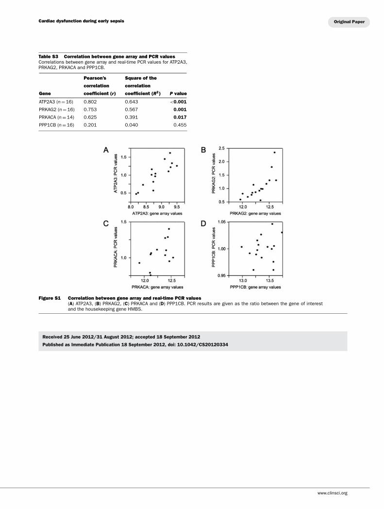
ate for PRKACA and absent for PPP1CB (see SupplementaryFigure S1 and Supplementary Table S3 at http://www.clinsci.org/cs/124/cs1240391add.htm).
DISCUSSION
In the present study, we describe a clinically relevant fluid-resuscitated long-term (3-day) rodent model of sepsis whereoutcome could be determined with high accuracy from haemody-namic measurements taken early in the illness. This demonstra-tion of an early difference in phenotype in animals that proceededto either recover fully or die occurred notwithstanding similar-ities in genotype, age, gender and upbringing of these animals,and receipt of a similar insult. They reflect patient data where,despite the marked heterogeneity of patient populations, baselinecardiovascular variables such as HR [8,17], stroke volume [18]and cardiac output [18,19] could differentiate between survivorsand non-survivors of septic shock. We used echocardiography-derived measurements of stroke volume for the present study,which predicted mortality with a high positive and negative pre-dictive value. Though our focus was on ventricular performance,we also found a similar predictive ability could be made for HR.This may offer a viable option for future studies if the capability tomeasure stroke volume is not available. Our findings suggest thatan increased systemic inflammatory response will compromisemyocardial function through circulating myocardial depressantfactors [20] including NO (nitric oxide) [21], and through alteredgene expression of proteins involved in cardiac contractile andrelaxation pathways (as demonstrated at 6 h in the present study)and, subsequently (at 12–48 h), in bioenergetic pathways [22].
The lower values of cardiac output in the predicted non-survivor group did not simply reflect more profound hypo-volaemia related to a greater degree of capillary leak, as fluidloading failed to generate either the maximal stroke volumes at-tained by the sham or sepsis survivor groups, nor did it reduceHR. Notably, all animals with sepsis at 6 h were normotensive(Table 1) and had received 40 ml/kg of body weight (equivalentto 3 litres in humans) between 2 and 6 h. Intrinsic myocardialdepression was, however, confirmed by a progressive decrease intolerance to large volume fluid loading in the animals with sepsis;this occurred to a much greater extent in predicted non-survivors.
Hollenberg et al. [23] reported myocardial depression in micewith sepsis, despite the co-existence of a high cardiac output.These findings reflect those made in clinical echocardiographicstudies, some of which could also prognosticate as early as the firstday of ICU (intensive care unit) admission [12,24,25]. As seenin this study, the failing thinner-walled right ventricle copes lesswell with excessive fluid loading; this will likely be compoundedin sepsis by co-existing (acute) lung injury, increasing pulmonaryartery pressures and right ventricular afterload. A careful balancemust therefore be reached between an avoidance of excess fluidthat will compromise myocardial function and worsen interstitialoedema [26], and sufficient fluid administration to rescue themacrocirculation, improve organ perfusion and enhance survival[26]. As our data demonstrate, the sicker the animals with sepsisbecome, the less fluid loading they are able to tolerate.
396 C© The Authors Journal compilation C© 2013 Biochemical Society

Cardiac dysfunction during early sepsis
Figure 4 Network-based gene expression analysis(A) Heatmap highlighting significant differences in 527 mean gene expression values between the four clinical groups(n = 4 per group). These are displayed as four clusters of transcripts with similar gene activity patterns. Associated coloursrepresent variance-normalized expression values relative to the mean expression of each transcript within this experiment.Red and blue indicate relative increase or decrease of transcript abundance, respectively. (B) Gene expression values ofone representative transcript for each of the four clusters, illustrating the relative transcript abundance in each cluster.For cluster 1: Cited4 [CBP (cAMP-response-element-binding protein) interacting transactivator-4], cluster 2: Nrep (neuronalprotein 3.1), cluster 3: SELP (selectin-P) and cluster-4: Mt1a (metallothionein-1a). (C) Signalling pathways most prominentlyactivated/altered in the sepsis cluster (cluster 3). Both TLR and IL-6 signalling lead to transcriptional activation of centralregulators of proliferation, cell death and immune response. MKK3/MKK6, mitogen-activated-protein-kinase kinase 3/6,PAI-1, plasminogen activator inhibitor-1, HAS-1, hyaluronan synthase-1; TRH, thyrotropin-releasing hormone, MCL, myeloidcell leukaemia sequence 1 (BCL2-related), IL4R: IL-4 receptor. The Ingenuity Pathway Analysis Figure is c© 2000–2008Ingenuity Systems, Inc. All rights reserved.
Histology demonstrated negligible cardiac tissue necrosis,supporting previous findings in animals with sepsis [15,27,28]and in non-survivors of human septic shock [29,30]. EM diddisplay evidence of mitochondrial swelling, suggesting organelle
damage, confirming previous work by ourselves and others in an-imals [21,22,28,31–33] and patients [34] with sepsis. The findingof glycogen accumulation in cardiomyocytes from animals withsepsis, as reported previously [35], has also been identified in
www.clinsci.org 397

A. Rudiger and others
Figure 5 Myocardial transcriptomics revealing transcript alterations in β-adrenergic signalling and Ca2 + flux pathwaysdepending on disease severityCardiomyocyte contraction depends on intracellular Ca2 + regulated by catecholamines acting on the ARβ2, with sub-sequent activation of Gs (stimulatory G-proteins) and elevation of cAMP. This activates PKA, phosphorylating PLN (phos-pholamban), an SR transmembrane protein. Depending on its own phosphorylation state, PLN promotes the active state ofthe Ca2 + ATPase SERCA, favouring Ca2 + uptake into the SR. PP1 dephosphorylates PLN, deactivating SERCA. Ca2 + entryinto the cell through L-type calcium channels triggers Ca2 + -mediated Ca2 + release from the SR via RyRs (ryanodine recept-ors). This dictates the degree of actin–myosin cross-bridge formation and the strength of contraction. Relaxation occurswhen cytosolic Ca2 + is lowered primarily by activity of SERCA, as well as plasmalemmal Ca2 + -ATPases and Na+ /Ca2 +exchangers. Compared with predicted survivors, predicted non-survivors showed increased (red) cardiac gene transcriptsfor PDE10 (phosphodiesterase 10), PP1 and RyR3, whereas SERCA transcript abundance was decreased (blue). Theβ2AR (β2-adrenergic receptor) and PKA transcript levels were decreased in hearts from animals with sepsis, but did notreach statistical significance between predicted survivors and non-survivors. The Ingenuity Pathway Analysis Figure is c©2000–2008 Ingenuity Systems, Inc. All rights reserved.
hibernating myocardium of pigs following ischaemia [36]. Insummary, structural tissue damage was minor, although changeswere apparent at the subcellular level. These observations sup-port our hypothesis that myocardial dysfunction is a functionalrather than a structural derangement [4,37].
This descriptive approach to myocardial depression in sepsiscan be systematically studied in the present model by applyinggenome-wide analysis of alterations in the myocardial transcrip-tome. To this end, we employed a structured network knowledge-based approach that can provide insights into regulation of cellfunction and interaction, as demonstrated in healthy volunteerstreated with endotoxin [38]. Although we cannot yet describeprecise mechanisms through which these changes occur, sepsisclearly elicits significantly different responses compared withsham operation alone, and that some of these are differentiallyexpressed and associated with poor outcome.
The genome-wide profiling revealed early up-regulation ofTLR2/MyD88 and JAK/STAT3-dependent signalling with sepsis.This supports findings of improved cardiac function, with nor-mal sarcomere shortening and peak change in intracellularCa2 + , in TLR2− / − mice undergoing caecal ligation and punc-
ture compared with their wild-type equivalents [39]. Althoughthe JAK/STAT3 pathway is generally considered to be cardio-protective [40], it too can contribute to cardiac dysfunction dur-ing ischaemia [41]. A study of rats undergoing cecal ligationand puncture reported attenuated organ failure and improved sur-vival rates following JAK/STAT3 pathway inhibition, althoughthe heart was not specifically examined [42].
Importantly, in relation to the physiological changes recordedin this model, myocardial transcriptomics revealed alterationsin transcript abundance linked to β-adrenergic signalling andCa2 + flux that depended on disease severity and prognosis (Fig-ure 5). Although mindful of the limitations in interpreting tran-scriptomic data, it is conceivable that such transcript differencescould impact on myocardial function. In our model, the PKAtranscript level was significantly decreased in the animals withsepsis, whereas PDE10 transcript abundance was elevated. PDEincreases breakdown of cAMP, further reducing PKA activity[43]. Increased activity of PP1 dephosphorylates phospholam-ban, reducing Ca2 + uptake into the SR (sarcoplasmic reticulum)[44]. Down-regulation of SERCA has a similar effect [45]. Up-regulation of PP1 and down-regulation of PKA also reduce Ca2 +
398 C© The Authors Journal compilation C© 2013 Biochemical Society

Cardiac dysfunction during early sepsis
entry into the cell by inhibiting L-type calcium channels [46].A diminution in L-type calcium current was reported recently inventricular myocytes isolated from pigs with sepsis [47]. Highcytosolic and low SR Ca2 + levels lead to failure of diastolic relax-ation and impaired systolic contraction, respectively. Both theseaspects are also recognized in septic cardiomyopathy [4,48].
We focused our transcriptomic analyses on heart tissue asour prognostic tool was based on an assessment of myocardialdysfunction. Analyses of liver tissue taken concurrently showsignificant differences in other signalling pathways [49]. It is thusreasonable to speculate that each organ has its own transcriptomicresponse to infection. A better understanding of each organ’sresponse, and interactions between organs, may provide usefulclues to developing novel therapies.
In summary, our findings emphasize both the presence andprognostic significance of cardiac dysfunction with intrinsicmyocardial depression during early sepsis, at a time when over-all clinical severity was not marked. The poor-prognosis animalshad reduced tolerance to i.v. fluid loading, highlighting the im-portance of adequate but not excessive fluid administration inclinical management. Our study implies that outcome is alreadydetermined at an early stage in sepsis. Greater or lesser degreesof gene up- or down-regulation, with the likely addition of post-transcriptional modifications that were not measured in this study,will distinguish eventual survivors and non-survivors. If con-firmed, this has major consequences for patient management asa proportion are clearly not benefiting from current approaches;new paradigms would need to be introduced to improve outcomesin such patients.
CLINICAL PERSPECTIVES
� Myocardial function is depressed in sepsis and is an importantprognosticator in patients.
� Using a long-term fluid-resuscitated rat model of faecal peri-tonitis, we demonstrate that significant differences in strokevolume and HR measured 6 h post-insult could predict 3-daymortality with positive and negative predictive values of 93and 80 % respectively. In separate studies, cardiac gene ex-pression analysis at 6 h detected 527 transcripts significantlyup- or down-regulated by the septic process, including genesrelated to inflammatory and cell cycle pathways. The degreeof change in these signalling pathways correlated with clinicaldysfunction.
� These findings suggest a crucial role for early cardiovascularperformance in determining subsequent outcome, with cleartherapeutic implications.
AUTHOR CONTRIBUTION
Alain Rudiger conceived and designed the study, performed the ex-perimental work, analysed and interpreted the data, and draftedand approved the paper. Alex Dyson, Karen Felsmann and JaneCarre performed the experimental work, and drafted and approvedthe paper. Valerie Taylor performed the experimental work. SianHughes performed the experimental work and interpreted the data.Innes Clatworthy, Jana Lemm and Ralf Claus performed the experi-
mental work and interpreted the data. Alessandro Protti and DenisPellerin assisted with experimental design and interpreted the data.Michael Bauer interpreted the data, and drafted and approved thepaper. Mervyn Singer conceived and designed the study, interpretedthe data, supervised the study, and drafted and approved the paper.
FUNDING
This work was supported by the Swiss National Science Founda-tion (Basel, Switzerland), the Stiefel Zangger Foundation (Zurich,Switzerland) and the Siegenthaler Foundation (Zurich, Switzerland)(grants to A.R.), and the U.K. Medical Research Council (grants toA.D., J.C. and V.T.). The echocardiography equipment was funded bythe British Heart Foundation. Transcriptomic analyses were fundedby the Krokus Foundation (Basel, Switzerland) (to A.R.), and theFederal Ministry for Education and Research (within the ‘Center forSepsis Control and Care’) [grant number 01 EO 1002, Project D1.2(to M.B.)]. This work was undertaken at UCL Hospitals/UCL, whichreceived support from the National Institute of Health ResearchBiomedical Research Centre funding scheme.
REFERENCES
1 Hotchkiss, R. S. and Karl, I. E. (2003) The pathophysiology andtreatment of sepsis. N. Engl. J. Med. 348, 138–150
2 Levy, R. J. and Deutschman, C. S. (2004) Evaluating myocardialdepression in sepsis. Shock 22, 1–10
3 Vieillard-Baron, A., Caille, V., Charron, C., Belliard, G., Page, B.and Jardin, F. (2008) Actual incidence of global left ventricularhypokinesia in adult septic shock. Crit. Care Med. 36,1701–1706
4 Rudiger, A. and Singer, M. (2007) Mechanisms of sepsis-inducedcardiac dysfunction. Crit. Care Med. 35, 1599–1608
5 Hein, O. V., Misterek, K., Tessmann, J. P., van Dossow, V.,Krimphove, M. and Spies, C. (2005) Time course of endothelialdamage in septic shock: prediction of outcome. Crit. Care 9,R323–R330
6 Angstwurm, M. W., Gaertner, R. and Schopohl, J. (2005)Outcome in elderly patients with severe infection is influenced bysex hormones but not gender. Crit. Care Med. 33, 2786–2793
7 Brueckmann, M., Huhle, G., Lang, S., Haase, K. K., Bertsch, T.,Weiss, C., Kaden, J. J., Putensen, C., Borggrefe, M. andHoffmann, U. (2005) Prognostic value of plasma N-terminalpro-brain natriuretic peptide in patients with severe sepsis.Circulation 112, 527–534
8 Parker, M. M., Shelhamer, J. H., Natanson, C., Alling, D. W. andParrillo, J. E. (1987) Serial cardiovascular variables in survivorsand nonsurvivors of human septic shock: heart rate as an earlypredictor of prognosis. Crit. Care Med. 15, 923–929
9 Brealey, D., Brand, M., Hargreaves, I., Heales, S., Land, J.,Smolenski, R., Davies, N. A., Cooper, C. E. and Singer, M. (2002)Association between mitochondrial dysfunction and severity andoutcome of septic shock. Lancet 360, 219–223
10 Ammann, P., Maggiorini, M., Bertel, O., Haenseler, E.,Joller-Jemelka, H. I., Oechslin, E., Minder, E. I., Rickli, H. andFehr, T. (2003) Troponin as a risk factor for mortality in criticallyill patients without acute coronary syndromes. J. Am. Coll.Cardiol. 41, 2004–2009
www.clinsci.org 399
A. Rudiger and others
11 Charpentier, J., Luyt, C-E., Fulla, Y., Visonneau, C., Cariou, A.,Grabar, S., Dhainaut, J-F., Mira, J-P. and Chiche, J-D. (2004) Brainnatriuretic peptide: a marker of myocardial dysfunction andprognosis during severe sepsis. Crit. Care Med. 32, 660–665
12 Jardin, F., Fourme, T., Page, B., Loubieres, Y., Vieillard-Baron, A.,Beauchet, A. and Bourdarias, J-P. (1999) Persistent preloaddefect in severe sepsis despite fluid loading: a longitudinalechocardiographic study in patients with septic shock. Chest116, 1354–1359
13 Parker, M. M., Shelhamer, J. H., Bacharach, S. L., Green, M.,Natanson, C., Frederick, T. M., Deamske, B. A. and Parrillo, J. E.(1984) Profound but reversible myocardial depression in patientswith septic shock. Ann. Intern. Med. 100, 483–490
14 Brealey, D., Karyampudi, S., Jacques, T. S., Novelli, M., Stidwill,R., Taylor, V., Smolenski, R. T. and Singer, M. (2004)Mitochondrial dysfunction in a long-term rodent model of sepsisand organ failure. Am. J. Physiol. Regul. Integr. Comp. Physiol.286, R491–R497
15 Slama, M., Susic, D., Varagic, J., Ahn, J. and Frohlich, E. D.(2002) Echocardiographic measurement of cardiac output inrats. Am. J. Physiol. Heart Circ. Physiol. 284, H691–H697
16 Huber, W., von Heydebreck, A., Sultmann, H., Poustka, A. andVingron, M. (2002) Variance stabilization applied to microarraydata calibration and to the quantification of differentialexpression. Bioinformatics 18, S96–S104
17 Azimi, G. and Vincent, J-L. (1986) Ultimate survival from septicshock. Resuscitation 14, 245–253
18 Kumar, A., Schupp, E., Bunnell, E., Ali, A., Milcarek, B. andParrillo, J. E. (2008) Cardiovascular response to dobutaminestress predicts outcome in severe sepsis and septic shock. Crit.Care 12, R35
19 Tuchschmidt, J., Fried, J., Astiz, M. and Rackow, E. (1992)Elevation of cardiac output and oxygen delivery improvesoutcome in septic shock. Chest 102, 216–220
20 Parrillo, J. E., Burch, C., Shelhamer, J. H., Parker, M. M.,Natanson, C. and Schuette, W. (1985) A circulating mycoardialdepressant substance in humans with septic shock. J. Clin.Invest. 76, 1539–1553
21 dos Santos, C. C., Gattas, D. J., Tsoporis, J. N., Smeding, L.,Kabir, G., Masoom, H., Akram, A., Plotz, F., Slutsky, A. S.,Husain, M. et al. (2010) Sepsis-induced myocardial depressionis associated with transcriptional changes in energy metabolismand contractile related genes: a physiological and geneexpression-based approach. Crit. Care Med. 38, 894–902
22 Hinkelbein, J., Kalenka, A., Schubert, C., Peterka, A. andFeldmann, Jr, R. E. (2010) Proteome and metabolome alterationsin heart and liver indicate compromised energy production duringsepsis. Protein Pept. Lett. 17, 18–31
23 Hollenberg, S. M., Dumasius, A., Easington, C., Colilla, S. A.,Neumann, A. and Parrillo, J. E. (2001) Characterization of ahyperdynamic murine model of resuscitated sepsis usingechocardiography. Am. J. Respir. Crit. Care Med. 164, 891–895
24 Jones, A. E., Craddock, P. A., Tayal, V. S. and Kline, J. A. (2005)Diagnostic accuracy of left ventricular function for identifyingsepsis among emergency department patients with nontraumaticsymptomatic undifferentiated hypotension. Shock 24, 513–517
25 Poelaert, J., Declerck, C., Vogelaers, D., Colardyn, F. and Visser,C. A. (1997) Left ventricular systolic and diastolic function inseptic shock. Intensive Care Med. 23, 553–560
26 Zanotti-Cavazzoni, S. L., Guglielmi, M., Parrillo, J. E., Walker, T.,Dellinger, R. P. and Hollenberg, S. M. (2009) Fluid resuscitationinfluences cardiovascular performance and mortality in a murinemodel of sepsis. Intensive Care Med. 35, 748–754
27 Hotchkiss, R. S., Swanson, P. E., Cobb, J. P., Allyson, J.,Buchman, T. G. and Karl, I. E. (1997) Apoptosis in lymphoid andparenchymal cells during sepsis: findings in normal and T- andB-cell-deficient mice. Crit. Care Med. 25, 1298–1307
28 Solomon, M. A., Correa, R., Alexander, H. R., Koev, L. A., Corb,J. P., Kim, D. K., Roberts, W. C., Quezado, Z. M. N., Scholz, T. D.,Cunnion, R. E. et al. (1994) Myocardial energy metabolism andmorphology in a canine model of sepsis. Am. J. Physiol. HeartCirc. Physiol. 266, 757–768
29 Hotchkiss, R. S., Swanson, P. E., Freeman, B. D., Tinsley, K. W.,Cobb, J. P., Matuschak, G. M., Buchman, T. G. and Karl, I. E.(1999) Apoptotic cell death in patients with sepsis, shock, andmultiple organ dysfunction. Crit. Care Med. 27, 1230–1248
30 Rossi, M. A., Celes, M. R., Prado, C. M. and Saggioro, F. P.(2007) Myocardial structural changes in long-term human severesepsis/septic shock may be responsible for cardiac dysfunction.Shock 27, 10–18
31 Hersch, M., Gnidec, A. A., Bersten, A. D., Troster, M., Rutledge,F. S. and Sibbald, W. J. (1990) Histologic and ultrastructuralchanges in nonpulmonary organs during early hyperdynamicsepsis. Surgery 107, 397–410
32 Suliman, H. B., Welty-Wolf, K. E., Carraway, M. S., Tatro, L. andPiantadosi, C. A. (2004) Lipopolysaccharide induces oxidativecardiac mitochondrial damage and biogenesis. Cardiovasc. Res.64, 279–288
33 Watts, J. A., Kline, J. A., Thornton, L. R., Grattan, R. M. andBrar, S. S. (2004) Metabolic dysfunction and depletion ofmitochondria in hearts of septic rats. J. Mol. Cell. Cardiol. 36,141–150
34 Cowley, R. A., Merger, W. J., Fisher, R. S., Jones, R. T. and Trump,B. F. (1979) The subcellular pathology of shock in traumapatients: studies using immediate autopsy. Am. J. Surg. 45,255–269
35 Levy, R. J., Piel, D. A., Acton, P. D., Zhou, R., Ferrari, V. A., Karp,J. S. and Deutschman, C. S. (2005) Evidence of myocardialhibernation in the septic heart. Crit. Care Med. 33, 2752–2756
36 Thomas, S. A., Fallavollita, J. A., Suzuki, G., Borgers, M. andCanty, J. M. Jr. (2002) Dissociation of regional adaptations toischemia and global myolysis in an accelerated swine model ofchronic hibernating myocardium. Circ. Res. 91, 970–977
37 Singer, M., De Santis, V., Vitale, D. and Jeffcoate, W. (2004)Multiorgan failure is an adaptive, endocrine-mediated, metabolicresponse to overwhelming systemic inflammation. Lancet 364,545–548
38 Calvano, S. E., Xiao, W., Richards, D. R., Felciano, R. M., Baker,H. V., Cho, R. J., Chen, R. O., Brownstein, B. H., Cobb, J. P.,Tschoeke, S. K. et al. (2005) A network-based analysis ofsystemic inflammation in humans. Nature 437, 1032–1037
39 Zou, L., Feng, Y., Chen, Y. J., Si, R., Shen, S., Zhou, Q., Ichinose,F., Scherrer-Crosbie, M. and Chao, W. (2010) Toll-like receptor 2plays a critical role in cardiac dysfunction during polymicrobialsepsis. Crit. Care Med. 38, 1335–1342
40 Booz, G. W., Day, J. N. and Baker, K. M. (2002) Interplay betweenthe cardiac renin angiotensin system and JAK-STAT signaling:role in cardiac hypertrophy, ischemia/reperfusion dysfunction,and heart failure. J. Mol. Cell. Cardiol. 34, 1443–1453
41 Mascareno, E., El-Shafei, M., Maulik, N., Sato, M., Guo, Y., Das,D. K. and Siddiqui, M. A. (2001) JAK/STAT signaling isassociated with cardiac dysfunction during ischemia andreperfusion. Circulation 104, 325–329
42 Hui, L., Yao, Y., Wang, S., Yu, Y., Dong, N., Li, H. and Sheng, Z.(2009) Inhibition of Janus kinase 2 and signal transduction andactivator of transcription 3 protect against cecal ligation andpuncture-induced multiple organ damage and mortality. J. Trauma66, 859–865
43 Kass, D. A., Takimoto, E., Nagayama, T. and Champion, H. C.(2007) Phosphodiesterase regulation of nitric oxide signaling.Cardiovasc. Res. 75, 303–314
44 Pathak, A., del Monte, F., Zhao, W., Schultz, J. E., Lorenz, J. N.,Bodi, I., Weiser, D., Hahn, H., Carr, A. N., Syed, F. et al. (2005)Enhancement of cardiac function and suppression of heartfailure progression by inhibition of protein phosphatase 1. Circ.Res. 96, 756–766
400 C© The Authors Journal compilation C© 2013 Biochemical Society
Cardiac dysfunction during early sepsis
45 Periasamy, M. and Huke, S. (2001) SERCA pump level is acritical determinant of Ca2 + homeostasis and cardiaccontractility. J. Mol. Cell. Cardiol. 33, 1053–1063
46 du Bell, W. H. and Rogers, T. B. (2004) Protein phosphatase 1and an opposing protein kinase regulate steady-state L-typeCa2 + current in mouse cardiac myocytes. J. Physiol. 556, 79–93
47 Stengl, M., Bartak, F., Sykora, R., Chvojka, J., Benes, J.,Krouzecky, A., Novak, I., Sviglerova, J., Kuncova, J. andMatejovic, M. (2010) Reduced L-type calcium current inventricular myocytes from pigs with hyperdynamic septic shock.Crit. Care Med. 38, 579–587
48 Duncan, D. J., Yang, Z., Hopkins, P. M., Steele, D. S. andHarrison, S. M. (2010) TNF-α and IL-1β increase Ca2 + leakfrom the sarcoplasmic reticulum and susceptibility toarrhythmia in rat ventricular myocytes. Cell Calcium 47,378–386
49 Recknagel, P., Gonnert, F. A, Westermann, M., Lambeck, S.,Lupp, A., Rudiger, A., Dyson, A., Carre, J. E., Kortgen, A.,Krafft, C. et al. (2012) Liver dysfunction andphosphatidylinositol-3-kinase signaling in early sepsis:experimental studies in rodent models of peritonitis. PLoS Med.9, e1001338
Received 25 June 2012/31 August 2012; accepted 18 September 2012
Published as Immediate Publication 18 September 2012, doi: 10.1042/CS20120334
www.clinsci.org 401

Clinical Science (2013) 124, 391–401 (Printed in Great Britain) doi: 10.1042/CS20120334
SUPPLEMENTARY ONLINE DATA
Early functional and transcriptomic changes inthe myocardium predict outcome in a long-termrat model of sepsisAlain RUDIGER*, Alex DYSON*, Karen FELSMANN†, Jane E. CARRE*, Valerie TAYLOR*, Sian HUGHES‡,Innes CLATWORTHY§, Alessandro PROTTI*, Denis PELLERIN‖, Jana LEMM¶, Ralf A. CLAUS¶,Michael BAUER¶1 and Mervyn SINGER*
*Bloomsbury Institute of Intensive Care Medicine, Division of Medicine, University College London, Gower Street, London WC1E 6BT, U.K.†SIRS-Lab GmbH, Otto-Schott-Straße 15, D-07745 Jena, Germany‡Department of Histopathology, Royal Free and University College Medical School, University College London, Rockefeller Building, University Street,London WC1E 6JJ, U.K.§Imaging Facility, UCL Institute of Opthalmology, 11–43 Bath Street, London EC1V 9EL. U.K.‖The Heart Hospital, UCL Hospitals NHS Foundation Trust, 16-18 Westmoreland Street, London W1G 8PH, U.K.¶Integrated Research and Treatment Center–Center for Sepsis Control and Care, Jena University Hospital, Allee 101, D-07747 Jena, Germany
Table S1 Reproducibility of echocardiography parametersIntra-observer variability is the variability between two analyses of the same study. Inter-observer variability is the variabilitybetween two observers analysing the same study. Inter-study variability is the variability between data acquired at twotime-points within 24 h, recorded and analysed by one investigator. Variability is calculated as the difference between twoobservations divided by the mean of the two observations and expressed as percentage. LV EDV, LV end-diastolic volume; LVEF, LV ejection fraction; IVC inferior vena cava.
Naıve animals (n = 10) Animals with sepsis at 6 h (n = 10)
Intra-observer Inter-observer Inter-study Intra-observer Inter-observer
Paramter variability (%) variability (%) variability (%) variability (%) variability (%)
HR (beats/min) 0.39 (0.14) 0.63 (0.10) 6.7 (1.4) 0.70 (0.13) 0.85 (0.19)
LV EDV (ml) 4.5 (1.2) 7.7 (1.7) 8.8 (2.3) 8.1 (1.5) 8.9 (2.0)
Stroke volume (ml) 2.1 (0.35) 3.6 (0.7) 6.1 (1.7) 7.8 (1.6) 6.4 (1.1)
Cardiac output (ml/min) 1.9 (0.35) 3.9 (0.79) 3.9 (0.78) 7.5 (1.5) 5.9 (1.1)
LV EF (%) 5.1 (1.2) 5.8 (1.2) 8.2 (1.9) 11 (2.3) 14 (2.8)
IVC diameter (mm) 5.4 (1.1) 7.6 (2.4) 13 (3.7) 6.8 (2.1) 3.5 (1.3)
1 A member of the scientific advisory board of SIRS-Lab GmbH, Jena, Germany, where the microarray experiments were performed.
Correspondence: Professor Mervyn Singer (email [email protected]).
www.clinsci.org

A. Rudiger and others
Table S2 Genes significantly up-regulated or down-regulated in predicted sepsis non-survivors compared with survivors(clusters 1 and 3)
Gene
Molecule regulation Description Location Type
ABCG3 (includes EG:27405) Down ABC (ATP-binding cassette), sub-family G(WHITE), member 3
Plasma membrane Transporter
AFF4 Up AF4/FMR2 family, member 4 Nucleus Transcription regulator
ALMS1 Down Alstrom syndrome 1 Cytoplasm Other
APLN Down apelin, AGTRL1 ligand Extracellular space Other
APOBEC3F Up Apo B (apolipoprotein B) mRNA editingenzyme, catalytic polypeptide-like 3F
Unknown Enzyme
ARHGEF9 Down Cdc42 GEF (guanine-nucleotide-exchangefactor) 9
Cytoplasm Other
ARL5A Up Arf (ADP-ribosylation factor)-like 5A Unknown Enzyme
ATP2A3 Down ATPase, Ca2 + transporting, ubiquitous Cytoplasm Transporter
BCL10 Up B-cell CLL/lymphoma 10 Cytoplasm Transcription regulator
BCR Up Breakpoint cluster region Cytoplasm Kinase
BTG2 Up BTG family, member 2 Nucleus Transcription regulator
C11ORF54 Down Chromosome 11 open reading frame 54 Nucleus Other
C14ORF172 Up Chromosome 14 open reading frame 172 Unknown Enzyme
C4ORF14 Up Chromosome 4 open reading frame 14 Cytoplasm Other
CC2D2A Down Coiled-coil and C2 domain containing 2A Unknown Other
CD55 Up CD55 molecule, decay accelerating factor forcomplement (Cromer blood group)
Plasma membrane Other
CDC42SE1 Up CDC42 small effector 1 Plasma membrane Other
CDK5 Down Cyclin-dependent kinase 5 Nucleus Kinase
CDKN1A Up cyclin-dependent kinase inhibitor 1A (p21,Cip1)
Nucleus Kinase
CES1 (includes EG:1066) Up Carboxylesterase 1 (monocyte/macrophageserine esterase 1)
Cytoplasm Enzyme
CH25H Up Cholesterol 25-hydroxylase Cytoplasm Enzyme
CLCN3 Up Chloride channel 3 Plasma membrane Ion channel
CYB5R2 Down Cytochrome b5 reductase 2 Unknown Enzyme
CYP2U1 Down Cytochrome P-450, family 2, subfamily U,polypeptide 1
Unknown Enzyme
DDB2 Down Damage-specific DNA binding protein 2,48kDa
Nucleus Other
DDIT4L Down DNA-damage-inducible transcript 4-like Unknown Other
DEPDC7 Up DEP domain containing 7 Unknown Other
DTWD1 Down DTW domain containing 1 Unknown Other
DUSP11 Up Dual specificity phosphatase 11 (RNA/RNPcomplex 1-interacting)
Nucleus Phosphatase
EGFLAM Down EGF-like, fibronectin type III and laminin Gdomains
Unknown Other
ENPP1 Down Ectonucleotidepyrophosphatase/phosphodiesterase 1
Plasma membrane Enzyme
EVL Down Enah/Vasp-like Cytoplasm Other
FAM81A Down Family with sequence similarity 81, member A Unknown Other
FHL1 Up four and a half LIM domains 1 Cytoplasm Other
FLJ21865 Down Endo-β -N-acetylglucosaminidase Unknown Enzyme
FLNC Up Filamin C, gamma (actin binding protein 280) Cytoplasm Other
FOXK2 Up Forkhead box K2 Nucleus Transcription regulator
GADD45G Up Growth arrest and DNA-damage-inducible,gamma
Nucleus Other
GALM Down Galactose mutarotase (aldose 1-epimerase) Cytoplasm Enzyme
GALNACT-2 Up Chondroitin sulfate GalNAcT-2 Cytoplasm Enzyme
C© The Authors Journal compilation C© 2013 Biochemical Society

Cardiac dysfunction during early sepsis
Table S2 Continued
Gene
Molecule regulation Description Location Type
GCGR Down Glucagon receptor Plasma membrane G-protein coupled receptor
GEM Up GTP-binding protein overexpressed in skeletalmuscle
Plasma membrane Enzyme
GRN Down Granulin Extracellular space Growth factor
HAS1 Up Hyaluronan synthase 1 Plasma membrane Enzyme
HIP1 Down Huntingtin interacting protein 1 Cytoplasm Other
HRSP12 Down Heat-responsive protein 12 Cytoplasm Other
ITPKC Up Inositol 1,4,5-trisphosphate 3-kinase C Unknown Kinase
JAG2 Down jagged 2 Extracellular space Growth factor
KCNK2 Down Potassium channel, subfamily K, member 2 Plasma membrane Ion channel
KRCC1 Up Lysine-rich coiled-coil 1 Unknown Other
LOC499828 Up Similar to copine VIII isoform 1 Unknown Other
MAGED2 Down melanoma antigen family D, 2 Plasma membrane Other
MAP2K6 Down Mitogen-activated protein kinase kinase 6 Cytoplasm Kinase
MATN2 Down Matrilin 2 Extracellular space Other
MDM1 Down Mdm4, transformed 3T3 cell double minute1, p53 binding protein (mouse)
Unknown Other
METTL6 Up Methyltransferase like 6 Unknown Enzyme
MGC70857 Down Similar to RIKEN cDNA C030006K11 gene Unknown Other
MLL5 Down Myeloid/lymphoid or mixed-lineage leukemia5 (trithorax homologue, Drosophila)
Nucleus Other
MMD Down Monocyte to macrophagedifferentiation-associated
Plasma membrane Other
MRTO4 Up mRNA turnover 4 homologue (S. cerevisiae) Cytoplasm Other
MYC Up v-myc myelocytomatosis viral oncogenehomologue (avian)
Nucleus Transcription regulator
MYOT Up Myotilin Cytoplasm Other
NCF4 Up Neutrophil cytosolic factor 4, 40 kDa Cytoplasm Enzyme
NR0B2 Down Nuclear receptor subfamily 0, group B,member 2
Nucleus Ligand-dependent nuclearreceptor
NRM Down Nurim (nuclear envelope membrane protein) Nucleus Other
NSUN5 Up NOL1/NOP2/Sun domain family, member 5 Unknown Other
OLR1117 PREDICTED Up Olfactory receptor 1117 (predicted) Plasma membrane G-protein coupled receptor
OPN4 Up Opsin 4 (melanopsin) Plasma membrane G-protein coupled receptor
OR51E2 Down Olfactory receptor, family 51, subfamily E,member 2
Plasma membrane G-protein coupled receptor
PDE10A Up Phosphodiesterase 10A Cytoplasm Enzyme
PIK3IP1 Down Phosphoinositide-3-kinase interacting protein1
Unknown Other
PPP1CB Up Protein phosphatase 1, catalytic subunit, β
isoformCytoplasm Phosphatase
PPP1R15A (includes EG:23645) Up Protein phosphatase 1, regulatory (inhibitor)subunit 15A
Cytoplasm Other
PUS7 Up Pseudouridylate synthase 7 homologue (S.cerevisiae)
Unknown Other
PVR Up poliovirus receptor Plasma membrane G-protein coupled receptor
RAB40B Down RAB40B, member RAS oncogene family Plasma membrane Enzyme
RBM13 Up RNA binding motif protein 13 Nucleus Other
REL Up v-rel reticuloendotheliosis viral oncogenehomologue (avian)
Nucleus Transcription regulator
RGD1559728 PREDICTED Up RGD1559728 (predicted) Unknown Other
RGD1559957 PREDICTED Up RGD1559957 (predicted) Unknown Other
RGD1561949 PREDICTED Up Similar to spermidine synthase (predicted) Unknown Other
www.clinsci.org

A. Rudiger and others
Table S2 Continued
Gene
Molecule regulation Description Location Type
RGD1562388 PREDICTED Down Similar to ORF2 consensus sequence encodingendonuclease and reverse transcriptaseminus RNaseH (predicted)
Unknown Other
RGD1564483 PREDICTED Up Similar to ARP2/3 complex 16 kDa subunit(p16-ARC) (predicted)
Unknown Other
RGD1564567 PREDICTED Up Similar to CG14998-PC, isoform C (predicted) Unknown Other
RGS2 Up Regulator of G-protein signalling 2, 24kDa Nucleus Other
RND1 Up Rho family GTPase 1 Cytoplasm Enzyme
RYR3 Up Ryanodine receptor 3 Plasma membrane Ion channel
SCRIB Down Scribbled homologue (Drosophila) Cytoplasm Other
SCRN1 Down Secernin 1 Cytoplasm Other
SELP Up Selectin P (granule membrane protein 140 kDa,antigen CD62)
Plasma membrane Other
SH3BGRL2 Up SH3 domain binding glutamic acid-rich proteinlike 2
Plasma membrane Other
SIAH2 Up Seven in absentia homologue 2 (Drosophila) Nucleus Transcription regulator
SLC20A1 Up Solute carrier family 20 (phosphate transporter),member 1
Plasma membrane Transporter
SLC37A2 Down Solute carrier family 37 (glycerol-3-phosphatetransporter), member 2
Unknown Transporter
SLC37A4 Down Solute carrier family 37 (glucose-6-phosphatetransporter), member 4
Cytoplasm Transporter
SNRPA1 Up Small nuclear ribonucleoprotein polypeptide A’ Nucleus Other
SOX4 Down SRY (sex-determining region Y)-box 4 Nucleus Transcription regulator
SP3 Up Sp3 transcription factor Nucleus Transcription regulator
STAB1 Down Stabilin 1 Plasma membrane Transporter
STAG1 Up Stromal antigen 1 Nucleus Other
STAT3 Up Signal transducer and activator of transcription 3(acute-phase response factor)
Nucleus Transcription regulator
TCF3 Down Transcription factor 3 (E2A immunoglobulinenhancer binding factors E12/E47)
Nucleus Transcription regulator
TGS1 Up Trimethylguanosine synthase homologue (S.cerevisiae)
Nucleus Transcription regulator
TMEM53 Down trans-Membrane protein 53 Unknown Other
TP53I11 Down Tumour protein p53 inducible protein 11 Unknown Other
TREM2 Down Triggering receptor expressed on myeloid cells 2 Plasma membrane trans-membrane receptor
TRH Up Thyrotropin-releasing hormone Extracellular space Other
TRIB1 Up Tribbles homologue 1 (Drosophila) Cytoplasm Kinase
TUBB6 Up Tubulin, β 6 Cytoplasm Other
UAP1 Up UDP-N-acetylglucosamine pyrophosphorylase 1 Nucleus Enzyme
UNC5C Down unc-5 homologue C (C. elegans) Plasma membrane trans-membrane receptor
UROD Down Uroporphyrinogen decarboxylase Cytoplasm Enzyme
USPL1 Up Ubiquitin specific peptidase like 1 Unknown Other
VPS45 Down Vacuolar protein sorting 45 homologue (S.cerevisiae)
Cytoplasm Transporter
WDR60 Down WD repeat domain 60 Unknown Other
WDR67 Down WD repeat domain 67 Unknown Other
XIRP1 Up Xin actin-binding repeat containing 1 Plasma membrane Other
ZFP472 Down Zinc finger protein 472 Unknown Other
ZFP61 Down Zinc finger protein 61 Unknown Peptidase
ZMYND19 Up Zinc finger, MYND-type containing 19 Plasma membrane Other
C© The Authors Journal compilation C© 2013 Biochemical Society
Cardiac dysfunction during early sepsis
Table S3 Correlation between gene array and PCR valuesCorrelations between gene array and real-time PCR values for ATP2A3,PRKAG2, PRKACA and PPP1CB.
Pearson’s Square of the
correlation correlation
Gene coefficient (r) coefficient (R2) P value
ATP2A3 (n = 16) 0.802 0.643 <0.001
PRKAG2 (n = 16) 0.753 0.567 0.001
PRKACA (n = 14) 0.625 0.391 0.017
PPP1CB (n = 16) 0.201 0.040 0.455
Figure S1 Correlation between gene array and real-time PCR values(A) ATP2A3, (B) PRKAG2, (C) PRKACA and (D) PPP1CB. PCR results are given as the ratio between the gene of interestand the housekeeping gene HMBS.
Received 25 June 2012/31 August 2012; accepted 18 September 2012
Published as Immediate Publication 18 September 2012, doi: 10.1042/CS20120334
www.clinsci.org