Early Burn Mortality in a Rodent Model Choriodecidual ...
13
Page 1/13 A Single Intravenous Infusion of Human Choriodecidual Mesenchymal Stem Cell Decreases Early Burn Mortality in a Rodent Model Eng-Kean Yeong ( [email protected] ) National Taiwan University https://orcid.org/0000-0001-5082-9771 Thai-Yen Ling National Taiwan University College of Medicine Research Article Keywords: human choriodecidual mesenchymal stem cell, early burn mortality, rodent model Posted Date: March 1st, 2021 DOI: https://doi.org/10.21203/rs.3.rs-251335/v1 License: This work is licensed under a Creative Commons Attribution 4.0 International License. Read Full License
Transcript of Early Burn Mortality in a Rodent Model Choriodecidual ...

Page 1/13
A Single Intravenous Infusion of HumanChoriodecidual Mesenchymal Stem Cell DecreasesEarly Burn Mortality in a Rodent ModelEng-Kean Yeong ( [email protected] )
National Taiwan University https://orcid.org/0000-0001-5082-9771Thai-Yen Ling
National Taiwan University College of Medicine
Research Article
Keywords: human choriodecidual mesenchymal stem cell, early burn mortality, rodent model
Posted Date: March 1st, 2021
DOI: https://doi.org/10.21203/rs.3.rs-251335/v1
License: This work is licensed under a Creative Commons Attribution 4.0 International License. Read Full License

Page 2/13
AbstractBackground:
Systemic in�ammatory responses (SIR) are the main cause of pulmonary dysfunction leading tomortality within hours of extensive burns. Based on previous studies showing that cell entrapment occursin the lungs following the infusion of human choriodecidual mesenchymal stem cells (hcMSCs), wehypothesize that the intravenous infusion of hcMSCs, with an immunomodulatory potential, will decreasethe risk of SIR induced pulmonary failure leading to mortality in burn patients.
Methods:
Forty adult male Sprague-Dawley rats were randomized into two groups. Group A (sham control, n = 10)received no injury or intervention; the remaining rats (n = 30) were subjected to burns covering 40 % of thetotal body surface area by immersion of the dorsum in 100 °C water for 15 s under general anesthesia.Injured rats were further randomized into different treatment groups: Group B (saline only control, n = 10),Group C (saline plus culture medium control, n = 10), and Group D (saline plus 2 × 10⁶ hcMSCs, n = 10).Culture medium or hcMSCs were given in a single infusion via the tail vein immediately after burns.Mortality was evaluated on post-burn days 7 and 14.
Results:
The overall mortality among injured rats was 30 % (9/30). In the �rst week post-injury, four rats in Group Cand three in Group B versus none in Group D died. In the second week, one rat in both Groups C and Ddied. Altogether, mortality among Group D rats was 10 %, signi�cantly lower than that in groups B and Ccombined (40 %; p<0.001).
Conclusions:
We show that a single intravenous infusion of 2 × 10⁶ hcMSCs decreased burn mortality in a severelyburned animal model. However, clinical translation requires additional studies to exclude potentialadverse effects and to determine the optimal dosage and timing of administration.
BackgroundPrevious studies have mentioned that burns extending over 40 % of total body surface area can trigger asystemic in�ammatory response syndrome (SIRS) in the �rst few hours after injuries, affecting variousfunctions of the systemic organs [1, 2]. Although there has been a great advancement in modern burncare with early burn excision and grafting, aggressive �uid resuscitation, metabolic and nutritionalsupport, and infection control [3–5], mortality remains high even within specialized units, especially in theaged and in cases of truly extensive deep burns [6, 7]. Pulmonary failure is the common cause ofmortality. Besides the abovementioned treatments, various therapeutic agents, including high-dose

Page 3/13
vitamin C infusions have been used in an attempt to attenuate the in�ammation [8–18], but their clinicale�cacy remains debatable. Persistent and intractable SIRS has become a challenging clinical problem.
In recent decades, although the immunomodulation and anti-in�ammation properties of mesenchymalstem cells (MSCs) has ignited a hope in the treatment of SIRS [19–28], their bene�cial effect in reducingthe high mortality risk in extensive burns are seldom reported in the literature. Using an animal burnmodel, the present study investigated the clinical e�cacy of human choriodecidual mesenchymal stemcell (hcMSC) in treating extensive burns.
Methods
Rodent burn modelThe study protocol was approved by the National Taiwan University College of Medicine and College ofPublic Health Instituitional Animal Care and Use Committee (IACUC 20130331). Animal care and handlingwas performed in accordance with comprehensive institutional guidelines, including continual animalmonitoring and precautions to minimize pain and distress. Adult male Sprague-Dawley rats which were 6to 8 weeks of age having weight range between 250–300 g were housed in cages at ambient temperaturefor 7 days to allow acclimatization prior to initiating experimental procedures. In preparation for generalanesthesia, rats received nil per os for 4–6 h. Zoletil 50 (Tiletamine combined with Zolezepam) at adosage of 20 mg/kg was administered via intraperitoneal injection, and satisfactory depth of anesthesiawas con�rmed using the toe-pinch test. The dorsal aspect of each rat was shaved after securing theanimal in a prefabricated mold. Exposed skin was then immersed in 100 °C water for 15 s, followed by�uid resuscitation (intraperitoneal injection of 40 mL/kg of Ringer’s lactate solution). Each rat washoused individually with ad libitum access to laboratory rodent chow (MFG; BioLASCO Taiwan Co.,Ltd)and water. Cage temperature was maintained at 32 °C and rats were monitored for signs of distress. Thetoe pinch test was used to monitor post-burn pain, and subcutaneous injection of 0.02 mg/kgbuprenorphine was used for analgesia. Early euthanasia was proposed for rats exhibiting �uid shock,respiratory distress, pulselessness, or loss of more than 20 % of body weight. Tissue samples wereobtained on post-burn day 0 to con�rm burn depth. Mortality was evaluated on post-burn days 7 and 14.The study terminated on post-burn day 14. Early mortality was de�ned as death occurring within the �rstpost-burn week. Surface area of burned skin was measured using ImageJ, and calculation of total bodysurface area (TBSA) was based on the method employed by Gouma et al. (2012) [29]. Mortality was theprimary outcome measure.
hcMSC isolation and expansionHuman placentas were donated by women who had undergone cesarean sections with proceduresapproved by the local ethics committees of Taipei Medical University Hospital (TMU-JIRB 201501063).Written informed consent was obtained from all donors and experiments were performed in accordance

Page 4/13
with relevant guidelines and regulations. To isolate hcMSCs, choriodecidual membrane was �rstphysically separated from the placentas and washed with Hank’s buffer to remove obvious blood clots.Clear choriodecidual tissues were then shredded with a surgical knife in a digestion buffer (SMEMmedium supplemented with 0.5 mg/mL protease, 0.5 mg/mL collagenase B, and 1 mg/mL DNase I) andkept overnight at 4 °C. After pipetting and �ltering the digestion buffer containing the tissue fractionsthrough a 100 µm cell strainer, cells were collected by centrifugation and washed with blank mediumseveral times. The cells were subsequently re-suspended in culture medium (MCDB201 supplementedwith 1 % insulin transferrin selenium, 10 ng/mL epidermal growth factor, and 1% penicillin/streptomycin)and planted in culture dishes coated with human collagen type IV. After 24 h, the dishes were shackedhorizontally and washed with blank medium to remove non-adherent cells. Finally, the adherent cells werekept in the culture medium which was changed every 3–4 days.
Control and experimental groupsRats (N = 40) were randomized to two groups. Group A (sham control, n = 10) received no injury orintervention. Remaining rats were subjected to burns covering 40 % of TBSA, and further randomized toinfusion treatment groups: Group B (saline only control, n = 10), Group C (saline plus culture mediumcontrol, n = 10), and Group D (saline plus 2 × 10⁶ hcMSCs, n = 10). Culture medium which was consistedof MCDB-201 medium supplemented with L-glutamine and 30 mM HEPES (product number M6770,Sigma-Aldrich) or hcMSCs were given in a single infusion via the tail vein immediately after burns.
Statistical analysisAnalysis of variance (ANOVA) was used for mortality comparisons among independent groups, while thelog-rank (Mantel-Cox) test was used to compare survival distributions. Values of p < 0.05 were consideredstatistically signi�cant.
ResultsRat mean body weight (n = 40) was 258 ± 23 g, and mean burn surface area was 49.6 ± 12.9 cm2
(constituting 38 ± 4 % of TBSA). Histology con�rmed deep dermal to full-thickness burns. Theexperimental results were shown in Fig. 1 and the survival distributions among groups were comparedusing log-rank test as in Fig. 2. The survival rate in Group D was signi�cantly higher than that in Groups Bor C (p < 0.01), and was lowest in Group C. In the �rst week post-injury, mortality was zero in Group D,while it was 35 % in Groups B (3/10) and C (4/10) combined. In the second week post-injury, one rat eachin Group C and Group D died. The overall mortality rate in the present study was 30 % (9/30): 10 % (1/10)of injured rats received an hcMSC infusion, and 40 % (8/20) of injured rats did not receive an hcMSCinfusion. The burn healing course was demonstrated by a representative rat in each group as shown inFig. 3.

Page 5/13
DiscussionIn the present study, a rat model with a mean full-thickness burn area of 38 ± 4 % of TBSA was generatedby immersing the rat dorsum in 100°C water for 15 s. Without therapeutic intervention, the modelexhibited a 40 % mortality rate, simulating a clinical picture of severe burn injuries. Given that dataregarding the relationship between burn size and mortality in small mammals is lacking [28, 29], it isalways challenging to create a regular and uniform reproducible burn model for mortality study. Thevariations in the burn depth in different model designs and post-burn care protocol resulted in differentmortality rates as reported in the literature. While one study demonstrated 62.5 % mortality rate in ratswith burns covering 26–30 % of TBSA [30], the others reported 10 % mortality in a 30–40 % of TBSA burnmodel [31, 32].
Large burns cause an elevation of serum cytokines levels which have been demonstrated both in humansand animals after thermal burns and associated with mortality. Our results showing that 77.8 % (7/9) ofmortalities occurred in the �rst week after the onset of burn injury was compatible with the statement thatthe serum IL-6 levels peaked during the �rst hours after burn injury and were proportionate to the burnedsize [33]. In animal studies, the serum levels of proin�ammatory cytokines were found to increase fromdays 2 to 7 after in�iction of burns of varying degrees [8, 34]. The elevated serum cytokines cause anincrease of systemic capillary permeability resulting in protein leakage into the interstitial space,generalized edema, and eventually hypovolemic shock [35, 36].
The present study proved that intravenous infusion of hcMSC attenuated SIRS in large burns. Althoughsome might argue that there was a lack of evidence in serum cytokine level measurement, our studyresults were supported by cytokine data from other similar studies. Carolina et al. (2016) demonstratedthat intradermal subcutaneous injections of MSCs altered plasma cytokine levels in burned rats [37].Using a 30 % TBSA burned rat model, Liu L. et al transplanted 5×10⁶ GFP-labeled human umbilical cordmesenchymal stem cells (HUCMSCs) at day 3 after burn via a tail vein injection [22] and concluded thatHUCMSCs remarkably decreased the quantity of in�ltrated in�ammatory cells and levels of IL-1, IL-6,TNF-α and increased levels of IL-10 and TSG-6 in wound. In another study, a reduction in the plasmalevels of proin�ammatory cytokines IL-6, IL-1β and TNF-α was proved to result in a low mortality rate [21].
Although the immune modulation and immune suppression properties of MSCs have been proved inanimal studies, their bio-distribution following intravenous injection is a critical issue of discussion. Asthey are relatively large cells and express various adhesion molecules, our previous study has shown thatthe majority are trapped within capillaries of various organs, especially in the lungs before the cells reachtheir target following injections. Although Liu L. et. al mentioned that HUCMSCs migrated to the burnwounds two weeks after injection via the tail vein, Su, LJ et al., by injecting albumin-conjugated�uorescent nanodiamonds (FNDs) pcMSCs via internal jugular vein in a miniature pig for quantitativetracking, mentioned that 80 % of the injected pcMSCs was found in the lung 24 h after intravenousdelivery, and decreased to 75 % after a week [38]. Based on a previous study result showing that elevatedIL-1 beta was found in the lung tissue after severe burns [39], the entrapment of hcMSCs in lung has

Page 6/13
become a therapeutic advantage in treating the deteriorating pulmonary functions caused by cytokinestorms [40]. Because of the bioactivities of hcMSCs in the wound [37], we assumed that the entrappedMSCs in the lung decrease neutrophil and macrophage in�ltration, as well as proin�ammatory cytokineproduction including levels of IL-1, IL-6 and TNF-α [38, 39, 41, 42], con�rming the bene�cial effects of theintravenous infusion of hcMSCs on the severely burned rats.
The present study proved that a single dosage of 2 × 10⁶ hcMSCs intravenous infusion was su�cient tohave bene�cial effects on the burn outcome. Due to the absence of standardized dosage, the dosageused in the present study was based on our previous animal study [40]. As over-dosage may lead topulmonary embolism and the problem of progressive cell apoptosis may affect e�cacy, furtherinvestigations are necessary for the study of optimal dosage based on burn severity, half-life of theinfused hcMSCs, and body weight of the recipient. Moreover, some may question our choice of usinghsMSCs in the study. The reason was because hsMSCs was the only source of stem cells available in ourstem cell culture research laboratory. Its advantages include ready availability as a waste product ofdelivery, low major histocompatibility complex molecule expression, ease of cell isolation and culture, andno requirement of invasive surgical procedures in the donors. However, the associated risk ofprotumorigenic effects is the main complication that deters its future clinical applications [22, 43],although some argue that the risk is relatively low in hsMSC compared to adipose or bone marrowderived MSCs. Further investigations are necessary to assess the safety and e�cacy of the hsMSCtreatment and our study results will be useful for the design of future translational researches on infusionstem cell therapy in extensive burns.
The major limitation of the present study was the lack of data showing the impact of hcMSC treatmenton serum cytokine levels, although previous studies in the literature have proved that hcMSC decreasedthe serum cytokine levels. Another drawback was the decrease in sample size resulting from successivemortalities. In addition, in the study, late mortality after 14 days was not investigated and the status ofwound healing was not assessed.
ConclusionWe have shown a single intravenous infusion of 2 × 10⁶ hcMSCs decreased burn mortality in a severelyburned animal model. However, clinical translation requires additional studies to exclude potentialadverse effects, and determine optimal dosage and timing of administration.
List Of Abbreviationssystemic in�ammatory response syndrome (SIRS)
mesenchymal stem cells (MSCs)
human choriodecidual mesenchymal stem cell (hcMSC)

Page 7/13
College of Medicine and College of Public Health Instituitional Animal Care and Use Committee (IACUC)
total body surface area (TBSA)
Spinner Minimum Essential Medium (SMEM)
Deoxyribonuclease I (Dnase I)
human umbilical cord mesenchymal stem cells (HUCMSCs)
�uorescent nanodiamonds (FNDs)
Declarations
Ethics approval and consent to participateThe study protocol was approved by the National Taiwan University College of Medicine and College ofPublic Health Instituitional Animal Care and Use Committee (IACUC 20130331). Animal care and handlingwas performed in accordance with comprehensive institutional guidelines, including continual animalmonitoring and precautions to minimize pain and distress.
Human placentas were donated by women who had undergone cesarean sections with proceduresapproved by the local ethics committees of Taipei Medical University Hospital (TMU-JIRB 201501063).Written informed consent was obtained from all donors and experiments were performed in accordancewith relevant guidelines and regulations.
Consent for publicationNot applicable
Data availability statementRaw data were generated at National Taiwan University Hospital. Derived data supporting the �ndings ofthis study are available from the corresponding author Ek Yeong on request
Competing interestsThe authors declare that they have no competing interests
Funding

Page 8/13
Funding from Taoyuan Hospital, Ministry of Health and Welfare
Author contributions:Eng-Kean Yeong: Conception and design, provision of study material or patients, collection and assemblyof data, data analysis and interpretation, manuscript writing.
Thai-Yen Ling: Conception and design, provision of study material, data analysis and interpretation.
Acknowledgement:We thank Chin-Hao Chang PhD and Shang-Jie Tsai for statistical consultation.
References1. Amol Dhopte, Rahul Bamal, Vinay Kumar Tiwari. A prospective analysis of risk factors for pediatric
burn mortality at a tertiary burn center in North India. Burns Trauma. 2017; 5: 30.
2. Finnerty CC, Przkora R, Herndon DN, Jeschke MG. Cytokine expression pro�le over time in burnedmice. Cytokine. 2009 Jan;45(1):20-5
3. Church D, Elsayed S, Reid O, Winston B, Lindsay R. Burn wound infections. Clin MicrobiolRev. 2006;19(2):403–434
4. Zavlin D, Chegireddy V, Boukovalas S, Nia AM, Branski LK, Jeffrey D. Friedman JD et al. Multi-institutional analysis of independent predictors for burn mortality in the United StatesBurnsTrauma. 2018; 6: 24.
5. Marc G. Jeschke, Margriet E. van Baar, Mashkoor A. et al., Burn injury. Nat Rev Dis Primers. 2020;6(1): 11.
�. Matthew P. Rowan, Leopoldo C. Cancio, Eric A. Elster et al. Burn wound healing and treatment: reviewand advancements. Crit Care. 2015;19:243.
7. Mile Stanojcic, Peter Chen, Fangming Xiu, Marc G. Jeschke. Impaired immuneresponse in elderly burn patients: new insights into the immune-senescence phenotype. Ann Surg.2016 Jul; 264(1): 195–202.
�. Rizzo JA, Rowan MP, Driscoll IR et al. Vitamin C in Burn Resuscitation. Crit Care Clin.2016; 32:539-546.
9. Tao K, Bai X, Jia W et al. Effects of resveratrol on the treatment of in�ammatory response induced bysevere burn. In�ammation.2015; 38:1273-1280.
10. Zhang W, Xie Y, Liu W et al. Role of Metallothionein in Post-Burn In�ammation. In�ammation.2016;39:768-774.
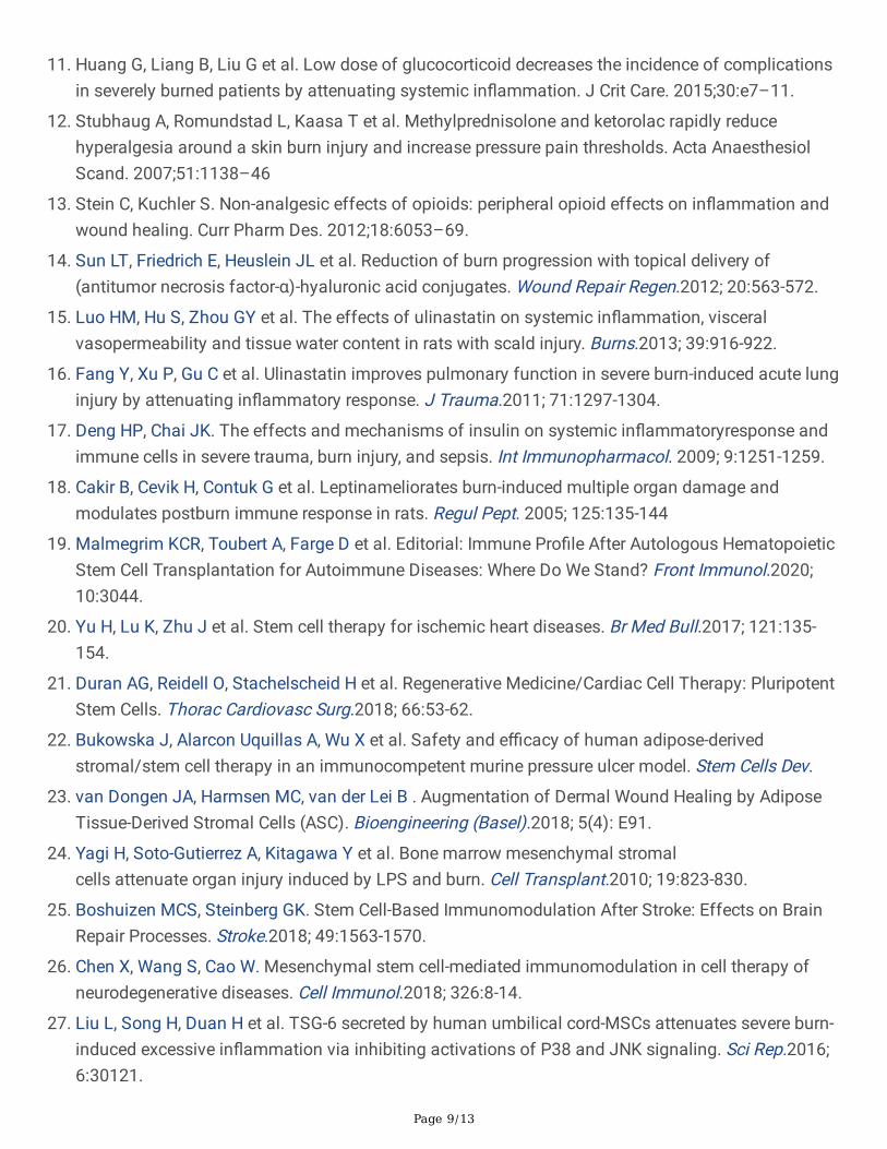
Page 9/13
11. Huang G, Liang B, Liu G et al. Low dose of glucocorticoid decreases the incidence of complicationsin severely burned patients by attenuating systemic in�ammation. J Crit Care. 2015;30:e7–11.
12. Stubhaug A, Romundstad L, Kaasa T et al. Methylprednisolone and ketorolac rapidly reducehyperalgesia around a skin burn injury and increase pressure pain thresholds. Acta AnaesthesiolScand. 2007;51:1138–46
13. Stein C, Kuchler S. Non-analgesic effects of opioids: peripheral opioid effects on in�ammation andwound healing. Curr Pharm Des. 2012;18:6053–69.
14. Sun LT, Friedrich E, Heuslein JL et al. Reduction of burn progression with topical delivery of(antitumor necrosis factor-α)-hyaluronic acid conjugates. Wound Repair Regen.2012; 20:563-572.
15. Luo HM, Hu S, Zhou GY et al. The effects of ulinastatin on systemic in�ammation, visceralvasopermeability and tissue water content in rats with scald injury. Burns.2013; 39:916-922.
1�. Fang Y, Xu P, Gu C et al. Ulinastatin improves pulmonary function in severe burn-induced acute lunginjury by attenuating in�ammatory response. J Trauma.2011; 71:1297-1304.
17. Deng HP, Chai JK. The effects and mechanisms of insulin on systemic in�ammatoryresponse andimmune cells in severe trauma, burn injury, and sepsis. Int Immunopharmacol. 2009; 9:1251-1259.
1�. Cakir B, Cevik H, Contuk G et al. Leptinameliorates burn-induced multiple organ damage andmodulates postburn immune response in rats. Regul Pept. 2005; 125:135-144
19. Malmegrim KCR, Toubert A, Farge D et al. Editorial: Immune Pro�le After Autologous HematopoieticStem Cell Transplantation for Autoimmune Diseases: Where Do We Stand? Front Immunol.2020;10:3044.
20. Yu H, Lu K, Zhu J et al. Stem cell therapy for ischemic heart diseases. Br Med Bull.2017; 121:135-154.
21. Duran AG, Reidell O, Stachelscheid H et al. Regenerative Medicine/Cardiac Cell Therapy: PluripotentStem Cells. Thorac Cardiovasc Surg.2018; 66:53-62.
22. Bukowska J, Alarcon Uquillas A, Wu X et al. Safety and e�cacy of human adipose-derivedstromal/stem cell therapy in an immunocompetent murine pressure ulcer model. Stem Cells Dev.
23. van Dongen JA, Harmsen MC, van der Lei B . Augmentation of Dermal Wound Healing by AdiposeTissue-Derived Stromal Cells (ASC). Bioengineering (Basel).2018; 5(4): E91.
24. Yagi H, Soto-Gutierrez A, Kitagawa Y et al. Bone marrow mesenchymal stromalcells attenuate organ injury induced by LPS and burn. Cell Transplant.2010; 19:823-830.
25. Boshuizen MCS, Steinberg GK. Stem Cell-Based Immunomodulation After Stroke: Effects on BrainRepair Processes. Stroke.2018; 49:1563-1570.
2�. Chen X, Wang S, Cao W. Mesenchymal stem cell-mediated immunomodulation in cell therapy ofneurodegenerative diseases. Cell Immunol.2018; 326:8-14.
27. Liu L, Song H, Duan H et al. TSG-6 secreted by human umbilical cord-MSCs attenuates severe burn-induced excessive in�ammation via inhibiting activations of P38 and JNK signaling. Sci Rep.2016;6:30121.
https://www.ncbi.nlm.nih.gov/pubmed/?term=Bukowska%20J%5BAuthor%5D&cauthor=true&cauthor_uid=31950878
https://www.ncbi.nlm.nih.gov/pubmed/?term=Harmsen%20MC%5BAuthor%5D&cauthor=true&cauthor_uid=30373121

Page 10/13
2�. Shumakov VI, Onishchenko NA, Rasulov MF et al. Mesenchymal bone marrow stemcells more effectively stimulate regeneration of deep burn wounds than embryonic �broblasts. BullExp Biol Med.2003; 136(2):192-195.
29. Gouma E, Simos Y, Verginadis I et al. A simple procedure for estimation of total body surface area and determination of a new value of Meeh's constant in rats. LabAnim. 2012; 46:40-45.
30. Abdullahi, S. Amini-Nik, M.G Jeschke Animal models in burn research. Cell Mol Life Sci. 2014 Sep;71(17): 3241–3255.
31. Elijah ZC, Chuan HA, Ashvin R et al. Creation of Consistent Burn Wounds: A Rat Model. Arch PlastSurg. 2014 Jul; 41(4): 317–324.
32. Meyer TN, Silva Al. A standard burn model using rats. Acta Cir Bras 199 Oct-Dec;14(4).
33. Liu L, Yu Y, Hou Y et al. Human umbilical cord mesenchymal stem cellstransplantation promotes cutaneous wound healing of severe burned rats. PLoS One.2014;9:e88348.
34. Zhang J, La X, Fan L et al. Immunosuppressive effects of mesenchymalstem cell transplantation in rat burn models. Int J Clin Exp Pathol.2015; 8:5129-5136.
35. Farina JA Jr, Rosique MJ, Rosique RG. Curbing in�ammation in burn patients Int J In�am. 2013;2013: 715645
3�. Jewo PI, Fadeyibi IO. Progress in burns research: A review of advances in burn pathophysiology. AnnBurns Fire Disasters.2015; 28:105-115.
37. Caliari-Oliveira C, Yaochite JN, Ramalho LN et al. XenogeneicMesenchymal Stromal Cells Improve Wound Healing and Modulate the Immune Response inan Extensive Burn Model. Cell Transplant.2016; 25:201-215
3�. Su, LJ., Wu, MS., Hui, Y. et al. Fluorescent nanodiamonds enable quantitative tracking of humanmesenchymal stem cells in miniature pigs. Sci Rep 7, 45607 (2017)
39. Diane A., Maud AS., Richard C., Laurence T. Josaine D. Anne-Marie R. et al. Interleukine -6, TNF-alphaand interleujkin-1 beta levels in blood and tissue in severely burned rats. Eur Cytokine Netw.2008Mar;19(1):1-7.
40. Gur�nkel R, Singer AJ, Cagnano E, Rosenberg L. Development of a novel animal burn model using ratheat in rats and swine. Acad Emerg Med. 2010 May;17(5):514-20.
41. Barrow RE, Meyer NA, Jeschke MG Effect of varying burn sizes and ambient temperature on thehypermetabolic rate in thermally injured rats. J Surg Res. 2001 99(2):253-7.
42. Jeschke MG, Gauglitz GG, Kulp GA, Finnerty CC, Williams FN, Kraft R, Suman OE, Mlcak RP, HerndonDN. Long-term persistence of the pathophysiologic response to severe burn injury. PLoS One.2011;6(7):e21245.
43. Djouad F., Plence P., Bony C., et al. Immunosuppressive effect of mesenchymal stem cells favorstumor growth in allogeneic animals. Blood. 2003;102(10):3837–3844.
https://www.ncbi.nlm.nih.gov/pubmed/?term=Rasulov%20MF%5BAuthor%5D&cauthor=true&cauthor_uid=14631508

Page 11/13
Figures
Figure 1
Experimental results of each group.

Page 12/13
Figure 2
Survival distribution among the groups as per the log-rank test.

Page 13/13
Figure 3
Representative images showing the burn healing course in each group