Ear surgery
43
In The Name Of GOD
-
Upload
emad-sayahi -
Category
Health & Medicine
-
view
22 -
download
1
Transcript of Ear surgery
In The Name Of GOD
Surgery of the Ear Dr. Sayahi
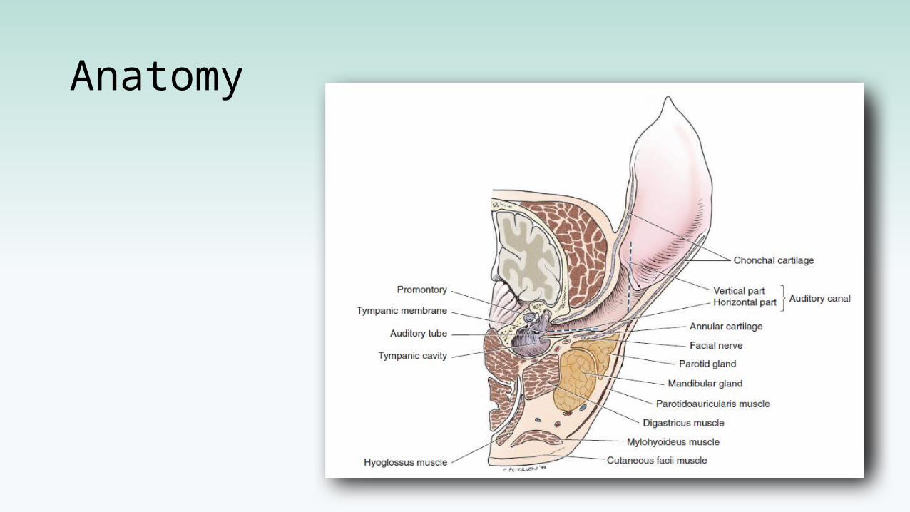
Anatomy
What can happen to ears?
pre
sbycu
sis
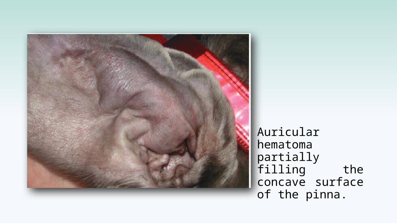
Auricular hematoma partially filling the concave surface of the pinna.
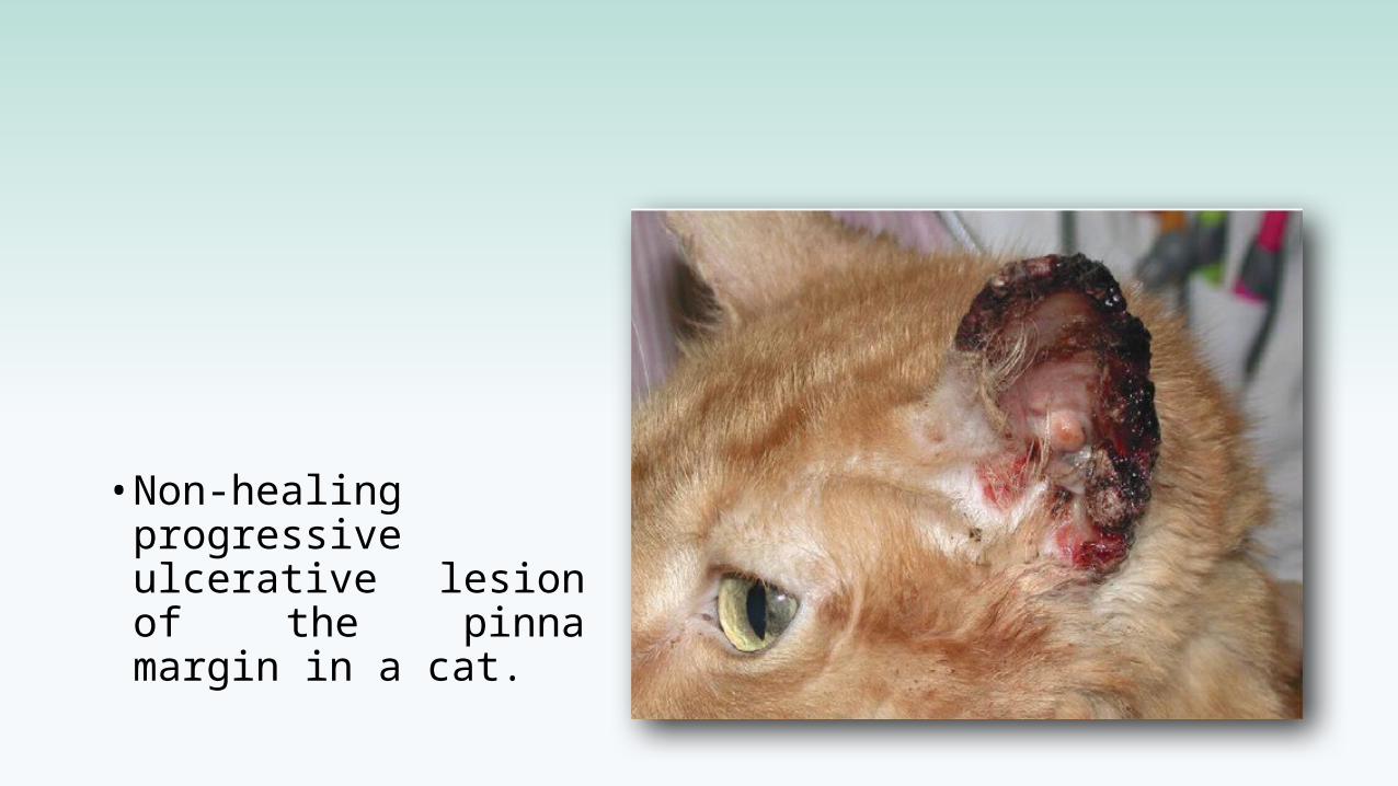
• Non-healing progressive ulcerative lesion of the pinna margin in a cat.
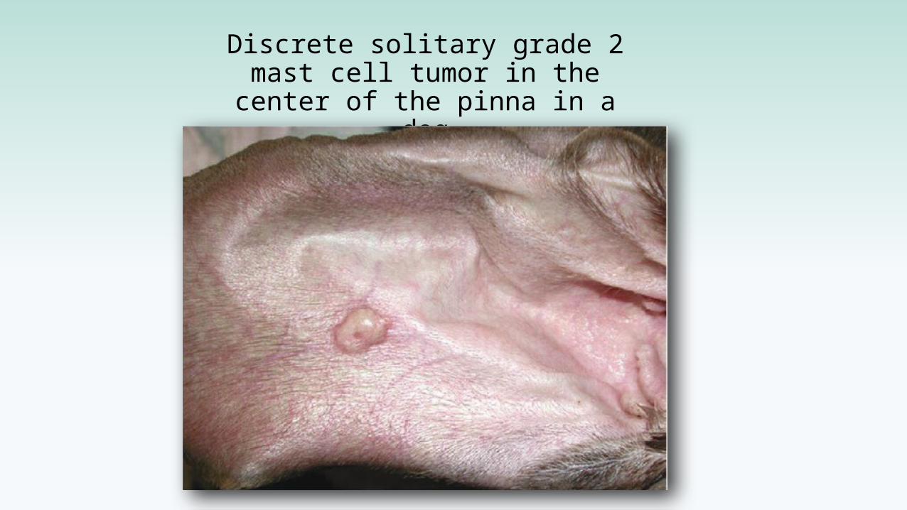
Discrete solitary grade 2 mast cell tumor in the center of the
pinna in a dog
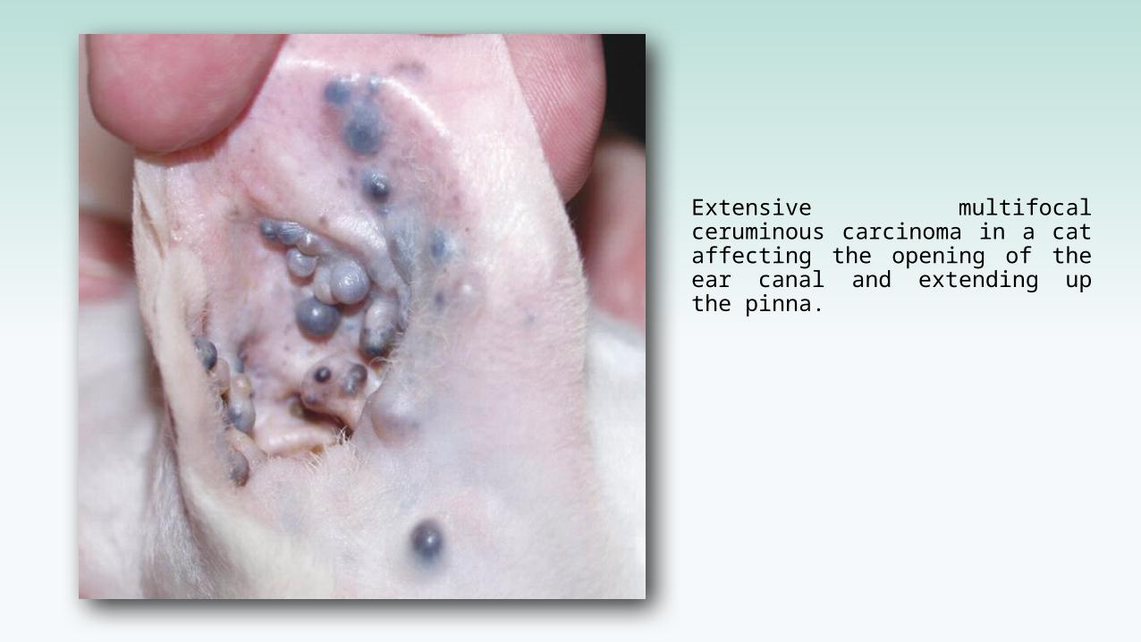
Extensive multifocal ceruminous carcinoma in a cat affecting the opening of the ear canal and extending up the pinna.
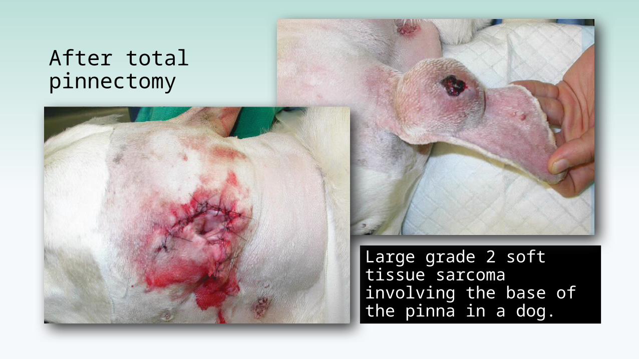
After total pinnectomy
Large grade 2 soft tissue sarcoma involving the base of the pinna in a dog.
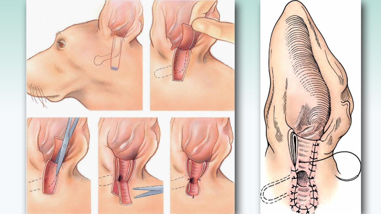
Lateral ear canal resection
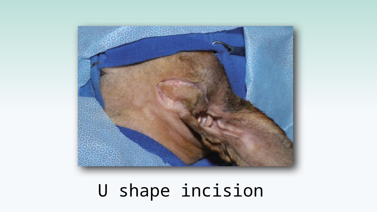
U shape incision
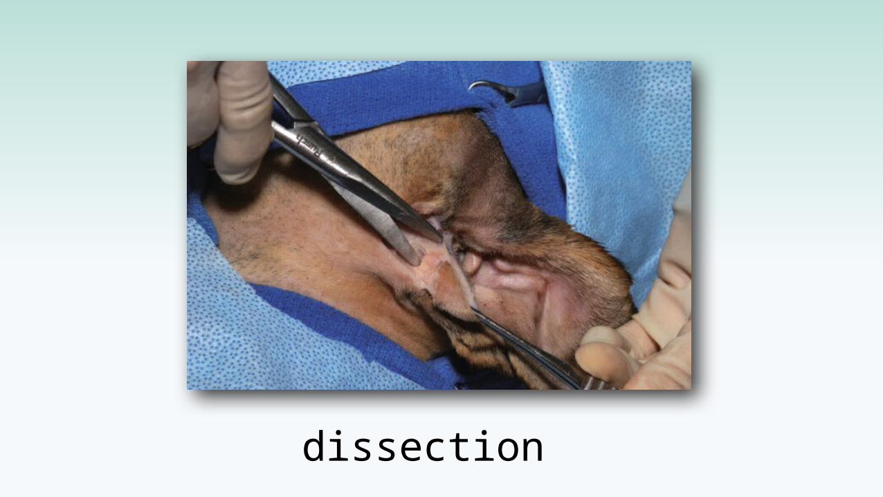
dissection
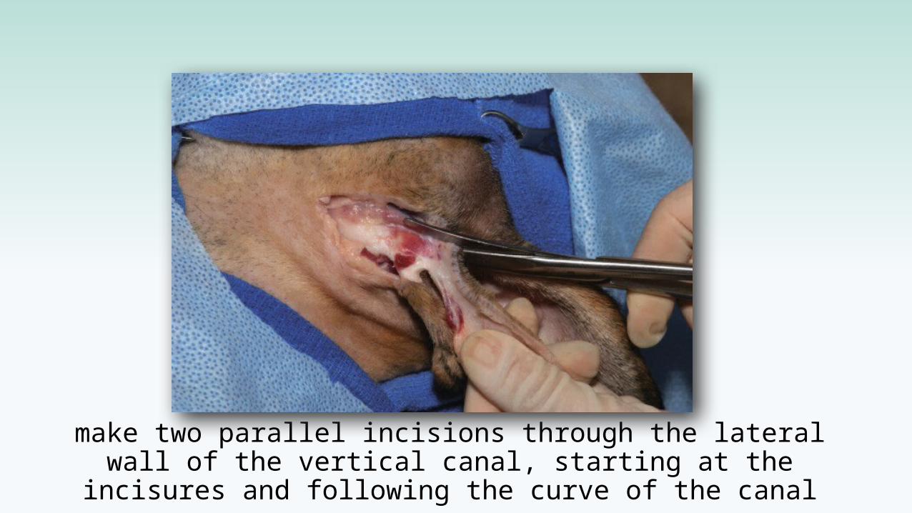
make two parallel incisions through the lateral wall of the vertical canal, starting at the incisures and following the
curve of the canal
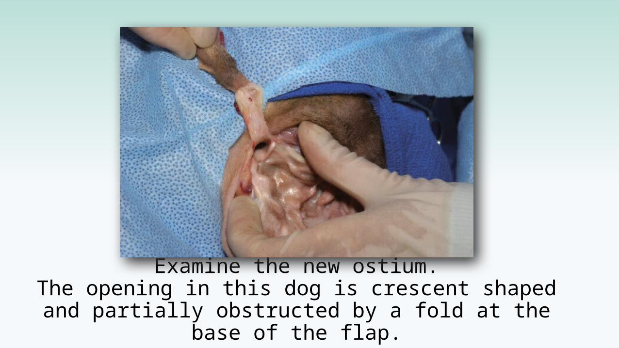
Examine the new ostium.The opening in this dog is crescent shaped and partially obstructed by a fold at the base of the
flap.
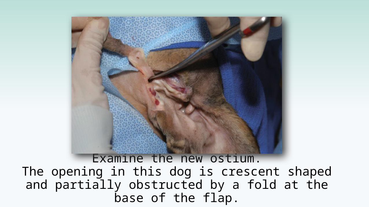
Examine the new ostium.The opening in this dog is crescent shaped and partially obstructed by a fold at the base of the
flap.
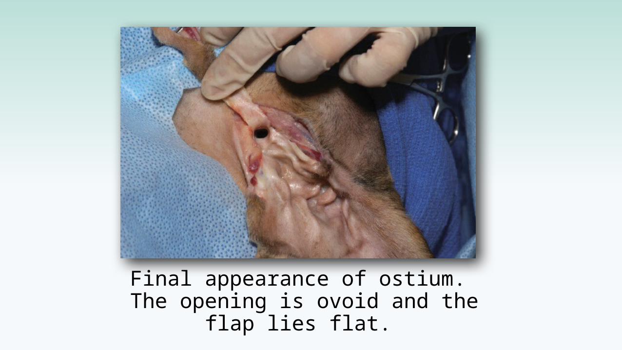
Final appearance of ostium. The opening is ovoid and the
flap lies flat.
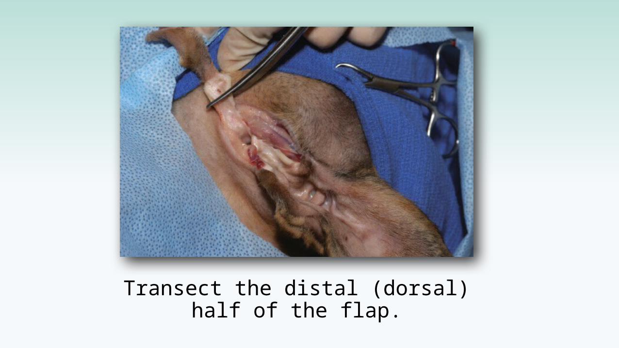
Transect the distal (dorsal)half of the flap.
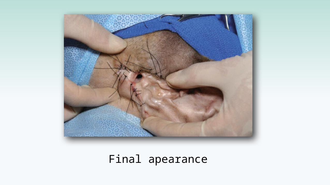
Final apearance
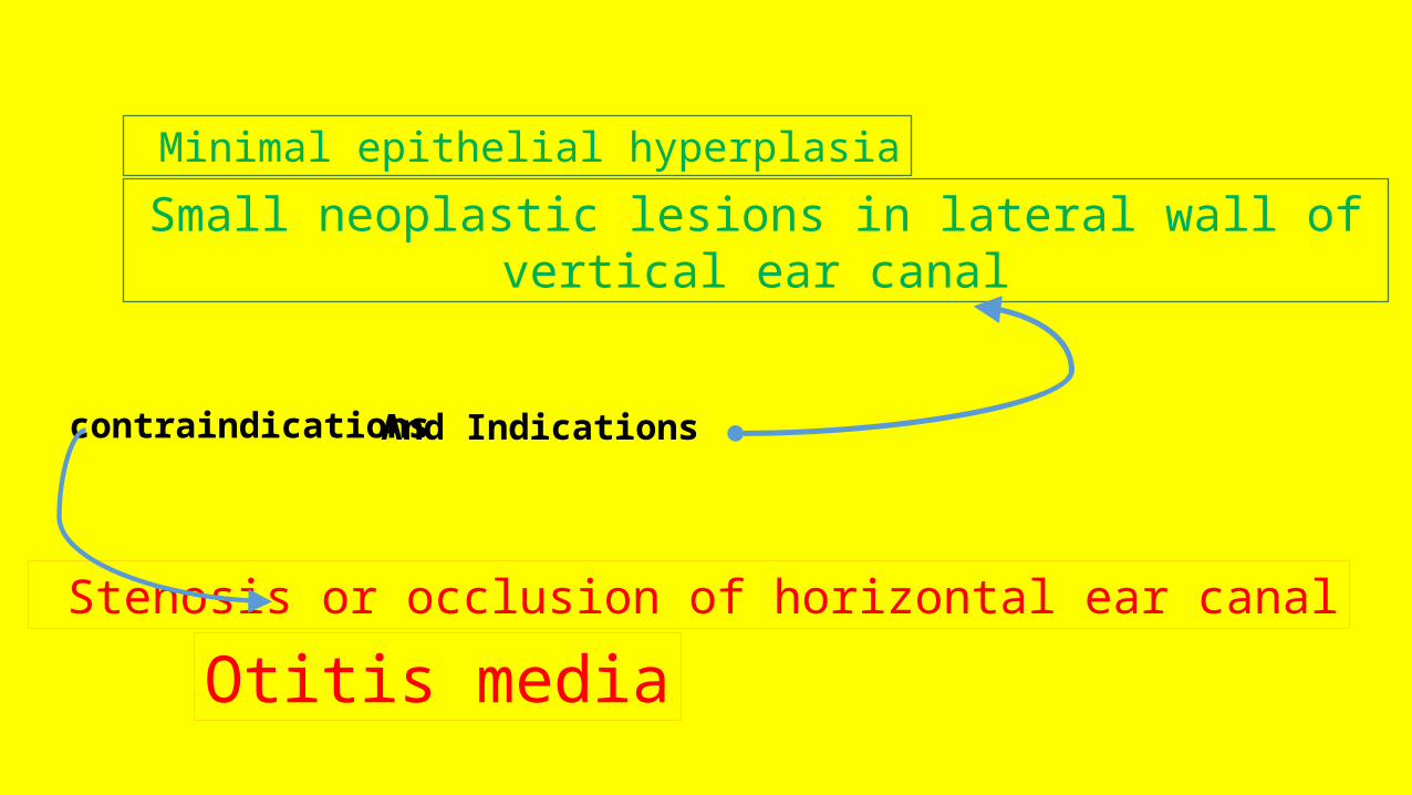
Minimal epithelial hyperplasia
Small neoplastic lesions in lateral wall of vertical ear canal
Stenosis or occlusion of horizontal ear canal
Otitis media
And Indicationscontraindications
Owner MUST acknowledged about final appearance

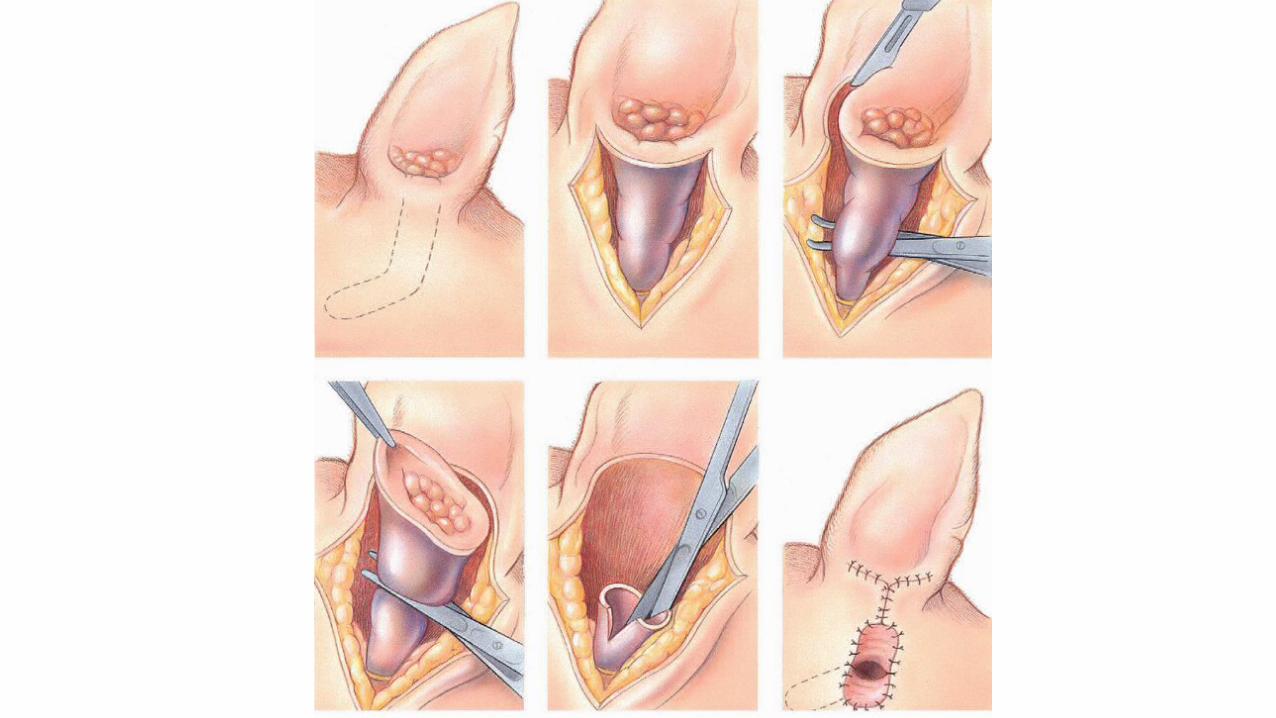
vertical ear canal ablation
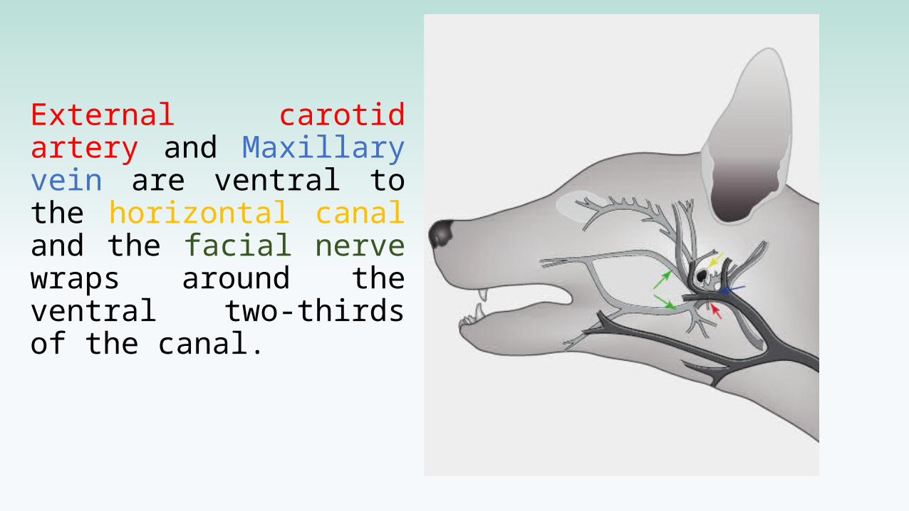
External carotid artery and Maxillary vein are ventral to the horizontal canal and the facial nerve wraps around the ventral two-thirds of the canal.
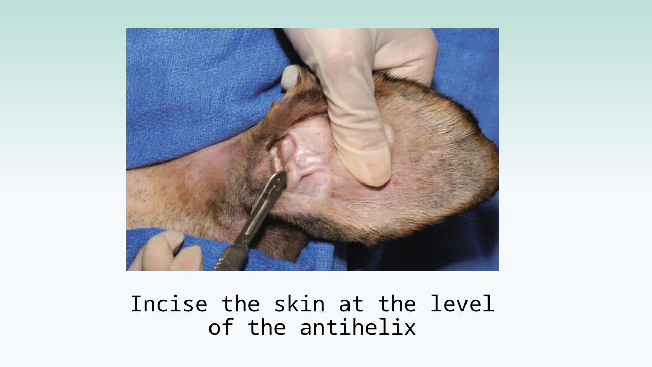
Incise the skin at the levelof the antihelix
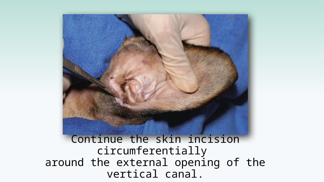
Continue the skin incision circumferentially around the external opening of the vertical canal.
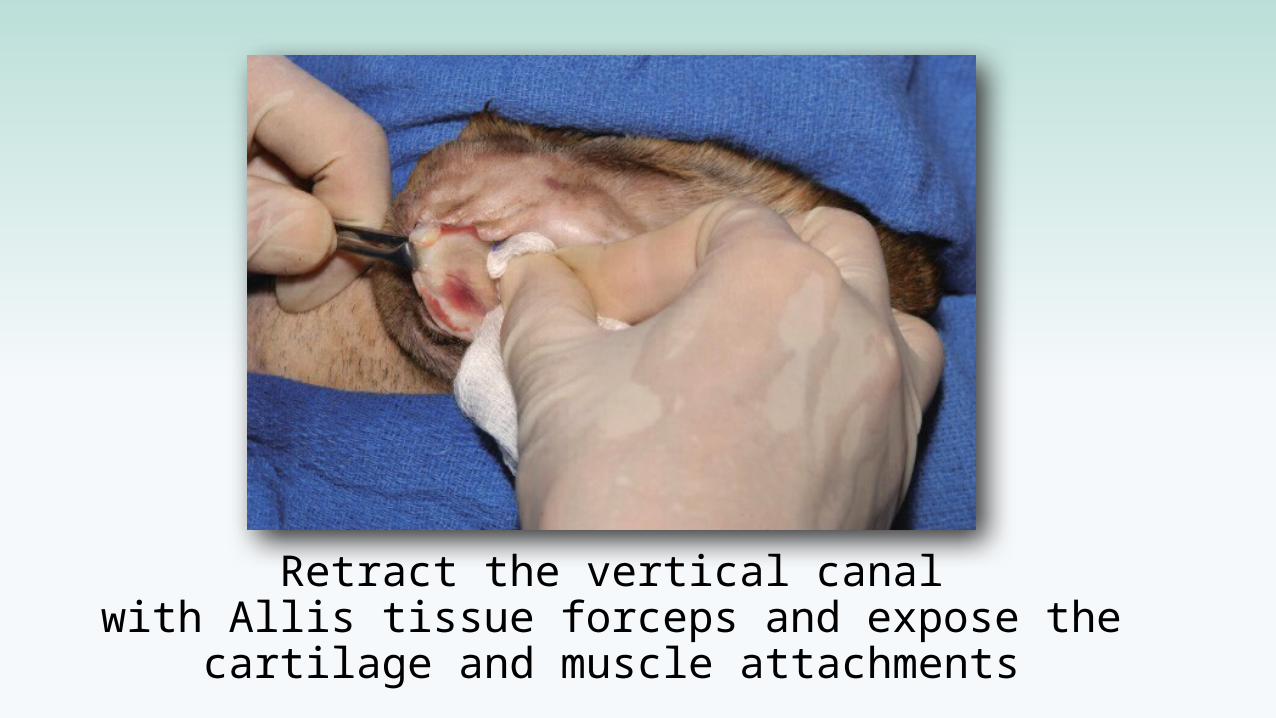
Retract the vertical canalwith Allis tissue forceps and expose the
cartilage and muscle attachments
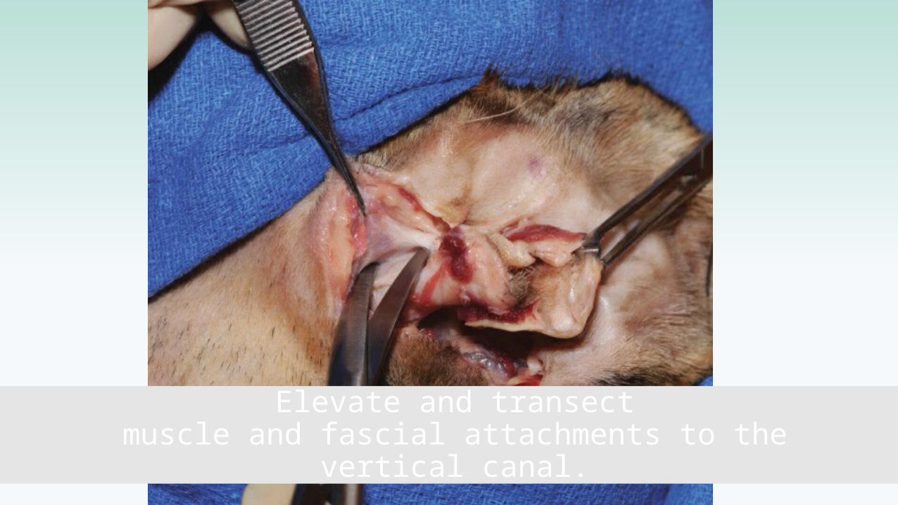
Elevate and transectmuscle and fascial attachments to the
vertical canal.
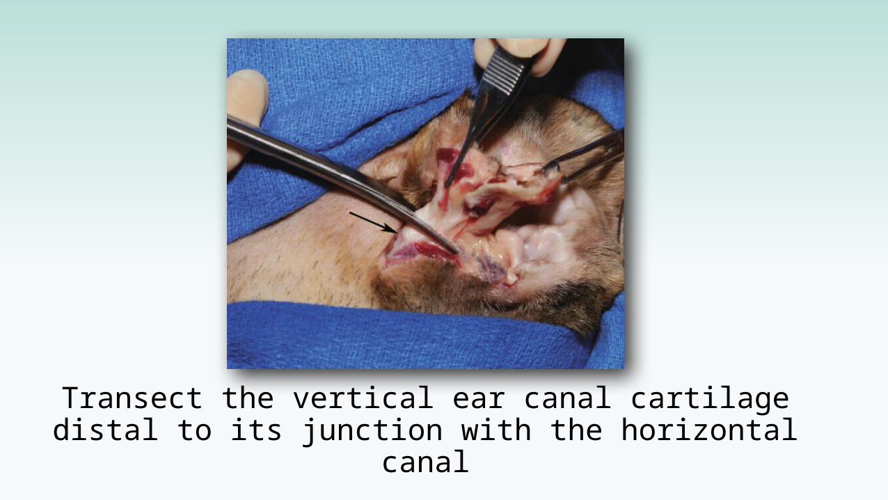
Transect the vertical ear canal cartilage distal to its junction with the horizontal canal
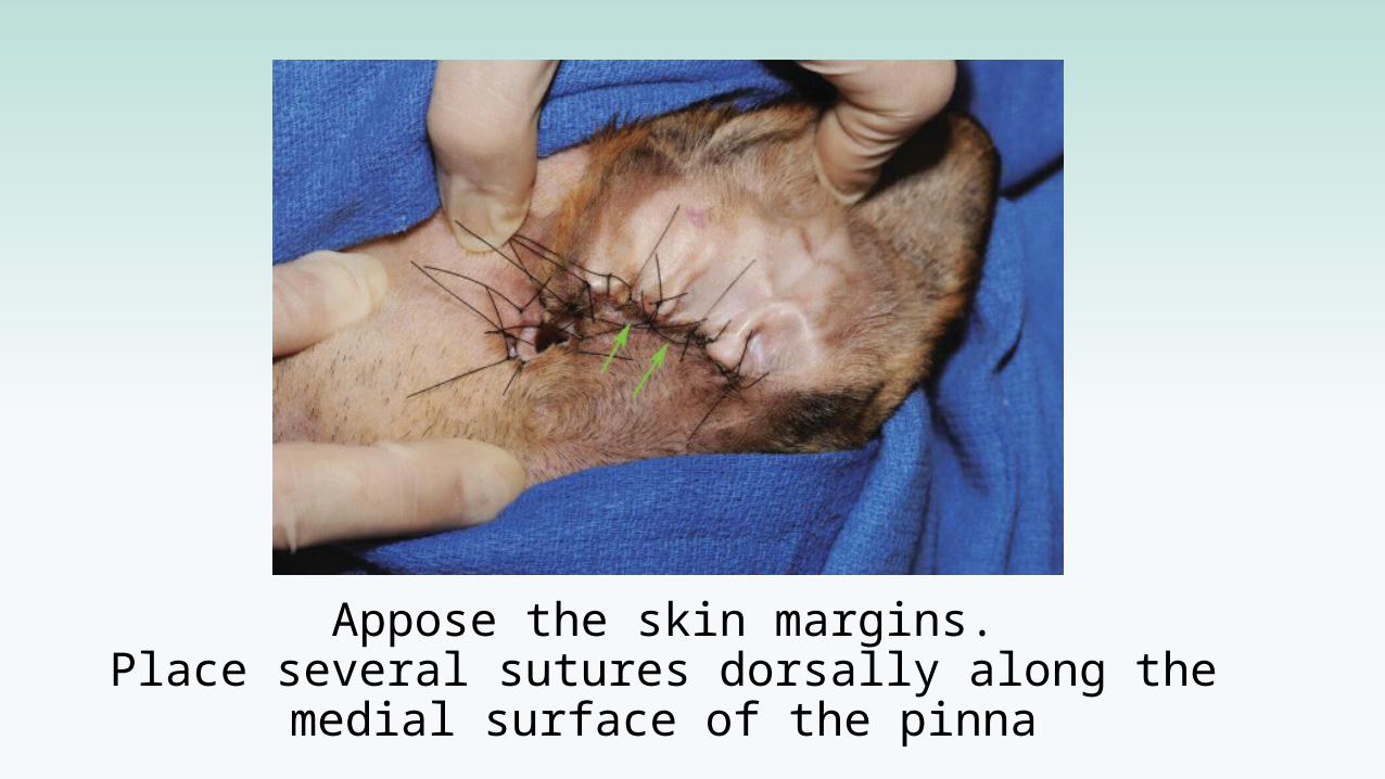
Appose the skin margins.Place several sutures dorsally along the
medial surface of the pinna
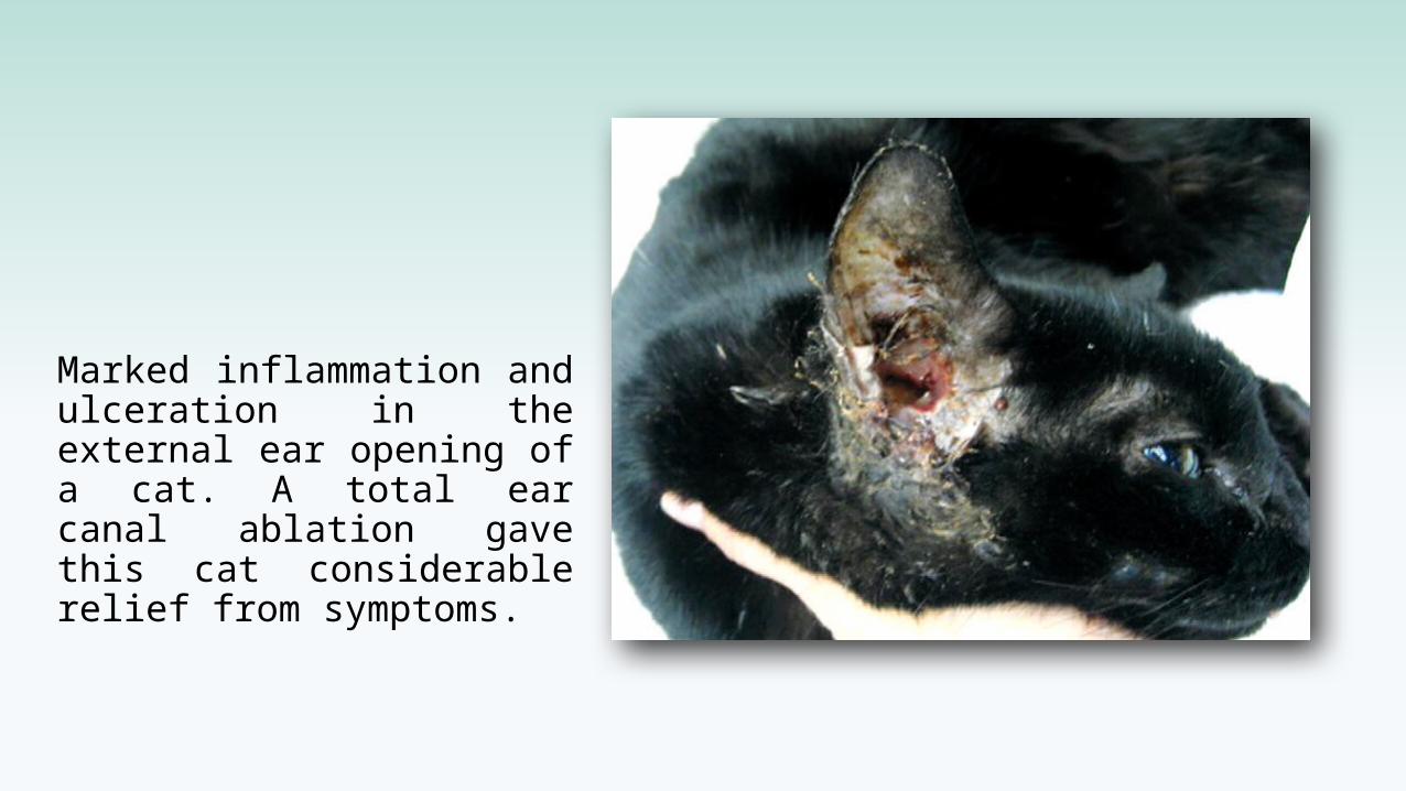
Marked inflammation and ulceration in the external ear opening of a cat. A total ear canal ablation gave this cat considerable relief from symptoms.
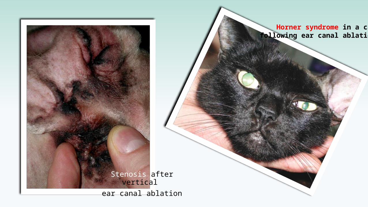
Stenosis after vertical ear canal ablation
Horner syndrome in a cat following ear canal ablation
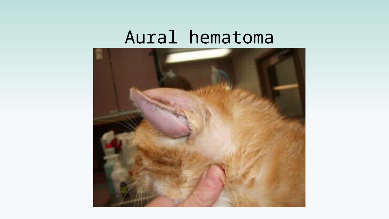
Aural hematoma
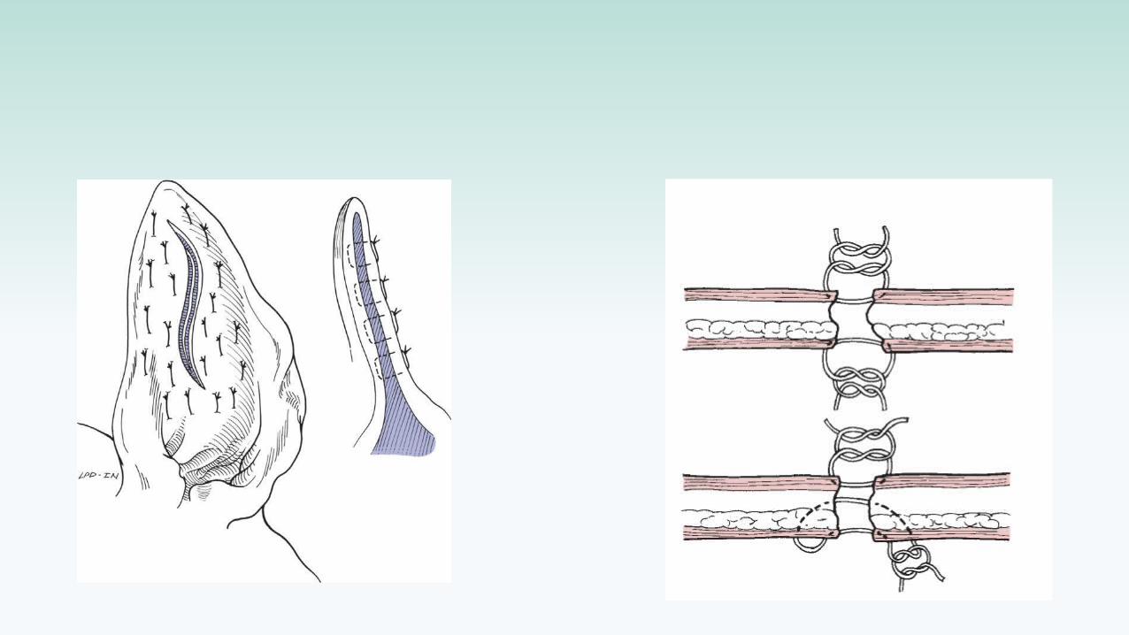
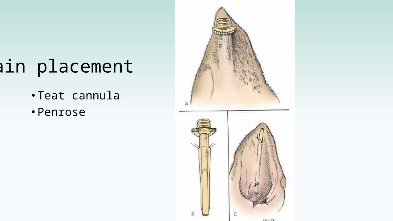
Drain placement
• Teat cannula• Penrose
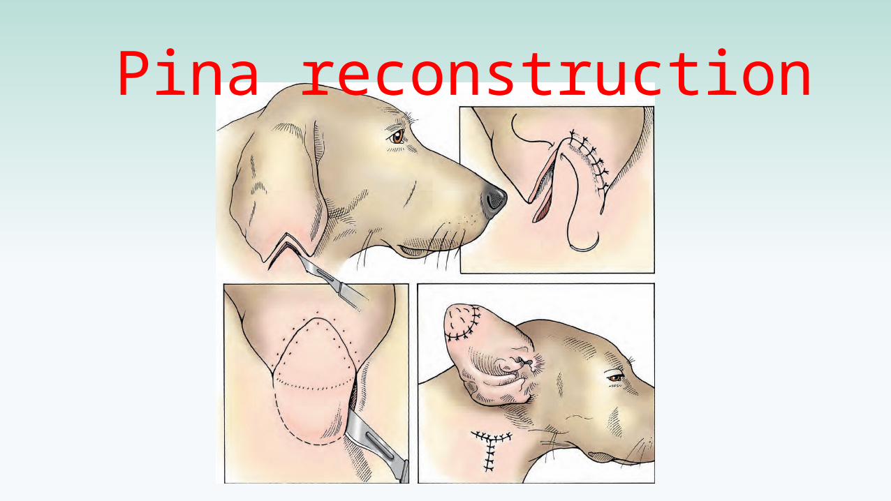
Pina reconstruction
Do you want to know how trim ear and tail?
Thanks for your attention