E XPOSURE FACTORS : Contrast, Density, Image Quality, technique math 1.
57
EXPOSURE FACTORS: Contrast, Density, Image Quality, technique math 1
-
Upload
issac-otwell -
Category
Documents
-
view
216 -
download
0
Transcript of E XPOSURE FACTORS : Contrast, Density, Image Quality, technique math 1.

EXPOSURE FACTORS:Contrast, Density, Image Quality, technique math
1

REVIEW SLIDE-WHAT DO WE KNOW ABOUT X-RAYS SO FAR?Properties of X-rays
Are highly penetrating invisible rays which are a form of electromagnetic radiation.
Are electrically neutral and therefore not affected by either electric or magnetic fields
Can be produced over a wide variety of energies and wavelengths (polyenergetic & heterogeneous).
Release very small amounts of heat upon passing through matter.
Travel in straight lines.
Travel at the speed of light in a vacuum.
Can ionize matter.
Cause fluorescence of certain crystals.
Cannot be focused by a lens.
Affects photographic film.
Produce chemical and biological changes in matter through ionization and excitation.
Produce secondary and scatter radiation.
2

UNITS OF MEASURE-REVIEW
Measure of current, amount of electron charges passing through a point
Measure of potential difference, value of potential electrical difference as one unit of current passes 3

UNITS OF MEASURE-REVIEW
Table 5. SI prefixes
Factor Name Symbol 1024 yotta Y
1021 zetta Z
1018 exa E
1015 peta P
1012 tera T
109 giga G
106 mega M
103 kilo k
102 hecto h
101 deka da
Factor Name Symbol 10-1 deci d
10-2 centi c
10-3 milli m
10-6 micro µ
10-9 nano n
10-12 pico p
10-15 femto f
10-18 atto a
10-21 zepto z
10-24 yocto y
4

EXPOSURE FACTORS
mAs Milliamps-Amount of
electrons burned off filament
Time-measured in seconds, fractions of seconds, determines how long the electrons will flow across to anode(another amount)
Kilovoltage Determines the
strength of the x-ray Determines the
wavelength of the x-ray
Determines the power of the x-ray
Determines the penetrating ability of x-ray
mA, time kVp, kV
5

EXPOSURE FACTORS
Controls the density of the image by controlling the amount of electrons sent to anode target
Controls the contrast of the image by controlling the penetrating power of the x-ray photon
Also controls the density of the image because more photons are able to penetrate the part being imaged
mAs kVp
6

DENSITY
7

CONTRAST
8

What changed?....contrast or density? 9

Now what changed?....very subtle, often subjective
10

1
2
3
4
11

1
2
4
5
3
12

DENSITY
Anatomic Density Body part/object being x-rayed Atomic # Thickness of part
Optical Density Amount of x-ray photons reaching the image receptor
The mA applied The time applied Also referred to as x-ray output
13

14

MILLIAMPERAGE
mA
One milliampere is equal to one thousandth of an ampere.
The amount of current supplied to the x-ray tube
Range 10 to 1200 mA15

TIME
In seconds
How long x-rays will be produced
0.001 to 6 seconds
16

MASmA X s = mAs
17

MAS RECIPROCITY
100 mA x 1/4 = 25 mAs
200 mA x 1/8 = 25 mAs
400 mA x 1/16 = 25 mAs
This works ONLY when you are trying to keep the mAs the SAME………
18

WHAT CHANGES TO MAS ARE NEEDED FOR HUMAN EYE TO DETECT?
19
TO CHANGE DENSITY
The human eye needs a 20-30% change in density on an image in order to visibly see it.
Most frequently radiographers will change the density by doubling or by halving the density.
What do you do in order to double density on an image?
20

DENSITY DIRECTLY PROPORTIONAL TO MAS
+ 25%mAS = 25% increase in density100 mAs
+50% mAs = 50% increase in density
21

22

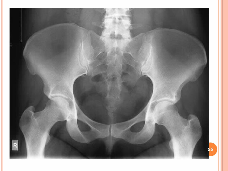
DENSITY
Density is like toast…too much and the toast is burned, too little and it is underdone.
The images differ in density only. Which one looks optimal to you?
23

WHAT WOULD YOU DO?
This image was taken at 60 mAs. What would you do to fix this image?
This image was taken at 300 mA. What was the time of the exposure?
If we wanted to change the mA but keepthe mAs the same, what would we do?
24

VARIABLES THAT AFFECT DENSITY Patient size
Thickness of body part Tissue composition
Z# Bone, muscle, soft tissue, water, air
mAs kVp Source image receptor distance (SID)
The distance from the tube to the image receptor The closer the tube, the more photons hit target
Beam modification The use of filter (we will cover later)
Image receptor The use of grid vs. non-grid, film , CR, DR (we will
cover later) Processing
Chemistry, time in chemistry (we will cover later25

DENSITY MATH WORK Milliamperage second Conversions
Math Review
Fraction to Decimal 1) 1/ 5 = _______ 2) 1/ 20 = _______ 3) 1/ 8 = _______ 4) 1/ 60 = _______ 5) 1/ 15 = _______
mA x Time (s) = mAs 10) 100 x 0.07 = _______ 11) 100 x 0.013 = _______ 12) 100 x 0.033 = _______ 13) 100 x 0.25 = _______ 14) 100 x 0.009 = _______ 15) 200 x 0.04 = _______ 16) 200 x 0.07 = _______ 17) 200 x 0.025 = _______ 18) 300 x 0.01 = _______ 19) 300 x 0.08 = _______ 20) 300 x 0.05 = _______
mA x Time (s) = mAs 21) 100 x 1/ 8 = _______ 22) 100 x 1/ 120 = _______ 23) 100 x 1/ 15 = _______ 24) 100 x 1/ 40 = _______ 25) 100 x 1/ 6 = _______ 26) 50 x 1/ 20 = _______ 27) 50 x 1/ 120 = _______ 28) 50 x 1/ 80 = _______ 29) 200 x 1/ 80 = _______ 30) 200 x 1/ 12 = _______ 31) 300 x 1/ 5 = _______ 32) 300 x 1/ 60 = _______ 33) 400 x 1/ 80 = _______ 34) 400 x 1/ 60 = _______ 35) 500 x 1/ 12 = _______ 36) 500 x 1/ 20 = _______ 37) 600 x 1/ 40 = _______ 38) 600 x 1/ 120 = _______ 39) 600 x 1/ 25 = _______ 40) 600 x 1/ 5 = _______
mA or S is unknown
1. 50 mA @ ______ S = 10 mAS 4. _____mA @ 0.1 S = 40 mAS
2. 100 mA @ _______ S = 4 mAS 5. _____ mA @ 0.2 S = 40 mAS
3. 200 mA @ _______ S = 5 mAS 6. _____ mA @ 0.3 = 60 mAS
mAs density changes & kVp contrast changes
1. mAs = 10 Double the optical density(OD) __________ ½ the OD ________
2. mAs = 15 Double the optical density(OD) ________ __ ½ the OD ________
3. kVp = 50 Narrow the contrast scale ___________ widen contrast ________
4. kVp = 75 Narrow the contrast scale ___________ widen contrast ________
This is posted on the website. PleaseDownload and turn in the next class Oct 2nd
26

SAMPLE PROBLEMSWhen mA is unknown…
The image was shot at 45mAs using a .75second exposure. What is the mA?
When s is unknown….
The image was shot at 80mAs using the 400mA station. What was the time of exposure?
mAmAs
mAsmA
mAsmA
60?75.
45?
45sec75.?
smA
mAs
mAsmA
2.sec?400
80sec?
80sec?400
27

CONTRAST
THE DIFFERENCES BETWEEN: Blacks Whites Dark gray Light grayTHERE IS A SCALE OF CONTRAST
• many colors of black, white, gray= long scale• Few colors of black, white, gray=short scale
28

SHORT SCALE OF CONTRAST
Not very many differences
Between grays
Also known as high contrast
29

1
2
3
4
30

1
2
4
5
3
31

LONG SCALE OF CONTRAST• Many
different shades of gray
• Also known as low contrast
32

KILOVOLTAGE PEAK (KVP)
33

BEAM ATTENUATION AKA ABSORPTION
High kVp Penetrates more easily Causes more grays Low contrast
Low kVp Decreases penetration Causes more black-
white High contrast
Different parts of body attenuate differently
The difference in attenuation is the basis for contrast
34

35

OPTIMAL KVPIS THERE SUCH A CONCEPT? YES and NO
Depends on the body part The anatomic area of interest
More energy is needed to penetrate through bony tissue (high z #) than soft tissue (low z #)
36

+ 15% KVP - 15% KVP
37

15% RULE
15% kVp = doubling of exposure to the image
receptor
15% kVp = halving of exposure to the image receptor
15% rule will always change the contrast of the image because kV is the primary method of changing image contrast.
Remember : 15% change ( ) KVP has the same effect as
doubling or ½ the MAS on density38

CONTRAST MATH WORK Milliamperage second Conversions
Math Review
Fraction to Decimal 1) 1/ 5 = _______ 2) 1/ 20 = _______ 3) 1/ 8 = _______ 4) 1/ 60 = _______ 5) 1/ 15 = _______
mA x Time (s) = mAs 10) 100 x 0.07 = _______ 11) 100 x 0.013 = _______ 12) 100 x 0.033 = _______ 13) 100 x 0.25 = _______ 14) 100 x 0.009 = _______ 15) 200 x 0.04 = _______ 16) 200 x 0.07 = _______ 17) 200 x 0.025 = _______ 18) 300 x 0.01 = _______ 19) 300 x 0.08 = _______ 20) 300 x 0.05 = _______
mA x Time (s) = mAs 21) 100 x 1/ 8 = _______ 22) 100 x 1/ 120 = _______ 23) 100 x 1/ 15 = _______ 24) 100 x 1/ 40 = _______ 25) 100 x 1/ 6 = _______ 26) 50 x 1/ 20 = _______ 27) 50 x 1/ 120 = _______ 28) 50 x 1/ 80 = _______ 29) 200 x 1/ 80 = _______ 30) 200 x 1/ 12 = _______ 31) 300 x 1/ 5 = _______ 32) 300 x 1/ 60 = _______ 33) 400 x 1/ 80 = _______ 34) 400 x 1/ 60 = _______ 35) 500 x 1/ 12 = _______ 36) 500 x 1/ 20 = _______ 37) 600 x 1/ 40 = _______ 38) 600 x 1/ 120 = _______ 39) 600 x 1/ 25 = _______ 40) 600 x 1/ 5 = _______
mA or S is unknown
1. 50 mA @ ______ S = 10 mAS 4. _____mA @ 0.1 S = 40 mAS
2. 100 mA @ _______ S = 4 mAS 5. _____ mA @ 0.2 S = 40 mAS
3. 200 mA @ _______ S = 5 mAS 6. _____ mA @ 0.3 = 60 mAS
mAs density changes & kVp contrast changes
1. mAs = 10 Double the optical density(OD) __________ ½ the OD ________
2. mAs = 15 Double the optical density(OD) ________ __ ½ the OD ________
3. kVp = 50 Narrow the contrast scale ___________ widen contrast ________
4. kVp = 75 Narrow the contrast scale ___________ widen contrast ________
This is posted on the website. PleaseDownload and turn in the next class (Oct 2nd) 39

CONTRAST MATH WORK
Request: Widen the contrastAsking for a long scale contrastLooking for more grays, more grays that look alike
Solution: Increase kV
Request: Narrow the contrastAsking for a short scale contrastLooking for less grays, more black and whites
Solution: Decrease kV
Use the 15% rule 40

CONTRAST MATH WORK
Image was shot at 75 kV. What is the new kV if you want to narrow the contrast?
75.6325.1175
25.117515.
kV
The new kV should be 63.75In order to narrow the contrast, you must reduce kV.
41

CONTRAST MATH WORK
Image was shot at 65kV. What wouldthe new kV be if you wanted to widen the contrast?
75.7475.965
75.96515.
To widen the contrast, you must increase kV. Add 15% of 65 to 65. The new kV would be 74.75 42

VARIABLES THAT AFFECT DENSITY Patient size
Thickness of body part Tissue composition
Z# Bone, muscle, soft tissue, water, air
mAs kVp Source image receptor distance (SID)
The distance from the tube to the image receptor The closer the tube, the more photons hit target
Beam modification The use of filter (we will cover later)
Image receptor The use of grid vs. non-grid, film , CR, DR (we will
cover later) Processing
Chemistry, time in chemistry (we will cover later43

SOURCE TO IMAGE RECEPTOR DISTANCE
SID -controlled and manipulated in the x-ray room
44

INTENSITY OF THE BEAM
1. As distance _______: intensity ________
2. As distance _______: intensity ________
3. Inverse relation
45

INTENSITY IS SPREAD OUT…
46

INVERSE SQUARE LAW
Farther the distance of the x-ray tube to the IR
Photons have less chance of getting to IR
Due to divergent beam
47

HOW DOES DISTANCE AFFECT IR EXPOSURE?1. Increased distance: decreased exposure
________________
2. Decreased distance: increased exposure ________________
3. Inversely proportional to the square of the distance
________________ Intensity is ¼ of original
________________ Intensity increases to 4 x’s the original exposure
48

INVERSE SQUARE LAW
Used for RADIATION PROTECTION
When you change your distance from the “radiation source”
The intensity of radiation will be reduced by a square of the distance MOVING AWAY FROM THE SOURCE
INCREASED – CLOSER TO SOURCE 49

INVERSE SQUARE LAW
1
2
2
1
d
d
I
I
2
2
50

APPLICATION OF INVERSE SQUARE LAW PRINCIPLES CAN YIELD SIGNIFICANT REDUCTIONS IN PATIENT AND OPERATOR
RADIATION EXPOSURE.
51

52

53

54
55

56

57