E-learning System for Studying Dento-maxillo-facial Radiology
4
E-learning System for Studying Dento-maxillo-facial Radiology Danisia Haba * , Ana-Elena Petcu * , Cristian-Gyozo Haba ** * University of Medecine and Pharmacy “Gr.T.Popa” Iasi, Romania, ** Technical University of Iasi, Romania E-mail: [email protected] , [email protected] Abstract The alternatives to the classic university teaching systems represented by the e-learning systems may bring important contributions to the university curricula. This paper contains information and results of a project which is intended to improve the quality of studying Dento- maxillo-facial Radiology for undergraduate and postgraduate students from Dental Faculty of University of Medicine and Pharmacy “Gr.T.Popa” Iasi, Romania. 1. Introduction The e-learning can be a method to increase the efficacy of teaching and learning process at all levels of education [1]. In recent years, we have seen a major improvement of web based technologies, increasing speed of Internet connections and a positive attitude of undergraduate students to experiment new methods of learning. Undergraduate students enrolled in dental medicine studies make no exception therefore different project were started to provide e-learning experience in this field [2, 3, 4, 5]. Dental radiology is an important discipline taught in Dental Medicine faculties and its preponderant visual characteristics makes it a good candidate in order to be complemented with new rich graphical and multimedia based web techniques. Positive results reported in the literature [6,7] have inspired the start of an e-learning system to improve the quality of studying Dento-maxillo- facial Radiology for undergraduate and postgraduate students from Dental Faculty of University of Medicine and Pharmacy “Gr.T.Popa” Iasi, Romania [8]. 2. The dento-maxillo-facial radiology course The Radiology courses for the students at the Faculty of Dental Medicine have suffered changes in the last years. In the period 1996-2000 the activity for the Radiology discipline was organized in an integrated modular structure. Students started to learn radiology beginning with the second year. Introductory notions of radiology were presented in the following modules: • Radiation history and the radiographic image; • The production, properties and interactions of X- rays; • The biologic effects and risks associated with X- rays; • Dose units and dosimetry. For each course, practical works of two hours were allocated with the intention to introduce students into medical radiology, followed by the specific issues that are present in dental branches of radiology: dental radiology, maxillo-facial radiology and the specialized branch of ondonto-maxilo-facial radiology. Practical works were conceived to teach students the nature and techniques to produce X-rays, proprieties of X- rays, biologic effect of radiations and their cumulative effect on patients. Next module was intended to introduce knowledge related to general radiology (classic techniques) that was integrated with the modules of medical and surgical courses. These modules were presenting radiological explorations used for the explorations of: • diseases of respiratory system, • diseases of cardiovascular system and mediastinum, • diseases of gastrointestinal system, • diseases of liver and biliary system, • diseases of pancreas and spleen, • diseases of urinary tract, • diseases of musculo-skeletal system. In the third year a module was allocated to dental and maxillo-facial radiology, integrated with the module I9-B- Odontal pathology. In this module students were presented: • Dental X-ray Equipment; • Dental X-ray Film and processing; • Dental X-ray Image characteristics and Quality assurance in the Dental Office; • Intraoral Radiography: The Bisecting and Paralleling Technique, The Bite-Wing and Occlusal Tehniques; • Interpretation of Dental Caries. The practical works associated within this module were intended for the students to learn: • component parts of dental radiological equipment, • types of radiological films and their processing techniques,
Transcript of E-learning System for Studying Dento-maxillo-facial Radiology
E-learning System for Studying Dento-maxillo-facial Radiology
Danisia Haba*, Ana-Elena Petcu*, Cristian-Gyozo Haba** * University of Medecine and Pharmacy “Gr.T.Popa” Iasi, Romania,
** Technical University of Iasi, Romania E-mail: [email protected], [email protected]
Abstract
The alternatives to the classic university teaching systems represented by the e-learning systems may bring important contributions to the university curricula. This paper contains information and results of a project which is intended to improve the quality of studying Dento-maxillo-facial Radiology for undergraduate and postgraduate students from Dental Faculty of University of Medicine and Pharmacy “Gr.T.Popa” Iasi, Romania.
1. Introduction
The e-learning can be a method to increase the efficacy of teaching and learning process at all levels of education [1]. In recent years, we have seen a major improvement of web based technologies, increasing speed of Internet connections and a positive attitude of undergraduate students to experiment new methods of learning. Undergraduate students enrolled in dental medicine studies make no exception therefore different project were started to provide e-learning experience in this field [2, 3, 4, 5]. Dental radiology is an important discipline taught in Dental Medicine faculties and its preponderant visual characteristics makes it a good candidate in order to be complemented with new rich graphical and multimedia based web techniques. Positive results reported in the literature [6,7] have inspired the start of an e-learning system to improve the quality of studying Dento-maxillo-facial Radiology for undergraduate and postgraduate students from Dental Faculty of University of Medicine and Pharmacy “Gr.T.Popa” Iasi, Romania [8].
2. The dento-maxillo-facial radiology course
The Radiology courses for the students at the Faculty of Dental Medicine have suffered changes in the last years. In the period 1996-2000 the activity for the Radiology discipline was organized in an integrated modular structure. Students started to learn radiology beginning with the second year. Introductory notions of radiology were presented in the following modules:
• Radiation history and the radiographic image;
• The production, properties and interactions of X-rays;
• The biologic effects and risks associated with X-rays;
• Dose units and dosimetry. For each course, practical works of two hours were allocated with the intention to introduce students into medical radiology, followed by the specific issues that are present in dental branches of radiology: dental radiology, maxillo-facial radiology and the specialized branch of ondonto-maxilo-facial radiology. Practical works were conceived to teach students the nature and techniques to produce X-rays, proprieties of X-rays, biologic effect of radiations and their cumulative effect on patients. Next module was intended to introduce knowledge related to general radiology (classic techniques) that was integrated with the modules of medical and surgical courses. These modules were presenting radiological explorations used for the explorations of:
• diseases of respiratory system, • diseases of cardiovascular system and
mediastinum, • diseases of gastrointestinal system, • diseases of liver and biliary system, • diseases of pancreas and spleen, • diseases of urinary tract, • diseases of musculo-skeletal system.
In the third year a module was allocated to dental and maxillo-facial radiology, integrated with the module I9-B-Odontal pathology. In this module students were presented:
• Dental X-ray Equipment; • Dental X-ray Film and processing; • Dental X-ray Image characteristics and Quality
assurance in the Dental Office; • Intraoral Radiography: The Bisecting and
Paralleling Technique, The Bite-Wing and Occlusal Tehniques;
• Interpretation of Dental Caries. The practical works associated within this module were intended for the students to learn:
• component parts of dental radiological equipment,
• types of radiological films and their processing techniques,
• techniques for obtaining correct images and ways to assure image and diagnostic quality in current medical practice,
• different radiographic techniques: bisector technique, paralleling technique, bite-wing technique and occlusal techniques,
• radiographic aspects of different types of caries, recognition of influence factors that affect the interpretation of radiographic images (technique and exposition errors).
Dental and maxillo-facial radiology was continued in fourth year with two modules, the first one being integrated with the second pathology module I-12- Odontal pathology, containing the following courses:
• Panoramic Radiography • Radiography of Patients with Special Needs • Interpretation of Developmental Disturbance of
Teeth and Bone • Interpretation of Pulpal and Periapical Lesions
Practical works were teaching students: • component part of panoramic radiological
equipment, related film types and their processing,
• techniques to obtain correct panoramic images • techniques used to obtain correct images in the
case of patients with special needs (gag reflex, disabilities or with specific dental needs),
• radiological aspect of different types of dental and bone anomalies, recognition of influence factors for these image types,
• radiological aspect of pulp necrosis and periapical processes as well as differences between normal and pathologic aspect.
Second module in the fourth year was integrated with I- 13-Reduced partial edentation containing the following courses:
• Digital Radiography • Interpretation of Periodontal Disease • Infection Control and The Dental Radiographer • Identification of Restorations, Dental Materials
and Foreign Objects and Implant assessment. The practical works were based on:
• basic concepts and types of digital imaging, step by step procedures and advantages and disadvantages of method
• description, detection and radiographic interpretation of periodontal disease
• guidelines for infection control and dental radiography
• identification of restorations and identification of materials used in dentistry and radiographic examination for implants.
Last module was taught in the fifth year and was integrated with I-15-Oro-maxilo-facial pathology module containing following:
• Extraoral radiography • Interpretation of trauma of the teeth and facial
skeleton • Interpretation of cysts and inflammatory process
of the jaw and maxillary sinus • Interpretation of benign and malign tumors of
the jaw and maxillary sinus • Interpretation of the temporomandibular joint
disorders • Interpretation of disorders of the salivary glands
The practical works for this last module were dedicated to the following topics:
• basic concepts and types of extraoral radiography, step by step procedures and advantages and disadvantages of method
• description, detection and radiographic interpretation of trauma of the teeth and facial skeleton
• description, detection and radiographic interpretation of cysts and inflammatory process of the jaw and maxillary sinus
• description, detection and radiographic interpretation of benign and malign tumors of the jaw and maxillary sinus
• description, detection and radiographic interpretation of the temporomandibular joint disorders
• description, detection and radiographic interpretation of salivary glands disorders
Starting this year, 2007, the structure of Dental Radiology and its position in the curricula of Faculty of Dental Medicine has changed. The course was programmed to be taught in the fourth year, in a single module covering an entire semester and was allocated the same number of hours (30 hours of course and 30 hours of practical works) as for the previous structure. In addition to the activity within the undergraduate program, our department organizes postgraduate courses for dentists that want to follow continuous education and gain specialization in dental medicine or dental radiology. One of these courses, “Dento-maxillo-facial radiology” addresses dentists that want to use in their dental offices radiographic equipment or want to develop a better practice in understanding radiographic images obtained with modern techniques such as digital dental radiographic equipment, volume tomography (Cone Beam CT) and sectional imaging (CT, IRM and PET-CT). For the enrollment in these courses applicants have to pass a test which verifies that participants have the basic knowledge necessary to understand topics in the course. During the course, the information is presented using different learning techniques that make use of slide presentations, presentation and discussion of case studies, ending with rapid quizzes that test applicants receptivity and understanding of topics presented in the course. The
course is complemented with practical trainings that demonstrate the new radiographic techniques, diagnostic methods that make used of the new imaging techniques and indications on interpreting the various pathologies in the dento-maxillo-facial area. The certification for the attending the course is obtained based on a final test that is scheduled at a minimum one month from the end of the course. The certificate is required by those dentists that want to install and use radiographic equipment in their dental offices and need therefore a license for working in an environment with level II radiations issued by the Romanian National Council for Regulation of Nuclear Activity (CNCAN).
3. The dento-maxillo-facial radiology online course
At the beginning, the e-learning course was intended to be a complement tool for both teachers and students in order to improve the results of the learning activities related to the Dento-maxillo-facial Radiology course. The organization of the e-learning course was tailored to parallel the structure of the normal course with the activities scheduled over the three study years and in correlation with the integrated modules in the undergraduate curricula. The changes in the course structure and schedule have required the redesign of the on-line course in order to gather together all the teaching material as the course was to be taught in a single semester. From the point of view of student and content management, the new structure has simplified the task for the teachers. With the first structure, there was a different entry for each of the study year where students could find information related to the current module. Actually, the course has several teaching sections supporting e-learning activities such as Dento-maxillo-facial radiology course and practical works, case reports and interactive evaluation of the knowledge achieved by the student. The actual course structure now includes:
• Chapter 1: Technique Imaging, Quality Assurance.
• Chapter 2: Patient Management, Special Problems, Normal Results.
• Chapter 3: Principles of Radiographic Interpretation and Signs in Dento-maxillo-facial Images.
All chapters have multiples educational objectives. For example, for Radiographic Interpretation, students are taught how to:
• choose the best diagnostic algorithm; • correlate information from images obtained with
different imaging techniques;
• describe radiographic images with sufficient details.
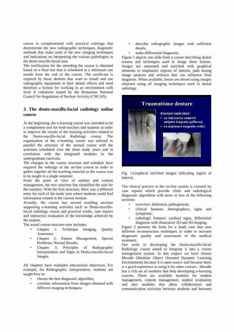
• make differential diagnostic. Figure 1 depicts one slide from a course describing dental trauma and techniques used to image these lesions. Images are annotated and enriched with graphical elements to emphasize regions of interest, path during image analysis and artifacts that can influence final diagnosis. When available, lesion are shown using images obtained using all imaging techniques used in dental radiology.
Fig. 1.Graphical enriched images indicating region of interest. The clinical practice in the on-line system is covered by case reports which provide clinic and radiological diagnostic algorithms with more or less of the following sections:
• overview: definition, pathogenesis; • clinical features: demographics, signs and
symptoms; • radiologic features: cardinal signs, differential
diagnosis with illustrative 2D and 3D imaging. Figure 2 presents the form for a study case that uses different reconstruction techniques in order to increase diagnostic quality and assessment of the medical treatment. Our work in developing the Dento-maxillo-facial Radiology course aimed to integrate it into a course management system. In this respect we have chosen Moodle (Modular Object Oriented Dynamic Learning Environment) because it is open source and because there is a good experience in using it for other courses. Moodle has a rich set of modules that help developing e-learning courses. There are available modules for student management, content management, student evaluation and also modules that allow collaboration and communication activities between students and between
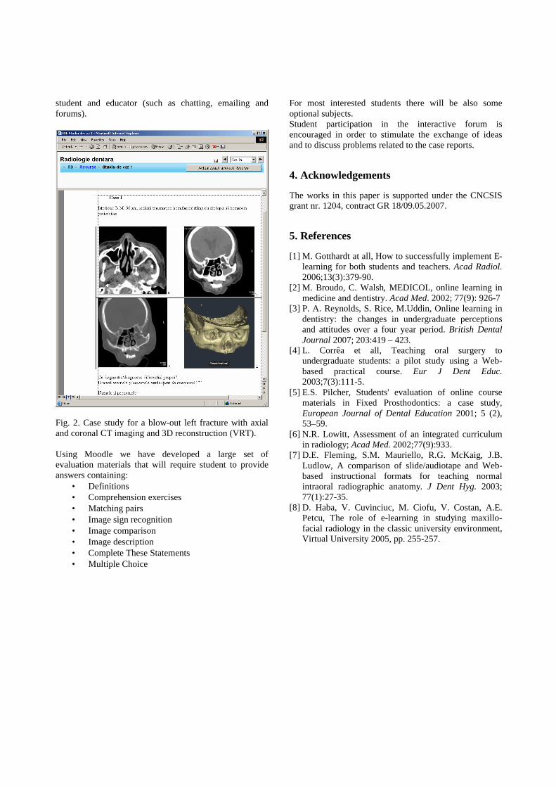
student and educator (such as chatting, emailing and forums).
Fig. 2. Case study for a blow-out left fracture with axial and coronal CT imaging and 3D reconstruction (VRT). Using Moodle we have developed a large set of evaluation materials that will require student to provide answers containing:
• Definitions • Comprehension exercises • Matching pairs • Image sign recognition • Image comparison • Image description • Complete These Statements • Multiple Choice
For most interested students there will be also some optional subjects. Student participation in the interactive forum is encouraged in order to stimulate the exchange of ideas and to discuss problems related to the case reports.
4. Acknowledgements
The works in this paper is supported under the CNCSIS grant nr. 1204, contract GR 18/09.05.2007.
5. References
[1] M. Gotthardt at all, How to successfully implement E-learning for both students and teachers. Acad Radiol. 2006;13(3):379-90.
[2] M. Broudo, C. Walsh, MEDICOL, online learning in medicine and dentistry. Acad Med. 2002; 77(9): 926-7
[3] P. A. Reynolds, S. Rice, M.Uddin, Online learning in dentistry: the changes in undergraduate perceptions and attitudes over a four year period. British Dental Journal 2007; 203:419 – 423.
[4] L. Corrêa et all, Teaching oral surgery to undergraduate students: a pilot study using a Web-based practical course. Eur J Dent Educ. 2003;7(3):111-5.
[5] E.S. Pilcher, Students' evaluation of online course materials in Fixed Prosthodontics: a case study, European Journal of Dental Education 2001; 5 (2), 53–59.
[6] N.R. Lowitt, Assessment of an integrated curriculum in radiology; Acad Med. 2002;77(9):933.
[7] D.E. Fleming, S.M. Mauriello, R.G. McKaig, J.B. Ludlow, A comparison of slide/audiotape and Web-based instructional formats for teaching normal intraoral radiographic anatomy. J Dent Hyg. 2003; 77(1):27-35.
[8] D. Haba, V. Cuvinciuc, M. Ciofu, V. Costan, A.E. Petcu, The role of e-learning in studying maxillo-facial radiology in the classic university environment, Virtual University 2005, pp. 255-257.