E-ISSN: 2090-5009 Immunome Research · Immunome Research- An Open Access online journal that uses...
13
Immunome Research- An Open Access online journal that uses online manuscript submission, review and tracking system for quality and quick review processing. Submit your manuscript at http://www.omicsonline.com/open-access/submitmanuscriptIMR.php OMICS Publishing Group 5716 Corsa Ave., Suite 110, Westlake, Los Angeles, CA 91362-7354, USA, Phone: +1- 650-268-9744, Fax: +1-650-618-1414, Toll free: +1-800-216-6499 http://www.omicsonline.com/open-access/immunome-research.php Immunome Research E-ISSN: 2090-5009 András Falus Semmelweis University Hungary Alessandro Sette La Jolla Institute for Allergy andImmunology USA Yongqun “Oliver” He University of Michigan Medical School USA Paul Zarogoulidis Aristotle University of Thessaloniki Thessaloniki, Greece Di Francesco S GD’Annunzio University of Chieti-Pescara Italy Timothy Ravasi King Abdullah University, Saudi Arabia L Tenaglia R G.D’Annunzio University,Italy Kaid Darwiche University Duisburg-Essen Germany Lonny Yarmus Johns Hopkins University, USA Marie-Paule Lefranc University of Montpellier France T he Immunome Research (IMR) is presented with a vision of providing the young talents a platform to encourage them to develop and globalize their skills and talents for the welfare of the society they are associated with and ultimately to the whole world. The Journal also aims to encourage every young talents to pursue research in fields they are interested and also makes every citizen accessible for the new technologies through their innovative ideas and extraordinary thoughts. The Journal aims to provide the highest possible standards of publication by scrutinizing the papers through peer-review assisted by the eminent experts from all parts of the globe. The journal includes a wide range of fields in its discipline to create a platform for the authors to make their contribution towards the journal and the editorial office promises a peer review process for the submitted manuscripts for the quality of publishing.
Transcript of E-ISSN: 2090-5009 Immunome Research · Immunome Research- An Open Access online journal that uses...

Immunome Research- An Open Access online journal that uses online manuscript submission, review and tracking system for quality and quick review processing. Submit your manuscript at http://www.omicsonline.com/open-access/submitmanuscriptIMR.php
OMICS Publishing Group5716 Corsa Ave., Suite 110, Westlake, Los Angeles, CA 91362-7354, USA, Phone: +1- 650-268-9744, Fax: +1-650-618-1414, Toll free: +1-800-216-6499
http://www.omicsonline.com/open-access/immunome-research.php
Immunome ResearchE-ISSN: 2090-5009
András FalusSemmelweis University
Hungary
Alessandro SetteLa Jolla Institute for
Allergy andImmunologyUSA
Yongqun “Oliver” HeUniversity of Michigan
Medical SchoolUSA
Paul ZarogoulidisAristotle University of
ThessalonikiThessaloniki, Greece
Di Francesco SGD’Annunzio University
of Chieti-PescaraItaly
Timothy RavasiKing Abdullah
University, Saudi Arabia
L Tenaglia R G.D’Annunzio University,Italy
Kaid DarwicheUniversity
Duisburg-EssenGermany
Lonny YarmusJohns Hopkins University, USA
Marie-Paule LefrancUniversity of Montpellier
France
The Immunome Research (IMR) is presented with a vision of providing the young talents a platform to encourage them
to develop and globalize their skills and talents for the welfare of the society they are associated with and ultimately to the whole world. The Journal also aims to encourage every young talents to pursue research in fields they are interested and also makes every citizen accessible for the new technologies through their innovative ideas and extraordinary thoughts. The Journal aims to provide the highest possible standards of publication by scrutinizing the papers through peer-review assisted by the eminent experts from all parts of the globe.
The journal includes a wide range of fields in its discipline to create a platform for the authors to make their contribution towards the journal and the editorial office promises a peer review process for the submitted manuscripts for the quality of publishing.
BioMed CentralImmunome Research
ss
Open AcceResearchRibosomal protein mRNAs are translationally-regulated during human dendritic cells activation by LPSMaurizio Ceppi1,2,3,6, Giovanna Clavarino1,2,3, Evelina Gatti1,2,3, Enrico K Schmidt1,2,3, Aude de Gassart1,2,3, Derek Blankenship5, Gerald Ogola5, Jacques Banchereau4, Damien Chaussabel4 and Philippe Pierre*1,2,3Address: 1Centre d'Immunologie de Marseille-Luminy, Université de la Méditerranée, Case 906, 13288 Marseille cedex 9, France, 2INSERM, U631, 13288 Marseille, France, 3CNRS, UMR6102, 13288 Marseille, France, 4Baylor Institute for Immunology Research (BIIR), 3434 Live Oak, Dallas, TX 75204, USA, 5Baylor Institute for Health Care Research and Improvement, 8080 North Central Expressway, Dallas, TX 75206, USA and 6Genomic Vision, Paris Santé Cochin, 75014 Paris, France
Email: Maurizio Ceppi - [email protected]; Giovanna Clavarino - [email protected]; Evelina Gatti - [email protected]; Enrico K Schmidt - [email protected]; Aude de Gassart - [email protected]; Derek Blankenship - [email protected]; Gerald Ogola - [email protected]; Jacques Banchereau - [email protected]; Damien Chaussabel - [email protected]; Philippe Pierre* - [email protected]
* Corresponding author
AbstractBackground: Dendritic cells (DCs) are the sentinels of the mammalian immune system,characterized by a complex maturation process driven by pathogen detection. Although multiplestudies have described the analysis of activated DCs by transcriptional profiling, recent findingsindicate that mRNAs are also regulated at the translational level. A systematic analysis of themRNAs being translationally regulated at various stages of DC activation was performed usingtranslational profiling, which combines sucrose gradient fractionation of polysomal-bound mRNAswith DNA microarray analysis.
Results: Total and polysomal-bound mRNA populations purified from immature, 4 h and 16 h LPS-stimulated human monocyte-derived DCs were analyzed on Affymetrix microarrays U133 2.0. Agroup of 375 transcripts was identified as translationally regulated during DC-activation. In additionto several biochemical pathways related to immunity, the most statistically relevant biologicalfunction identified among the translationally regulated mRNAs was protein biosynthesis itself. Wesingled-out a cluster of 11 large ribosome proteins mRNAs, which are disengaged from polysomesat late time of maturation, suggesting the existence of a negative feedback loop regulatingtranslation in DCs and linking ribosomal proteins to immuno-modulatory function.
Conclusion: Our observations highlight the importance of translation regulation during theimmune response, and may favor the identification of novel protein networks relevant forimmunity. Our study also provides information on the potential absence of correlation betweengene expression and protein production for specific mRNA molecules present in DCs.
Published: 27 November 2009
Immunome Research 2009, 5:5 doi:10.1186/1745-7580-5-5
Received: 30 September 2009Accepted: 27 November 2009
This article is available from: http://www.immunome-research.com/content/5/1/5
© 2009 Ceppi et al; licensee BioMed Central Ltd. This is an Open Access article distributed under the terms of the Creative Commons Attribution License (http://creativecommons.org/licenses/by/2.0), which permits unrestricted use, distribution, and reproduction in any medium, provided the original work is properly cited.
Page 1 of 12(page number not for citation purposes)
Immunome Research 2009, 5:5 http://www.immunome-research.com/content/5/1/5
BackgroundDendritic cells (DCs) are haematopoietic cells specializedin antigen capture and presentation for initiation of pri-mary and secondary immune responses. Due to this cen-tral role in induction and regulation of immunity, theyrepresent an attractive target for immunotherapy againstvarious diseases, including cancer and microbial infec-tions [1]. We recently demonstrated that translation regu-lation is required for function and survival of mouseactivated DCs [2]. Moreover, emerging evidence indicatethat translation plays a major role in immune regulationand its dysfunction can lead to pathology [3-5]. Althoughseveral seminal studies have described the use of microar-rays to define the gene expression and functional signa-ture of DCs upon pathogen detection [6,7], there were noattempts to include the additional layer of complexitybrought by translational regulation. As the relationshipbetween inflammation, innate immunity, and post-tran-scriptional regulation is becoming clearer [8], we have ina recent study used a microarray-based screen to identifythe immunologically relevant pathways regulated by miR-155 in lipopolysaccharide (LPS)-activated human mono-cyte-derived DC (moDC) [9]. To increase further ourunderstanding of post-transcriptional regulation andestablish the contribution of translation in the control ofimmune response, we carried-out, using Affymetrixmicroarrays, a systematic and comparative analysis ofpolysome-bound mRNA [10-12] purified from differentlyLPS-activated moDCs.
Using this approach, and in addition to several immuno-logically relevant mRNAs, we identified a network ofribosomal protein mRNAs being strongly down-modu-lated at the translational level at late time of DC matura-tion. Ribosomal proteins are integral components of thebasal cellular machinery involved in protein synthesis,whose roles have been regarded collectively as important,but individually disregarded. Recent findings, however,have demonstrated that components of the translationalapparatus are multifunctional and that several individualribosomal proteins play a role in regulating cell growth,transformation and death [13]. Our results clearly supportthese views and underline the importance of these pro-teins for DC function.
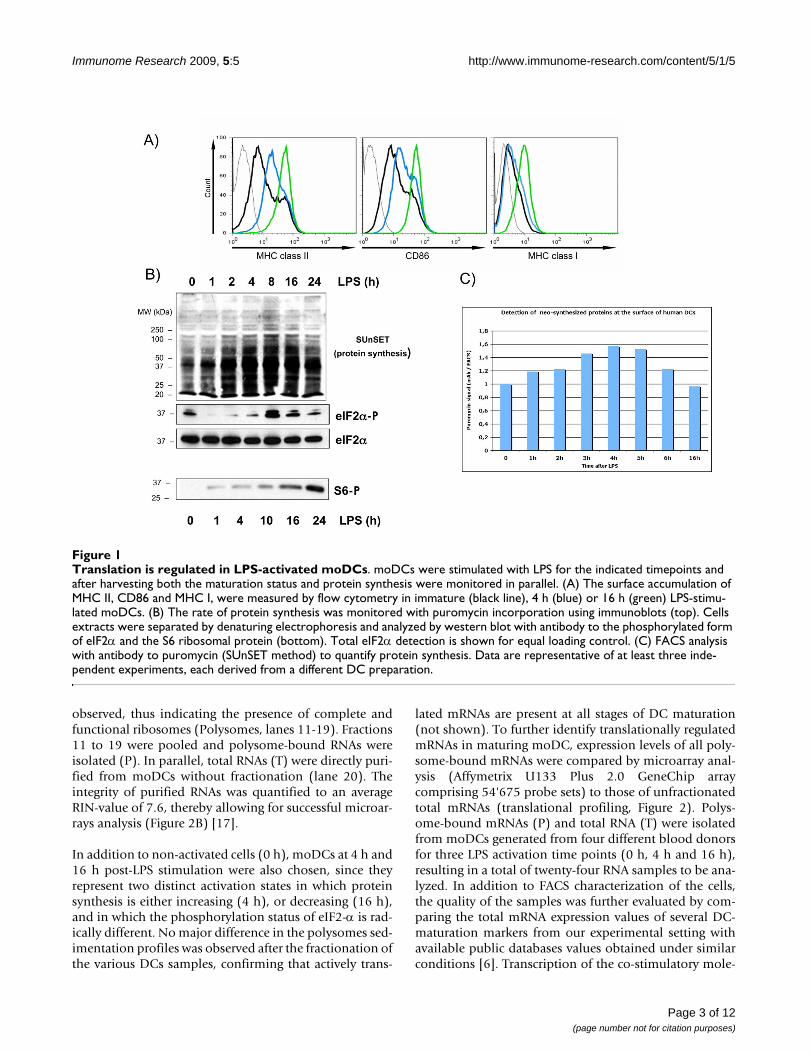
Results and discussionTranslation is regulated in LPS-activated human moDCsHuman monocyte-derived DCs were activated with LPSand displayed the expected cell surface accumulation ofMHC I, MHC II and CD86 as measured by flow cytometry(Figure 1A). The rate of protein synthesis in activatedmoDCs was monitored with puromycin incorporationusing immunoblot or FACS analysis (SUnSET) [14]. Pro-tein synthesis intensity was strongly increased upon LPS-stimulation and peaked at 4 h of activation, prior a steady
decrease to basal levels (Figure 1B and 1C). As the rate-limiting step in protein synthesis is the initiation ofmRNA translation, we investigated the status of transla-tion factors involved in the regulation of protein synthe-sis. Thus, we monitored by immunoblot thephosphorylation of the alpha-subunit of eukaryotic trans-lation initiation factor 2 (eIF2α), which prevents theassembly of ribosomal pre-initiation complexes [15].Almost complete eIF2α dephosphorylation was detectedbetween 1 h and 4 h after LPS-stimulation, correlatingwith the increase in protein synthesis. Interestingly, afterseveral hours of LPS-stimulation eIF2α phosphorylationwas recovered and even increased compared to the imma-ture DC levels. Thus eIF2α phosphorylation is regulatedduring the maturation of human moDCs and correlateswell with the regulation of mRNA translation in thesecells.
Translation initiation is also controlled through the stim-ulation of the PI3 kinase/AKT/mTOR signal transductioncascade, which leads to the phosphorylation of eIF4E-binding proteins (4E-BPs) and of the S6 ribosomal pro-tein (S6) by the ribosomal protein S6 kinase (S6K1) [16].Both molecules have an important role in regulating cap-mediated translation and S6 phosphorylation represents ahallmark of protein synthesis activation. S6 phosphoryla-tion was monitored in LPS-activated moDCs by immuno-blot (Figure 1B). We confirmed that S6 phosphorylationis steadily and strongly increased over time of LPS-expo-sure and this in a correlated manner to the moDCs matu-ration phenotype.
Thus, protein synthesis is tightly controlled upon LPS-sensing by MoDCs and is likely to represent a functionallyimportant aspect of human DC maturation. Furthermore,this process implies that specific mRNA molecules mightbe translationally regulated in response to eIF2α or S6phosphorylation under these conditions.
mRNA translational engagement changes at late time points of DC-activationIn order to identify mRNA molecules potentially regu-lated at the translational level, the global engagement ofmRNA molecules on polysomes was measured on LPS-activated moDCs by translational profiling [10-12].Actively translated polysome-bound mRNAs (P) were sep-arated from messenger free ribonucleoproteins (mRNPs,F) using sucrose gradient fractionation and visualized bydenaturing agarose gel (Figure 2A). Efficient polysomeseparation was confirmed by the presence of 5S rRNA inthe lighter fractions of the gradient (lanes 1-3), followedby fractions where only 18S rRNA was visible (lanes 4-5)and fractions in which an excess of large ribosomal subu-nit was detected (28S rRNA >> 18S rRNA, lanes 6-9). Fromfraction 11, a constant ratio of 28S over 18S rRNA was
Page 2 of 12(page number not for citation purposes)
Immunome Research 2009, 5:5 http://www.immunome-research.com/content/5/1/5
observed, thus indicating the presence of complete andfunctional ribosomes (Polysomes, lanes 11-19). Fractions11 to 19 were pooled and polysome-bound RNAs wereisolated (P). In parallel, total RNAs (T) were directly puri-fied from moDCs without fractionation (lane 20). Theintegrity of purified RNAs was quantified to an averageRIN-value of 7.6, thereby allowing for successful microar-rays analysis (Figure 2B) [17].
In addition to non-activated cells (0 h), moDCs at 4 h and16 h post-LPS stimulation were also chosen, since theyrepresent two distinct activation states in which proteinsynthesis is either increasing (4 h), or decreasing (16 h),and in which the phosphorylation status of eIF2-α is rad-ically different. No major difference in the polysomes sed-imentation profiles was observed after the fractionation ofthe various DCs samples, confirming that actively trans-
lated mRNAs are present at all stages of DC maturation(not shown). To further identify translationally regulatedmRNAs in maturing moDC, expression levels of all poly-some-bound mRNAs were compared by microarray anal-ysis (Affymetrix U133 Plus 2.0 GeneChip arraycomprising 54'675 probe sets) to those of unfractionatedtotal mRNAs (translational profiling, Figure 2). Polys-ome-bound mRNAs (P) and total RNA (T) were isolatedfrom moDCs generated from four different blood donorsfor three LPS activation time points (0 h, 4 h and 16 h),resulting in a total of twenty-four RNA samples to be ana-lyzed. In addition to FACS characterization of the cells,the quality of the samples was further evaluated by com-paring the total mRNA expression values of several DC-maturation markers from our experimental setting withavailable public databases values obtained under similarconditions [6]. Transcription of the co-stimulatory mole-
Translation is regulated in LPS-activated moDCsFigure 1Translation is regulated in LPS-activated moDCs. moDCs were stimulated with LPS for the indicated timepoints and after harvesting both the maturation status and protein synthesis were monitored in parallel. (A) The surface accumulation of MHC II, CD86 and MHC I, were measured by flow cytometry in immature (black line), 4 h (blue) or 16 h (green) LPS-stimu-lated moDCs. (B) The rate of protein synthesis was monitored with puromycin incorporation using immunoblots (top). Cells extracts were separated by denaturing electrophoresis and analyzed by western blot with antibody to the phosphorylated form of eIF2α and the S6 ribosomal protein (bottom). Total eIF2α detection is shown for equal loading control. (C) FACS analysis with antibody to puromycin (SUnSET method) to quantify protein synthesis. Data are representative of at least three inde-pendent experiments, each derived from a different DC preparation.
Page 3 of 12(page number not for citation purposes)

Immunome Research 2009, 5:5 http://www.immunome-research.com/content/5/1/5
cules CD80 and CD86, as well as the MHC I and IImRNAs were found to be all up-regulated after DC stimu-lation by LPS (see Additional file 1), thus confirming thequality of our samples and reliability of our analysis.
Global alterations of total and polysomal-bound mRNAas a function of time post-LPS was investigated, to obtaina comprehensive view of translation regulation inmoDCs. The 54'675 probe sets present were first filteredon expression (signal > 100 in all tested conditions) toobtain a preliminary list of 7'709 probe sets. The 7'709probe sets were then selected on fold change (applying a2-fold cut-off), comparing Polysomal and Total mRNA atdifferent timepoints. Between 0 h and 4 h post-LPS,among the 783 transcriptionally up-regulated genes (sig-nal 0 h < signal 4 h), 662 genes (84%) were sharedbetween total and polysomal RNA, 51 genes (6%) were
unique to polysomal RNA, and 70 genes (10%) wereunique to total RNA (Figure 3A). Among the 959 tran-scriptionally down-regulated genes (signal 0 h > signal 4h), 597 genes (63%) were shared between total and poly-somal RNA, 165 genes (17%) were unique to polysomalRNA, and 188 genes (20%) were unique to total RNA (Fig-ure 3B). Between 4 h and 16 h post-LPS, among the 536transcriptionally up-regulated genes (signal 4 h < signal16 h), 456 genes (65%) were shared between total andpolysomal RNA, 91 genes (13%) were unique to polyso-mal RNA, and 151 genes (22%) were unique to total RNA(Figure 3C). Similarly, among the 698 transcriptionallydown-regulated genes (signal 4 h > signal 16 h), 355 genes(66%) were shared between total and polysomal RNA, 55genes (10%) were unique to polysomal RNA, and 127genes (24%) were unique to total RNA (Figure 3D). Theseresults indicate that during DC maturation, transcription
Efficient profiling and isolation of polysomes out of human moDCsFigure 2Efficient profiling and isolation of polysomes out of human moDCs. Polysomes sedimentation profiles (A) and RNA-integrity profiles (B) after sucrose gradient fractionation of untreated (0 h) or 4 h and 16 h LPS-stimulated moDCs.
Page 4 of 12(page number not for citation purposes)

Immunome Research 2009, 5:5 http://www.immunome-research.com/content/5/1/5
and translation intensity are relatively well coupled uponLPS-sensing, as suggested by the overall increase in pro-tein synthesis at the onset of maturation. However, weevaluate to 30% (the mean percentage of the genes uniqueto polysomal RNA and total RNA) the proportion of trans-lationally-engaged mRNA molecules, in which transcrip-tion and translation are not linearly connected. Thepolysome-bound (that is, translated) mRNAs were up-reg-ulated and down-regulated with the same proportionsindicating that translation regulation in DCs is probablytargeting discrete subsets of genes, whereas the majority ofgenes are regulated by transcription and mRNA stability, adetailed description of these genes subsets is given inAdditional file 2. Most of the identified genes were distrib-uted in all biological activities with no clear functionalclustering capable of unraveling a distinct pattern of regu-lation. However, an over-representation of protein syn-
thesis genes was found in the genes identified astranslationaly down-regulated at late time of maturation(see Additional file 2D).
Biological functions of translationally regulated mRNAs in activated moDCsWe decided to take a statistically unbiased approach tofurther identify entire functional pathways, which couldbe regulated at the translational level. Importantly, thedata obtained from four different donors were homoge-nous and no major pattern variation for both total andpolysomal-bound RNA expression was found, thus allow-ing statistical analysis (see Additional file 3, compare thefour columns within each T or P).
The 54'675 probe sets present on the Affymetrix Gene-Chip array were first filtered on flags (P in 50% 0 h or P in
Global alterations of total and polysomal-bound mRNA in LPS-activated moDCsFigure 3Global alterations of total and polysomal-bound mRNA in LPS-activated moDCs. The 54'675 probe sets present on the Affymetrix U133 Plus 2.0 GeneChip array were first filtered on expression (signal > 100 in all tested conditions) to obtain a preliminary list of 7'709 probe sets. The 7'709 probe sets were then filtered on fold change (applying a 2-fold cut-off) comparing Polysomal (Poly, red) and Total (Tot, blue) mRNA, between 0 h and 4 h (A and B) or 4 h and 16 h (C and D) post-LPS. For effective Venn diagram visualization, the transcriptionally up-regulated probe sets (A and C) were distinguished from the transcriptionally down-regulated probe sets (B and D). See Additional file 3 for a detailed description of the different genes subsets.
Page 5 of 12(page number not for citation purposes)
Immunome Research 2009, 5:5 http://www.immunome-research.com/content/5/1/5
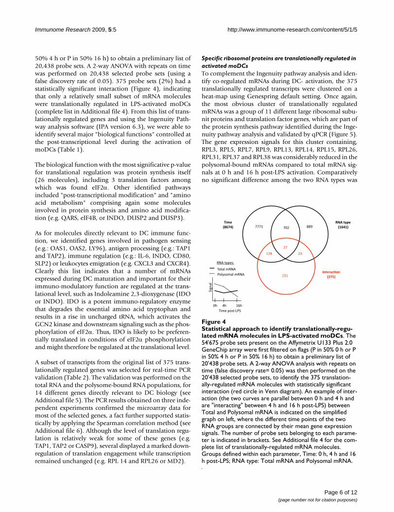
50% 4 h or P in 50% 16 h) to obtain a preliminary list of20,438 probe sets. A 2-way ANOVA with repeats on timewas performed on 20,438 selected probe sets (using afalse discovery rate of 0.05). 375 probe sets (2%) had astatistically significant interaction (Figure 4), indicatingthat only a relatively small subset of mRNA moleculeswere translationally regulated in LPS-activated moDCs(complete list in Additional file 4). From this list of trans-lationally regulated genes and using the Ingenuity Path-way analysis software (IPA version 6.3), we were able toidentify several major "biological functions" controlled atthe post-transcriptional level during the activation ofmoDCs (Table 1).
The biological function with the most significative p-valuefor translational regulation was protein synthesis itself(26 molecules), including 3 translation factors amongwhich was found eIF2α. Other identified pathwaysincluded "post-transcriptional modification" and "aminoacid metabolism" comprising again some moleculesinvolved in protein synthesis and amino acid modifica-tion (e.g. QARS, eIF4B, or INDO, DUSP2 and DUSP3).
As for molecules directly relevant to DC immune func-tion, we identified genes involved in pathogen sensing(e.g.: OAS1, OAS2, LY96), antigen processing (e.g.: TAP1and TAP2), immune regulation (e.g.: IL-6, INDO, CD80,SLP2) or leukocytes emigration (e.g. CXCL3 and CXCR4).Clearly this list indicates that a number of mRNAsexpressed during DC maturation and important for theirimmuno-modulatory function are regulated at the trans-lational level, such as Indoleamine 2,3-dioxygenase (IDOor INDO). IDO is a potent immuno-regulatory enzymethat degrades the essential amino acid tryptophan andresults in a rise in uncharged tRNA, which activates theGCN2 kinase and downstream signaling such as the phos-phorylation of eIF2α. Thus, IDO is likely to be preferen-tially translated in conditions of eIF2α phosphorylationand might therefore be regulated at the translational level.
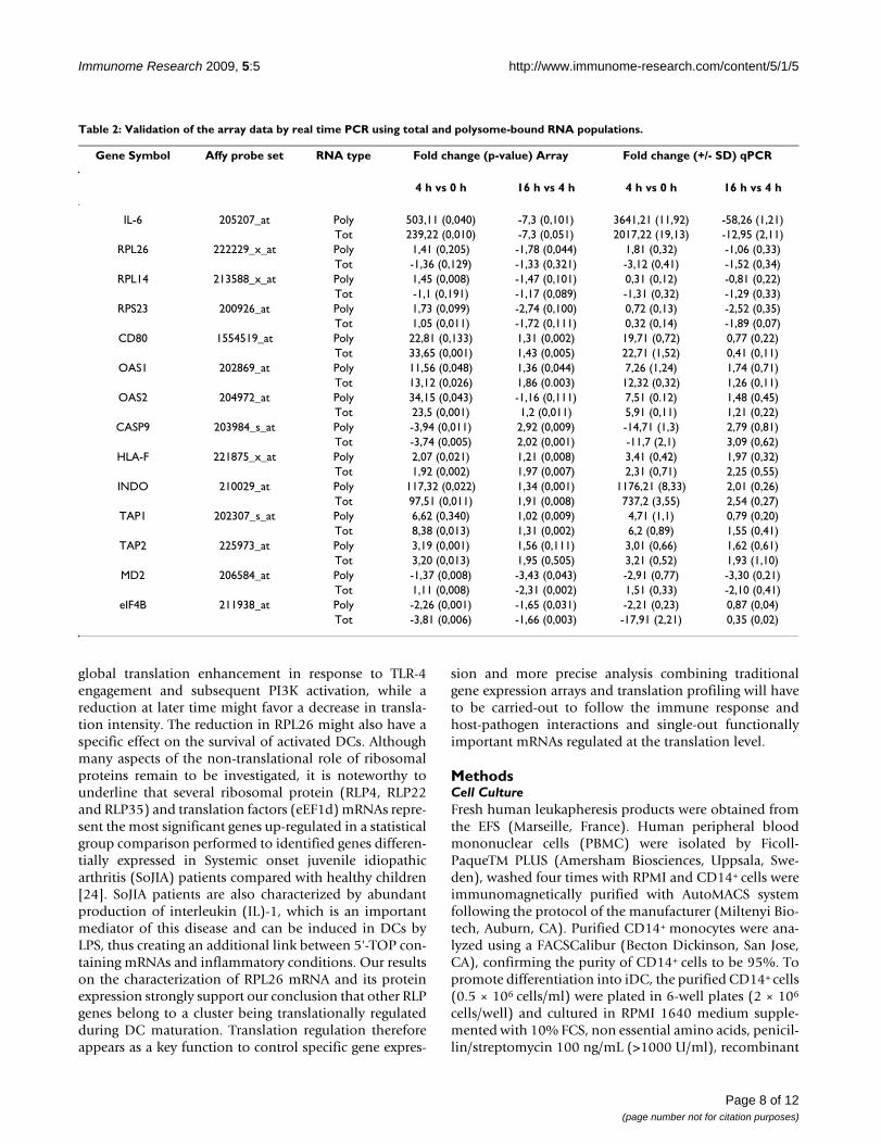
A subset of transcripts from the original list of 375 trans-lationally regulated genes was selected for real-time PCRvalidation (Table 2). The validation was performed on thetotal RNA and the polysome-bound RNA populations, for14 different genes directly relevant to DC biology (seeAdditional file 5). The PCR results obtained on three inde-pendent experiments confirmed the microarray data formost of the selected genes, a fact further supported statis-tically by applying the Spearman correlation method (seeAdditional file 6). Although the level of translation regu-lation is relatively weak for some of these genes (e.g.TAP1, TAP2 or CASP9), several displayed a marked down-regulation of translation engagement while transcriptionremained unchanged (e.g. RPL 14 and RPL26 or MD2).
Specific ribosomal proteins are translationally regulated in activated moDCsTo complement the Ingenuity pathway analysis and iden-tify co-regulated mRNAs during DC- activation, the 375translationally regulated transcripts were clustered on aheat-map using Genespring default setting. Once again,the most obvious cluster of translationally regulatedmRNAs was a group of 11 different large ribosomal subu-nit proteins and translation factor genes, which are part ofthe protein synthesis pathway identified during the Inge-nuity pathway analysis and validated by qPCR (Figure 5).The gene expression signals for this cluster containing,RPL3, RPL5, RPL7, RPL9, RPL13, RPL14, RPL15, RPL26,RPL31, RPL37 and RPL38 was considerably reduced in thepolysomal-bound mRNAs compared to total mRNA sig-nals at 0 h and 16 h post-LPS activation. Comparativelyno significant difference among the two RNA types was
Statistical approach to identify translationally-regulated mRNA molecules in LPS-activated moDCsFigure 4Statistical approach to identify translationally-regu-lated mRNA molecules in LPS-activated moDCs. The 54'675 probe sets present on the Affymetrix U133 Plus 2.0 GeneChip array were first filtered on flags (P in 50% 0 h or P in 50% 4 h or P in 50% 16 h) to obtain a preliminary list of 20'438 probe sets. A 2-way ANOVA analysis with repeats on time (false discovery rate= 0.05) was then performed on the 20'438 selected probe sets, to identify the 375 translation-ally-regulated mRNA molecules with statistically significant interaction (red circle in Venn diagram). An example of inter-action (the two curves are parallel between 0 h and 4 h and are "interacting" between 4 h and 16 h post-LPS) between Total and Polysomal mRNA is indicated on the simplified graph on left, where the different time points of the two RNA groups are connected by their mean gene expression signals. The number of probe sets belonging to each parame-ter is indicated in brackets. See Additional file 4 for the com-plete list of translationally-regulated mRNA molecules. Groups defined within each parameter, Time: 0 h, 4 h and 16 h post-LPS; RNA type: Total mRNA and Polysomal mRNA.
�����
�����
��������
������
�����������
�����
∀∀#�!!!��
����
!���
����!�
�!�
���� ������������ �
�� ����
�����������
���������������
��������������
�����������
Page 6 of 12(page number not for citation purposes)

Immunome Research 2009, 5:5 http://www.immunome-research.com/content/5/1/5
detected after 4 h of LPS stimulation, suggesting that theseRPL mRNAs were engaged in translation at this early timeof DC activation. Thus, RPL transcripts seem to undergo"translational engagement" between 0 h and 4 h, fol-lowed by "translational disengagement" between 4 h and16 h, thus matching the observed rate of protein synthesisin DCs. Transcription of ribosomal protein genes starts ata C-residue that is embedded in an oligopyrimidine tractof length 5-25 base pairs, also known as the 5'-TOP signal.The 5'-TOP signal is an essential cis-regulatory motif forthe translational control of most ribosomal proteinmRNAs expression [18,19]. A preponderance of evidencesuggests that translation of mRNAs containing a 5'-TOPsignal is strongly up-regulated in response to ribosomeprotein S6 phosphorylation triggered by mTORC1 stimu-lation and subsequent S6K1 activation. In addition toribosomal proteins mRNAs, TOP mRNAs include alsothose coding for several translation elongation factorssuch as eEF2, indicating that most of the translationallydown-regulated genes identified in DCs during theirresponse to LPS contain a 5'-TOP signal.
In recent years, several reports have also suggested thattranslation of TOP mRNAs can be independent of S6phosphorylation [20,21]. In activated DCs, S6 phosphor-ylation correlates well with the translational engagementof the ribosomal proteins after 4 h of LPS stimulation(Figure 5, 4h). However even if the intensity of S6 phos-phorylation keeps increasing during the following 12 h ofDC maturation, several of the large ribosomal proteinmRNAs are nevertheless clearly disengaged from polys-omes. Thus, although S6 phosphorylation is likely to
favor translational engagement of 5'-TOP containingmRNAs, it does not counteract their increased releasefrom the ribosomes.
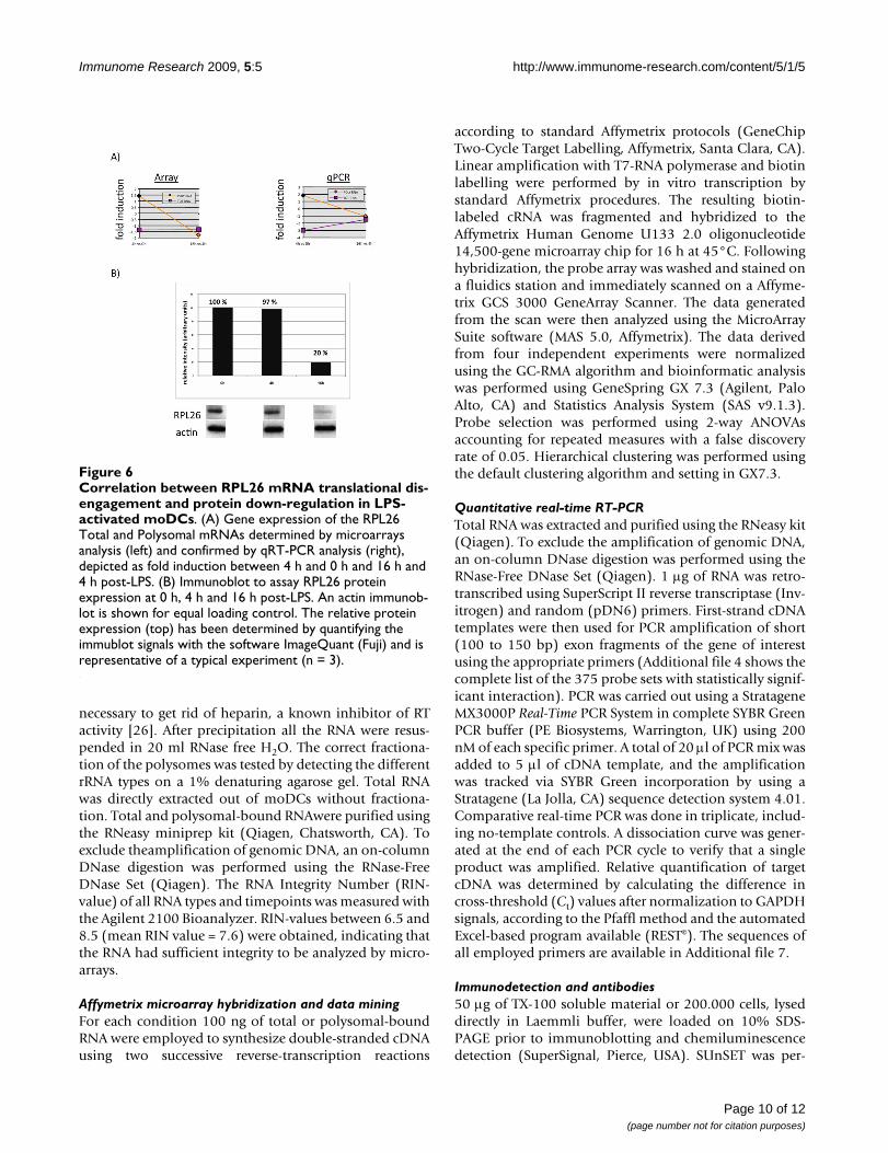
A role for ribosome proteins (RLP) outside of translationis emerging, however most of the current knowledgeabout these proteins links them with specific translationregulation activity including ER-binding for RLP7 andcontrol of survival for RPL5, 11, 23 and 26 [22]. Indeed,these particular RLPs are involved in the murine doubleminute protein (MDM2)-mediated p53 pathway regula-tion [23]. Mdm2 is a E3-ubiquitin ligase known to regu-late p53 levels directly by promoting its degradation andindirectly by targeting ribosomal protein L26, which nor-mally binds the p53 mRNA and augments its translation.As the ribosomal protein L26 (RPL26) represents animportant component of the auto-regulatory feedbackloop that maintains low p53 activity in non-stressed cells[22], we further investigated the regulation of RPL26 atthe mRNA and protein levels in activated DCs. AlthoughRPL26 mRNA gene expression was stable upon-LPS stim-ulation, its polysomal-bound engagement was lost at 16h, as identified by microarrays and confirmed by qPCR(Figure 6A). More importantly, RPL26 protein expressionwas down-regulated by 80% after 16 h of LPS- mediatedactivation, correlating with the down-regulation of polys-omal-bound mRNAs (Figure 6B).
The translation regulation of ribosomal protein mRNAs isone of the features of LPS-activated human DCs. In theearly phase of DC maturation, a specific increase in ribos-osomal proteins synthesis might impact positively on the
Table 1: Top "biological functions" of mRNA molecules affected by translation regulation in LPS-activated moDCs
Category (nr. of molecules)
Biological Function p-value Molecules (gene symbols)
Protein Synthesis (26) biosynthesis of proteins and translation regulation
1.73E-04 ADAMTS5, CASP9, EEF2, EIF2A, EIF4A2, EIF4B, NCK1, PLAU, PSMC2, RBM3, RPL3, RPL5, RPL7, RPL13, RPL14,
RPL15, RPL26, RPL31, RPL37, RPL38, RPL9, RPS11, SERPINB1, SRGN, UBC, UBE2K
RNA Post-Transcriptional Modification (12)
modification of mRNA 8.67E-03 CDC5L, CPSF1, DBR1, EIF4A2, EIF4B, GRSF1, HNRNPR, CPSF1, GRSF1, MAPKAPK2, ZFP36, QARS
Amino Acid Metabolism (11) catabolism of L-tryptophan and dephosphorylation of amino acids
8.57E-08 CD80, IL6, INDO, KMO, KYNU, DUSP2, DUSP3, PPM1A, PPM1F, PPP1CB, PTPN6
Cell Morphology (10) transmembrane potential of mitochondria
1.26E-03 AP2A2, BNIP3, CASP9, CFLAR, IL6, MAPK9, MOAP1, NFE2L2, PTPN6, SRGN
Immune Cell Trafficking (8) emigration of leukocytes 2.08E-03 CD44, CXCL3, CXCR4, CYTIP, GFI1, LDLR, PLAUCancer (7) tumorigenesis of intestinal tissue 2.64E-03 APC, MLH1, IFI16, SMARCA2, HSPA1A, IFI16, IGFBP4
Nucleic Acid Metabolism (5) metabolism of nucleic acid component 6.15E-04 ATIC, MTAP, OAS1, OAS2, REXO2Cell-mediated Immune
Response (4)secretion of cytokine 3.60E-03 CADM1, LCP2, SRGN, GFI1
Antigen Presentation (2) antigen peptide transporter 8.84E-03 TAP1, TAP2Small molecule biochemistry
(1)activation by LPS 4.21E-02 LY96 (MD2)
The 375 probe sets with statistically significant interaction (Additional file 4) have been analysed with the Ingenuity Pathway software. The table displays the 10 most significant biological "categories" affected by translation regulation (out of 29 found), ranked by the number of molecules per category. The significance is expressed as p-value and is based on a Fisher's exact test, calculated by the software itself. The translationally regulated mRNA molecules validated by qPCR are indicated in bold (Table 2). RPL26 has been also validated at the protein level and is underlined (Figure 6).
Page 7 of 12(page number not for citation purposes)
Immunome Research 2009, 5:5 http://www.immunome-research.com/content/5/1/5
global translation enhancement in response to TLR-4engagement and subsequent PI3K activation, while areduction at later time might favor a decrease in transla-tion intensity. The reduction in RPL26 might also have aspecific effect on the survival of activated DCs. Althoughmany aspects of the non-translational role of ribosomalproteins remain to be investigated, it is noteworthy tounderline that several ribosomal protein (RLP4, RLP22and RLP35) and translation factors (eEF1d) mRNAs repre-sent the most significant genes up-regulated in a statisticalgroup comparison performed to identified genes differen-tially expressed in Systemic onset juvenile idiopathicarthritis (SoJIA) patients compared with healthy children[24]. SoJIA patients are also characterized by abundantproduction of interleukin (IL)-1, which is an importantmediator of this disease and can be induced in DCs byLPS, thus creating an additional link between 5'-TOP con-taining mRNAs and inflammatory conditions. Our resultson the characterization of RPL26 mRNA and its proteinexpression strongly support our conclusion that other RLPgenes belong to a cluster being translationally regulatedduring DC maturation. Translation regulation thereforeappears as a key function to control specific gene expres-
sion and more precise analysis combining traditionalgene expression arrays and translation profiling will haveto be carried-out to follow the immune response andhost-pathogen interactions and single-out functionallyimportant mRNAs regulated at the translation level.
MethodsCell CultureFresh human leukapheresis products were obtained fromthe EFS (Marseille, France). Human peripheral bloodmononuclear cells (PBMC) were isolated by Ficoll-PaqueTM PLUS (Amersham Biosciences, Uppsala, Swe-den), washed four times with RPMI and CD14+ cells wereimmunomagnetically purified with AutoMACS systemfollowing the protocol of the manufacturer (Miltenyi Bio-tech, Auburn, CA). Purified CD14+ monocytes were ana-lyzed using a FACSCalibur (Becton Dickinson, San Jose,CA), confirming the purity of CD14+ cells to be 95%. Topromote differentiation into iDC, the purified CD14+ cells(0.5 × 10⁶ cells/ml) were plated in 6-well plates (2 × 10⁶
cells/well) and cultured in RPMI 1640 medium supple-mented with 10% FCS, non essential amino acids, penicil-lin/streptomycin 100 ng/mL (>1000 U/ml), recombinant
Table 2: Validation of the array data by real time PCR using total and polysome-bound RNA populations.
Gene Symbol Affy probe set RNA type Fold change (p-value) Array Fold change (+/- SD) qPCR
4 h vs 0 h 16 h vs 4 h 4 h vs 0 h 16 h vs 4 h
IL-6 205207_at Poly 503,11 (0,040) -7,3 (0,101) 3641,21 (11,92) -58,26 (1,21)Tot 239,22 (0,010) -7,3 (0,051) 2017,22 (19,13) -12,95 (2,11)
RPL26 222229_x_at Poly 1,41 (0,205) -1,78 (0,044) 1,81 (0,32) -1,06 (0,33)Tot -1,36 (0,129) -1,33 (0,321) -3,12 (0,41) -1,52 (0,34)
RPL14 213588_x_at Poly 1,45 (0,008) -1,47 (0,101) 0,31 (0,12) -0,81 (0,22)Tot -1,1 (0,191) -1,17 (0,089) -1,31 (0,32) -1,29 (0,33)
RPS23 200926_at Poly 1,73 (0,099) -2,74 (0,100) 0,72 (0,13) -2,52 (0,35)Tot 1,05 (0,011) -1,72 (0,111) 0,32 (0,14) -1,89 (0,07)
CD80 1554519_at Poly 22,81 (0,133) 1,31 (0,002) 19,71 (0,72) 0,77 (0,22)Tot 33,65 (0,001) 1,43 (0,005) 22,71 (1,52) 0,41 (0,11)
OAS1 202869_at Poly 11,56 (0,048) 1,36 (0,044) 7,26 (1,24) 1,74 (0,71)Tot 13,12 (0,026) 1,86 (0.003) 12,32 (0,32) 1,26 (0,11)
OAS2 204972_at Poly 34,15 (0,043) -1,16 (0,111) 7,51 (0.12) 1,48 (0,45)Tot 23,5 (0,001) 1,2 (0,011) 5,91 (0,11) 1,21 (0,22)
CASP9 203984_s_at Poly -3,94 (0,011) 2,92 (0,009) -14,71 (1,3) 2,79 (0,81)Tot -3,74 (0,005) 2,02 (0,001) -11,7 (2,1) 3,09 (0,62)
HLA-F 221875_x_at Poly 2,07 (0,021) 1,21 (0,008) 3,41 (0,42) 1,97 (0,32)Tot 1,92 (0,002) 1,97 (0,007) 2,31 (0,71) 2,25 (0,55)
INDO 210029_at Poly 117,32 (0,022) 1,34 (0,001) 1176,21 (8,33) 2,01 (0,26)Tot 97,51 (0,011) 1,91 (0,008) 737,2 (3,55) 2,54 (0,27)
TAP1 202307_s_at Poly 6,62 (0,340) 1,02 (0,009) 4,71 (1,1) 0,79 (0,20)Tot 8,38 (0,013) 1,31 (0,002) 6,2 (0,89) 1,55 (0,41)
TAP2 225973_at Poly 3,19 (0,001) 1,56 (0,111) 3,01 (0,66) 1,62 (0,61)Tot 3,20 (0,013) 1,95 (0,505) 3,21 (0,52) 1,93 (1,10)
MD2 206584_at Poly -1,37 (0,008) -3,43 (0,043) -2,91 (0,77) -3,30 (0,21)Tot 1,11 (0,008) -2,31 (0,002) 1,51 (0,33) -2,10 (0,41)
eIF4B 211938_at Poly -2,26 (0,001) -1,65 (0,031) -2,21 (0,23) 0,87 (0,04)Tot -3,81 (0,006) -1,66 (0,003) -17,91 (2,21) 0,35 (0,02)
Page 8 of 12(page number not for citation purposes)

Immunome Research 2009, 5:5 http://www.immunome-research.com/content/5/1/5
human GM-CSF and 20 ng/mL (>100 U/ml) IL-4 for 5days (both from PeproTech, Rocky Hill, NJ). At days 2 and4, half of the volume of the medium was replaced by freshmedium supplemented with GM-CSF and IL-4. For DCmaturation, 100 ng/mL LPS (Escherichia coli type 026:B6;Sigma-Aldrich, St. Louis, MO) was added to the cells atday 5, for the indicated number of hours.
Polysomal profiling by sucrose gradient fractionationPolysome-bound mRNA molecules were enriched bysucrose gradient fractionation following the protocol orig-inally developed by Garcia-Sanz and collaborators [25].Briefly, 60 to 80 × 106 day 5 human moDCs were lysed in1 ml of polysome buffer (10 mM Tris-HCl (pH 8), 140mM NaCl, 1.5 mM MgCl2, 0.5% NP40, 0.1 mg/mlcycloheximide, and 500 units/mL RNasin (Promega,
Madison, WI). After 10 min on ice, lysates were quicklycentrifuged (10.000 × g for 10 sec at 4°C) and the super-natant was resuspended in a stabilizing solution (0.2 mg/ml cycloheximide, 0.7 mg/ml heparin, 1 mM phenyl-methanesulfonyl fluoride). After a quick centrifugation(12.000 × g for 2 min at 4°C) to remove mitochondriaand membrane debris, the resulting supernatant was lay-ered on a 15% to 40% sucrose gradient. Gradients werethen ultracentrifuged (35.000 × g for 2 h at 4°C, SW41rotor) and after centrifugation 20 × 550 ml fractions werecollected, starting from the top of the gradient. All thefractions were then digested with Proteinase K (200 mg/ml) in presence of 1%SDS and 10 mM EDTA. RNA wasthen extracted with Phenol/Chloroform/Isoamylalcohol(volume ratio 25:24:1) and precipitated with 2.5 Volumesof 100% Ethanol in presence of 0.8 M lithium chloride,
Specific ribosomal protein mRNAs are translationally-regulated in LPS-activated moDCsFigure 5Specific ribosomal protein mRNAs are translationally-regulated in LPS-activated moDCs. The 375 mRNA mole-cules with statistically significant interaction (see Figure 4) were clustered on a heat-map using the software GeneSpring GX 7.3. The translationally regulated ribosomal protein mRNAs appear as a specific signature (left panel), which has been extracted and enlarged (right panel). See the text for more details.
Page 9 of 12(page number not for citation purposes)
Immunome Research 2009, 5:5 http://www.immunome-research.com/content/5/1/5
necessary to get rid of heparin, a known inhibitor of RTactivity [26]. After precipitation all the RNA were resus-pended in 20 ml RNase free H2O. The correct fractiona-tion of the polysomes was tested by detecting the differentrRNA types on a 1% denaturing agarose gel. Total RNAwas directly extracted out of moDCs without fractiona-tion. Total and polysomal-bound RNAwere purified usingthe RNeasy miniprep kit (Qiagen, Chatsworth, CA). Toexclude theamplification of genomic DNA, an on-columnDNase digestion was performed using the RNase-FreeDNase Set (Qiagen). The RNA Integrity Number (RIN-value) of all RNA types and timepoints was measured withthe Agilent 2100 Bioanalyzer. RIN-values between 6.5 and8.5 (mean RIN value = 7.6) were obtained, indicating thatthe RNA had sufficient integrity to be analyzed by micro-arrays.
Affymetrix microarray hybridization and data miningFor each condition 100 ng of total or polysomal-boundRNA were employed to synthesize double-stranded cDNAusing two successive reverse-transcription reactions
according to standard Affymetrix protocols (GeneChipTwo-Cycle Target Labelling, Affymetrix, Santa Clara, CA).Linear amplification with T7-RNA polymerase and biotinlabelling were performed by in vitro transcription bystandard Affymetrix procedures. The resulting biotin-labeled cRNA was fragmented and hybridized to theAffymetrix Human Genome U133 2.0 oligonucleotide14,500-gene microarray chip for 16 h at 45°C. Followinghybridization, the probe array was washed and stained ona fluidics station and immediately scanned on a Affyme-trix GCS 3000 GeneArray Scanner. The data generatedfrom the scan were then analyzed using the MicroArraySuite software (MAS 5.0, Affymetrix). The data derivedfrom four independent experiments were normalizedusing the GC-RMA algorithm and bioinformatic analysiswas performed using GeneSpring GX 7.3 (Agilent, PaloAlto, CA) and Statistics Analysis System (SAS v9.1.3).Probe selection was performed using 2-way ANOVAsaccounting for repeated measures with a false discoveryrate of 0.05. Hierarchical clustering was performed usingthe default clustering algorithm and setting in GX7.3.
Quantitative real-time RT-PCRTotal RNA was extracted and purified using the RNeasy kit(Qiagen). To exclude the amplification of genomic DNA,an on-column DNase digestion was performed using theRNase-Free DNase Set (Qiagen). 1 μg of RNA was retro-transcribed using SuperScript II reverse transcriptase (Inv-itrogen) and random (pDN6) primers. First-strand cDNAtemplates were then used for PCR amplification of short(100 to 150 bp) exon fragments of the gene of interestusing the appropriate primers (Additional file 4 shows thecomplete list of the 375 probe sets with statistically signif-icant interaction). PCR was carried out using a StratageneMX3000P Real-Time PCR System in complete SYBR GreenPCR buffer (PE Biosystems, Warrington, UK) using 200nM of each specific primer. A total of 20 μl of PCR mix wasadded to 5 μl of cDNA template, and the amplificationwas tracked via SYBR Green incorporation by using aStratagene (La Jolla, CA) sequence detection system 4.01.Comparative real-time PCR was done in triplicate, includ-ing no-template controls. A dissociation curve was gener-ated at the end of each PCR cycle to verify that a singleproduct was amplified. Relative quantification of targetcDNA was determined by calculating the difference incross-threshold (Ct) values after normalization to GAPDHsignals, according to the Pfaffl method and the automatedExcel-based program available (REST©). The sequences ofall employed primers are available in Additional file 7.
Immunodetection and antibodies50 μg of TX-100 soluble material or 200.000 cells, lyseddirectly in Laemmli buffer, were loaded on 10% SDS-PAGE prior to immunoblotting and chemiluminescencedetection (SuperSignal, Pierce, USA). SUnSET was per-
Correlation between RPL26 mRNA translational disengage-ment and protein down-regulation in LPS-activated moDCsFigure 6Correlation between RPL26 mRNA translational dis-engagement and protein down-regulation in LPS-activated moDCs. (A) Gene expression of the RPL26 Total and Polysomal mRNAs determined by microarrays analysis (left) and confirmed by qRT-PCR analysis (right), depicted as fold induction between 4 h and 0 h and 16 h and 4 h post-LPS. (B) Immunoblot to assay RPL26 protein expression at 0 h, 4 h and 16 h post-LPS. An actin immunob-lot is shown for equal loading control. The relative protein expression (top) has been determined by quantifying the immublot signals with the software ImageQuant (Fuji) and is representative of a typical experiment (n = 3).
Page 10 of 12(page number not for citation purposes)
Immunome Research 2009, 5:5 http://www.immunome-research.com/content/5/1/5
formed as previously described [14] using A647-labelledmouse IgG2a anti-puromycin antibody (12D10). Anti-body against RPL26 was from Abnova. For flow cytometryanalysis, APC-conjugated anti-HLA-DR (L243 clone) andPE-conjugated anti-CD86 (clone IT2.2) were from BDPharMingen (San Jose, CA, USA). Anti- eIF2, phospho-eIF2 and -phospho-S6 were from Cell Signaling Technol-ogy (Beverly, MA, USA). For immunofluorescence analy-sis, MoDCs were let adhere on Alcyan blue coated-coverslips and surface stained with unlabelled antibody at4°C for 30 min. After washes, coverslips were placed inwarm medium for the indicated time and then fixed in 3%PFA for conventional staining with secondary antibodies.
GEO Reviewers LinkFollowing link has been created to allow read-only accessreview of record GSE14000 and associated accessions,which are the Affymetrix gene expression data related tothe submitted manuscript.
http://www.ncbi.nlm.nih.gov/geo/query/acc.cgi?token=jlalxuycqigwqnw&acc=GSE14000
Competing interestsThe authors declare that they have no competing interests.
Authors' contributionsAll authors read and approved the final manuscript. MC,GC, ES, AdG performed experiments. DB, GO, JB and DCcontributed to bioinformatics and statistical analysis. MC,EG and PP designed experiments and wrote the manu-script.
Additional material
AcknowledgementsThis work is supported by grants to PP from the ANR- DC-Trans, Réseau National Génopole, La Ligue Nationale Contre le Cancer and the Human Frontier of Science Program. MC is supported by the Swiss National Sci-ence Foundation (SNF). PP is part of the DC-THERA FP6 NoE. We are par-ticularly indebted to the CNRS Affimetrix facility and to the PICsL imaging core facility for expert technical assistance.
References1. Steinman RM: Lasker Basic Medical Research Award. Den-
dritic cells: versatile controllers of the immune system. NatMed 2007, 13:1155-1159.
2. Lelouard H, Schmidt EK, Camosseto V, Clavarino G, Ceppi M, HsuHT, Pierre P: Regulation of translation is required for dendriticcell function and survival during activation. J Cell Biol 2007,179:1427-1439.
3. Scheu S, Stetson DB, Reinhardt RL, Leber JH, Mohrs M, Locksley RM:Activation of the integrated stress response during T helpercell differentiation. Nat Immunol 2006, 7:644-651.
4. Sonenberg N, Hinnebusch AG: New modes of translational con-trol in development, behavior, and disease. Mol Cell 2007,28:721-729.
5. Pierre P: Immunity and the regulation of protein synthesis:surprising connections. Curr Opin Immunol 2009, 21:70-77.
6. Huang Q, Liu do N, Majewski P, Schulte le AC, Korn JM, Young RA,Lander ES, Hacohen N: The plasticity of dendritic cell responsesto pathogens and their components. Science 2001,294:870-875.
7. Robbins SH, Walzer T, Dembele D, Thibault C, Defays A, Bessou G,Xu H, Vivier E, Sellars M, Pierre P, et al.: Novel insights into therelationships between dendritic cell subsets in human andmouse revealed by genome-wide expression profiling.Genome Biol 2008, 9:R17.
Additional file 1Shows the validation of our microarray analysis in the human moDCs system.Click here for file[http://www.biomedcentral.com/content/supplementary/1745-7580-5-5-S1.PDF]
Additional file 2(A-D) shows a detailed description of the genes subsets related to the global alterations of total and polysomal-bound mRNA in LPS-acti-vated moDCs described in Fig. 3.Click here for file[http://www.biomedcentral.com/content/supplementary/1745-7580-5-5-S2.PDF]
Additional file 3Shows a heatmap of the preliminary list of 20'438 probe sets filtered on flags described in Fig. 4.Click here for file[http://www.biomedcentral.com/content/supplementary/1745-7580-5-5-S3.PDF]
Additional file 4Shows the complete list of the 375 probe sets with statistically signifi-cant interaction and is related to Fig. 4.Click here for file[http://www.biomedcentral.com/content/supplementary/1745-7580-5-5-S4.PDF]
Additional file 5(A-E) shows the validation of the array data by real time qPCR using Total and Polysomal RNA populations, and is related to Table 2.Click here for file[http://www.biomedcentral.com/content/supplementary/1745-7580-5-5-S5.PDF]
Additional file 6(A-D) shows correlations between Array and PCR Data after Spear-man correlations.Click here for file[http://www.biomedcentral.com/content/supplementary/1745-7580-5-5-S6.PDF]
Additional file 7Shows the sequences of all employed primers for quantitative real-time PCR.Click here for file[http://www.biomedcentral.com/content/supplementary/1745-7580-5-5-S7.PDF]
Page 11 of 12(page number not for citation purposes)
Immunome Research 2009, 5:5 http://www.immunome-research.com/content/5/1/5
Publish with BioMed Central and every scientist can read your work free of charge
"BioMed Central will be the most significant development for disseminating the results of biomedical research in our lifetime."
Sir Paul Nurse, Cancer Research UK
Your research papers will be:
available free of charge to the entire biomedical community
peer reviewed and published immediately upon acceptance
cited in PubMed and archived on PubMed Central
yours — you keep the copyright
Submit your manuscript here:http://www.biomedcentral.com/info/publishing_adv.asp
BioMedcentral
8. Todd DJ, Lee AH, Glimcher LH: The endoplasmic reticulumstress response in immunity and autoimmunity. Nat RevImmunol 2008, 8:663-674.
9. Ceppi M, Pereira PM, Dunand-Sauthier I, Barras E, Reith W, SantosMA, Pierre P: MicroRNA-155 modulates the interleukin-1 sig-naling pathway in activated human monocyte-derived den-dritic cells. Proc Natl Acad Sci USA 2009, 106:2735-2740.
10. Pradet-Balade B, Boulme F, Beug H, Mullner EW, Garcia-Sanz JA:Translation control: bridging the gap between genomics andproteomics? Trends Biochem Sci 2001, 26:225-229.
11. Beilharz TH, Preiss T: Translational profiling: the genome-widemeasure of the nascent proteome. Brief Funct Genomic Proteomic2004, 3:103-111.
12. Parent R, Beretta L: Translational control plays a prominentrole in the hepatocytic differentiation of HepaRG liver pro-genitor cells. Genome Biol 2008, 9:R19.
13. Warner JR, McIntosh KB: How common are extraribosomalfunctions of ribosomal proteins? Mol Cell 2009, 34:3-11.
14. Schmidt EK, Clavarino G, Ceppi M, Pierre P: SUnSET, a nonradio-active method to monitor protein synthesis. Nat Methods2009, 6:275-277.
15. Ron D, Walter P: Signal integration in the endoplasmic reticu-lum unfolded protein response. Nat Rev Mol Cell Biol 2007,8:519-529.
16. Sabatini DM: mTOR and cancer: insights into a complex rela-tionship. Nat Rev Cancer 2006, 6:729-734.
17. Kiewe P, Gueller S, Komor M, Stroux A, Thiel E, Hofmann WK: Pre-diction of qualitative outcome of oligonucleotide microarrayhybridization by measurement of RNA integrity using the2100 Bioanalyzer capillary electrophoresis system. AnnHematol 2009, 88:1177-1183.
18. Levy S, Avni D, Hariharan N, Perry RP, Meyuhas O: Oligopyrimi-dine tract at the 5' end of mammalian ribosomal proteinmRNAs is required for their translational control. Proc NatlAcad Sci USA 1991, 88:3319-3323.
19. Meyuhas O: Synthesis of the translational apparatus is regu-lated at the translational level. Eur J Biochem 2000,267:6321-6330.
20. Stolovich M, Tang H, Hornstein E, Levy G, Cohen R, Bae SS, BirnbaumMJ, Meyuhas O: Transduction of growth or mitogenic signalsinto translational activation of TOP mRNAs is fully relianton the phosphatidylinositol 3-kinase-mediated pathway butrequires neither S6K1 nor rpS6 phosphorylation. Mol Cell Biol2002, 22:8101-8113.
21. Ruvinsky I, Meyuhas O: Ribosomal protein S6 phosphorylation:from protein synthesis to cell size. Trends Biochem Sci 2006,31:342-348.
22. Ofir-Rosenfeld Y, Boggs K, Michael D, Kastan MB, Oren M: Mdm2regulates p53 mRNA translation through inhibitory interac-tions with ribosomal protein L26. Mol Cell 2008, 32:180-189.
23. Dai MS, Zeng SX, Jin Y, Sun XX, David L, Lu H: Ribosomal proteinL23 activates p53 by inhibiting MDM2 function in response toribosomal perturbation but not to translation inhibition. MolCell Biol 2004, 24:7654-7668.
24. Allantaz F, Chaussabel D, Stichweh D, Bennett L, Allman W, Mejias A,Ardura M, Chung W, Wise C, Palucka K, et al.: Blood leukocytemicroarrays to diagnose systemic onset juvenile idiopathicarthritis and follow the response to IL-1 blockade. J Exp Med2007, 204:2131-2144.
25. Garcia-Sanz JA, Mikulits W, Livingstone A, Lefkovits I, Mullner EW:Translational control: a general mechanism for gene regula-tion during T cell activation. FASEB J 1998, 12:299-306.
26. del Prete MJ, Vernal R, Dolznig H, Mullner EW, Garcia-Sanz JA: Iso-lation of polysome-bound mRNA from solid tissues amena-ble for RT-PCR and profiling experiments. Rna 2007,13:414-421.
Page 12 of 12(page number not for citation purposes)
http://www.ncbi.nlm.nih.gov/entrez/query.fcgi?cmd=Retrieve&db=PubMed&dopt=Abstract&list_uids=2014251
http://www.ncbi.nlm.nih.gov/entrez/query.fcgi?cmd=Retrieve&db=PubMed&dopt=Abstract&list_uids=2014251
http://www.ncbi.nlm.nih.gov/entrez/query.fcgi?cmd=Retrieve&db=PubMed&dopt=Abstract&list_uids=2014251
http://www.ncbi.nlm.nih.gov/entrez/query.fcgi?cmd=Retrieve&db=PubMed&dopt=Abstract&list_uids=9506473
http://www.ncbi.nlm.nih.gov/entrez/query.fcgi?cmd=Retrieve&db=PubMed&dopt=Abstract&list_uids=9506473