E Comparing results of clinical versus ultrasonographic...
5
Journal of Research in Medical Sciences | December 2013 | 1051 Comparing results of clinical versus ultrasonographic examination in developmental dysplasia of hip Hamidreza Arti 1 , Seyed Abdoulhossein Mehdinasab 1 , Sara Arti 2 1 Associate Professor, Department of Orthopedic Surgery and Trauma Research Center, School of Medicine, Ahvaz Jundishapur University of Medical Sciences, Ahvaz, Iran, 2 Reasercher in Vice Chancellery for Research, Isfahan University of Medical Sciences, Isfahan, Iran Background: Developmental dysplasia of hip (DDH) is one of the congenital anomalies in newborns that if not diagnosed and treated on time can lead to a severe disability. Although clinical examination is a very useful way for screening, but in some patients, a confirmatory diagnostic method such as ultrasonography is needed. e aim of the present study is to compare the sensitivity and specificity of clinical examination and ultrasonography in early detecting of DDH. Materials and Methods: A total of 5800 of newborns were examined by orthopedic surgeon as a screening method. e newborns with risk factors or suspicious on clinical examination were introduced to repeat clinical and ultrasonographic examination of hip. e results were collected and recorded by a check list and then the sensitivity and specificity of clinical examination were calculated. Results: Of 5701 newborns (11402 hips) who were studied by two methods of clinical examination and ultrasonography (by Graf method), the overall incidence of DDH was 29 per 1000. Only 94 hips (13.5%) of 694 disordered ones according to clinical examination were involved on ultrasonographic evaluation. A total of 240 hips of 334 (72%) involved hips according to ultrasonography (Graf type IIb or more) were diagnosed normal on clinical examination, considering ultrasonography as a gold standard method of evaluating DDH, the sensitivity and specificity of clinical examination were calculated 28.1% and 94.5%, respectively. Conclusion: According to the present study, ultrasonogeraphic examination has a high valuable in screening of DDH and the clinical examination done by an experienced orthopedic surgeon has an acceptable value in primary screening of DDH in developing countries for detecting of healthy neonates, but if the newborn has a risk factor or is suspicious on clinical examination, it will be necessary to get assistance from ultrasonography by an experienced sonographer. Key words: Developmental dysplasia of hip (DDH), instability, dislocation, subluxation, ultrasonography Address for correspondence: Sara Arti, Vice Chancellery for Research, Isfahan University of Medical Sciences, Isfahan, Iran. E-mail: [email protected] Received: 25-02-2013; Revised: 16-07-2013; Accepted: 09-09-2013 The primary reason of instability and dysplasia in neonatal hip is laxity in joint capsule. Familial or inherent generalized laxity exists in a high proportion of DDH patients, also hormonal and mechanical are the other etiologic factor. The females to males ratio is 4-6 times. Effective risk factors in DDH are as follows: Breech presentation, first delivery, sibling, female gender, oligohydramnios, torticollis, plagiocephaly, pescalcaneovalgus, calcaneovalgus, cesarean section, talipes equinovarus, generalized laxity, absence of flexion in knee and hip, low birth weight (<2500 g), prematurity (before 37 weeks), restricted hip abduction, asymmetrical gluteal folds, wide perinea, and use of swaddling. [4] All neonates should be examined in the nursery by an experienced person, for example, an orthopedist, INTRODUCTION Developmental dysplasia of hip (DDH) consists of subluxation to complete dislocation of femoral head and acetabular dysplasia. [1] Hip dysplasia and instability includes a vast range of disorders from hip instability, joint capsular laxity, and prematurity of neonatal hip to complete dislocation of femoral head out of the acetabulum. The word dysplasia consists of a disorder occurred during growth and development of hip in which, acetabulum, proximal of femur, and joint capsule are dynamically changing and making deformation. The incidence of the disease reported from 188 per 1000 in Canadian Indians to 0.1 per 1000 in Hong Kong, and 0 in African natives. Stability takes place so rapidly that if examination is accomplished a week aſter birth, the incidence will be reduced to half. [2-3] ORIGINAL ARTICLE How to cite this article: Arti HR, Mehdinasab SA, Arti S. Comparing results of clinical versus ultrasonographic examination in developmental dysplasia of hip. J Res Med Sci 2013;18:1051-5.
Transcript of E Comparing results of clinical versus ultrasonographic...

Journal of Research in Medical Sciences | December 2013 |1051
Comparing results of clinical versus ultrasonographic examination in developmental dysplasia of hip
Hamidreza Arti1, Seyed Abdoulhossein Mehdinasab1, Sara Arti2
1Associate Professor, Department of Orthopedic Surgery and Trauma Research Center, School of Medicine, Ahvaz Jundishapur University of Medical Sciences, Ahvaz, Iran, 2Reasercher in Vice Chancellery for Research, Isfahan University of Medical Sciences, Isfahan, Iran
Background: Developmental dysplasia of hip (DDH) is one of the congenital anomalies in newborns that if not diagnosed and treated on time can lead to a severe disability. Although clinical examination is a very useful way for screening, but in some patients, a confirmatory diagnostic method such as ultrasonography is needed. The aim of the present study is to compare the sensitivity and specificity of clinical examination and ultrasonography in early detecting of DDH. Materials and Methods: A total of 5800 of newborns were examined by orthopedic surgeon as a screening method. The newborns with risk factors or suspicious on clinical examination were introduced to repeat clinical and ultrasonographic examination of hip. The results were collected and recorded by a check list and then the sensitivity and specificity of clinical examination were calculated. Results: Of 5701 newborns (11402 hips) who were studied by two methods of clinical examination and ultrasonography (by Graf method), the overall incidence of DDH was 29 per 1000. Only 94 hips (13.5%) of 694 disordered ones according to clinical examination were involved on ultrasonographic evaluation. A total of 240 hips of 334 (72%) involved hips according to ultrasonography (Graf type IIb or more) were diagnosed normal on clinical examination, considering ultrasonography as a gold standard method of evaluating DDH, the sensitivity and specificity of clinical examination were calculated 28.1% and 94.5%, respectively. Conclusion: According to the present study, ultrasonogeraphic examination has a high valuable in screening of DDH and the clinical examination done by an experienced orthopedic surgeon has an acceptable value in primary screening of DDH in developing countries for detecting of healthy neonates, but if the newborn has a risk factor or is suspicious on clinical examination, it will be necessary to get assistance from ultrasonography by an experienced sonographer.
Key words: Developmental dysplasia of hip (DDH), instability, dislocation, subluxation, ultrasonography
Address for correspondence: Sara Arti, Vice Chancellery for Research, Isfahan University of Medical Sciences, Isfahan, Iran. E-mail: [email protected]: 25-02-2013; Revised: 16-07-2013; Accepted: 09-09-2013
The primary reason of instability and dysplasia in neonatal hip is laxity in joint capsule. Familial or inherent generalized laxity exists in a high proportion of DDH patients, also hormonal and mechanical are the other etiologic factor.
The females to males ratio is 4-6 times. Effective risk factors in DDH are as follows:
Breech presentation, first delivery, sibling, female gender, oligohydramnios, torticollis, plagiocephaly, pescalcaneovalgus, calcaneovalgus, cesarean section, talipes equinovarus, generalized laxity, absence of flexion in knee and hip, low birth weight (<2500 g), prematurity (before 37 weeks), restricted hip abduction, asymmetrical gluteal folds, wide perinea, and use of swaddling.[4]
All neonates should be examined in the nursery by an experienced person, for example, an orthopedist,
IntRoductIon
Developmental dysplasia of hip (DDH) consists of subluxation to complete dislocation of femoral head and acetabular dysplasia. [1]
Hip dysplasia and instability includes a vast range of disorders from hip instability, joint capsular laxity, and prematurity of neonatal hip to complete dislocation of femoral head out of the acetabulum.
The word dysplasia consists of a disorder occurred during growth and development of hip in which, acetabulum, proximal of femur, and joint capsule are dynamically changing and making deformation. The incidence of the disease reported from 188 per 1000 in Canadian Indians to 0.1 per 1000 in Hong Kong, and 0 in African natives. Stability takes place so rapidly that if examination is accomplished a week after birth, the incidence will be reduced to half. [2-3]
or
igin
al a
rt
icl
E
How to cite this article: Arti HR, Mehdinasab SA, Arti S. Comparing results of clinical versus ultrasonographic examination in developmental dysplasia of hip. J Res Med Sci 2013;18:1051-5.

Arti, et al.: Comparing results of clinical versus ultrasonographic
Journal of Research in Medical Sciences| December 2013 | 1052
a pediatrician, or an obstetrician or midwifery through doing Ortolani and Barlow tests. If there is any risk factor in the neonate, the suspicion of an abnormal hip will be increased.[5] Through Barlow test, the dislocated hip is relocated by flexion and abduction and a click sound can be heard.[6] Through Ortolani test, an unstable hip is dislocated by flexion and adduction and a clunk sound will be heard.[7]
Although their presence is helpful and shows a hip dislocation or subluxation, the absence does not rule out the diagnosis of DDH[6] and it needs a confirmatory tool-like ultrasonography.[8,9]
Also, clinical findings are different according to the child’s age and the type of the dislocation; diagnostic tools have different values in different ages, for example ultrasonography is useful from birth to 6-9 months of age and not after 9-12 months of age.[10]
The precise role of ultrasonography is not well-determined because of its cost and dependency on the examiner, especially in medical societies of developing countries, so the aim of the present study is comparison of the sensitivity and specificity of clinical versus ultrasonographic examination in diagnosing neonatal hip dislocation.
MateRIals and Methods
In a prospective study, between years 2007 and 2009, 5800 of newborns were examined by an orthopedic surgeon after being examine by an obstetrician or a pediatrician in Hajar hospital of Shahrekord university at the end of the 1st and 6th weeks and then underwent ultrasonographic evaluation at the end 6 weeks of birth, all the infants underwent clinical examination and ultrasonographic evaluation again and were followed to 2 years of age.
During the study, three neonates died and 96 ones did not take part in follow-up programs, so were omitted from the study and the rest of 5701 patients were evaluated by two methods of clinical and ultrasonographic examination.
The inclusion criteria were as follows: 1. All the neonates were born between years 2007 till 2009 in Hajar hospital in Shahrekord. 2. The neonates with risk factor.[4]
The exclusion criteria were as follows: Musculoskeletal disorders like arthrogryposis, teratological hip dysplasia, neural tube defects, neonates admitted in intensive care unit ward, and parental discontent of participation in the study. The informed written consent was taken from the parents of all neonates and infants.
After doing clinical examination and recording the results, ultrasonographic examination was done by Toshiba 7.5 MHz linear transducer sonolayer SSA-270. The ultrasonographic examination was done dynamically and statically by an experienced sonographer and the results were determined according to Graf classification (Graf type IIb and more than 6 mm displacement considered pathologic).[8] The sonographist was not aware of the results of clinical examinations in this study. The normal developing hips (Graf type Ia or Ib) did not need follow-up, but hips with delay development (Graf type IIa) were followed up for 3 months and if no development occurred during this period, were included in Graf type IIb group. Neonates with Graf type IIb and the ones who were included in Graf types IIc, IId, III, or IV in the first ultrasonographic examination, considered DDH.
The clinical examinations included Barlow and Ortolani were accomplished by the authors.
Tonis system was used for evaluating hip instability as follows: • Grade I: Joint capsular instability without any sound
on the examination or up to 70° restriction of abduction from midline.
• Grade II: Hips having possibility of dislocation (Ortolani’s snapping).
• Grade III: Dislocatable and reducible hips (dislocation sign).
• Grade IV: Fully dislocated and irreducible hips.[11] After gathering and recording of data in check lists. All data were analyzed by using spss18 software and sensitivity, specificity were calculated. For comparing of sex between groups x2 and Fisher’s exact tests were used. P< 0.05 considered significant.
Results
A total of 5800 newborns underwent clinical examination from which 3 ones died and 96 did not participate in follow-up program and so were omitted from the study and the remaining of 5701 neonates (11402 hips) were studied by two methods clinical and ultrasonographic examinations.[12] A total of 2518 neonates (44%) were males and 3183 (56%) were females. A total of 694 hips had degrees of instability on the clinical examination from which 334 hips (154 neonates with unilateral hip involvement and 26 neonates with bilateral hip involvement) had Graf type IIb or more severe instability on the ultrasonographic evaluations.
The overall incidence of the disease was 29 per 1000 by ultrasonographic screening (23.2 per 1000 in females and 5.8 per 1000 in males) Graf type IIa (physiologically immature hip) was seen in 1324 hips which were followed.
Arti, et al.: Comparing results of clinical versus ultrasonographic
Journal of Research in Medical Sciences | December 2013 |1053
had left-side DDH, and 100 hips in 50 neonates (30%) were bilateral DDH.
All the patients were followed to 2 years of age and the ones needed treatment (Graf type IIb or more) were treated and followed. Comparing female and male gender in group I by means of x2 test showed significant differences (P = 0.002). Also, x2 test was not showed significant differences between male and female gender in group II (P = 0.449). Fisher’s exact test did not showed significant differences between male and female gender in group III (P = 0.733), also Fisher’s exact test did not show significant differences between male and female in group IV (P = 0.4).
dIscussIon
The clinical examination of the neonatal’s hip is a part of neonatal and infantile examinations but is not always enough in diagnosing DDH alone, because in some dysplastic, unstable, subluxated, or dislocated hips specially if examined by an inexperienced person diagnosis can not be made or a normal hip may falsely be considered pathologic (false positive).[13,14]
According to multiple studies, ultrasonography is a very precise, sensitive, and noninvasive method in diagnosis of DDH and abundance of joint cartilage in femoral head and acetabulum increases the sensitivity of ultrasonography but if done in the first days after birth leads to false positive results due to joint capsular laxity.[15,16] To prevent this, clinical and ultrasonographic examinations was repeated in 6 weeks after birth and the results of final examination were evaluated. Considering relativity large sample size (5701 neonates and 11402 hips), the sensitivity and specificity of our study measured 28.1% and 94.5%, respectively. These were similar to some of the studies[17] but different from others.[18] The reason of the difference is the large sample size of our study because small sample size leads to change sensitivity. Besides the experience of the examiner and sonographer has an essential role in making differences.[19] In our study, we took assistance from a 10 years experienced orthopedic surgeon and sonographer.
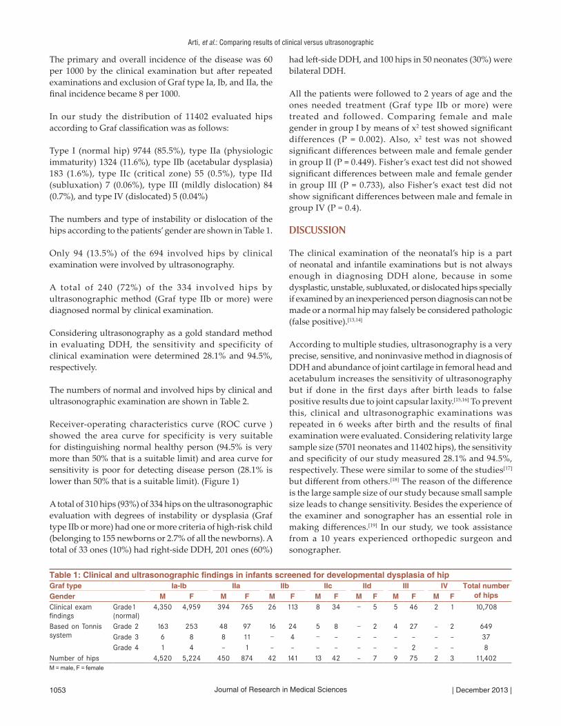
The primary and overall incidence of the disease was 60 per 1000 by the clinical examination but after repeated examinations and exclusion of Graf type Ia, Ib, and IIa, the final incidence became 8 per 1000.
In our study the distribution of 11402 evaluated hips according to Graf classification was as follows:
Type I (normal hip) 9744 (85.5%), type IIa (physiologic immaturity) 1324 (11.6%), type IIb (acetabular dysplasia) 183 (1.6%), type IIc (critical zone) 55 (0.5%), type IId (subluxation) 7 (0.06%), type III (mildly dislocation) 84 (0.7%), and type IV (dislocated) 5 (0.04%)
The numbers and type of instability or dislocation of the hips according to the patients’ gender are shown in Table 1.
Only 94 (13.5%) of the 694 involved hips by clinical examination were involved by ultrasonography.
A total of 240 (72%) of the 334 involved hips by ultrasonographic method (Graf type IIb or more) were diagnosed normal by clinical examination.
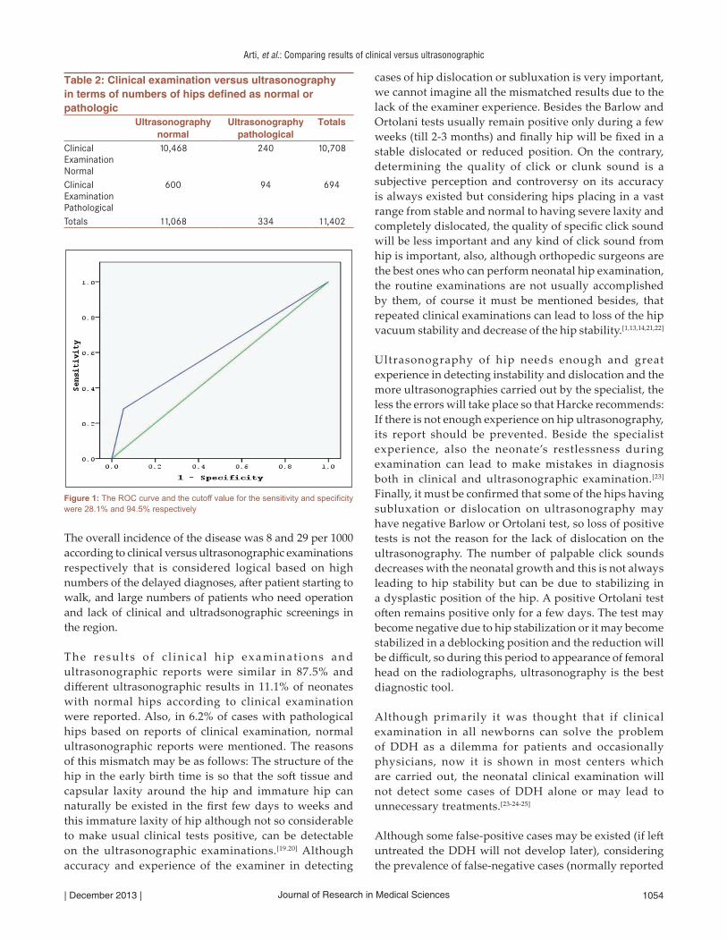
Considering ultrasonography as a gold standard method in evaluating DDH, the sensitivity and specificity of clinical examination were determined 28.1% and 94.5%, respectively.
The numbers of normal and involved hips by clinical and ultrasonographic examination are shown in Table 2.
Receiver-operating characteristics curve (ROC curve ) showed the area curve for specificity is very suitable for distinguishing normal healthy person (94.5% is very more than 50% that is a suitable limit) and area curve for sensitivity is poor for detecting disease person (28.1% is lower than 50% that is a suitable limit). (Figure 1)
A total of 310 hips (93%) of 334 hips on the ultrasonographic evaluation with degrees of instability or dysplasia (Graf type IIb or more) had one or more criteria of high-risk child (belonging to 155 newborns or 2.7% of all the newborns). A total of 33 ones (10%) had right-side DDH, 201 ones (60%)
Table 1: Clinical and ultrasonographic findings in infants screened for developmental dysplasia of hipGraf type Ia-Ib IIa IIb IIc IId III IV Total number
of hipsGender M F M F M F M F M F M F M FClinical exam findings
Grade1 (normal)
4,350 4,959 394 765 26 113 8 34 — 5 5 46 2 1 10,708
Based on Tonnis system
Grade 2 163 253 48 97 16 24 5 8 — 2 4 27 – 2 649Grade 3 6 8 8 11 — 4 — – – – – – – – 37Grade 4 1 4 – 1 – – – – – – – 2 – – 8
Number of hips 4,520 5,224 450 874 42 141 13 42 – 7 9 75 2 3 11,402M = male, F = female
Arti, et al.: Comparing results of clinical versus ultrasonographic
Journal of Research in Medical Sciences| December 2013 | 1054
The overall incidence of the disease was 8 and 29 per 1000 according to clinical versus ultrasonographic examinations respectively that is considered logical based on high numbers of the delayed diagnoses, after patient starting to walk, and large numbers of patients who need operation and lack of clinical and ultradsonographic screenings in the region.
The resul t s o f c l in ica l h ip examinat ions and ultrasonographic reports were similar in 87.5% and different ultrasonographic results in 11.1% of neonates with normal hips according to clinical examination were reported. Also, in 6.2% of cases with pathological hips based on reports of clinical examination, normal ultrasonographic reports were mentioned. The reasons of this mismatch may be as follows: The structure of the hip in the early birth time is so that the soft tissue and capsular laxity around the hip and immature hip can naturally be existed in the first few days to weeks and this immature laxity of hip although not so considerable to make usual clinical tests positive, can be detectable on the ultrasonographic examinations.[19.20] Although accuracy and experience of the examiner in detecting
cases of hip dislocation or subluxation is very important, we cannot imagine all the mismatched results due to the lack of the examiner experience. Besides the Barlow and Ortolani tests usually remain positive only during a few weeks (till 2-3 months) and finally hip will be fixed in a stable dislocated or reduced position. On the contrary, determining the quality of click or clunk sound is a subjective perception and controversy on its accuracy is always existed but considering hips placing in a vast range from stable and normal to having severe laxity and completely dislocated, the quality of specific click sound will be less important and any kind of click sound from hip is important, also, although orthopedic surgeons are the best ones who can perform neonatal hip examination, the routine examinations are not usually accomplished by them, of course it must be mentioned besides, that repeated clinical examinations can lead to loss of the hip vacuum stability and decrease of the hip stability.[1,13,14,21,22]
Ultrasonography of hip needs enough and great experience in detecting instability and dislocation and the more ultrasonographies carried out by the specialist, the less the errors will take place so that Harcke recommends: If there is not enough experience on hip ultrasonography, its report should be prevented. Beside the specialist experience, also the neonate’s restlessness during examination can lead to make mistakes in diagnosis both in clinical and ultrasonographic examination.[23] Finally, it must be confirmed that some of the hips having subluxation or dislocation on ultrasonography may have negative Barlow or Ortolani test, so loss of positive tests is not the reason for the lack of dislocation on the ultrasonography. The number of palpable click sounds decreases with the neonatal growth and this is not always leading to hip stability but can be due to stabilizing in a dysplastic position of the hip. A positive Ortolani test often remains positive only for a few days. The test may become negative due to hip stabilization or it may become stabilized in a deblocking position and the reduction will be difficult, so during this period to appearance of femoral head on the radiolographs, ultrasonography is the best diagnostic tool.
Although primarily it was thought that if clinical examination in all newborns can solve the problem of DDH as a dilemma for patients and occasionally physicians, now it is shown in most centers which are carried out, the neonatal clinical examination will not detect some cases of DDH alone or may lead to unnecessary treatments.[23-24-25]
Although some false-positive cases may be existed (if left untreated the DDH will not develop later), considering the prevalence of false-negative cases (normally reported
Figure 1: The ROC curve and the cutoff value for the sensitivity and specificity were 28.1% and 94.5% respectively
Table 2: Clinical examination versus ultrasonography in terms of numbers of hips defined as normal or pathologic
Ultrasonography normal
Ultrasonography pathological
Totals
Clinical Examination Normal
10,468 240 10,708
Clinical Examination Pathological
600 94 694
Totals 11,068 334 11,402
Arti, et al.: Comparing results of clinical versus ultrasonographic
Journal of Research in Medical Sciences | December 2013 |1055
on the screening but developed dislocation later) and the incidence of DDH in the study about 29 per 1000 newborns it can be recommended that the best, fastest, least expensive, and most useful method of DDH screening in developing countries is clinical examination by an experienced person. If possible, all the neonates or suspicious and with risk factors neonates should be reexamined by an orthopedic surgeon after primary examination by the obstetrician in the delivery ward or the midwifery or pediatrician.
Although now today for detecting of DDH a clinical sound or click or absent of it is not confirmatory, then ultrasonography should no longer be regarded as a screening method but should be used as an investigative and confirmatory tools in neonates to clarify and distinguish the diagnosis. Ideal screening test must be simple, reliable with high levels of sensitivity and specificity, and providing cost-effective results. Since these criteria are not fulfilled in cases of DDH, the screening term may be converted to surveillance as a more appropriate suggestive alternative word. On the basis of our data and relevant literature[26-27-28-29] and taking into consideration all the possible conditions related to DDH, we conclude that, along with the clinical examination to detect DDH, ultrasonography screening should be added to high-risk neonatal screening program.
acKnoWledGMents
We gratefully thanks to Mehdi Haghighat M.D. for performing meticulous ultrsonography of newborns in his private office and Hassan Shakerian M.D. for collecting these numerous data respectively, also appreciate the Vice Chancellor for research of Ahwaz Jundishapur, and Shahrekord University of Medical Sciences for supporting this research. This research is derived from thesis no. 443.
RefeRences
1. Herring JA. Developmental dysplasia of the hip: Tachdjian›s pediatric orthopaedics from the Texas Scottish Rite hospital for children. 4th ed. Philadelphia: Saunders Elsevier; 2008. p. 637-8.
2. Kokavec M, Bialik V. Developmental dysplasia of the hip. Prevention and real incidence. Bratisl Lek Listy 2007;108:251-4.
3. Ortolani M. Congenital hip dysplasia in the light of early and very early diagnosis. Clin Orthop Relat Res 1976:6-10.
4. Herring JA. Developmental Dysplasia of the Hip: Tachdjian’s Pediatric Orthopaedics From The Texas Scottish Rite Hospital For Children. 4th ed. Philadelphia: Saunders Elsevier; 2008. p. 639-41.
5. Paton RW, Hinduja K, Thomas CD. The significance of at-risk factors in ultrasound surveillance of developmental dysplasia of the hip. A ten-year prospective study. J Bone Joint Surg Br 2005;87B:1264-6.
6. Barlow TG. Early diagnosis and treatment of congenital dislocation of the hip. J Bone Joint Surg Br 1962;44:292-301.
7. Ortolani M. Un segno poco noto e sue importanza per la diagnosi precoce di preussasione congenita dell’anca. Pediatria 1937:129-36.
8. Graf R. Classification of hip joint dysplasia by means of sonography. Arch Orthop Trauma Surg 1984;102:248-52.
9. Graf R. Hip sonography: 20 years experience and results. Hip Int 2007;17 Suppl 5:S8-14.
10. Harcke HT. Screening newborns for developmental dysplasia of the hip: The role of sonography. AJR Am J Roentgenol 1994;162:395-7.
11. Tonnis D, Storch K, Ulbrich H. Results of newborn screening for CDH with and without sonography and correlation of risk factors. J Pediatr Orthop 1990;10:145-52.
12. Harcke HT, Grissom LE. Pediatric hip sonography: diagnosis and differential diagnosis. Radiol Clin North Am 1999; 37:787–796.
13. Holen KJ, Tegnander A, Bredland T, Johansen OJ, Sæther OD, Eik-Nes SH, et al. Universal or selective screening of the neonatal hip using ultrasound? A prospective, randomized trial of 15,529 newborn infants. J Bone Joint Surg Br 2002;84:886-91.
14. Synder M, Harcke HT, Domzalski M. Role of ultrasound in the diagnosis and management of developmental dysplasia of the hip: An international perspective. Orthop Clin North Am 2006;37:141-7.
15. Finnbogason T, Jorulf H, Söderman E, Rehnberg L. Neonatal hip instability: A prospective comparison of clinical examination and anterior dynamic ultrasound. Acta Radiol 2008;49:212-9.
16. Dogruel H, Atalar H, Yavuz OY, Sayli U. Clinical examination versus ultrasonography in detecting developmental dysplasia of the hip. Int Orthop 2008;32:415-9.
17. Koşar P, Ergun E, Ünlübay D, Koşar U. Comparison of morphologic and dynamic US methods in examination of the newborn hip. Diagn Interv Radiol 2009;15:284-9.
18. Woolacott NF, Puhan MA, Steurer J, Kleijnen J. Ultrasonography in screening for developmental dysplasia of the hip in newborns: Systematic review. BMJ 2005;18;330:1413.
19. Peled E, Eidelman M, Katzman A, Bialik V. Neonatal incidence of hip dysplasia: ten years of experience. Clin Orthop Relat Res 2008;466:771-5.
20. Rosenberg N, Bialik V. The effectiveness of combined clinical-sonographic screening in the treatment of neonatal hip instability. Eur J Ultrasound 2002;15:55-60.
21. Mahan ST, Katz JN, Kim YJ. To screen or not to screen? A decision analysis of the utility of screening for developmental dysplasia of the hip. J Bone Joint Surg Am 2009;91:1705-19.
22. Finne PH, Dalen I, Ikonomou N, Ulimoen G, Hansen TW. Diagnosis of congenital hip dysplasia in the newborn. Acta Orthop 2008;79:313-20.
23. Lee J. Developmental dysplasia of the hip: Universal or selective ultrasound screening? Ann Acad Med Singapore 2008;37(12 Suppl):101-3.
24. Köse N, Omeroğlu H, Ozyurt B, Akçar N, Ozçelik A, Inan U, Seber S. Our three-year experience with an ultrasonographic hip screening program conducted in infants at 3 to 4 weeks of age. Acta Orthop Traumatol Turc 2006;40:285-90.
25. Desprechins B, Ernst C, de Mey J. Screening for developmental dysplasia of the hip. JJBR-BTR 2007;90:4-5.
26. Toma P, Valle M, Rossi U, Brunenghi GM. Paediatric hip-ultrasound screening for developmental dysplasia of the hip: A review. Eur J Ultrasound 2001;4:45-55.
27. Zdravkovic N, Stojanovic S, Early Diagnosis of developmental hip dysplasia in the district of Pirot, Sebia, Facta Universitatis Series. Med Biology 2004;11:26-30.
28. Eastwood DM. Neonatal hip screening. Lancet 2003;361:595-7.29. Rosendahl, K. Toma P. Ultrasound in the diagnosis of
developmental dysplasia of the hip in newborns. The European approach. A review of methods, accuracy and clinical validity.European Radiology 2007;17(8), 960-1967.
Source of Support: Nil, Conflict of Interest: None declared.