dysci.webhosting.cals.wisc.edudysci.webhosting.cals.wisc.edu/.../uploads/sites/94/20… · Web...
9
Serotonin (5-HT) and calcium homeostasis during the transition period J. Laporta, S. A. E. Moore, S. R. Weaver, and L. L. Hernandez Department of Dairy Science University of Wisconsin-Madison Corresponding Author: [email protected] Summary 5-HT (5-hydroxytryptamine) is an autocrine/paracrine factor produced by the mammary gland The bovine mammary gland possesses five 5-HT receptor subtypes through which 5-HT can exert its actions 5-HT has been demonstrated to induce parathyroid hormone related-protein (PTHrP) expression in the mammary gland PTHrP is responsible for the bone mobilization that occurs during lactation Increased levels of 5-HT during the transition period increase osteoclast activity and circulating ionized calcium concentrations 5-HT increases mRNA expression of calcium pumps in the mammary gland Circulating 5-HT concentrations are positively correlated with ionized calcium on day 1 of lactation in dairy cattle Infusion of 5-HTP to lactating dairy cows significantly affects ionized calcium concentrations Introduction On the day of parturition, a dairy cow will produce 10 liters or more of colostrum containing at least 23 g of calcium (Goff, 2008). This challenges the ability of the cow to maintain adequate blood calcium levels at the onset of lactation. The requirement for maintenance of adequate calcium continues throughout lactation, but homeostatic balance is typically restored after a few days. The calcium demand required to support adequate milk synthesis drastically alters the bone and mineral metabolism of the animal. Periparturient hypocalcemia is one of the most common metabolic diseases of dairy cattle (Oetzel, 1988). Due to inadequate blood calcium concentrations at the onset of lactation, animals experience a range of clinical symptoms, depending on the extent of the decreased calcium levels (Adams et al., 1996). Clinical hypocalcemia is defined as a total blood calcium concentration of less than 1.4 mM, and subclinical hypocalcemia defined as total blood calcium concentration of 1.4 to 2.0 mM (DeGaris and Lean, 2009). Approximately 25% of primiparous and 50% of multiparous cows will succumb to subclinical hypocalcemia and between 5 to 10% of animals will develop clinical hypocalcemia (Goff, 2008; Reinhardt et al., 2011). Older cows have greater risk of developing hypocalcemia associated with a decrease in their capacity to mobilize calcium from the bone. In fact, the risk for milk fever increases by 9% per lactation (Lean et al., 2006). Unfortunately, the early symptoms (stage I) of milk fever often go undetected because they are short-lived. Identification of cows with subclinical hypocalcemia is not practical because the cows do not display evident clinical signs (Oetzel and Miller, 2012). Not until the animal begins to exhibit decreases in body temperature, incoordination when walking, and muscle trembling (stage II) do the signs of milk fever become increasingly noticeable to the producer (Adams et al., 1996). Incidences of subclinical and clinical hypocalcemia are long known to result in a variety of other disorders in dairy cattle during the transition period (Goff, 2008; Reinhardt et al., 2011). Particularly, cows with subclinical hypocalcemia have a higher risk of developing fever, metritis, retained fetal membranes, displaced abomasum, dystocia, ketosis, and to develop infectious diseases (Oetzel, 1988; Kimura et al., 2006;
Transcript of dysci.webhosting.cals.wisc.edudysci.webhosting.cals.wisc.edu/.../uploads/sites/94/20… · Web...
Serotonin (5-HT) and calcium homeostasis during the transition period
J. Laporta, S. A. E. Moore, S. R. Weaver, and L. L. HernandezDepartment of Dairy Science
University of Wisconsin-MadisonCorresponding Author: [email protected]
Summary 5-HT (5-hydroxytryptamine) is an autocrine/paracrine factor produced by the mammary gland The bovine mammary gland possesses five 5-HT receptor subtypes through which 5-HT can exert its actions 5-HT has been demonstrated to induce parathyroid hormone related-protein (PTHrP) expression in the mammary gland PTHrP is responsible for the bone mobilization that occurs during lactation Increased levels of 5-HT during the transition period increase osteoclast activity and circulating ionized calcium
concentrations 5-HT increases mRNA expression of calcium pumps in the mammary gland Circulating 5-HT concentrations are positively correlated with ionized calcium on day 1 of lactation in dairy cattle Infusion of 5-HTP to lactating dairy cows significantly affects ionized calcium concentrations
IntroductionOn the day of parturition, a dairy cow will produce 10 liters or more of colostrum containing at least 23 g of calcium (Goff,
2008). This challenges the ability of the cow to maintain adequate blood calcium levels at the onset of lactation. The requirement for maintenance of adequate calcium continues throughout lactation, but homeostatic balance is typically restored after a few days. The calcium demand required to support adequate milk synthesis drastically alters the bone and mineral metabolism of the animal. Periparturient hypocalcemia is one of the most common metabolic diseases of dairy cattle (Oetzel, 1988). Due to inadequate blood calcium concentrations at the onset of lactation, animals experience a range of clinical symptoms, depending on the extent of the decreased calcium levels (Adams et al., 1996). Clinical hypocalcemia is defined as a total blood calcium concentration of less than 1.4 mM, and subclinical hypocalcemia defined as total blood calcium concentration of 1.4 to 2.0 mM (DeGaris and Lean, 2009). Approximately 25% of primiparous and 50% of multiparous cows will succumb to subclinical hypocalcemia and between 5 to 10% of animals will develop clinical hypocalcemia (Goff, 2008; Reinhardt et al., 2011). Older cows have greater risk of developing hypocalcemia associated with a decrease in their capacity to mobilize calcium from the bone. In fact, the risk for milk fever increases by 9% per lactation (Lean et al., 2006). Unfortunately, the early symptoms (stage I) of milk fever often go undetected because they are short-lived. Identification of cows with subclinical hypocalcemia is not practical because the cows do not display evident clinical signs (Oetzel and Miller, 2012). Not until the animal begins to exhibit decreases in body temperature, incoordination when walking, and muscle trembling (stage II) do the signs of milk fever become increasingly noticeable to the producer (Adams et al., 1996).
Incidences of subclinical and clinical hypocalcemia are long known to result in a variety of other disorders in dairy cattle during the transition period (Goff, 2008; Reinhardt et al., 2011). Particularly, cows with subclinical hypocalcemia have a higher risk of developing fever, metritis, retained fetal membranes, displaced abomasum, dystocia, ketosis, and to develop infectious diseases (Oetzel, 1988; Kimura et al., 2006; Goff, 2008). Furthermore, cows with subclinical hypocalcemia also exhibit reduced feed intake and pregnancy rates, as well as longer intervals to pregnancy compared with normocalcemic cows (Martinez et al., 2012). The prevalence of milk fever and subclinical hypocalcemia is more common in older cows, which are less efficient in mobilizing calcium, and in Jersey cattle, likely due to their higher milk production per unit body weight, as well as increased calcium content of milk (Oetzel, 1988). On average, treatment of each incidence of clinical and subclinical hypocalcemia costs approximately $300 and $125, respectively (Guard, 1996). This estimate includes the direct cost of treating the animal, along with production losses, which are estimated to be approximately 14%.
Calcium Homeostasis During Lactation and the SkeletonApproximately 99% of calcium body reserves are stored in the bone tissue, and the main mechanism responsible for
maintaining maternal calcium homeostasis in the lactating dam is bone calcium mobilization. Therefore, bone resorption is the primary contributor of calcium in the milk of mammals, as opposed to increased calcium absorption in the intestine, and decreased calcium excretion by the kidney (Kovacs, 2011). Calcium mobilization from bone during lactation is stimulated by mammary secretion of parathyroid hormone-related protein (PTHrP) (Kovacs, 2011; Wysolmerski, 2012), which is present in human, rat, goat, and bovine milk and in the serum of the lactating mother. The skeletal system maintains structural and functional roles based on the communication between two cell types, osteoblasts (OB), which are responsible for bone formation, and osteoclasts (OC), which are responsible for bone resorption, and therefore calcium mobilization. Communication between these cell types can occur in three ways: (1) direct contact (juxtacrine), in which membrane-bound ligands and receptors interact in response to intracellular signaling; (2) gap junctions that facilitate passage of water-soluble molecules between the cell types; (3) paracrine communication through soluble secreted ligands (Matsuo and Irie, 2008). Initially, OB cells are responsible for production of precursors that are necessary for the differentiation and activation of OC (Asagiri and Takayanagi, 2007). Specifically, PTHrP, like parathyroid hormone (PTH), signals through a G-protein coupled receptor (GPCR; PTH1R), to decrease OB cell proliferation and up-regulate genes responsible for OC differentiation. In OB, PTHrP increases gene expression of receptor-activated nuclear factor kappa-B ligand (RANKL), matrix metalloproteinase 13 (MMP13), macrophage-colony stimulating factor (M-CSF), interleukin-6 (IL-6), and monocyte chemotactic

protein-1 (MCP1), which are known factors that activate OC differentiation (Matsuso and Irie, 2008; Datta and Abou-Samra, 2009). These factors produced by OB cells are then responsible for the induction of numerous genes including: metalloproteinase (MMP9), receptor-activated nuclear factor kappa-B (RANK), cathepsin K, and tartrate-resistant alkaline phosphatase (TRAP) in OC (Datta and Abou-Samra, 2009). The OC-induced genes and their associated proteins are directly related to the physical degradation of bone via their roles in initiation of collagen degradation and mineral dissolution. Thus, this PTHrP/OB-OC regulated transcriptional cascade provides the cellular cues to increase calcium mobilization from bone during the physiological conditions of lactation and the pathological condition of humoral hypercalcemia of malignancy (HHM).
Elevation of mammary PTHrP production is responsible for the bone mobilization typical during lactation (VanHouten, 2005; Wysolmerski, 2010). Initially, PTHrP was discovered as a factor responsible for the clinical syndrome humoral hypercalcemia of malignancy (HHM). HHM is a common metabolic complication that occurs in metastatic epithelial cancers that are osteolytic in nature (Stewart, 2002; Fiaschi-Taesch and Stewart, 2003; Stewart, 2005). Except in instances of malignancy, PTHrP is only detectable in the systemic circulation during lactation (Wysolmerksi, 2010). Additionally, mammary-synthesized PTHrP provides important roles in mammary gland development, development of the fetal mammary gland, and other autocrine-paracrine roles within the gland (Thiede, 1994; Wysolmerski et al., 1995; Philbrick et al., 1996; VanHouten et al., 2003; VanHouten et al., 2004). In rodent models, it has been demonstrated that mammary-specific deletion of PTHrP results in decreased concentrations of bone turnover markers (VanHouten et al., 2003). The decrease in bone turnover markers is accompanied by increased bone mass, but does not significantly impact milk calcium levels. Decreased levels of estrogen during this time can exacerbate PTHrP-mediated bone loss, however decreased estrogen cannot fully explain the effects of PTHrP on bone metabolism (Sasser et al., 1979; Ardenshipour et al., 2010).
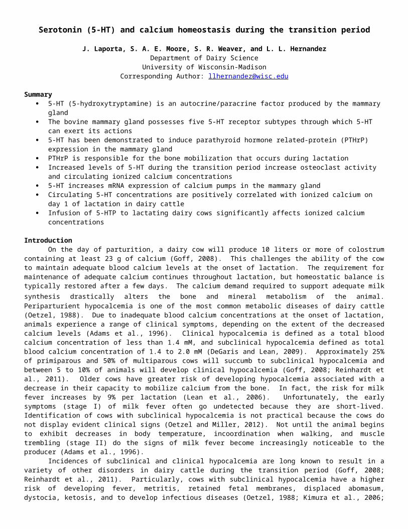
5-HT and Mammary Gland Function5-HT is a known homeostatic regulator of the mammary gland that is synthesized from the amino acid L-tryptophan in a two-
step process, with the production of 5-hydroxytryptophan (5-HTP) via the rate-limiting enzyme, tryptophan hydroxylase 1 (TPH1) in the first step, and 5-HTP conversion to 5-HT by aromatic amino acid decarboxylase in the second step (Figure 1; Wang et al., 2002; Matsuda et al., 2004; Stull et al., 2007; Hernandez et al., 2008; Hernandez et al., 2009).
Thus far, 5-HT has been demonstrated to regulate milk protein gene expression, as well as the disassembly of tight junctions that occurs during the involution process (Matsuda et al., 2004; Stull et al., 2007; Hernandez et al., 2008; Pai and Horseman, 2008). Furthermore, the
mammary glands of different species express unique 5-HT receptor patterns (Hernandez et al., 2009; Pai et al., 2009). To date, only the role of the 5-HT7 receptor in the mammary gland has been described in human, rodent and bovine mammary epithelial cells (Stull et al., 2007; Pai and Horseman, 2008). The 5-HT7 receptor and 5-HT2B receptors are expressed in the mammary glands of mice, humans, and cattle, whereas expression of other 5-HT receptor types varies among species (Hernandez et al., 2009; Pai et al., 2009; Hernandez et al., 2012). The 5-HT receptors are expressed from 14 unique genes and several alternatively spliced variants, and are
grouped into seven families. The type 3 receptors are ligand-gated ion channels, and all the other 5-HT receptors signal through various GPCR pathways (Raymond et al., 2001). Due to the complexity of the receptors associated with the functions of 5-HT in the mammary gland, the possibilities for additional roles (aside from the regulation of milk protein gene expression and tight junction regulation) are numerous. In fact, the bovine mammary gland expresses 5 receptor isoforms (5-HT1B, 2A, 2B, 4 and 7) within the epithelial component of the gland (Hernandez et al., 2009). However, aside from 5-HT7 the functions of these receptors in the mammary gland have not been reported.
In addition to functions of 5-HT in the mammary gland, various other physiological functions have been identified throughout the body. Several studies have shown that 5-HT decreased bone mass in laboratory animal and human models (Bliziotes et al., 2001; Yadav et al., 2008; Modder et al., 2010; Chabbi-Achengli et al., 2012). Patients administered selective 5-HT reuptake inhibitors (elevated 5-HT activity) typically exhibit decreases in bone density (Modder et al., 2010). Mice deficient for TPH-1 exhibit reduced osteoclastogenesis (Chabbi-Achengli et al., 2012). Additionally, lactating mice that are deficient for TPH-1 have demonstrated reduced mammary and
circulating PTHrP levels, and decreased osteoclast number and size, and increased bone strength (Hernandez et al., 2012; Laporta et al., unpublished results). Increased concentrations of the 5-HT2B receptor have been observed during OB cell differentiation (Collet et al., 2008). In a knockout mouse model for the 5-HT2B receptor, there was a significant reduction in bone mineral density in females exclusively (Collet et al., 2008). Additionally, OB isolated from these animals displayed markedly lower rates of proliferation in culture compared to wild type animals. Moreover, 5-HT2B may be responsible for the induction of PTHrP expression in the mammary gland (Hernandez et al., 2012; Laporta et al., 2013a,
Figure 2. 5-HT synthesized and secreted by mammary epithelial cells acts through the 5-HT2B receptor to induce PTHrP secretion from the mammary gland during lactation, which is a regulator of calcium mobilization from the bone during lactation to support the mammary gland demand for calcium needed for milk formation (Horseman and Hernandez, 2013).
Figure 1. 5-HT synthesis pathway
Laporta et al., unpublished results). The 5-HT2B receptor subtype is GPCR in the Gq/11 family. The primary mode of 5-HT2B action is activation of phospholipase C- (PLC-), which signals through inositol phosphate (IP3) and protein kinase C (PKC; Raymond et al., 2001).
Evidence that PTHrP is responsible for the calcium mobilization from bone that occurs during lactation, and the role of 5-HT in regulating bone formation, presents the strong possibility that 5-HT is involved in governing this mechanism during lactation. The following model for the regulation of calcium mobilization from bone during the transition period is proposed (Figure 2).
Manipulation of the Serotonergic System for the Management of Hypocalcemia in Dairy CattleThe long-term focus of our research is to determine the
effectiveness of utilizing the intermediate in 5-HT synthesis (5-hydroxy-L-tryptophan; 5-HTP) to manage subclinical and clinical hypocalcemia in dairy cattle. Our initial studies were conducted in a rat model, in which we supplemented 0.2% 5-HTP in the diet of pregnant rats beginning on d 13 of pregnancy through d 9 of lactation (Laporta et al., 2013a). Results of this research indicate that serum 5-HT and ionized calcium concentrations were elevated in rats receiving the diet enriched in 5-HTP compared to control fed dams (Figure 3). Additionally, we demonstrated increased plasma PTHrP concentrations on d 9 of lactation in the 5-HTP fed dams. Additionally, we saw increased production of 5-HT and PTHrP within the mammary gland and increased calcium in the milk on d 9 of lactation. Due to the observation that calcium was increase in the milk, we measured expression of various calcium pumps within the mammary gland and determined that their expression was significantly increased in 5-HTP supplemented dams. Upon examination of femurs collected from these dams we detected increases in OC number and size in the 5-HTP supplemented animals, along with increases in gene expression of several bone turnover markers (Laporta et al., 2013a). This
supports the concept that 5-HT is indeed involved in increasing bone turnover during the periparturient period and this modulated through increases in PTHrP.
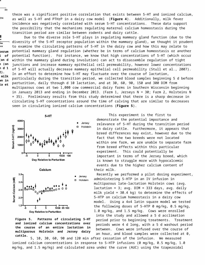
In order to elucidate whether this phenomenon was unique to rodents, we performed a preliminary study in dairy cattle. We obtained serum and plasma samples from multiparous Holstein cattle (N=42) on d 1 of lactation (within 24 h of calving) from within the University of Wisconsin Research Herd. We analyzed the samples collected for serum 5-HT and ionized calcium, and plasma PTHrP. Interestingly, we were able to determine that there was a significant positive correlation that exists between 5-HT and ionized calcium, as well as 5-HT and PTHrP in a dairy cow model (Figure 4). Additionally, milk fever incidence was negatively correlated with serum 5-HT concentrations. These data support the possibility that the mechanisms regulating maternal calcium homeostasis during the transition period are similar between rodents and dairy cattle.
Due to the diverse role 5-HT plays in regulating mammary gland function (due to the diversity of the 5-HT receptor population within the mammary gland), we thought it prudent to examine the circulating patterns of 5-HT in the dairy cow and how this may relate to potential mammary gland regulation (whether be in terms of calcium homeostasis or another potential function). For instance, we know that high concentrations of 5-HT (which occur within the mammary gland during involution) can act to disassemble regulation of tight junctions and increase mammary epithelial cell permeability, however lower concentrations of 5-HT will actually decrease mammary epithelial cell permeability (Stull et al., 2007). In an effort to determine how 5-HT may fluctuate over the course of lactation, particularly during the transition period, we collected blood samples beginning 5 d before parturition, daily through d 10 lactation, and at 30, 60, 90, 150 and 300 DIM from multiparous
Figure 3. Serum 5-HT and plasma PTHrP concentrations on d 1 and d 9 of lactation (A,B), mammary gland 5-HT and PTHrP protein concentration on d 9 lactation (C,D) and calcium concentrations on d 1 (E) and d 9 of lactation (F) in serum and milk of Sprague-Dawley rats fed control diet (n = 15) and diet enriched with 5-HTP (0.2%; n = 15). Data are represented as mean ± SEM. *P < 0.05, **P < 0.01, ***P < 0.001, ****P < 0.0001. 5-HT; PTHrP; TPH1. (Laporta et al., 2013a)
Figure 4. Spearman correlations between serum circulating 5-HT concentrations on d 1 of lactation with: (A) milk fever incidence (on d 1 of lactation), (B) serum ionized calcium (on d 1 of lactation), and (C) plasma PTHrP (on d 1 of lactation) in Holstein cattle (n=42). Correlation between serum calcium and milk fever incidence on d 1 of lactation is shown in (D). Milk fever incidence (yes/no) (Laporta et al., 2013b)

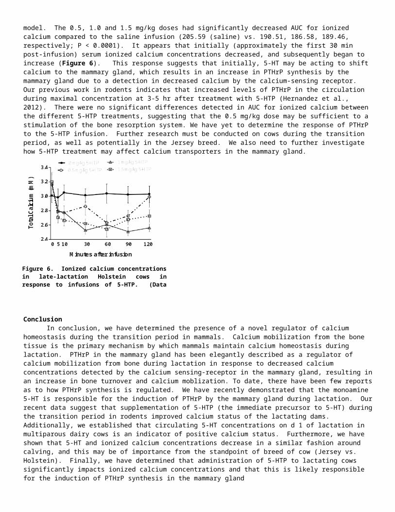
cows at two 1,000 cow commercial dairy farms in Southern Wisconsin beginning in January 2013 and ending in December 2013. (Farm 1, Jerseys N = 30; Farm 2, Holsteins N = 35). Preliminary results from this study determined that there is a sharp decrease in circulating 5-HT concentrations around the time of calving that are similar to decreases seen in circulating ionized calcium concentrations (Figure 5).
This experiment is the first to demonstrate the potential importance and relevance of 5-HT during the transition period in dairy cattle. Furthermore, it appears that breed differences may exist, however due to the fact that the two breeds were not located within one farm, we are unable to separate farm from breed effects within this particular experiment. This could potentially be important in terms of the Jersey breed, which is known to struggle more with hypocalcemic events due to the higher calcium content of their milk.Recently we performed a pilot dosing experiment, administering 5-HTP in an IV infusion in multiparous late-lactation Holstein cows (avg. lactation = 3; avg. DIM = 333 days, avg. daily milk yield = 30.4 kg) to determine the effects of 5-HTP on calcium homeostasis in a dairy cow model. Using a 4x4 latin square model we tested the following doses of 5-HTP 0 mg/kg, 0.5 mg/kg, 1.0 mg/kg, and 1.5 mg/kg. Cows were
enrolled into the study and allowed a 5 d acclimation period prior to beginning treatments. Treatment periods were 4 d long, with a 5 d washout period between. Cows were infused over the course of an hour, and blood samples were collected at 0, 5, 10, 30, 60, 90 and 120 min after cessation of the infusion. We measured ionized calcium concentrations in response to 5-HTP infusions (0 mg/kg, 0.5 mg/kg, 1.0 mg/kg, and 1.5 mg/kg) and calculated area under the curve (AUC) using the trapezoidal model. The 0.5, 1.0 and 1.5 mg/kg doses had significantly decreased AUC for ionized calcium compared to the saline infusion (205.59 (saline) vs. 190.51, 186.58, 189.46, respectively; P < 0.0001). It appears that initially (approximately the first 30 min post-infusion) serum ionized calcium concentrations decreased, and subsequently began to increase
(Figure 6). This response suggests that initially, 5-HT may be acting to shift calcium to the mammary gland, which results in an increase in PTHrP synthesis by the mammary gland due to a detection in decreased calcium by the calcium-sensing receptor. Our previous work in rodents indicates that increased levels of PTHrP in the circulation during maximal concentration at 3-5 hr after treatment with 5-HTP (Hernandez
et al., 2012). There were no significant differences detected in AUC for ionized calcium between the different 5-HTP treatments, suggesting that the 0.5 mg/kg dose may be sufficient to a stimulation of the bone resorption system. We have yet to determine the response of PTHrP to the 5-HTP infusion. Further research must be conducted on cows during the transition period, as well as potentially in the Jersey breed. We also need to further investigate how 5-HTP treatment may affect calcium transporters in the mammary gland.
Figure 6. Ionized calcium concentrations in late-lactation Holstein cows in response to infusions of 5-HTP. (Data unpublished).
Figure 5. Patterns of circulating 5-HT and ionized calcium concentrations over the course of an entire lactation in multiparous Holstein and Jersey dairy cattle.
ConclusionIn conclusion, we have determined the presence of a novel regulator of calcium homeostasis during the transition period in
mammals. Calcium mobilization from the bone tissue is the primary mechanism by which mammals maintain calcium homeostasis during lactation. PTHrP in the mammary gland has been elegantly described as a regulator of calcium mobilization from bone during lactation in response to decreased calcium concentrations detected by the calcium sensing-receptor in the mammary gland, resulting in an increase in bone turnover and calcium moblization. To date, there have been few reports as to how PTHrP synthesis is regulated. We have recently demonstrated that the monoamine 5-HT is responsible for the induction of PTHrP by the mammary gland during lactation. Our recent data suggest that supplementation of 5-HTP (the immediate precursor to 5-HT) during the transition period in rodents improved calcium status of the lactating dams. Additionally, we established that circulating 5-HT concentrations on d 1 of lactation in multiparous dairy cows is an indicator of positive calcium status. Furthermore, we have shown that 5-HT and ionized calcium concentrations decrease in a similar fashion around calving, and this may be of importance from the standpoint of breed of cow (Jersey vs. Holstein). Finally, we have determined that administration of 5-HTP to lactating cows significantly impacts ionized calcium concentrations and that this is likely responsible for the induction of PTHrP synthesis in the mammary gland
Literature Cited1. Adams, R., V. Ishler, and D. Moore. 1996. Trouble-shooting milk fever and downer cow problems. DAS 96-27. IVE1f.
PENpages 2890216: 1-7.2. Ardenshipour, L., S. Brian, P. Dann, J. VanHouten, and J. Wysolmerski. 2010. Increased PTHrP and decreased estrogens
alter bone turnover but do not reproduce the full effects of lactation on the skeleton. Endocrinology. 151(12):5591-5601.3. Asagiri, M., and H. Takayanagi. 2007. The molecular understanding of osteoclast differentiation. Bone. 40(2):251-264.4. Bliziotes, M. M., A. J. Eshleman, X.-W. Zhang, and K. M. Wiren. 2001. Neurotransmitter action in osteoblasts: expression
of a functional system for serotonin receptor activation and reuptake. Bone. 29(5):477-486.5. Chabbi-Achengli, Y., A. E. Coudert, J. Callebert, V. Geoffroy, F. Cote, C. Collet, and M.C. de Vernejoul. 2012. Decreased
osteoclastogenesis in serotonin-deficient mice. Proc. Acad. Natl. Sci. USA. 109(7):2567-2572.6. Collet, C., C. Schiltz, V. Geoffrey, L. Maroteaux, J.-M. Launay, and M.-C. de Vernejoul. 2008. The serotonin 5-HT2B
receptor controls bone mass via osteoblast recruitment and proliferation. FASEB J. 22:418-427.7. Datta, N. S., and A. B. Abou-Samra. 2009. PTH and PTHrP signaling in osteoblasts. Cell Signal. 21(8): 1245-1254.8. DeGaris, P. J., and I. J. Lean. 2009. Milk fever in dairy cows: a review of pathophysiology and control principles. Vet. J.
176:58-69.9. Fiaschi-Taesch, N. M., and A. F. Stewart. 2003. Minireview: parathyroid hormone related-protein as an intracrine factor—
trafficking mechanisms and functional consequences. Endocrinology. 144(2): 407-411.10. Goff, J. P. 2008. The monitoring, prevention, and treatment of milk fever and subclinical hypocalcemia in dairy cows. Vet.
J. 176:50-57.11. Guard, C. 1996. Fresh cow problems are costly: culling hurts the most. Page 100 in Proc. 1994 Annu. Conf. Vet., Cornell
Univ., Ithaca, NY.12. Hernandez, L. L., C. M. Stiening, J. B. Wheelock, L. H. Baumgard, A. M. Parkhurst, and R. J. Collier. 2008. Evaluation of
serotonin as a feedback inhibitor of lactation in the bovine. J. Dairy Sci. 91:1834-1844.13. Hernandez, L. L., S. W. Limesand, J. L. Collier, N. D. Horseman, and R. J. Collier. 2009. The bovine mammary gland
expressed multiple functional isoforms of serotonin receptors. J. Endocrinol. 203:123-131.14. Hernandez, L. L., K. A. Gregerson, and N. D. Horseman. 2012. Mammary gland serotonin regulates parathyroid hormone-
related protein and other bone-related signals. Am. J. Physiol. Endocrinol. Metab. 302(8):E1009-1015. 15. Horseman, N. D., and L. L. Hernandez. 2013. New concepts of breast cell communication to bone. Trends Endocrinol.
Metab. Sep 18 epub ahead of print.16. Kimura, K., T. A. Reindhardt, and J. P. Goff. 2006. Parturition and hypocalcemia blunts calcium signals in immune cells of
dairy cattle. J. Dairy Sci. 89:2588-2595.17. Kovacs, C. S. 2011. Calcium and bone metabolism disorders during pregnancy and lactation. Endocrinol. Metab. Cli. North
Am. 40(4):795-826.18. Laporta, J., T. L. Peters, S. R. Weaver, K. E. Merriman, and L. L. Hernandez. 2013a. Feeding 5-hydroxy-L-tryptophan
during the transition from pregnancy to lactation increases calcium mobilization from bone in rats. Dom. Anim. Endocrinol. 44(4):176-184.
19. Laporta, J. S. A. Moore, M. W. Peters, T. L. Peters, and L. L. Hernandez. 2013b. Short communication: Circulating serotonin (5-HT) concentrations on day 1 of lactation as a potential predictor of transition-related disorders. 96(8):5146-5150.
20. Lean, I. J., P. J. DeGaris, D. M. McNeil, and E. Block. 2006. Hypocalcemia in dairy cows: meta-analysis and dietary cation anion difference theory revisited. J. Dairy Sci. 89(20):669-684.
21. Martinez, N., C. A. Risco, F. S. Lima, R. S. Bisinooto, L. F. Greco, E. S. Ribeiro, F. Maunsell, K. Galvao, and J. E. P. Santos. 2012. Evaluation of peripartal calcium status, energetic profile, and neutrophil function in dairy cows at low or high risk of developing uterine diseases. J. Dairy Sci. 95:7158-7172.
22. Matsuda, M., T. Imaoka, A. J. Vomachka, G. A. Gudelsky, Z. Hou, M. Mistry, J. P. Bailey, K. M. Nieport, D. J. Walther, M. Bader, and N. D. Horseman. 2004. Serotonin regulated mammary gland development via an autocrine-paracrine loop. Dev. Cell. 6:193-203.
23. Matsuo, K., and N. Irie. 2008. Osteoclast-osteoblast communication. Arch. Biochem. Biophys. 473(2):201-209.
24. Modder, U. I., S. J. Achenbach, S. Amin, B. L. Riggs, L. J. Melton III, and S. Khosla. 2010. Relation of serum serotonin levels to bone density and structural parameters in women. J. Bone Mineral Res. 25(2):415-422.
25. Oetzel, G. R. 1988. Parturient paresis and hypocalcemia in ruminant livestock. Vet. Clin. North Am. Food Anim. Pract. 4(2):351-364.
26. Oetzel, G. R., and B. E. Miller. 2012. Effect of oral calcium bolus supplementation on early-lactation health and milk yield in commercial dairy herds. J. Dairy Sci. 95:7051-7065.
27. Pai, V. P., and N. D. Horseman. 2008. Biphasic regulation of mammary epithelial resistance by serotonin through activation of multiple pathways. J. Biol. Chem. 283(45):30901-30910.
28. Pai, V. P., A. M. Marshall, L. L. Hernandez, A. R. Buckley, and N. D. Horseman. 2009. Altered serotonin physiology in human breast cancers favors paradoxical growth and cell survival. Breast Cancer Res. 11(6):R81.
29. Philbrick, W. M., J. J. Wysolmerski, S. Galbraith, E. Holt, J. J. Orloff, K. H. Yang, R. C. Vasavada, E. C. Weir, A. E. Broadus, and A. F. Stewart. 1996. Defining the role of parathyroid hormone-related protein in normal physiology. Physiol. Rev. 76(1): 127-173.
30. Raymond, J. R., Y. V. Mukhin, A. Gelasco, J. Turner, G. Collinsworth, T. W. Gettys, J. S. Grewal, and M. N. Garnovskaya. 2001. Multiplicity of mechanisms of serotonin receptor signal transduction. Pharmacol. Therap. 92:179-212.
31. Reinhardt, T. A., J. D. Lippolis, B. J. McCluskey, J. P. Goff, and R. L. Horst. 2011. Prevalence of subclinical hypocalcemia in dairy herds. Vet. J. 188:122-124.
32. Sasser, R. G., D. E. Falk, and R. H. Ross. 1979. Estrogen in plasma of parturient paretic and normal cows. J. Dairy Sci. 62:551-556.
33. Stewart, A. F. 2002. Hyperparathyroidism, humoral hypercalcemia of malignancy, and the anabolic actions of parathyroid hormone and parathyroid hormone related-protein on the skeleton. J. Bone Miner. Res. 17(5):758-762.
34. Stewart, A. F. 2005. Clinical practice. Hypercalcemia associated with cancer. N. Eng. J. Med. 352(4):373-379.35. Stull, M. A., V. Pai, A. J. Vomachka, A. M. Marshall, G. A. Jacob, and N. D. Horseman. 2007. Mammary gland
homeostasis employs serotonergic regulation of epithelial tight junctions. Proc. Natl. Acad. Sci. 104(42):16708-16713.36. Thiede, M. A. 1994. Parathyroid hormone-related protein: a regulated calcium-mobilizing product of the mammary gland.
J. Dairy Sci. 77:1952-1963.37. VanHouten, J. N., P. Dann, A. F. Stewart, C. J. Watson, M. Pollak, A. C. Karaplis, and J. J. Wysolmerski. 2003. Mammary-
specific deletion of parathyroid hormone-related protein preserves bone mass during lactation. J. Clin. Invest. 112(9):1429-1436.
38. VanHouten, J. N., P. Dann, G. McGeoch, E. M. Brown, K. Krapcho, M. Neville, and J. J. Wysolmerski. 2004. The calcium-sensing receptor regulates mammary gland parathyroid hormone-related protein production and calcium transport. J. Clin. Invest. 113:598-608.
39. VanHouten, J. N. 2005. Calcium sensing by the mammary gland. J. Mammary Gland Bio. Neoplasia. 10(2):129-139.40. Wang, L. H. Erlandsen, J. Haavik, P. M. Knappskog, and R. C. Stevens. 2002. Three-dimensional structure of human
tryptophan hydroxylase and its implications for the biosynthesis of the neurotransmitters serotonin and melatonin. Biochemistry. 41(42):12569-12574.
41. Wysolmerski, J. J., J. F. McCaughern-Carucci, A. G. Daifotis, A. E. Broadus, and W. M. Philbrick. 1995. Overexpression of parathyroid hormone-related protein or parathyroid hormone in transgenic mice impairs branching morphogenesis during mammary gland development. Development. 121: 3539-3547.
42. Wysolmerski, J. J. 2010. Interactions between breast, bone, and brain regulate mineral and skeletal metabolism during lactation. Ann. N. Y. Acad. Sci. 1192:161-169.
43. Wysolmerski, J. J. 2012. Parthyroid hormone-related protein: an update. J. Clin. Endocrinol. Metab. 97(9):2947-2956.44. Yadav, V. K., J. H. Ryu, N. Suda, K. F. Tanaka, J. A. Gringrich, G. Schutz, F. H. Glorieux, C. Y. Chiang, J. D. Zajac, K. L.
Insogna, J. J. Mann, R. Hen, P. Ducy, and G. Karsenty. 2008. Lrp5 controls bone formation by inhibiting serotonin synthesis in the duodenum. Cell. 135(5):825-837.