OBJECTIVE: Identify Different Interactions among species Interactions.

Dynamic PIP2 interactions with voltage sensorelements contribute to KCNQ2 channel gatingQiansen Zhanga,1, Pingzheng Zhoub,1, Zhuxi Chena, Min Lib,c, Hualiang Jianga, Zhaobing Gaob,2, and Huaiyu Yanga,2
aDrug Discovery and Design Center, State Key Laboratory of Drug Research and bChinese Academy of Sciences Key Laboratory of Receptor Research, ShanghaiInstitute of Materia Medica, Chinese Academy of Sciences, Shanghai 201203, China; and cThe Solomon H. Snyder Department of Neuroscience, HighThroughput Biology Center and Johns Hopkins Ion Channel Center, School of Medicine, Johns Hopkins University, Baltimore, MD 21205
Edited by Michael L. Klein, Temple University, Philadelphia, PA, and approved November 4, 2013 (received for review July 1, 2013)
The S4 segment and the S4–S5 linker of voltage-gated potassium(Kv) channels are crucial for voltage sensing. Previous studies on theShaker and Kv1.2 channels have shown that phosphatidylinositol-4,5-bisphosphate (PIP2) exerts opposing effects on Kv channels, up-regulating the current amplitude, while decreasing the voltage sen-sitivity. Interactions between PIP2 and the S4 segment or the S4–S5linker in the closed state have been highlighted to explain the effectsof PIP2 on voltage sensitivity. Here, we show that PIP2 preferentiallyinteracts with the S4–S5 linker in the open-state KCNQ2 (Kv7.2) chan-nel, whereas it contacts the S2–S3 loop in the closed state. Theseinteractions are different from the PIP2–Shaker and PIP2–Kv1.2 inter-actions. Consistently, PIP2 exerts different effects on KCNQ2 relativeto the Shaker and Kv1.2 channels; PIP2 up-regulates both the currentamplitude and voltage sensitivity of the KCNQ2 channel. Disruptionof the interaction of PIP2 with the S4–S5 linker by a single mutationdecreases the voltage sensitivity and current amplitude, whereas dis-ruption of the interaction with the S2–S3 loop does not alter voltagesensitivity. These results provide insight into the mechanism of PIP2action on KCNQ channels. In the closed state, PIP2 is anchored at theS2–S3 loop; upon channel activation, PIP2 interacts with the S4–S5linker and is involved in channel gating.
lipid | membrane | membrane protein | M current
Aseries of ion channels, such as inward rectifier K+ (Kir)channels, transient receptor potential channels, and voltage-
gated channels, are sensitive to the presence of phosphatidyli-nositol-4,5-bisphosphate (PIP2) in membranes (1–4). Structuralstudies on Kir channels (1, 2, 5) demonstrated that PIP2 directlyinteracts with the channels. Subsequent studies supported thatPIP2 also interacts directly with voltage-gated potassium (Kv)channels (6–19). Several positive residues that may be criticalfor PIP2 activity have been identified (7, 11, 18, 20–24). Previousstudies on Kv1.2 and Shaker channels showed that PIP2 exertsopposing effects on Kv channels, up-regulating the current am-plitude, while leading to a decrease in voltage sensitivity (7, 18).The S4 segment and the S4–S5 linker of Kv channels are crucialfor voltage sensing. The interactions of PIP2 with the S4 seg-ments and the S4–S5 linkers of the closed-state Shaker andKv1.2 channels underlie the loss-of-function effect of PIP2 onvoltage sensitivity (7, 18).The KCNQ (Kv7) family of slowly activated outwardly recti-
fying potassium channels is one of the Kv channel families thatare sensitive to the presence of PIP2 in the membrane. KCNQchannels have been widely studied because of their importantbiological and pharmacological functions. Retigabine, a first-in-class K+ channel opener used for the treatment of epilepsy,adopts a unique mechanism to enhance the activity of KCNQchannels (25). PIP2 is important for the functions of KCNQchannels. Reduction of PIP2 affinity caused by congenic muta-tions of KCNQ channels is associated with long QT syndrome,suggesting critical physiological implications of PIP2 onKCNQ channels (23, 26). We reported that PIP2 also altersthe pharmacological selectivity of KCNQ potassium channels(6). Zaydman et al. (27) showed that the coupling of voltage
sensing and pore opening in the KCNQ1 channel requiresPIP2 and suggested there is a PIP2 interaction site at the in-terface between the voltage-sensing domain (VSD) and thecentral pore domain (PD). However, the effects and inter-actions of PIP2 on KCNQ channels are not well understood.Here, by combining molecular dynamics (MD) simulations,
mutagenesis, and electrophysiological determinations, we observedthat the effects and interactions of PIP2 on KCNQ2 are differentrelative to the Shaker and Kv1.2 channels. PIP2 up-regulates boththe current amplitude and voltage sensitivity of the KCNQ2channel. PIP2 preferentially interacts with the S4–S5 linker of theopen-state KCNQ2 channel and does not interact with the S4segment or S4-S5 linker of the closed state. In the closed state,PIP2 only interacts with the S2–S3 loop. Furthermore, our elec-trophysiological experiments suggest that disruption of the in-teraction of PIP2 with the S4–S5 linker may decrease the voltagesensitivity and current amplitude, whereas disruption of the in-teraction with the S2–S3 loop only alters the current amplitude ofthe channel. These results provide insights into the mechanism ofPIP2 action on Kv channels.
ResultsAgonistic Effects of PIP2 on the Voltage Sensitivity of the KCNQ2Channel. Depletion of PIP2 suppresses the current of the KCNQ2channel (6, 11, 19). However, the effects of PIP2 on the voltagesensitivity of the KCNQ2 channel remain unclear. Here, the es-sential roles of PIP2 in KCNQ2 channel activity were examined and
Significance
Many membrane protein functions require phosphatidylinosi-tol-4,5-bisphosphate (PIP2), a minor phospholipid in the innerleaflet of plasma membranes; however the underlying mech-anisms have not been well elucidated. Here, we show that PIP2might differentially regulate the activities of highly homolo-gous proteins. The voltage-gated potassium (Kv) channelsShaker, Kv1.2 and KCNQ2 have similar structural arrangementsand voltage sensing mechanisms. Previous studies showed thatPIP2 decreases the voltage sensitivity of the Shaker and Kv1.2channels; we observed that PIP2 up-regulates the voltagesensitivity of the KCNQ2 channel. The differential interactionsof PIP2 with these channels contribute to the diversity of PIP2regulations. Our data suggest that the effects of PIP2 and itsinteractions with membrane proteins should be studied at afiner scale.
Author contributions: Z.G. and H.Y. designed research; Q.Z., P.Z., Z.G., and H.Y. performedresearch; Q.Z., P.Z., Z.C., M.L., H.J., Z.G., and H.Y. analyzed data; and Q.Z., P.Z., Z.G., and H.Y.wrote the paper.
The authors declare no conflict of interest.
This article is a PNAS Direct Submission.1Q.Z. and P.Z. contributed equally to this work.2To whom correspondence may be addressed. E-mail: [email protected] or [email protected].
This article contains supporting information online at www.pnas.org/lookup/suppl/doi:10.1073/pnas.1312483110/-/DCSupplemental.
www.pnas.org/cgi/doi/10.1073/pnas.1312483110 PNAS | December 10, 2013 | vol. 110 | no. 50 | 20093–20098
BIOPH
YSICSAND
COMPU
TATIONALBIOLO
GY

validated in CHO–K1 cells. We first cotransfected KCNQ2 andMusicrinic receptor 1 (M1) in the cells, where PIP2 is reducedthrough the phospholipase C (PLC)-mediated lipid hydrolysis (3,14). Consistent with previous reports (6, 11, 19), we observed thatactivation of the M1 receptor by Oxo-M (5 μM) induces a dramaticsuppression of the current amplitude (Fig. 1A). Next, we decreasedthe PIP2 concentration using a voltage-sensitive phosphatase fromDanio rerio (Dr–VSP), which hydrolyzes PIP2 at highly depolarizedvoltages (e.g., +120 mV) and transiently reduces the PIP2 level(28). In the cells cotransfected with KCNQ2 and Dr–VSP, thecurrent is significantly reduced upon +120 mV depolarization (Fig.1B). The closing of the KCNQ2 channel in response to decreasedPIP2 concentration is consistent with previous observations (6). TheM1 and Dr–VSP experiments support the positive effects of PIP2on current amplitude. However, because the depletion of PIP2 byboth methods completely abolishes the function of the KCNQ2channels, the influences of PIP2 on voltage sensitivity could not beanalyzed. Therefore, we evaluated the effects of increased PIP2levels on the KCNQ2 channel. A phosphatidylinositol-5-kinase(PI5-K) was coexpressed with the KCNQ2 channel. Overexpressionof PI5-K can elevate the PIP2 membrane concentration to milli-molar levels (17). When coexpressed with PI5-K, the current am-plitude of the KCNQ2 channel at +50 mV (saturated voltagepotential) is significantly higher than the control (Fig. 1 C andE). Noticeably, the conductance-voltage (G-V ) curve is signifi-cantly left-shifted (Fig. 1D). Therefore, unlike the PIP2-mediateddecrease in the voltage sensitivity of the Shaker and Kv1.2channels, we observed that PIP2 up-regulates both the currentamplitude and voltage sensitivity of the KCNQ2 channel.
Interactions of PIP2 with the Open and Closed States of KCNQ2.Elucidation of the interactions between PIP2 and KCNQ2 chan-nels would facilitate the understanding of PIP2 regulation of thesechannels. Previous studies of the Shaker and Kv1.2 channels haveidentified the interactions of PIP2 with the S4–S5 linker of theclosed-state channels; the role of PIP2 in controlling the stability ofthe voltage sensor in the closed state could explain the loss-of-function effects of PIP2 on voltage sensitivity of the channels (7, 18).To predict the interactions of PIP2 with the KCNQ2 channel, weperformed all-atom MD simulations on both the open and closed
conformations of the KCNQ2 channel in the presence of PIP2molecules in the palmitoyloleoyl phosphatidylcholine (POPC) bi-layer. Simulations were performed using the program GROMACS4.6.1 (29). Simulations with the CHARMM36 force field (30) andthe CHARMM27 force field (31) yielded similar PIP2 interactions(Figs. 2 and 3 and Fig. S1), indicating the reproducibility of the MDsimulation results. For simplicity, only the simulations with theCHARMM36 force field are discussed in detail below.Similar to other Kv channels, the transmembrane (TM) do-
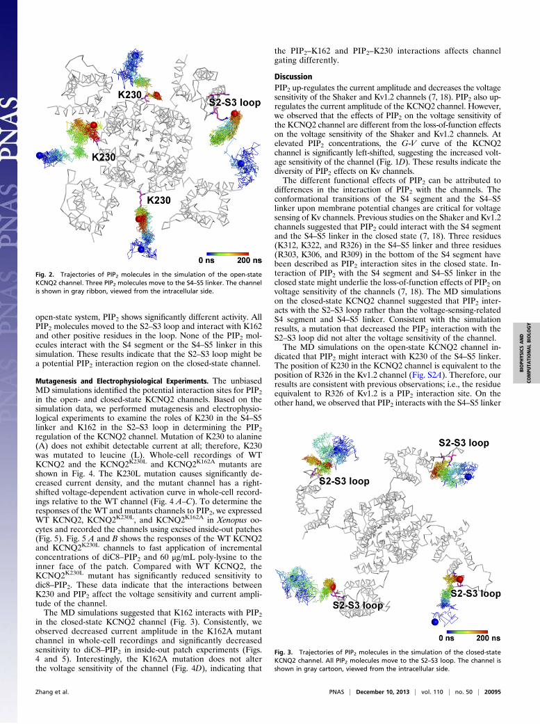
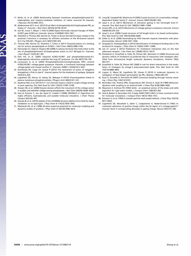
main of the KCNQ channel can be divided into three parts: theS1–S4 segments forming the VSD, a canonical PD (S5–P–S6),and the S4–S5 linker, which couples the movement of the VSDto the opening or closing of the PD. In addition to the TM do-main, positive residues in the cytoplasmic C terminus just afterthe end of S6 (referred to as the “CCT region”) are believed tocontribute to PIP2 sensitivity (3, 11, 20, 21, 24, 27). We modeledthe TM domain (residues 95–312) and CCT region (residues313–337) for the open-state KCNQ2 channel based on thecrystal structures of Kv1.2 (PDB code 2A79) (32) and an acti-vated KcsA K+ channel (PDB code 3PJS) (33). The TM domainof the closed-state KCNQ2 was modeled based on the closed-state conformation of the Kv1.2/Kv2.1 chimera channel reportedby Jensen et al. (34), and the CCT region was modeled based onthe crystal structure of the closed KcsA channel (PDB code3EFF) (35). We used various methods to test the reliability of themodeled structures (Figs. S2–S4) (SI Materials and Methodsprovides details). The structure of each state was simulated ina POPC bilayer in the presence of four PIP2 molecules (Fig. S5).In the initial simulation systems, the PIP2 molecules were placedin the inner leaflet of the bilayer, far from the channel. Theclosest distance between PIP2 and the channel was more than15 Å. Each system was subjected to a 200-ns MD simulation.Fig. 2 shows the diffusion trajectories of the four PIP2 mole-
cules in the simulation of the open-state KCNQ2 channel. ThreePIP2 molecules diffused to the S4–S5 linker, interacting withK230 at the linker. Statistically, the S4–S5 linker may be a pu-tative PIP2 interaction site with relatively higher potency. Fig. 3shows the diffusion trajectories of the four PIP2 molecules in thesimulation of the closed state. Although the initial positions ofthe PIP2 molecules in this system are similar to those in the
Fig. 1. Effects of manipulating PIP2 concentrations by M1 receptors, Dr–VSP, and PI5-K on KCNQ2 channels. (A) Representative traces and time course ofKCNQ channel currents in CHO–K1 cells transfected with KCNQ2 and the M1 receptors. Oxo-M at 5 μM suppresses the KCNQ2 channel current rapidly;representative traces are shown as indicated. (B) Representative traces and time course of KCNQ2 channel current in the cells cotransfected with KCNQ2 andDr–VSP. Protocol shown was used to elicit KCNQ2 channel currents and activate Dr–VSP. Depolarization to +120 mV caused a dramatic decrease of KCNQ2channel currents. (C) Representative traces of KCNQ2 channels in cells without (Left) and with (Right) cotransfection with PI5-K. The holding potential is −80mV, followed by a series of depolarization steps from −70 to +50 mV in 10-mV increments, followed by a 1000-ms hyperpolarization step to −120 mV torecord the tail current. (D) Voltage activation curves of the KCNQ2 channels in cells without and with cotransfection with PI5-K as indicated. (E) Histogramsummarizing the current densities of KCNQ2 channels when PIP2 concentrations were manipulated as indicated.
20094 | www.pnas.org/cgi/doi/10.1073/pnas.1312483110 Zhang et al.
open-state system, PIP2 shows significantly different activity. AllPIP2 molecules moved to the S2–S3 loop and interact with K162and other positive residues in the loop. None of the PIP2 mol-ecules interact with the S4 segment or the S4–S5 linker in thissimulation. These results indicate that the S2–S3 loop might bea potential PIP2 interaction region on the closed-state channel.
Mutagenesis and Electrophysiological Experiments. The unbiasedMD simulations identified the potential interaction sites for PIP2in the open- and closed-state KCNQ2 channels. Based on thesimulation data, we performed mutagenesis and electrophysio-logical experiments to examine the roles of K230 in the S4–S5linker and K162 in the S2–S3 loop in determining the PIP2regulation of the KCNQ2 channel. Mutation of K230 to alanine(A) does not exhibit detectable current at all; therefore, K230was mutated to leucine (L). Whole-cell recordings of WTKCNQ2 and the KCNQ2K230L and KCNQ2K162A mutants areshown in Fig. 4. The K230L mutation causes significantly de-creased current density, and the mutant channel has a right-shifted voltage-dependent activation curve in whole-cell record-ings relative to the WT channel (Fig. 4 A–C). To determine theresponses of the WT and mutants channels to PIP2, we expressedWT KCNQ2, KCNQ2K230L, and KCNQ2K162A in Xenopus oo-cytes and recorded the channels using excised inside-out patches(Fig. 5). Fig. 5 A and B shows the responses of the WT KCNQ2and KCNQ2K230L channels to fast application of incrementalconcentrations of diC8–PIP2 and 60 μg/mL poly-lysine to theinner face of the patch. Compared with WT KCNQ2, theKCNQ2K230L mutant has significantly reduced sensitivity todic8–PIP2. These data indicate that the interactions betweenK230 and PIP2 affect the voltage sensitivity and current ampli-tude of the channel.The MD simulations suggested that K162 interacts with PIP2
in the closed-state KCNQ2 channel (Fig. 3). Consistently, weobserved decreased current amplitude in the K162A mutantchannel in whole-cell recordings and significantly decreasedsensitivity to diC8–PIP2 in inside-out patch experiments (Figs.4 and 5). Interestingly, the K162A mutation does not alterthe voltage sensitivity of the channel (Fig. 4D), indicating that
the PIP2–K162 and PIP2–K230 interactions affects channelgating differently.
DiscussionPIP2 up-regulates the current amplitude and decreases the voltagesensitivity of the Shaker and Kv1.2 channels (7, 18). PIP2 also up-regulates the current amplitude of the KCNQ2 channel. However,we observed that the effects of PIP2 on the voltage sensitivity ofthe KCNQ2 channel are different from the loss-of-function effectson the voltage sensitivity of the Shaker and Kv1.2 channels. Atelevated PIP2 concentrations, the G-V curve of the KCNQ2channel is significantly left-shifted, suggesting the increased volt-age sensitivity of the channel (Fig. 1D). These results indicate thediversity of PIP2 effects on Kv channels.The different functional effects of PIP2 can be attributed to
differences in the interaction of PIP2 with the channels. Theconformational transitions of the S4 segment and the S4–S5linker upon membrane potential changes are critical for voltagesensing of Kv channels. Previous studies on the Shaker and Kv1.2channels suggested that PIP2 could interact with the S4 segmentand the S4–S5 linker in the closed state (7, 18). Three residues(K312, K322, and R326) in the S4–S5 linker and three residues(R303, K306, and R309) in the bottom of the S4 segment havebeen described as PIP2 interaction sites in the closed state. In-teraction of PIP2 with the S4 segment and S4–S5 linker in theclosed state might underlie the loss-of-function effects of PIP2 onvoltage sensitivity of the channels (7, 18). The MD simulationson the closed-state KCNQ2 channel suggested that PIP2 inter-acts with the S2–S3 loop rather than the voltage-sensing-relatedS4 segment and S4–S5 linker. Consistent with the simulationresults, a mutation that decreased the PIP2 interaction with theS2–S3 loop did not alter the voltage sensitivity of the channel.The MD simulations on the open-state KCNQ2 channel in-
dicated that PIP2 might interact with K230 of the S4–S5 linker.The position of K230 in the KCNQ2 channel is equivalent to theposition of R326 in the Kv1.2 channel (Fig. S2A). Therefore, ourresults are consistent with previous observations; i.e., the residueequivalent to R326 of Kv1.2 is a PIP2 interaction site. On theother hand, we observed that PIP2 interacts with the S4–S5 linker
Fig. 2. Trajectories of PIP2 molecules in the simulation of the open-stateKCNQ2 channel. Three PIP2 molecules move to the S4–S5 linker. The channelis shown in gray ribbon, viewed from the intracellular side.
Fig. 3. Trajectories of PIP2 molecules in the simulation of the closed-stateKCNQ2 channel. All PIP2 molecules move to the S2–S3 loop. The channel isshown in gray cartoon, viewed from the intracellular side.
Zhang et al. PNAS | December 10, 2013 | vol. 110 | no. 50 | 20095
BIOPH
YSICSAND
COMPU
TATIONALBIOLO
GY

only in the open state of the KCNQ2 channel, controlling thestability of the voltage sensor in the open state rather than theclosed state. Therefore, PIP2 increases the voltage sensitivity ofthe KCNQ2 channel. Consistently, disruption of the interactionof PIP2 with the S4–S5 linker by a single mutation decreasesvoltage sensitivity of the channel.Based on the MD simulations, mutagenesis, and electrophys-
iological experiments, we propose the following mechanism forthe action of PIP2 on the KCNQ2 channel (Fig. 6). Uponmembrane hyperpolarization, the open state transfers to theclosed state, and PIP2 loses contact with the S4–S5 linker;however, PIP2 is still retained in the vicinity due to interactionswith the S2–S3 loop. The S2–S3 loop of the KCNQ2 channel islong and contains several positive residues. As indicated bymultiple sequence alignments with well-known Kv channels (Fig.S6), the HERG (Kv11.1) and hyperpolarization-activated cyclicnucleotide-gated (HCN) channels also have long S2–S3 loopswith several positive residues, whereas the S2–S3 loops of otherchannels are short; this is a potential basis for the sensitivity ofthe KCNQ, HERG, and HCN channels to PIP2 (10, 11, 36).K230 is conserved in KCNQ2-5 channels but not in KCNQ1.
The corresponding residue in the KCNQ1 channel is glutamine(Q260). The S4–S5 linker of the KCNQ1 channel contains apositive residue (R259) in a forward position. To test whether theopen-state KCNQ1 channel and other isoforms interact differentlywith PIP2, we performed out an additional 200-ns simulation onthe open-state KCNQ1 channel. As shown in Fig. S7, the PIP2molecules interact with R259 of the KCNQ1 channel. This resultsuggests that the S4–S5 linker of the open-state KCNQ1 channel isalso a PIP2 interaction site. However, in the simulation of theKCNQ1 channel, the PIP2 molecules simultaneously interact withthe S2–S3 loop. Structurally, PIP2 diffuses to the VSD–PD in-terface in the simulations of both channels, but the detailedinteractions are different. This difference indicates that PIP2 mayexert specific effects on the KCNQ1 channel.Positive residues after the S6 segment are proposed to be
critical for PIP2 interaction (3, 11, 20, 21, 24, 27). However, our
simulations do not provide any evidence that the CCT region isa PIP2 interaction site. We performed inside-out patch experimentsto test the roles of two positively charged residues in the CCT re-gion, K327 and K331, in PIP2 regulation. The KCNQ2K327A andKCNQ2K331A mutant channels do not show significantly reducedsensitivity to dic8–PIP2 (Fig. S8). These results indicate that the roleof the CCT region in the KCNQ channel might be not related toPIP2 interaction.MD simulation is of increasing importance to analyzing pro-
tein–lipid interactions. For example, MD simulations have beensuccessfully applied to identify cholesterol binding or interactionsites for a variety of G protein-coupled receptors (37–40). Here,we show that MD simulation is a powerful method to predictPIP2 interactions with membrane proteins. However, the 200-nstime scale of the current simulations is not sufficient to resolveall uncertainties. Comparison of the strengths of PIP2 interactionwith distinct sites requires long-time MD simulations. Further-more, long-time MD simulations in combination with otherexperiments are needed to investigate PIP2 interactions duringthe conformational transitions between open and closed states.These interactions are crucial for the effects of PIP2 on the ac-tivation kinetics of the channels, such as the effect on the VSD–
PD coupling. Our future studies will focus on such simulationsand related experiments.
Materials and MethodsHomology Modeling. The crystal structure of Kv1.2 (PDB code 2A79) (32)provides information on the open-state conformation. Using very long-timeall-atom MD simulations, Jensen et al. (34) reported the closed-state con-formation of the Kv1.2/Kv2.1 “paddle chimera” channel. Based on thesestructures, we modeled the structures of the transmembrane domain inopen- and closed-state KCNQ2 channels. The structures of the cytoplasmic Cterminus of the open- and closed-state KCNQ2 channels were modeledbased on the open- and closed-state KcsA structures (PDB codes 3PJS and3EFF) (33, 35), respectively. We used various methods to test the open- andclosed-state homology models of the KCNQ2 channel.
Simulation Systems. The open- and closed-state KCNQ2 channel models wereembedded separately in a 130 × 130 Å POPC bilayer by aligning the protein’saxis of symmetry with the bilayer normal. In each system, lipids locatedwithin 1 Å of the KCNQ2 channel were removed, and four PIP2 molecules wereadded manually to the inner leaflet of the bilayer. The initial position of eachPIP2 molecule was more than 15 Å away from any atom of the channel (Fig.S5). Subsequently, each system was solvated by TIP3P waters with 0.15 M KCl.Each simulation system included ∼172,000 atoms (130 × 130 × 110 Å).
MD Simulation. MD simulations were performed using the GROMACS 4.6.1package (29) with the Isobaric-Isothermal (NPT) ensemble and periodic
Fig. 4. Distinct roles of K230 and K162 in KCNQ2 channel function. (A)Representative traces of WT and mutant KCNQ2 channels. (B) Histogramsummarizing the current densities of WT and mutant channels. (C and D) G-Vcurves of the WT and K230L and K162A mutant channels (*P < 0.05).
Fig. 5. PIP2 sensitivities of the WT and mutant channels. (A–C) Represen-tative traces of inside-out patch recordings showing the PIP2 sensitivitiesof the WT and mutant channels as indicated. Application of poly-lysine (P–L)60 μg/mL inhibits the current of all of the tested channels. (D) Histogramsummarizing the dic8–PIP2 (100 μM) responses of the indicated channels(*P < 0.05).
20096 | www.pnas.org/cgi/doi/10.1073/pnas.1312483110 Zhang et al.

boundary condition. The CHARMM36–CAMP force field (30) was applied forthe protein and the POPC phospholipids, and Lupyan et al.’s (41) PIP2 modelwas used. Energy minimizations were first performed to relieve unfavorablecontacts, followed by equilibration steps of 27 ns in total to equilibrate thelipid bilayer and the solvent, with restraints on PIP2 and the main chain ofthe transmembrane domain. We simultaneously relaxed all of the loopsduring the equilibration steps to obtain more reasonable loop con-formations. After the equilibration steps, the PIP2 molecules were still dis-tant from the KCNQ2 channel; the minimum distances between PIP2molecules and the KCNQ2 channel were still more than 15 Å. Subsequently,a 200-ns production run was performed for each system. The KCNQ2 channelcontains many cytoplasmic domains (536 residues) after residue 337. Themotions of the C-terminal residues 313–337 should be restrained by thesecytoplasmic domains. However, we could not build the structures of thesecytoplasmic domains. Therefore, to reflect the effects of the missing cyto-plasmic domains on the motion of residues 313–337, conformational restraintswere applied on the Cα atoms of these residues. The temperature of eachsystem was maintained at 300 K using the v-rescale method (42) with a cou-pling time of 0.1 ps. The pressure was kept at 1 bar using the Berendsenbarostat (43) with τp = 1.0 ps and a compressibility of 4.5 × 10⁻⁵ bar−1. SETTLE(44) constraints and LINCS (45) constraints were applied on the hydrogen-in-volved covalent bonds in water molecules and in other molecules, respectively,and the time step was set to 2 fs. Electrostatic interactions were calculated
with the Particle-Mesh Ewald (PME) algorithm (46) with a real-space cutoff of1.4 nm.
cDNA and Mutagenesis. The voltage-gated potassium channel KCNQ2 cDNAwas a gift from M. Sanguinetti (University of Utah, Salt Lake City, UT). Themuscarinic receptor 1 (M1) cDNA was a gift from Hailin Zhang (University ofHebei, Shijiazhuang, China). The Danio rerio voltage-sensitive phosphatase(Dr–VSP) cDNA was a gift from Yasushi Okamura (Okazaki Institute for In-tegrative Bioscience, Okazaki, Japan). The PI(4)P-5 kinase cDNA was a giftfrom Dominik Oliver (Institute for Physiology and Pathophysiology, Marburg,Germany). Point mutations were introduced using the Quick Change II site-directed mutagenesis kit (Stratagene) and verified by DNA sequencing.
Electrophysiological Recording in CHO–K1 Cells. The ruptured whole-cell patchvoltage clamp recordings were performed using cultured CHO–K1 cells atroom temperature with an Axopatch-200B amplifier (Molecular Devices).The electrodes were pulled from borosilicate glass capillaries (TW150-4,World Precision Instruments). When filled with the intracellular solution, theelectrodes had an average resistance of 3–5 Mohms. The pipette solutioncontained 145 mM KCl, 1 mM MgCl2, 5 mM EGTA, 10 mM Hepes (pH set to7.3 using KOH), and 5 mM MgATP. During the recording, constant perfusionof extracellular solution was maintained using a BPS perfusion system (ALAScientific Instruments). The extracellular solution contained 140 mM NaCl,5 mM KCl, 2 mM CaCl2, 1.5 mM MgCl2, 10 mM Hepes (pH set to 7.4 usingNaOH), and 10 mM glucose. The signals were filtered at 2 kHz, digitizedusing a DigiData 1440A, and analyzed with pClamp 9.2 software (MolecularDevices). The series resistance was compensated by 60–80%.
Oocyte Preparation and Macropatch Recording. The Xenopus laevis oocyteswere prepared and injected according to previously reported protocols (47).The mRNA for each channel or mutant was generated using the Ambion’smMESSAGE mMACHINE T7 Kit. The mRNA was injected at 10–30 ng/oocyte,and recordings were performed 3–7 d later. For macropatch recordings,electrodes with resistances 1.5–2.0 Mohms were filled with filter ND96 so-lution containing 2 mM KCl, 91 mM NaCl, 1 mM MgCl2, 5 mM NaOH, and5 mM Hepes (pH set to 7.4 using NaOH). The internal (bath solution) wasa high-potassium solution containing 96 mM KCl, 5 mM EDTA, and 10 mMHepes (pH set to 7.4 using KOH). Before establishing an inside-out patch, thebath solution was switched to the recording solution, which contained60 mM KCl, 5 mM EGTAK2, 5 mM KF, 0.1 mM Na3VO4, 10 mM K4P2O7, and10 mM Hepes (pH 7.4). The recordings were performed using an EPC10 am-plifier and the “Pulse” software (Heka) at room temperature. The solutionswere applied using the DAD12 (ALA Instruments) fast perfusion system.
Details of the homology modeling, cell culture, and transient transfectionexperiments are provided in the SI Materials and Methods.
ACKNOWLEDGMENTS. The authors thank National Supercomputing Centerin Tianjin (Tianhe 1A), Tianhe research and development team of NationalUniversity of Defense Technology (Tianhe 2), and National SupercomputingCenter in Jinan for computational resources. We gratefully acknowledge thefinancial support from the State Key Program of Basic Research of ChinaGrants (2013CB910604 and 2009CB918502), the National Natural ScienceFoundation of China (81173027, 81072579, 81230076, and 21210003),National Institutes of Health (U54 MH084691), Shanghai Science andTechnology Development Funds (12QA1404000 and 13JC1406700), SpecialResearch Foundation of Chinese Academy of Sciences (61327014), the Hi-Tech Research and Development Program of China (2012AA020302 and2012AA01A305), and SA–SIBS Scholarship Program.
1. Whorton MR, MacKinnon R (2011) Crystal structure of the mammalian GIRK2 K+
channel and gating regulation by G proteins, PIP2, and sodium. Cell 147(1):199–208.2. Hansen SB, Tao X, MacKinnon R (2011) Structural basis of PIP2 activation of the
classical inward rectifier K+ channel Kir2.2. Nature 477(7365):495–498.3. Suh BC, Hille B (2008) PIP2 is a necessary cofactor for ion channel function: How and
why? Annu Rev Biophys 37:175–195.4. Gamper N, Shapiro MS (2007) Regulation of ion transport proteins by membrane
phosphoinositides. Nat Rev Neurosci 8(12):921–934.5. Whorton MR, MacKinnon R (2013) X-ray structure of the mammalian GIRK2-βγ
G-protein complex. Nature 498(7453):190–197.6. Zhou P, et al. (2013) Phosphatidylinositol 4,5-bisphosphate alters pharmacological
selectivity for epilepsy-causing KCNQ potassium channels. Proc Natl Acad Sci USA110(21):8726–8731.
7. Rodriguez-Menchaca AA, et al. (2012) PIP2 controls voltage-sensor movement andpore opening of Kv channels through the S4-S5 linker. Proc Natl Acad Sci USA 109(36):E2399–E2408.
8. Decher N, et al. (2008) Structural determinants of Kvbeta1.3-induced channel in-activation: A hairpin modulated by PIP2. EMBO J 27(23):3164–3174.
9. Oliver D, et al. (2004) Functional conversion between A-type and delayed rectifier K+
channels by membrane lipids. Science 304(5668):265–270.10. Bian JS, Kagan A, McDonald TV (2004) Molecular analysis of PIP2 regulation of HERG
and IKr. Am J Physiol Heart Circ Physiol 287(5):H2154–H2163.11. Zhang HL, et al. (2003) PIP(2) activates KCNQ channels, and its hydrolysis underlies
receptor-mediated inhibition of M currents. Neuron 37(6):963–975.12. Suh BC, Hille B (2002) Recovery from muscarinic modulation of M current channels
requires phosphatidylinositol 4,5-bisphosphate synthesis. Neuron 35(3):507–520.13. Bian JS, Cui J, McDonald TV (2001) HERG K(+) channel activity is regulated by changes
in phosphatidyl inositol 4,5-bisphosphate. Circ Res 89(12):1168–1176.14. Li Y, Gamper N, Hilgemann DW, Shapiro MS (2005) Regulation of Kv7 (KCNQ) K+
channel open probability by phosphatidylinositol 4,5-bisphosphate. J Neurosci 25(43):9825–9835.
15. Kruse M, Hammond GR, Hille B (2012) Regulation of voltage-gated potassium chan-nels by PI(4,5)P2. J Gen Physiol 140(2):189–205.
16. Hernandez CC, Zaika O, Tolstykh GP, Shapiro MS (2008) Regulation of neural KCNQchannels: signalling pathways, structural motifs and functional implications. J Physiol586(7):1811–1821.
Fig. 6. Mechanism of PIP2 action on the KCNQ2 channel. In the closed state,PIP2 is anchored at the S2–S3 loop (Bottom). Upon channel activation, PIP2interacts with the S4–S5 linker and is involved in channel gating (Top). PIP2 isshown in magenta.
Zhang et al. PNAS | December 10, 2013 | vol. 110 | no. 50 | 20097
BIOPH
YSICSAND
COMPU
TATIONALBIOLO
GY
17. Winks JS, et al. (2005) Relationship between membrane phosphatidylinositol-4,5-bisphosphate and receptor-mediated inhibition of native neuronal M channels.J Neurosci 25(13):3400–3413.
18. Abderemane-Ali F, et al. (2012) Dual effect of phosphatidyl (4,5)-bisphosphate PIP2 onShaker K+ channels. J Biol Chem 287(43):36158–36167.
19. Suh BC, Inoue T, Meyer T, Hille B (2006) Rapid chemically induced changes of PtdIns(4,5)P2 gate KCNQ ion channels. Science 314(5804):1454–1457.
20. Telezhkin V, Thomas AM, Harmer SC, Tinker A, Brown DA (2013) A basic residue in theproximal C-terminus is necessary for efficient activation of the M-channel subunitKv7.2 by PI(4,5)P₂. Pflugers Arch 465(7):945–953.
21. Thomas AM, Harmer SC, Khambra T, Tinker A (2011) Characterization of a bindingsite for anionic phospholipids on KCNQ1. J Biol Chem 286(3):2088–2100.
22. Hernandez CC, Zaika O, Shapiro MS (2008) A carboxy-terminal inter-helix linker as thesite of phosphatidylinositol 4,5-bisphosphate action on Kv7 (M-type) K+ channels.J Gen Physiol 132(3):361–381.
23. Park KH, et al. (2005) Impaired KCNQ1-KCNE1 and phosphatidylinositol-4,5-bisphosphate interaction underlies the long QT syndrome. Circ Res 96(7):730–739.
24. Loussouarn G, et al. (2003) Phosphatidylinositol-4,5-bisphosphate, PIP2, controlsKCNQ1/KCNE1 voltage-gated potassium channels: a functional homology betweenvoltage-gated and inward rectifier K+ channels. EMBO J 22(20):5412–5421.
25. Gunthorpe MJ, Large CH, Sankar R (2012) The mechanism of action of retigabine(ezogabine), a first-in-class K+ channel opener for the treatment of epilepsy. Epilepsia53(3):412–424.
26. Logothetis DE, Petrou VI, Adney SK, Mahajan R (2010) Channelopathies linked toplasma membrane phosphoinositides. Pflugers Arch 460(2):321–341.
27. ZaydmanMA, et al. (2013) Kv7.1 ion channels require a lipid to couple voltage sensingto pore opening. Proc Natl Acad Sci USA 110(32):13180–13185.
28. Hossain MI, et al. (2008) Enzyme domain affects the movement of the voltage sensorin ascidian and zebrafish voltage-sensing phosphatases. J Biol Chem 283(26):18248–18259.
29. Hess B, Kutzner C, van der Spoel D, Lindahl E (2008) GROMACS 4: Algorithms forhighly efficient, load-balanced, and scalable molecular simulation. J Chem TheoryComput 4(3):435–447.
30. Klauda JB, et al. (2010) Update of the CHARMM all-atom additive force field for lipids:Validation on six lipid types. J Phys Chem B 114(23):7830–7843.
31. MacKerell AD, et al. (1998) All-atom empirical potential for molecular modeling anddynamics studies of proteins. J Phys Chem B 102(18):3586–3616.
32. Long SB, Campbell EB, Mackinnon R (2005) Crystal structure of a mammalian voltage-dependent Shaker family K+ channel. Science 309(5736):897–903.
33. Uysal S, et al. (2011) Mechanism of activation gating in the full-length KcsA K+
channel. Proc Natl Acad Sci USA 108(29):11896–11899.34. Jensen MO, et al. (2012) Mechanism of voltage gating in potassium channels. Science
336(6078):229–233.35. Uysal S, et al. (2009) Crystal structure of full-length KcsA in its closed conformation.
Proc Natl Acad Sci USA 106(16):6644–6649.36. Zolles G, et al. (2006) Pacemaking by HCN channels requires interaction with phos-
phoinositides. Neuron 52(6):1027–1036.37. Sengupta D, Chattopadhyay A (2012) Identification of cholesterol binding sites in the
serotonin1A receptor. J Phys Chem B 116(43):12991–12996.38. Lee JY, Lyman E (2012) Predictions for cholesterol interaction sites on the A2A
adenosine receptor. J Am Chem Soc 134(40):16512–16515.39. Khelashvili G, Grossfield A, Feller SE, Pitman MC, Weinstein H (2009) Structural and
dynamic effects of cholesterol at preferred sites of interaction with rhodopsin iden-tified from microsecond length molecular dynamics simulations. Proteins 76(2):403–417.
40. Grossfield A, Feller SE, Pitman MC (2006) A role for direct interactions in the modu-lation of rhodopsin by omega-3 polyunsaturated lipids. Proc Natl Acad Sci USA103(13):4888–4893.
41. Lupyan D, Mezei M, Logothetis DE, Osman R (2010) A molecular dynamics in-vestigation of lipid bilayer perturbation by PIP2. Biophys J 98(2):240–247.
42. Bussi G, Donadio D, Parrinello M (2007) Canonical sampling through velocity rescal-ing. J Chem Phys 126(1):014101.
43. Berendsen HJC, Postma JPM, Vangunsteren WF, Dinola A, Haak JR (1984) Molecular-dynamics with coupling to an external bath. J Chem Phys 81(8):3684–3690.
44. Miyamoto S, Kollman PA (1992) Settle - an analytical version of the shake and rattlealgorithm for rigid water models. J Comput Chem 13(8):952–962.
45. Hess B, Bekker H, Berendsen HJC, Fraaije JGEM (1997) LINCS: A linear constraint solverfor molecular simulations. J Comput Chem 18(12):1463–1472.
46. Essmann U, et al. (1995) A smooth particle mesh ewald method. J Chem Phys 103(19):8577–8593.
47. Logothetis DE, Movahedi S, Satler C, Lindpaintner K, Nadal-Ginard B (1992) In-cremental reductions of positive charge within the S4 region of a voltage-gated K+
channel result in corresponding decreases in gating charge. Neuron 8(3):531–540.
20098 | www.pnas.org/cgi/doi/10.1073/pnas.1312483110 Zhang et al.