Dynamic longitudinal investigation of individual nerve ...
6
Dynamic longitudinal investigation of individual nerve endings in the skin of anesthetized mice using in vivo two- photon microscopy Mikhail Yuryev Leonard Khiroug Downloaded From: https://www.spiedigitallibrary.org/journals/Journal-of-Biomedical-Optics on 15 Dec 2021 Terms of Use: https://www.spiedigitallibrary.org/terms-of-use
Transcript of Dynamic longitudinal investigation of individual nerve ...

Dynamic longitudinal investigation ofindividual nerve endings in the skin ofanesthetized mice using in vivo two-photon microscopy
Mikhail YuryevLeonard Khiroug
Downloaded From: https://www.spiedigitallibrary.org/journals/Journal-of-Biomedical-Optics on 15 Dec 2021Terms of Use: https://www.spiedigitallibrary.org/terms-of-use

Dynamic longitudinal investigation of individual nerveendings in the skin of anesthetized mice using in vivotwo-photon microscopy
Mikhail Yuryev and Leonard KhirougUniversity of Helsinki, Neuroscience Center, Viikinkaari 4, 00790 Helsinki, Finland
Abstract. Visualization of individual cutaneous nerve endings has previously relied on laborious procedures oftissue excision, fixation, sectioning and staining for light or electron microscopy. We present a method fornon-invasive, longitudinal two-photon microscopy of single nerve endings within the skin of anesthetized trans-genic mice. Besides excellent signal-to-background ratio and nanometer-scale spatial resolution, this method offerstime-lapse “movies” of pathophysiological changes in nerve fine structure over minutes, hours, days or weeks.Structure of keratinocytes and dermal matrix is visualized simultaneously with nerve endings, providing clear land-marks for longitudinal analysis. We further demonstrate feasibility of dissecting individual nerve fibers with infra-redlaser and monitoring their degradation and regeneration. In summary, our excision-free optical biopsy technique isideal for longitudinal microscopic analysis of animal skin and skin innervations in vivo and can be applied widely inpreclinical models of chronic pain, allergies, skin cancers and a variety of dermatological disorders. © 2012 Society of
Photo-Optical Instrumentation Engineers (SPIE). [DOI: 10.1117/1.JBO.17.4.046007]
Keywords: two-photon microscopy; optical biopsy; skin innervation; cutaneous nerve endings; peripheral nervous system; thy1 promoter;thy1-YFP-H mouse line.
Paper 11682L received Nov. 23, 2011; revised manuscript received Jan. 18, 2012; accepted for publication Feb. 1, 2012; publishedonline Apr. 11, 2012.
1 IntroductionThe various types of sensory nerve structures present in skin canbe classified according to their modality as thermoreceptors,touch receptors and nociceptors.1 Under physiological condi-tions, the number of sensory neurons innervating skin appearsto be tightly regulated.2 However, the dynamics of nerve endingscan be significantly affected by different pathophysiologicalconditions.3 Indeed, changes in cutaneous nerves and nerve end-ings are involved in a variety of acute and chronic neurologicaldisorders including traumatic injuries,4 Morton’s neuroma5 andperipheral neuropathies such as Meralgia Paraesthetica.6
Optical microscopy is a commonly used method for inves-tigation of skin innervations. A major limitation of microscopictechniques is the necessity to excise skin tissue, fix it, preparesections and apply markers and antibodies for histochemical andimmunological staining. To allow tissue access for the antibo-dies during immunostaining in skin sheets ex vivo, mechanicalseparation of epidermis and dermis aided by chemical treatmenthas to be used.7 In addition to being tedious and literally painfulto the patients, the ex vivo microscopic analysis fails to provideany information on the time course of the multiple dynamic pro-cesses which occur in skin under pathophysiological conditions.Thus, the invasiveness and lengthiness of the ex vivo proceduresgreatly hinders longitudinal measurements and renders analysisof nerve ending dynamics unfeasible.8
The introduction of transgenic mice expressing fluorescentproteins in specific neuronal subpopulations9 significantly broa-dened the possibilities of dynamical investigations by allowing
three-dimensional imaging of neuronal structures without addi-tional staining.10 Application of transgenic mice has allowedlong-term investigation of nerves in the skin.11 However, struc-tural measurements of sensory neurons have been restricted toex vivo preparations. For instance, the thy1-YFP-H transgenicmice9 have been applied in the studies of axon regeneration12
as well as Wallerian degeneration.13 While it is possible, in prin-ciple, to visualize neurons in the skin of living transgenic miceusing conventional wide-field or confocal microscopy,9–11 thelight scattering nature of skin greatly reduces the quality ofimages and light penetration depth, thus hindering investigationof fine neuronal structures.
Two-photon microscopy (also known as multiphoton laser-scanning microscopy) is a break-through technology in the fieldof optical imaging.14 By exploiting longer near-infrared wave-lengths for non-linear excitation of fluorophores, two-photonmicroscopy dramatically reduces the undesirable effects oflight scattering in thick tissues such as skin. Moreover, dueto non-linear summation of the energy of two photons requiredfor exciting a fluorophore, fluorescence is only emitted strictlyfrom the focal point of the objective. This feature endows two-photon microscopy with the intrinsic optical scanning propertyand ensures excellent axial resolution while significantlydecreasing phototoxicity. These advantages make the techniqueparticularly suitable for measurements in living tissues,especially in skin. Besides working with extrinsic fluorophores(such as fluorescent proteins and organic dyes), two-photonmicroscopy can be used for investigation of skin’s endogenouschromophores.15 However, to the best of our knowledge, long-itudinal in vivomicroscopic imaging of cutaneous nerve endings
Address all correspondence to: Leonard Khiroug, University of Helsinki, Neuro-science Center, Viikinkaari 4, 00790, Helsinki, Finland. Tel: +358 45 635 2270;E-mail: [email protected]. 0091-3286/2012/$25.00 © 2012 SPIE
Journal of Biomedical Optics 17(4), 046007 (April 2012)
Journal of Biomedical Optics 046007-1 April 2012 • Vol. 17(4)
Downloaded From: https://www.spiedigitallibrary.org/journals/Journal-of-Biomedical-Optics on 15 Dec 2021Terms of Use: https://www.spiedigitallibrary.org/terms-of-use
has not been documented so far. Here, we report a dynamicinvestigation of skin innervations in vivo by means of two-photon microscopy in transgenic thy1-YFP-H mouse model.
2 Materials and MethodsTransgenic mice expressing yellow fluorescent protein (thy1-YFP-H transgenic line were purchased from Jackson Laboratory(Bar Harbor, ME, USA). Mice at the age of 2 to 4 months wereanaesthetized by i.p. injection of the mixture of ketamine0.08 mg per g body weight (Ketaminol, Intervet International)and xylazine 0.01 mg · g−1 (Rompun, Bayer), with additionalinjections of the same anesthetic as necessary. Eye drops (Vis-cotears, Novartis Pharmaceuticals) were applied for protectingthe eyes from dehydration. The body temperature during theexperiments was closely monitored and maintained at 37°Cusing a heating pad (Supertech) placed under the mouse. Thefootpad was cleaned with ethanol and positioned on a metal bar.A drop of water was placed on the skin and covered with amicroscope slide. The slide was attached to a metal bar withpaper clips to stabilize the footpad for subsequent imaging.
The two-photon imaging of nerve fibers, as well as two-photon nanosurgery of selected nerves, was performed usingthe FluoView 1000 MP system (Olympus). The excitationlight was generated by a mode-locked Ti:Sapphire Mai-TaiDeepSee femtosecond laser (Spectra Physics). The laser emis-sion was focused onto the specimen by the objective lensXLPlan N 25X (Olympus). The laser wavelength of 950 nmwas used for excitation of YFP. Fluorescence was collectedthrough the beam-splitting cube in four separate channels:1. 397 to 412 nm for autofluorescence detection and consequen-tial spectral unmixing of possible “bleed-through” of autofluor-escence into the YFP channel; 2. 450 to 480 nm for detection ofsecond harmonic generation (SHG) signal for collagen fibervisualization; 3. 520 to 550 nm for imaging of YFPþ nerves;and 4. 580 to 630 nm for detection of both YFP fluorescenceand autofluorescence of cornified layer of epidermis and ofhair. In those cases where strong autofluorescence from hairwas detected in the third channel (520 to 550 nm) and YFPfluorescence was partly detected in the fourth channel (580to 630 nm), spectral unmixing was performed using ImageJ(http://rsbweb.nih.gov/ij/).
During the investigation of thin structures, images wereacquired at the resolution of either 800 × 800 pixels or 1024 ×1024 pixels to obtain more detailed pictures. Axial step was var-ied between 1 and 5 μm according to the spatial resolutionneeded. The laser power was initially set at 100 mW but wasvaried from spot to spot, depending on optical properties ofeach skin spot, in order to obtain comparable fluorescence inten-sity between experiments. The images were processed off-lineusing Imaris software (Bitplane).
The laser lesions were produced using the same laser as usedfor image acquisition. First, a z-stack with a 5-μm vertical stepwas acquired to obtain three-dimensional (3D) reconstruction ofthe nerve fibers structure and select the x, y and z coordinates forthe desired area of lesion. Second, a micro-lesion was producedby parking the laser beam at the chosen point and increasing thelaser power from 100 to 1000 mW. The dose of laser exposurewas limited by setting the shutter open time to 1 s. Finally, thelaser power was decreased back to 100 mW, and a time-lapseseries of 3D image stacks of the same site was acquired overa period of minutes to days after lesion for dynamic investiga-tion of the consequences of laser microsurgery.
To enable longitudinal experiments with repetitive imagingof the same nerve ending over periods of days or weeks, wedeveloped the following procedure. The carpal pad was visuallyidentified and used as a reference point, with its coordinatesstored for future reference. A nerve fiber of interest was firstselected at low magnification and followed all the way to thenerve ending chosen for laser surgery and longitudinal imaging.The horizontal coordinates of the corresponding imaging fieldwere stored in relation to the carpal pad coordinates, while thevertical coordinate was calculated from the uppermost level ofepidermal autofluorescence {see Video 1 for illustration of spa-tial relationship between superficial epidermis, nerves and col-lagen (MPEG, 6.5 MB) [http://dx.doi.org/10.1117/1.JBO.17.4.046007.1]}. During each subsequent imaging session, thehind paw was repositioned the same way as in the first session.The carpal pad was visually identified and used as a zero point inthe horizontal coordinate system. For setting the vertical coor-dinate, autofluorescence of superficial epidermis was used.
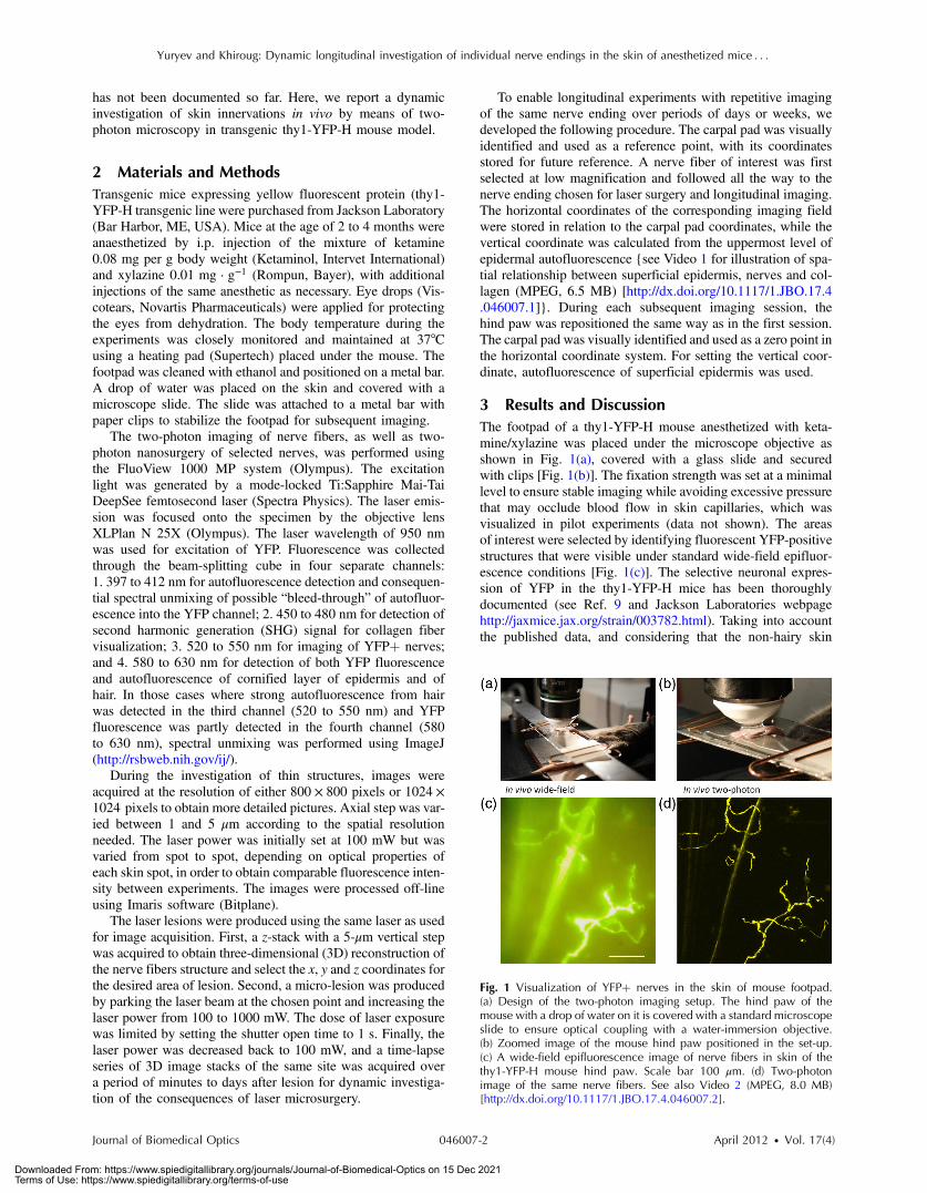
3 Results and DiscussionThe footpad of a thy1-YFP-H mouse anesthetized with keta-mine/xylazine was placed under the microscope objective asshown in Fig. 1(a), covered with a glass slide and securedwith clips [Fig. 1(b)]. The fixation strength was set at a minimallevel to ensure stable imaging while avoiding excessive pressurethat may occlude blood flow in skin capillaries, which wasvisualized in pilot experiments (data not shown). The areasof interest were selected by identifying fluorescent YFP-positivestructures that were visible under standard wide-field epifluor-escence conditions [Fig. 1(c)]. The selective neuronal expres-sion of YFP in the thy1-YFP-H mice has been thoroughlydocumented (see Ref. 9 and Jackson Laboratories webpagehttp://jaxmice.jax.org/strain/003782.html). Taking into accountthe published data, and considering that the non-hairy skin
Fig. 1 Visualization of YFPþ nerves in the skin of mouse footpad.(a) Design of the two-photon imaging setup. The hind paw of themouse with a drop of water on it is covered with a standard microscopeslide to ensure optical coupling with a water-immersion objective.(b) Zoomed image of the mouse hind paw positioned in the set-up.(c) A wide-field epifluorescence image of nerve fibers in skin of thethy1-YFP-H mouse hind paw. Scale bar 100 μm. (d) Two-photonimage of the same nerve fibers. See also Video 2 (MPEG, 8.0 MB)[http://dx.doi.org/10.1117/1.JBO.17.4.046007.2].
Yuryev and Khiroug: Dynamic longitudinal investigation of individual nerve endings in the skin of anesthetized mice : : :
Journal of Biomedical Optics 046007-2 April 2012 • Vol. 17(4)
Downloaded From: https://www.spiedigitallibrary.org/journals/Journal-of-Biomedical-Optics on 15 Dec 2021Terms of Use: https://www.spiedigitallibrary.org/terms-of-use

regions of hind paw foot pad essentially lack motor neuronaxons, we concluded that the YFPþ structures monitored inthe present study were part of a small subset of sensory nervesoriginating from DRG neurons.9,16,17
The z-stacks of optical sections were collected using two-photon excitation mode, and 3D projections were built revealingfine morphological details of YFP-positive nerve fibers andterminals [Fig. 1(d)]. For comparison, conventional wide-fieldimage of the same region is shown in Fig. 1(c). Clear opticalsections were obtained with two-photon microscopy at thedepths of up to 250 μm (for animated projections of theimage z-stack, see Video 2).
Fluorescence was collected using three separate emissionchannels. Emission of YFP fluorescence was collected in thespectral intervals of 520 to 550 nm [Fig. 2(a)] and 580 to630 nm [Fig. 2(b)]. At the edges of footpad, YFP fluorescenceoccasionally partly overlapped with the autofluorescence of hair.Despite the overlapping spectra, it was possible to distinguishthe nerve structures from hair and skin autofluorescence usingmathematical unmixing routines18 [Fig. 2(d) and (2e)]. Sim-ultaneous collection of the second harmonic generation (SHG)signal and fluorescence of YFP [Fig. 2(c) and 2(f)] allowed us toinvestigate spatial distribution of neuronal network intertwinedwith collagen fibers, which would not be possible using conven-tional histological techniques.
Spatial relationship between nerve endings and collagenlayers could be resolved as illustrated in Fig. 3(a) and Video 3.Thin structures that were situated in close proximity to eachother could be clearly distinguished [Fig. 3(b)]. Full-width athalf-maximum of the thinnest part of nerve is 750� 200 nm
(n ¼ 5) [Fig. 3(c)]. The fibers that were 3 μm apart fromeach other were fully discernible, as indicated by lack of overlapin their intensity profiles [Fig. 3(d)].
Moreover, it was possible to affect the nerves by producingselective lesions utilizing the same laser as for imaging (Fig. 4).To produce spatially restricted lesions to selected nerve fibers,we “parked” the laser beam at one spot to achieve continuousexposure of up to 500 ms. This technique allowed investigationof dynamic rearrangement of neuronal networks on a short timescale [Fig. 4(a) and 4(b)] as well as over the periods of days andweeks [Fig. 4(d) and 4(e)], potentially extendable to even longerperiods of several months. Immediately after the lesion, theintegrity of the targeted nerve fragment was lost and small struc-tures resembling vesicles filled with YFP became clearly visiblein the lesion area [Fig. 4(c)]. These round structures persisted for
Fig. 2 Simultaneous detection of YFP fluorescence (yellow), hair auto-fluorescence (red) and Second Harmonic Generation (SHG) signaloriginating from collagen fibers (grey) shown as raw images prior tospectral unmixing (a, b, and c) and as processed images resultingfrom spectral unmixing (d, e, and f). Scale bar 100 μm.
Fig. 3 Two- and three-dimensional image analysis. (a) Horizontal view of an optical section and the corresponding lateral projections of the YFPþnerve fibers. The diagram shows normalized total intensity of the respective lateral projections. Scale bar 100 μm. (b) Zoomed maximum intensityprojection of the z-stack. Scale bar 50 μm. Lines mark corresponding lateral intensity profiles of a single nerve fiber (c) and of two neighboring fibers(d) See also Video 3 (MPEG, 6.7 MB) [http://dx.doi.org/10.1117/1.JBO.17.4.046007.3].
Yuryev and Khiroug: Dynamic longitudinal investigation of individual nerve endings in the skin of anesthetized mice : : :
Journal of Biomedical Optics 046007-3 April 2012 • Vol. 17(4)
Downloaded From: https://www.spiedigitallibrary.org/journals/Journal-of-Biomedical-Optics on 15 Dec 2021Terms of Use: https://www.spiedigitallibrary.org/terms-of-use

at least 10 days after the lesion [Fig. 4(d) and 4(e)], when re-innervation began as indicated by appearance of thin filopodia-like elongatedYFPþ structures shown with an arrow in Fig. 4(e)and 4(f).
Interestingly, days later we observed diffuse cell-sizestructures with the spectral characteristics of YFP. It seems plau-sible to assume that these were phagocytes presumably partici-pating in clearance of the severed nerve fibers. An alternativeexplanation is injury-induced transient expression of thy1 inkeratinocytes and subsequent thy1-driven expression of YFPin non-neuronal structures surrounding the severed nerveending.10
“Nanosurgery,” or selective disruption of single axon hasbeen demonstrated in the brain of anesthetized transgenicmice several years ago.19 However, the underlying physical prin-ciples of this process still remain elusive, especially in the caseof its application to skin. The feasibility of producing highlyselective lesions and disruptions of axons strongly dependson the scattering properties of the tissue. Typically, nerveendings that are positioned upon the collagen layer are moresuitable for selective disruption. Although it is possible to pro-duce lesions using shorter wavelength, e.g., 800 nm where YFPexcitation is not optimal, precise disruption of single axonsseems to be feasible only at the peak of YFP absorption at950 nm (our unpublished observation).
After local injury the peripheral nerves may undergo signif-icant regeneration or degradation. Insufficient functional recov-ery of nerve sensitivity after the sensory nerve injury remains anunmet clinical need. The incomplete re-innervation followingthe disruption of nerves may lead to abnormal sensory functions.The method presented here could be useful as an assay for devel-oping new treatments for peripheral nerve injuries. It will beparticularly interesting to investigate inflammatory processesconcomitant to the nerve lesion by means of, for example,neutrophil staining in vivo via i.v. injection of antibodies.20
There are several ways to circumvent physical limitationsrestricting optical investigations in skin. One of them is mount-ing of special skin fold chamber on the back of the animal toprovide access to the deepest layers of skin for two-photonmicroscopic imaging.21 However, the skin fold chamberapproach is more invasive compared to the method presentedhere. Another method providing access to the deep skin layersutilizes small-diameter fiber-optic probes that could provide realtime images in the living animal, allowing access to peripheralnerve system as well as the brain. However, in addition to itsinvasive nature, the fiber-based method does not offer the spatialresolution required for investigation of subcellular structures.22
Peripheral neuropathy remains an important clinical pro-blem. This condition has been associated with a variety of dis-orders such as diabetes, immune diseases, drug toxicity(especially cancer chemotherapy) and HIV.8 Use of in vivotwo-photon optical biopsy for morphological analysis in livingtransgenic mice will provide new and significant insights inthese areas of research. Different lines of transgenic miceexpressing, e.g., GFP, DsRed or CFP could be used in thesetypes of experiments for convenient spectral separation.9,23
This is essential for experiments where, simultaneously withnerve endings, visualization of blood flow and extravasationof i.v.-injected tagged drug or blood plasma is desired. Usingthe mice with fluorescently marked mitochondria will enablefunctional imaging of nerve endings in skin. A promisingand flexible approach is the in vivo electroporation, whichallows expressing fluorescent proteins of interest in the nervoussystem of postnatal animals as we and others have previouslyshown.24,25
In conclusion, we designed the protocol for single nerve end-ings visualization in thy1-YFP-H transgenic mice using two-photon microscopy. The method allows monitoring dynamicchanges in thin structure of nerve fibers with submicron resolu-tion over the periods of days or weeks.
AcknowledgmentsThis work was partly funded by the Academy of Finland. MikhailYuryev gratefully acknowledges the financial support and meth-odological training he received from Neurotar Ltd (www.neurotar.com) in the course of this project.
References1. E. A. Lumpkin and M. J. Caterina, “Mechanisms of sensory transduc-
tion in the skin,” Nature 445(7130), 858–865 (2007).2. M. Koltzenburg, C. L. Stucky, and G. R. Lewin, “Receptive properties
of mouse sensory neurons innervating hairy skin,” J. Neurophysiol.78(4), 1841–1850 (1997).
3. W. R. Kennedy, G.Wendelschafer-Crabb, and T. Johnson, “Quantitationof epidermal nerves in diabetic neuropathy,” Neurology 47(4),1042–1048 (1996).
4. L. R. Robinson, “Traumatic injury to peripheral nerves,” Muscle Nerve23(6), 863–873 (2000).
5. K. K. Wu, “Mortons’s interdigital neuroma: a clinical review of its etiol-ogy, treatment, and results,” J. Foot Ankle Surg. 35(2), 112–119(1996).
6. P. H. Williams and K. P. Trzil, “Management of meralgia paresthetica,”J. Neurosurg. 74(1), 76–80 (1991).
7. E. Tschachler et al., “Sheet preparations expose the dermal nerve plexusof human skin and render the dermal nerve end organ accessible toextensive analysis,” J. Invest. Dermatol. 122(1), 177–182 (2004).
8. G. Lauria and R. Lombardi, “Skin biopsy: a new tool for diagnosingperipheral neuropathy,” BMJ 334(7604), 1159–1162 (2007).
Fig. 4 Dynamic restructuring of cutaneous nerve endings induced by alocal laser-mediated lesion. (a) Image of the nerves 1 min before thelesion. Arrow points to the area of damage. Scale bar 100 μm.(b) Image of the same spot 1 min after the lesion. (c) Zoomed areaof the damage. Arrow points to the vesicles that have emerged aroundthe site of injury. (d) The same region six days after the lesion. Arrowsmark the diffusely fluorescent cell-size structures, presumably phago-cytes. (e) The same region 10 days after the lesion. Arrow points onthe newly developed nerve fiber. (f) Zoomed image of the emergingnerve fiber.
Yuryev and Khiroug: Dynamic longitudinal investigation of individual nerve endings in the skin of anesthetized mice : : :
Journal of Biomedical Optics 046007-4 April 2012 • Vol. 17(4)
Downloaded From: https://www.spiedigitallibrary.org/journals/Journal-of-Biomedical-Optics on 15 Dec 2021Terms of Use: https://www.spiedigitallibrary.org/terms-of-use

9. G. Feng et al., “Imaging neuronal subsets in transgenic miceexpressing multiple spectral variants of GFP,” Neuron 28(1), 41–51(2000).
10. Y. A. Pan and J. R. Sanes, “Non-invasive visualization of epidermalresponses to injury using a fluorescent transgenic reporter,” J. Invest.Dermatol. 123(5), 888–891 (2004).
11. C. Cheng et al., “Dynamic plasticity of axons within a cutaneousmilieu,” J. Neurosci. 30(44), 14735–14744 (2010).
12. A. W. English, W. Meador, and D. I. Carrasco, “Neurotrophin-4∕5 isrequired for the early growth of regenerating axons in peripheralnerves,” Eur. J. Neurosci. 21(10), 2646–2634 (2005).
13. B. Beirowski et al., “The progressive nature of Wallerian degenerationin wild-type and slow Wallerian degeneration (Wlds) nerves,” BMCNeurosci. 6, 6 (2005).
14. F. Helmchen and W. Denk, “Deep tissue two-photon microscopy,”Nat. Meth. 2(12), 932–940 (2005).
15. K. Koenig and I. Riemann, “High-resolution multiphoton tomographyof human skin with subcellular spatial resolution and picoseconds timeresolution,” J. Biomed. Opt. 8(3), 432–439 (2003).
16. M. L. Groves et al., “Axon regeneration is peripheral nerves is enhancedby proteoglycan degradation” Exp. Neurol. 195(2), 278–292(2005).
17. C. M. Webb, E. M. Cameron, and J. P. Soundberg, “Fluorescence-labeled reporter gene in transgenic mice provides a useful tool for inves-tigating cutaneous innervation,” Vet. Pathol. 21 July 2011, (2011).
18. T. Zimmermann et al., “Spectral imaging and linear un-mixing enablesimproved FRET efficiency with a novel GFP2-YFP FRET pair,” FEBSLett. 531(2), 245–249 (2002).
19. L. Sacconi et al., “In vivo multiphoton nanosurgery of cortical neurons,”J. Biomed. Opt. 12(5), 050502 (2007).
20. M. Phillipson et al., “Intraluminal crawling of neutrophils to emigrationsites: a molecularly distinct process from adhesion in the recruitmentcascade,” J. Exp. Med. 203(12), 2569–2575 (2006).
21. F.-C. Li et al., “Dorsal skin fold chamber for high resolution multipho-ton imaging,” Opt. Quant. Elect. 37(13), 1439–1445 (2005).
22. P. Vincent et al., “Live imaging of neural structure and function byfibred fluorescence microscopy,” EMBO Rep. 7(11), 1154–1161 (2006).
23. T. Misgeld et al., “Imaging axonal transport of mitochondria in vivo,”Nat. Methods 4, 559–561 (2007).
24. C. Boutin et al., “Efficient in vivo electroporation of the postnatal rodentforebrain,” PloS One 3(4), e1883 (2008).
25. D. Molotkov et al., “Gene delivery to postnatal rat brain by non-ven-tricular plasmid injection and electroporation,” J. Vis. Exp. 43, 2244(2010).
Yuryev and Khiroug: Dynamic longitudinal investigation of individual nerve endings in the skin of anesthetized mice : : :
Journal of Biomedical Optics 046007-5 April 2012 • Vol. 17(4)
Downloaded From: https://www.spiedigitallibrary.org/journals/Journal-of-Biomedical-Optics on 15 Dec 2021Terms of Use: https://www.spiedigitallibrary.org/terms-of-use