Dynamic Light Scattering Microrheology Reveals...
10
Dynamic Light Scattering Microrheology Reveals Multiscale Viscoelasticity of Polymer Gels and Precious Biological Materials Brad A. Krajina, † Carolina Tropini, ‡ Audrey Zhu, † Philip DiGiacomo, § Justin L. Sonnenburg, ‡ Sarah C. Heilshorn, ∥ and Andrew J. Spakowitz* ,†,∥,⊥,# † Department of Chemical Engineering, Stanford University, Stanford, California 94305, United States ‡ Department of Microbiology and Immunology, Stanford University School of Medicine, Stanford, California 94305, United States § Department of Bioengineering, Stanford University, Stanford, California 94305, United States ∥ Department of Materials Science and Engineering, Stanford University, Stanford, California 94305, United States ⊥ Department of Applied Physics, Stanford University, Stanford, California 94305, United States # Biophysics Program, Stanford University, Stanford, California 94305, United States * S Supporting Information ABSTRACT: The development of experimental techniques capable of probing the viscoelasticity of soft materials over a broad range of time scales is essential to uncovering the physics that governs their behavior. In this work, we develop a microrheology technique that requires only 12 μL of sample and is capable of resolving dynamic behavior ranging in time scales from 10 −6 to 10 s. Our approach, based on dynamic light scattering in the single-scattering limit, enables the study of polymer gels and other soft materials over a vastly larger hierarchy of time scales than macrorheology measurements. Our technique captures the viscoelastic modulus of polymer hydrogels with a broad range of stiffnesses from 10 to 10 4 Pa. We harness these capabilities to capture hierarchical molecular relaxations in DNA and to study the rheology of precious biological materials that are impractical for macrorheology measurements, including decellularized extracellular matrices and intestinal mucus. The use of a commercially available benchtop setup that is already available to a variety of soft matter researchers renders microrheology measurements accessible to a broader range of users than existing techniques, with the potential to reveal the physics that underlies complex polymer hydrogels and biological materials. ■ INTRODUCTION Soft materials exhibit a rich range of rheological behaviors that play a central role in determining their processing behavior and practical functions. Examples include the engineered materials for biomedical applications, 1 self-healing materials for synthetic skin and flexible electronics, 2 self-assembling copolymers for nanopatterning, 3 and the active cytoskeleton of living cells. 4 The rheology of soft materials is governed by physical processes that occur over a vast range of time scales, 5 which presents a formidable challenge for unlocking the molecular under- pinnings of viscoelastic behavior. Thus, the development of rheological measurement techniques that interrogate the viscoelasticity of soft materials across disparate time scales offers the opportunity to gain deep insights into the molecular physics of soft matter with broad applications across scientific, engineering, and medical disciplines. Despite signi ficant progress that has been forged in developing microrheology techniques that surpass the capabilities of conventional rheology and are amenable to the small sample quantities often encountered for biological materials, 6−8 the requirements for specialized expertise, time-consuming data acquisition and analysis, or a limited range of detectable material properties pose substantial barriers to establishing these techniques as accessible characterization tools across a broader community of materials chemistry and biomaterials researchers. Conventional rheological techniques (macrorheology), which measure the material response to macroscopic external perturbations, typically provide access to a very limited range of time scales. In particular, oscillatory rheology provides access to frequencies ω of only up to about 10 2 s −1 . Although broader ranges of time scales can be accessed by combining oscillatory shear rheology with squeeze-flow and torsional resonance techniques, this requires the combined use of three separate measurements to access the frequency range of interest. 9,10 Furthermore, such macrorheology experiments typically require large volumes of material, which prohibits measurements on certain precious biological samples, where only microliter-scale volumes can be obtained and procurement is often time- consuming and costly. Received: September 27, 2017 Published: December 15, 2017 Research Article Cite This: ACS Cent. Sci. 2017, 3, 1294-1303 © 2017 American Chemical Society 1294 DOI: 10.1021/acscentsci.7b00449 ACS Cent. Sci. 2017, 3, 1294−1303 This is an open access article published under an ACS AuthorChoice License, which permits copying and redistribution of the article or any adaptations for non-commercial purposes.
-
Upload
nguyenduong -
Category
Documents
-
view
215 -
download
0
Transcript of Dynamic Light Scattering Microrheology Reveals...

Dynamic Light Scattering Microrheology Reveals MultiscaleViscoelasticity of Polymer Gels and Precious Biological MaterialsBrad A. Krajina,† Carolina Tropini,‡ Audrey Zhu,† Philip DiGiacomo,§ Justin L. Sonnenburg,‡
Sarah C. Heilshorn,∥ and Andrew J. Spakowitz*,†,∥,⊥,#
†Department of Chemical Engineering, Stanford University, Stanford, California 94305, United States‡Department of Microbiology and Immunology, Stanford University School of Medicine, Stanford, California 94305, United States§Department of Bioengineering, Stanford University, Stanford, California 94305, United States∥Department of Materials Science and Engineering, Stanford University, Stanford, California 94305, United States⊥Department of Applied Physics, Stanford University, Stanford, California 94305, United States#Biophysics Program, Stanford University, Stanford, California 94305, United States
*S Supporting Information
ABSTRACT: The development of experimental techniquescapable of probing the viscoelasticity of soft materials over abroad range of time scales is essential to uncovering the physicsthat governs their behavior. In this work, we develop amicrorheology technique that requires only 12 μL of sampleand is capable of resolving dynamic behavior ranging in timescales from 10−6 to 10 s. Our approach, based on dynamic lightscattering in the single-scattering limit, enables the study ofpolymer gels and other soft materials over a vastly largerhierarchy of time scales than macrorheology measurements. Ourtechnique captures the viscoelastic modulus of polymer hydrogelswith a broad range of stiffnesses from 10 to 104 Pa. We harnessthese capabilities to capture hierarchical molecular relaxations inDNA and to study the rheology of precious biological materials that are impractical for macrorheology measurements, includingdecellularized extracellular matrices and intestinal mucus. The use of a commercially available benchtop setup that is alreadyavailable to a variety of soft matter researchers renders microrheology measurements accessible to a broader range of users thanexisting techniques, with the potential to reveal the physics that underlies complex polymer hydrogels and biological materials.
■ INTRODUCTIONSoft materials exhibit a rich range of rheological behaviors thatplay a central role in determining their processing behavior andpractical functions. Examples include the engineered materialsfor biomedical applications,1 self-healing materials for syntheticskin and flexible electronics,2 self-assembling copolymers fornanopatterning,3 and the active cytoskeleton of living cells.4
The rheology of soft materials is governed by physical processesthat occur over a vast range of time scales,5 which presents aformidable challenge for unlocking the molecular under-pinnings of viscoelastic behavior. Thus, the development ofrheological measurement techniques that interrogate theviscoelasticity of soft materials across disparate time scalesoffers the opportunity to gain deep insights into the molecularphysics of soft matter with broad applications across scientific,engineering, and medical disciplines. Despite significantprogress that has been forged in developing microrheologytechniques that surpass the capabilities of conventionalrheology and are amenable to the small sample quantitiesoften encountered for biological materials,6−8 the requirementsfor specialized expertise, time-consuming data acquisition and
analysis, or a limited range of detectable material propertiespose substantial barriers to establishing these techniques asaccessible characterization tools across a broader community ofmaterials chemistry and biomaterials researchers.Conventional rheological techniques (macrorheology),
which measure the material response to macroscopic externalperturbations, typically provide access to a very limited range oftime scales. In particular, oscillatory rheology provides access tofrequencies ω of only up to about 102 s−1. Although broaderranges of time scales can be accessed by combining oscillatoryshear rheology with squeeze-flow and torsional resonancetechniques, this requires the combined use of three separatemeasurements to access the frequency range of interest.9,10
Furthermore, such macrorheology experiments typically requirelarge volumes of material, which prohibits measurements oncertain precious biological samples, where only microliter-scalevolumes can be obtained and procurement is often time-consuming and costly.
Received: September 27, 2017Published: December 15, 2017
Research Article
Cite This: ACS Cent. Sci. 2017, 3, 1294−1303
© 2017 American Chemical Society 1294 DOI: 10.1021/acscentsci.7b00449ACS Cent. Sci. 2017, 3, 1294−1303
This is an open access article published under an ACS AuthorChoice License, which permitscopying and redistribution of the article or any adaptations for non-commercial purposes.
To surmount these challenges, a variety of microrheologytechniques have emerged that probe the viscoelasticity of softmatter at time scales and sample volumes beyond the reach ofconventional macrorheology.6,7,11,12 Microrheology techniquestypically employ micrometer-scale probe particles that sensetheir local viscoelastic environment in response to thermal(passive) or external (active) forces. Among these techniques,video particle tracking (VPT)7,13,14 and quadrant detec-tion8,12,15−17 microrheology have enjoyed extensive implemen-tation for probing spatially localized viscoelasticity inheterogeneous samples and living cells and require very lowsample volumes (<1 μL). Spatially localized information fromthese techniques is achieved either by explicitly tracking particletrajectories by optical microscopy (VPT) or by focusing a laseronto individual probe particles and monitoring laser deflectionsonto a quadrant photodiode detector (quadrant detection). Forbulk-averaged viscoelasticity across broad time scales, dynamiclight scattering in the multiple-scattering limit, i.e., diffusingwave spectroscopy (DWS), has also been widely lever-aged.11,15,18 However, each of these techniques suffers fromdistinct requirements that limit their widespread adoption byusers beyond microrheology specialists, such as applicability toonly soft materials (G < 100 Pa for VPT19), technicallychallenging implementation (quadrant detection and VPT),low statistical power (quadrant detection), or large samplevolume requirements (at least 150 μL for DWS6,20−27). Thus,there exists a critical need for a technique that is readilyavailable to researchers with a broad range of expertise and thatmeasures precious soft and biological materials across a breadthof viscoelastic properties and time scales.Here, we develop a technique that is broadly accessible to
users with a range of expertise and that interrogatesviscoelasticity of small-volume samples over a vast spectrumof material properties and time scales. Our experimentalmethodology is based on dynamic light scattering (DLS) in thesingle-scattering limit and hence requires only dilute probeconcentrations (<0.5%). This DLS microrheology (DLSμR)technique extracts the frequency-dependent shear moduli ofmaterials with a broad range of stiffnesses up to 104 Pa and overa vastly broader range of time scales (10−1 to 106 s−1) thanoscillatory macrorheology (<102 s−1). This is achieved using acommercial benchtop instrument that is already available to avariety of materials chemists, involves minimal user inter-vention, and requires sample volumes as low as 12 μL. Weleverage this technique to measure the hierarchy of molecularrelaxations that occur in DNA solutions and to capture theviscoelastic behavior of precious biological materials, includingextracellular matrices and mucus, where conventional macro-rheology is impractical (Figure 1). This technique is accessibleto a wide range of users and opens opportunities for studying arange of complex biological and soft materials.The ranges of frequency and viscoelasticity that we explore in
this work have typically been viewed to be inaccessible to DLSin the single-scattering limit.18,28−31 However, as we demon-strate, this perceived limitation reflects misconceptions relatedto the statistics of photon correlation in single-scatteringdetection. We harness the highest spatial resolution accessibleto single-scattering DLS by operating in noninvasive back-scatter detection mode, and prove that commercial photoncorrelation instruments are capable of capturing probefluctuations at nanometer-scale resolution. This reveals accessto time scales and material stiffness previously viewed to be
accessible by microrheology only through DWS and quadrantdetection.
■ RESULTSDLSμR Offers an Accessible Technique for Rheolog-
ical Measurements of Soft Matter over a Wide Spectrumof Time Scales and Material Properties. Figure 2 illustratesour workflow for extracting the frequency-dependent shearmodulus G*(ω) of soft materials using a commercially availableDLS instrument. In contrast to DWS, by operating in thesingle-scattering limit, our methodology involves dilute
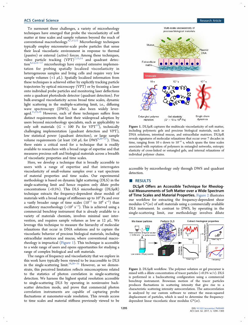
Figure 1. DLSμR captures the multiscale viscoelasticity of soft matter,including polymeric gels and precious biological materials, such asDNA solutions, intestinal mucus, and extracellular matrices. DLSμRreveals signatures of molecular relaxations that occur over 7 decades intime, ranging from 10 s down to 10−6 s, which spans the time scalesassociated with reptation of polymers in entangled networks, entropicelasticity of cross-linked or entangled gels, and internal relaxations ofindividual polymer chains.
Figure 2. DLSμR workflow. The polymer solution or gel precursor ismixed with a dilute concentration of tracer particles (<0.5% w/v). DLSis performed in a backscattering configuration using a commercialbenchtop instrument. Brownian motion of the tracer particlesproduces fluctuations in scattering intensity that give rise to acharacteristic scattering intensity autocorrelation. The autocorrelationis analyzed by our custom software to extract the mean-squareddisplacement of particles, which is used to determine the frequency-dependent linear viscoelastic shear modulus G*(ω).
ACS Central Science Research Article
DOI: 10.1021/acscentsci.7b00449ACS Cent. Sci. 2017, 3, 1294−1303
1295

concentrations of probe particles (<0.5% w/v) that serve aslight scatterers.The commercial benchtop instrument we leverage here
illuminates the sample with a 50 μm diameter laser beam, andscattered light is focused onto a photodiode detector using atranslating lens to collect photons from a specified scatteringvolume within the sample of about 1.4 nL. In practice, we use atotal sample volume of 12 μL. This strikingly contrasts with thevolume requirements of DWS, where at least 150 μL of sampleis typically required.6,20−27We perform DLS in backscatterdetection mode, which offers the smallest length scale ofresolution accessible to the wavelength of the laser, therebycapturing time scales that are inaccessible to oscillatory shearrheology and VPT. In contrast to DWS, the low probeconcentrations typically required for performing single-scattering DLS can present a challenge for separating thescattering by probe particles from background scattering by thematerial.32 Another key advantage of the backscattering opticsutilized here is that much higher probe concentrations can beused without exiting the single-scattering regime due to theshorter photon path length in the backscattering configurationand the ability to tune the photon path length using thetranslating lens system.33 Leveraging these capabilities enablesus to use probe concentrations (0.1% to 0.25%) that are 1−2orders of magnitude greater than those typically implementedfor single-scattering DLS,18,31,32 thereby ensuring dominance ofscattering from probe particles.Thermal fluctuations of the tracer particles give rise to
scattering intensity fluctuations, which are described by a timelag τ dependent scattering intensity autocorrelation g(2)(τ).Scattering photon autocorrelations are collected using a digitalcorrelator with delay times ranging from 0.5 μs to 70 s, and theexperimentally accessible frequency range within this window isdictated by the time scales over which particle displacementsare detectable. The intensity autocorrelation encodes theaverage mean-squared displacement of tracer particles withinthe scattering volume ⟨Δr2(τ)⟩ over the time lag τ. The mean-squared displacement of particles with radius a in turn can beused to extract the frequency-dependent shear modulusaccording to the generalized Stokes−Einstein relation, G*(ω)= kBT/[πa i ω⟨Δr2(ω)⟩]. Our custom software packageanalyzes raw scattering intensity autocorrelations to extractG*(ω) of the material.To investigate the capability of our technique to capture the
viscoelastic behavior of polymer gels, we study chemicallycross-linked polyacrylamide networks over a broad range ofstiffnesses (shear moduli G* from 10 to 104 Pa) and compareour DLSμR measurements to those obtained from conventionaloscillatory macrorheology. Over this full range of stiffnesses,our DLSμR measurements exhibit excellent agreement withmacrorheology in the frequency range in which the twotechniques overlap (Figure 3). This viscoelasticity spectrumaccessible to DLSμR spans the stiffnesses of a diverse range ofbiological tissues, ranging from mucus to skeletal muscle.34 Thisrange of biologically relevant stiffnesses is inaccessible to VPT,which would be limited to materials with |G*| < 102 Pa usingthe 100 nm diameter probe particles used in our experiments.19
We note that although generally good agreement is foundbetween our DLSμR measurements and macrorheology, themodulus in our DLSμR experiments underestimates themodulus by about a factor of 2 for the stiffer (5% and 10%)gels. This may be due to inhomogeneities formed during thefree-radical polymerization, which are likely to be more
prevalent in the more rapidly forming gels.35 Such inhomoge-neities may force the probe particles into more compliantregions of the network. A similar effect of inhomogeneities ingel microrheology has been previously described,7 and particletracking microrheology experiments have demonstrated theexistence of mechanical heterogeneity in polyacrylamide gels.31
Moreover, DLSμR captures a vastly broader range of timescales than conventional oscillatory macrorheology and VPT,ranging from frequencies ω of 10−1 to 106 s−1. Although similarfrequency ranges have been captured in macrorheologymeasurements that combine oscillatory rheology, squeeze-flow, and torsional resonance, this is achieved only with thecombined use of 3 separate measurements, in total requiringhundreds of microliters of material.9,10 Our technique enablesus to probe a hierarchy of viscoelastic processes in the materialin a single measurement with only 12 μL of sample. Ourmeasurements demonstrate that, at intermediate to long timescales, the gels behave as elastic solids in which G* isapproximately constant with respect to frequency. At short timescales, the tracer particles probe faster relaxation modes in thegel due to effective elastic chains that give rise to a power-lawscaling with G* ∼ ωα. Consistent with the decreasing molecularweight of effective elastic chains with increasing cross-linkingdensity, we find that the time scale at which these relaxationmodes emerge decreases by 2 orders of magnitude as the gel
Figure 3. DLSμR recapitulates macrorheology in cross-linkedpolyacrylamide gels with shear moduli G* spanning 101 to 104 Pa.Top: Comparison of the frequency ω dependence of the magnitude ofthe shear modulus |G*| obtained by DLSμR and macrorheology ofpolyacrylamide gels with varying polacrylamide concentrations (% w/v). The dashed lines represent the high-frequency scaling of a Rousepolymer G* ∼ ω1/2 and space-filling branched polymer fractals withnondraining hydrodynamics G* ∼ ω. Bottom: Illustration of typicalparticle trajectories occurring over a time interval corresponding to theangular frequency ω = 101 s for a 100 nm diameter tracer particleembedded in gels with the indicated polyacrylamide composition.
ACS Central Science Research Article
DOI: 10.1021/acscentsci.7b00449ACS Cent. Sci. 2017, 3, 1294−1303
1296

stiffness increases across the range spanned in our measure-ments (Supporting Information and Supporting Figure 1).For the most compliant gels, α ≈ 1/2, consistent with freely
draining Rouse dynamics of flexible partial chains betweencross-links.36 This suggests that, at low polymer concentrations,the gel network is formed from loosely cross-linked highmolecular weight polymers in which each effective elastic chainexperiences overlap with surrounding chains that screenhydrodynamic interactions. For the more densely cross-linkedgels, the high-frequency scaling behavior approaches α ≈ 1,which coincides with the critical scaling predicted bypercolation theory at the gel point for nondraining percolatingclusters37 as well as the expected scaling behavior fornondraining Zimm relaxation of self-similar branched chainswith a fractal dimension Df = 3.38 This relaxation can beinterpreted in terms of a network that consists of effectiveelastic chains that possess an internal fractal branched structurethat is space-filling at all length scales below the gel correlationlength and retains high-frequency signatures of the fractalstructure and dynamics present at the percolation point (seeSupporting Information for derivation). This is consistent withtime−temperature superposition experiments in which fullyformed gels often exhibit scaling of G″ that mirrors the fractaldynamics at the gel point.39
The transition in the high-frequency relaxation from Rouseto critically overlapped Zimm dynamics suggests that thegreater cross-linking efficiency in the higher concentration gelsproduces more tightly cross-linked networks consisting ofbranched effective elastic chains that do not overlap withsurrounding chains, and therefore do not experience screeningof hydrodynamic interactions. This is consistent with time-curesuperposition experiments just above the gel point, which showlarger critical relaxation scaling exponents with greater cross-linking efficiency (compare ref 40 and ref 41) or lowermolecular weight polymer precursors.42 Similarly, atomic forcemicroscopy microrheology experiments on polyacrylamide gelsat frequencies up to ω ≈ 103 rad/s have demonstrated a high-frequency scaling exponent that increases with polyacrylamidecontent, approaching α ≈ 1 for 10 kPa gels.43 Our techniquethus offers a substantial advantage over oscillatory shearrheology in terms of elucidating the hierarchy of physicalprocesses that govern the behavior of polymer gels.Unlike VPT and quadrant detection microrheology, which
involve explicit tracking of particle trajectories, DLSμR yieldsmeasurements of tracer particle fluctuations that are directlyaveraged over the 1.4 nL scattering volume. Hence, VPT andquadrant detection extract spatially heterogeneous viscoelas-ticity that is concealed by averaging inherent to DLSμR.However, our technique offers a more streamlined andaccessible work flow for diverse users than VPT and quadrantdetection microrheology. In contrast to VPT, in which eachtime point requires a full microscopy image of tracer particlepositions to be acquired, stored, and processed, DLSμRmeasurements directly provide the statistically averaged particlefluctuations in the form of the scattering intensity autocorre-lation function, thereby reducing the data footprint associatedwith each measurement by several orders of magnitude. UnlikeVPT, quadrant detection can access the range of viscoelasticityand time scales that are encompassed by DLSμR, but quadrantdetection requires tracking individual trajectories at a time byoptically trapping single probe particles. This comes at theexpense of vastly decreased statistical power, even compared toVPT, in which approximately 100 particles are typically present
in the field of view. Moreover, quadrant detection micro-rheology requires expertise with optical traps that is notcommonly found in many soft materials laboratories. Together,these limitations render quadrant detection a far less accessibleand more technically challenging approach. By comparison,DLSμR is readily implemented on a benchtop commercialinstrument that is already available to many materials chemistryand biomaterials researchers and can rapidly captureviscoelasticity with high statistical power while requiringminimal user intervention.Historically, DLSμR in the single-scattering limit has found
limited use in comparison to VPT, quadrant detection, andDWS, and it is conventionally considered to be inadequate forviscoelastic properties in the stiffness and time-scale rangesprobed in our measurements.18,28−31 By comparison toquadrant detection and DWS, previous studies in single-scattering detection have typically explored a much morelimited range of stiffnesses (G* < 100 Pa) and frequencies ω <104 s−1.18,31,32,44−46 The range of stiffnesses and time scalesaccessible is fundamentally limited by the length scale of tracerparticle fluctuations to which the light-scattering configurationis sensitive (visualized in Figure 3). This can be understood onthe basis of the generalized Stokes−Einstein relation, whereinthe mean-squared displacements of tracer particles in an elasticgel are inversely proportional to the shear modulus.11 Thus, thesensitivity is dictated by the smallest length scale of probefluctuations that produce detectable decreases in the scatteringintensity autocorrelation g(2)(τ).We find that the backscattering optics utilized here are
capable of resolving decreases in the scattering intensityautocorrelation that correspond to mean-squared displacementsof about 1 nm2, which are 2 orders of magnitude smaller thanthe mean-squared displacements detectable by VPT,19 as wellas those previously accessed using single-scattering DLS formicrorheology of soft polyacrylamide gels.31 In accordance withthe generalized Stokes−Einstein relation, the minimumdetectable displacement, together with the probe particle size,dictates the maximum measurable stiffness. The 1 nmresolution achieved here renders measurements on gels asstiff as 104 Pa possible using 100 nm beads, and the maximumaccessible stiffness scales inversely with the size of the probethat is required to satisfy the continuum assumption of theStokes−Einstein relation. In our experiments, the 100 nmbeads are much larger than the mesh size of the polyacrylamidenetwork, which will be about 10 nm or less in the range ofconcentrations we probe,47 and are therefore sufficiently largeto ensure that the continuum limit is reached.It is noteworthy that the length scale of fluctuations to which
we are sensitive is much smaller than the length scale thatwould be estimated based on the scattering wave vector q. Forthe backscattering angle θ (173°), the wavelength λ of the laser(633 nm), and a refractive index n equal to that of water, thescattering wave vector in our experiments q = 4πn sin(θ/2)/λcorresponds to a length scale q−1 = 38 nm.48 For this reason,DLSμR in single-scattering detection has often been viewed asunsuitable for measurements in the stiffness and frequencyranges that require the spatial resolution captured in ourexperiments.18,28 In addition to improved spatial resolutionthrough the use of backscattering optics compared to someprevious studies using forward or orthogonal scattering,18,31
higher spatial resolution is also achieved by accurate estimationof the correlation function zero-time intercept using themethods described in the Supporting Information. Accurate
ACS Central Science Research Article
DOI: 10.1021/acscentsci.7b00449ACS Cent. Sci. 2017, 3, 1294−1303
1297
estimation of the zero-time correlation intercept has beenpreviously reported to represent a limitation for DLSmicrorheology.18 Our results emphasize the importance ofconsidering the sensitivity of photon correlation detection andthe reliability of correlation intercept estimation in evaluatingthe limits of the DLSμR technique.Polymer gels exhibit broken ergodicity due to frozen-in
density fluctuations formed during the gelation process, whichmust be accounted for in interpretation of DLS autocorrelationdata.49 In principle, broken ergodicity can be corrected for byobtaining ensemble-averaged scattering intensity autocorrela-tion functions by collecting the time-averaged autocorrelationacross an ensemble of spatial positions in the sample, which istime-consuming and tedious. The broken-ergodicity correctionwe implement here (described in the Supporting Information)enables extraction of the contribution to the scattering intensitydue to dynamic fluctuations within the scattering volume ofinterest, while obviating the need for full ensemble averaging ofthe correlation function. However, we note that the acquisitiontimes we use here to collect high-quality photon statistics arenot amenable to rapidly evolving materials, such as during thegelation process.DLSμR Elucidates Hierarchical Molecular Relaxations
in DNA. The broad spectrum of time scales probed by DLSoffers the opportunity to gain insights into the physics ofmacromolecules that exhibit distinct relaxation behaviors atdifferent time scales. Semiflexible polymers constitute animportant class of macromolecules that exhibit distinct physicsat different length scales and time scales. The physical behaviorof such polymers is governed by bending elasticity on the scaleof their persistence length lp, but they can exhibit flexibleGaussian chain behavior at lengths much longer than theirpersistence length. DNA represents an excellent model systemfor exploring this relaxation hierarchy, since its persistencelength (lp ≈ 50 nm)50 is sufficiently short to produce flexibleGaussian chain statistics at experimentally accessible molecularweights, but provides sufficient bending stiffness to yieldsemiflexible polymer behavior at experimentally accessiblelength scales and time scales.DLSμR measurements were performed on semidilute,
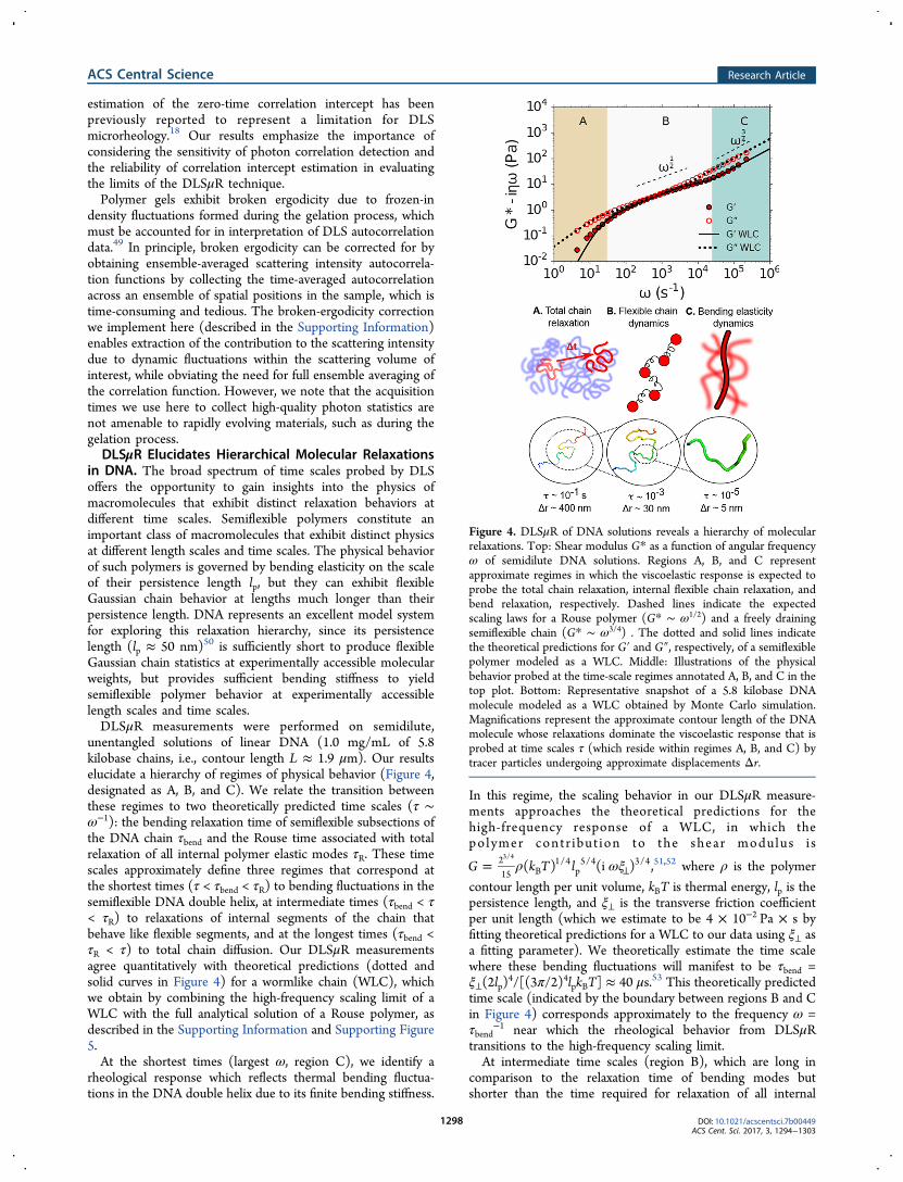
unentangled solutions of linear DNA (1.0 mg/mL of 5.8kilobase chains, i.e., contour length L ≈ 1.9 μm). Our resultselucidate a hierarchy of regimes of physical behavior (Figure 4,designated as A, B, and C). We relate the transition betweenthese regimes to two theoretically predicted time scales (τ ∼ω−1): the bending relaxation time of semiflexible subsections ofthe DNA chain τbend and the Rouse time associated with totalrelaxation of all internal polymer elastic modes τR. These timescales approximately define three regimes that correspond atthe shortest times (τ < τbend < τR) to bending fluctuations in thesemiflexible DNA double helix, at intermediate times (τbend < τ< τR) to relaxations of internal segments of the chain thatbehave like flexible segments, and at the longest times (τbend <τR < τ) to total chain diffusion. Our DLSμR measurementsagree quantitatively with theoretical predictions (dotted andsolid curves in Figure 4) for a wormlike chain (WLC), whichwe obtain by combining the high-frequency scaling limit of aWLC with the full analytical solution of a Rouse polymer, asdescribed in the Supporting Information and Supporting Figure5.At the shortest times (largest ω, region C), we identify a
rheological response which reflects thermal bending fluctua-tions in the DNA double helix due to its finite bending stiffness.
In this regime, the scaling behavior in our DLSμR measure-ments approaches the theoretical predictions for thehigh-frequency response of a WLC, in which thepolymer contr ibut ion to the shear modulus i s
ρ ωξ= ⊥G k T l( ) (i )215 B
1/4p
5/4 3/43/4
,51,52 where ρ is the polymer
contour length per unit volume, kBT is thermal energy, lp is thepersistence length, and ξ⊥ is the transverse friction coefficientper unit length (which we estimate to be 4 × 10⁻² Pa × s byfitting theoretical predictions for a WLC to our data using ξ⊥ asa fitting parameter). We theoretically estimate the time scalewhere these bending fluctuations will manifest to be τbend =ξ⊥(2lp)
4/[(3π/2)4lpkBT] ≈ 40 μs.53 This theoretically predictedtime scale (indicated by the boundary between regions B and Cin Figure 4) corresponds approximately to the frequency ω =τbend
−1 near which the rheological behavior from DLSμRtransitions to the high-frequency scaling limit.At intermediate time scales (region B), which are long in
comparison to the relaxation time of bending modes butshorter than the time required for relaxation of all internal
Figure 4. DLSμR of DNA solutions reveals a hierarchy of molecularrelaxations. Top: Shear modulus G* as a function of angular frequencyω of semidilute DNA solutions. Regions A, B, and C representapproximate regimes in which the viscoelastic response is expected toprobe the total chain relaxation, internal flexible chain relaxation, andbend relaxation, respectively. Dashed lines indicate the expectedscaling laws for a Rouse polymer (G* ∼ ω1/2) and a freely drainingsemiflexible chain (G* ∼ ω3/4) . The dotted and solid lines indicatethe theoretical predictions for G′ and G″, respectively, of a semiflexiblepolymer modeled as a WLC. Middle: Illustrations of the physicalbehavior probed at the time-scale regimes annotated A, B, and C in thetop plot. Bottom: Representative snapshot of a 5.8 kilobase DNAmolecule modeled as a WLC obtained by Monte Carlo simulation.Magnifications represent the approximate contour length of the DNAmolecule whose relaxations dominate the viscoelastic response that isprobed at time scales τ (which reside within regimes A, B, and C) bytracer particles undergoing approximate displacements Δr.
ACS Central Science Research Article
DOI: 10.1021/acscentsci.7b00449ACS Cent. Sci. 2017, 3, 1294−1303
1298
polymer elastic modes, the shear modulus reflects theviscoelastic relaxation of flexible subchains that behave likeentropic springs. For a Rouse-like polymer, in which hydro-dynamic interactions can be neglected, this is theoreticallypredicted to produce a shear modulus in which the storage andloss moduli scale as G′ = G″ ∼ ω1/2.5 Our measurements showthat the relaxation behavior of the DNA chains approaches thisscaling behavior over a limited range of frequencies (102 < ω <103) before transitioning to total chain diffusion at longer timescales and bend fluctuation dynamics at shorter time scales.At the longest time scales (smallest ω, region A), all internal
modes of the polymer chain have fully relaxed, and the shearmodulus captures the viscoelastic response due to translationaldiffusion of the polymer chains. For semidilute unentangledsolutions, this response is anticipated at times longer than theRouse relaxation time of the entire polymer contour length L:τR = 2lpξRL
2/(3π2kBT) ≈ 3 × 10⁻² s.5 At frequencies ω < τR−1,
our measurements exhibit a transition to viscous dynamics, withG″ > G′, which is consistent with relaxation of the internalentropic elastic modes of the polymers and a transition towardtotal chain diffusion. This transition is in excellent agreementwith theoretical predictions for a WLC that exhibits flexiblechain behavior at these time scales, which we obtain using aRouse monomer friction coefficient per unit length of ξR = 1.0× 10⁻² Pa × s based on the fitting procedure described inSupporting Information. The value of ξR we obtain is about 4times smaller than the transverse friction coefficient ξ⊥.In order to evaluate the consistency of our fitted friction
coefficients, we compare the value of ξR obtained from ourDLSμR data to existing diffusivity measurements and relate ourobservation that ξ⊥ ≈ 4ξR to polymer physics theory. In thesemidilute regime, the effective Rouse friction coefficient ξR isdependent on the polymer concentration, which defines thelength scale for hydrodynamic screening by overlappingpolymers. At length scales larger than the hydrodynamicscreening length, each polymer in the semidilute network canbe viewed as consisting of coarse-grained polymer “blobs” thatdiffuse like effective Rouse monomers36 and exhibit internalhydrodynamic interactions at smaller length and time scales.Measurements of the diffusivity D of fluorescently labeled 5.9kb DNA have confirmed this concentration dependence and ata concentration of 1.0 mg/mL provide a Rouse frictioncoefficient ξR = kBT/(DL) = 0.6 × 10⁻² Pa × s.54,55 This issomewhat lower than our value ξR = 1.0 × 10⁻² Pa × s, andfurther investigation is required to elucidate the origin of thisdiscrepancy.Below the hydrodynamic screening length, and within each
effective Rouse monomer, DNA may be expected to behaveaccording to slender body hydrodynamics.56 In this regime, thehydrodynamics are described by two friction coefficients, ξ⊥and ξ∥, which represent the transverse and longitudinal friction,respectively, and ξ⊥ = 2ξ∥. If the friction coefficient of eachRouse monomer corresponds to the total friction of eachsubsection that obeys slender body dynamics, then slenderbody theory implies that ξR = 3(2/ξ⊥ + 1/ξ∥)
−1 = 3ξ⊥/4.However, our fit to the DLSμR data instead yields ξR ≈ ξ⊥/4.The origin of this disagreement is unclear, and may reflectadditional many-body effects due to the semidilute solution notconsidered by slender body theory.Importantly, our results are only slightly dependent on probe
particle size for probe diameters of at least 500 nm (the probesize used in Figure 4), indicating that the probes sense thecontinuum viscoelasticity of the fluid (Supporting Figure 2).
This is consistent with previous microrheology studies of linearDNA solutions, which show that, at the concentration used inour work, and with DNA molecular weights where entangle-ments are weak or nonexistent, noncontinuum effects such aspolymer depletion are not substantial, provided the probes arelarger than the radius of gyration of the polymers.57,58 Unlikethe polyacrylamide gels, 100 nm probes do not satisfy thecontinuum assumption of the Stokes−Einstein relation, sincethe radius of gyration of a 5.8 kilobase Gaussian DNA chain isabout 180 nm, and the system resides close to the criticaloverlap concentration.The transition to an ω3/4 scaling behavior at high frequencies
has been previously demonstrated using quadrant detection andDWS microrheology of other semiflexible polymers, such as F-actin, wormlike micelles, and polysaccharides.8,59−62 However,despite the extensive history of DNA as a model polymer in thepolymer physics community, previous microrheology studieson DNA solutions were conducted at much lower frequenciesthan the maximum frequency accessed here.6,63,64 Ourmeasurements not only capture the expected high-frequencyscaling behavior that has been confirmed in experiments onother semiflexible polymers but also exhibit quantitativeagreement with polymer physics theory across the full rangeof molecular relaxations.
DLSμR Accesses Multiscale Relaxation in ExtracellularMatrices Where Time−Temperature Superposition Fails.Time−temperature superposition is a widely implementedtechnique for broadening the frequency range that is accessedin a conventional macrorheology experiment. In brief, thistechnique involves collecting the frequency-dependent shearmodulus at a range of temperatures and horizontally shiftingthe isotherms to construct a single “master curve” thatencompasses a substantially broader range of frequencies thanthose accessed at any particular temperature.65 However, thisconstruction is valid only for materials in which the relaxationrates and physical processes that govern the viscoelasticbehavior can be described by a single temperature dependence.Biological systems provide a variety of instances in which the
assumptions underlying time−temperature superposition fail.In fact, in general, time−temperature superposition does notsubstantially broaden the frequency range for aqueous polymersystems, since the available temperature variations do not leadto substantial frequency shift factors. In particular, extracellularmatrices represent an important class of biological materialsthat exhibit nontrivial thermal responsiveness that violatestime−temperature superposition. Furthermore, the rheologicalbehavior of extracellular matrices exhibits a profound impact ondirecting cellular behavior.66,67
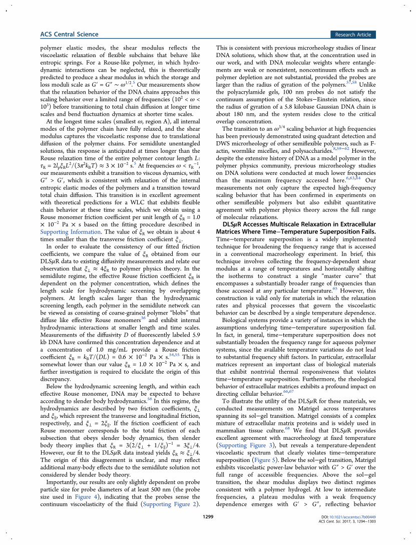
To illustrate the utility of the DLSμR for these materials, weconducted measurements on Matrigel across temperaturesspanning its sol−gel transition. Matrigel consists of a complexmixture of extracellular matrix proteins and is widely used inmammalian tissue culture.68 We find that DLSμR providesexcellent agreement with macrorheology at fixed temperature(Supporting Figure 3), but reveals a temperature-dependentviscoelastic spectrum that clearly violates time−temperaturesuperposition (Figure 5). Below the sol−gel transition, Matrigelexhibits viscoelastic power-law behavior with G″ > G′ over thefull range of accessible frequencies. Above the sol−geltransition, the shear modulus displays two distinct regimesconsistent with a polymer hydrogel. At low to intermediatefrequencies, a plateau modulus with a weak frequencydependence emerges with G′ > G″, reflecting behavior
ACS Central Science Research Article
DOI: 10.1021/acscentsci.7b00449ACS Cent. Sci. 2017, 3, 1294−1303
1299
reminiscent of an elastic solid. At high frequencies (ω > 104
s−1), the shear modulus increases with frequency andapproaches a power-law behavior that reflects coupling to theunderlying internal relaxation modes. These results demon-strate that DLSμR enables measurements of the broadfrequency viscoelasticity of biological materials where conven-tional oscillatory rheology with time−temperature super-position is not an option.
DLSμR Reveals Broad Frequency EntanglementDynamics in Intestinal Mucus for Which MacrorheologyIs Impractical. Intestinal mucus lines the luminal surface ofthe intestinal epithelium and serves as an essential barrierwhose functions include acting as a physical barrier againstmicrobial invasion and providing nutritional support to the hostmicrobiome.69−71 Disruptions to the mucosal layer areimplicated in a variety of diseases, which suggests that theability to quantify the physical properties of the mucosal layermay provide biophysical insights into the role of mucus in suchpathologies. Intestinal mucus is available in only smallquantities from small animal models and human patients, asonly a small amount can be recovered from a single animal in amouse model, and in human patients, mucus must be obtainedfrom biopsies. Thus, intestinal mucus represents an excellentexample of a biological material in which acquiring macro-rheology measurements is challenging, costly, and impractical.Our DLSμR measurements on intestinal mucus isolated from
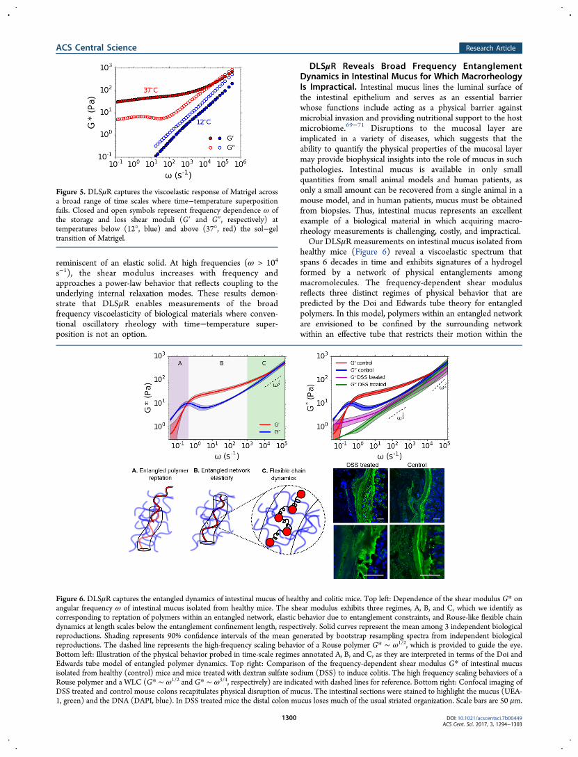
healthy mice (Figure 6) reveal a viscoelastic spectrum thatspans 6 decades in time and exhibits signatures of a hydrogelformed by a network of physical entanglements amongmacromolecules. The frequency-dependent shear modulusreflects three distinct regimes of physical behavior that arepredicted by the Doi and Edwards tube theory for entangledpolymers. In this model, polymers within an entangled networkare envisioned to be confined by the surrounding networkwithin an effective tube that restricts their motion within the
Figure 5. DLSμR captures the viscoelastic response of Matrigel acrossa broad range of time scales where time−temperature superpositionfails. Closed and open symbols represent frequency dependence ω ofthe storage and loss shear moduli (G′ and G″, respectively) attemperatures below (12°, blue) and above (37°, red) the sol−geltransition of Matrigel.
Figure 6. DLSμR captures the entangled dynamics of intestinal mucus of healthy and colitic mice. Top left: Dependence of the shear modulus G* onangular frequency ω of intestinal mucus isolated from healthy mice. The shear modulus exhibits three regimes, A, B, and C, which we identify ascorresponding to reptation of polymers within an entangled network, elastic behavior due to entanglement constraints, and Rouse-like flexible chaindynamics at length scales below the entanglement confinement length, respectively. Solid curves represent the mean among 3 independent biologicalreproductions. Shading represents 90% confidence intervals of the mean generated by bootstrap resampling spectra from independent biologicalreproductions. The dashed line represents the high-frequency scaling behavior of a Rouse polymer G* ∼ ω1/2, which is provided to guide the eye.Bottom left: Illustration of the physical behavior probed in time-scale regimes annotated A, B, and C, as they are interpreted in terms of the Doi andEdwards tube model of entangled polymer dynamics. Top right: Comparison of the frequency-dependent shear modulus G* of intestinal mucusisolated from healthy (control) mice and mice treated with dextran sulfate sodium (DSS) to induce colitis. The high frequency scaling behaviors of aRouse polymer and a WLC (G* ∼ ω1/2 and G* ∼ ω3/4, respectively) are indicated with dashed lines for reference. Bottom right: Confocal imaging ofDSS treated and control mouse colons recapitulates physical disruption of mucus. The intestinal sections were stained to highlight the mucus (UEA-1, green) and the DNA (DAPI, blue). In DSS treated mice the distal colon mucus loses much of the usual striated organization. Scale bars are 50 μm.
ACS Central Science Research Article
DOI: 10.1021/acscentsci.7b00449ACS Cent. Sci. 2017, 3, 1294−1303
1300

tube diameter (see illustrations in Figure 6). At short times(large ω, region C), the viscoelastic spectrum is dominated bythe internal relaxation of flexible subsections of the polymersthat are smaller than the effective tube diameter and have yet to“discover” that they are confined by entanglements within thesurrounding network. In this regime, G*(ω) possesses afrequency scaling behavior that reflects single chain dynamicsthat are intermediate between a Rouse polymer and a WLC. Atintermediate times (region B), polymer relaxations aregoverned by entanglement constraints with the surroundingpolymer matrix, which produces an extended elastic plateaumodulus in which G′ > G″ and G′ exhibits a weak frequencydependence. At sufficiently long times (small ω, region A), thepolymers are able to escape their local entanglements byreptation, which enables the material to flow, yielding a viscousresponse with G″ > G′.The rheological properties of intestinal mucus may play a
pivotal role in gut barrier function as well as the growth ofmicrobes embedded within the matrix.71,72 To demonstrate thecapability of DLSμR to capture changes in mucus rheologyassociated with loss of barrier function, we investigate intestinalmucus in a mouse colitis model. Incorporation of dextransulfate sodium (DSS) into the drinking water of mice disruptsbarrier function of intestinal mucus and induces inflammationof the underlying epithelium.73 Figure 6 illustrates the changein mucus rheology between healthy and DSS treated (colitic)mice measured by DLSμR. Our results exhibit a striking changein mucus rheology in the colitis model in which the mucus issubstantially softened by DSS treatment, suggestive of asubstantial reduction in the overall cross-linking density ofthe matrix (Figure 6). This perturbation to mucus viscoelas-ticity coincides with physical disruption of mucus morphologywithin the colon, as visualized by confocal microscopy (Figure6). Furthermore, although we cannot obtain mucus from micein sufficient quantities for macrorheology, to validate ourDLSμR measurements on mucus samples, we performedDLSμR on reconstituted porcine gastric mucus and foundquantitative agreement with macrorheology (Supporting Figure4). These findings suggest that DLSμR may serve as a valuabletool for investigating biophysical connections betweenintestinal mucus viscoelasticity and gut barrier status.
■ CONCLUSIONSIn this work, we present a microrheology technique based onDLS in the single-scattering limit that provides access to thefrequency-dependent viscoelasticity of soft materials across upto 7 decades in time scale. This technique requires only 12 μLof sample, which renders it amenable to precious materialswhere sample volume requirements for macrorheology andbroad-frequency DWS are impractical. Moreover, we demon-strate that, contrary to common belief, DLSμR in single-scattering detection mode is capable of probing theviscoelasticity of polymer hydrogels with a broad range ofmaterial properties, with shear moduli spanning 10 to 104 Pa,which encompasses stiffnesses across a variety of biologicaltissues. Importantly, this technique is implemented with acommercial benchtop instrument and is therefore accessible tousers with a broad range of expertise.DLSμR represents a valuable contribution to a suite of
rheological tools that are available to researchers, and extendsthe domain of microrheology to a broader base of users. Forapplications where rapid characterization of ensemble-averagedviscoelasticity of precious materials is desired, DLSμR provides
a powerful and accessible alternative to existing techniques.Although VPT and quadrant detection microrheology extractspatially heterogeneous viscoelasticity that is lost by ensembleaveraging in DLSμR, our technique overcomes crucialchallenges in these established approaches that present barriersto their widespread implementation in the materials chemistryand biomaterials communities. By comparison to VPT, DLSμRreveals viscoelasticity in materials 2 orders of magnitude stifferand across a range of time scales that is 4 orders of magnitudegreater than those accessible to VPT, while obviating the needfor the large data footprint and cumbersome data analysis thatis required of optical microscopy. In contrast to quadrantdetection and related optical trap techniques, which present asubstantial technical challenge for many users and provide lowstatistical power, DLSμR is readily implemented on acommercial benchtop platform and delivers statisticallyaveraged quantities with minimal user “hands-on” time.We exploit these capabilities to interrogate the hierarchical
molecular relaxations that occur in polymeric gels and preciousbiological materials, including DNA, extracellular matrix, andintestinal mucus. Our measurements capture physical processesranging from the rapid bending fluctuations of individualpolymers to the long-time-scale relaxations in entangledmacromolecular networks, and are rationalized in terms ofexisting polymer physics theories. Together, these findingsdemonstrate that DLSμR is a powerful tool for exploring therheological behavior of a variety of precious soft and biologicalmaterials that can be readily adopted by an expansive scope ofresearchers.
■ METHODS
Detailed experimental methods are provided in the SupportingInformation.
■ ASSOCIATED CONTENT
*S Supporting InformationThe Supporting Information is available free of charge on theACS Publications website at DOI: 10.1021/acscentsci.7b00449.
Figures, detailed experimental methods and analysis, aderivation for the predicted high-frequency scalingbehavior of polyacrylamide gels, theoretical analysis ofwormlike chains, comparison between microrheologyand macrorheology for Matrigel, and DLSμR experi-ments (PDF)
■ AUTHOR INFORMATION
Corresponding Author*E-mail: [email protected].
ORCIDSarah C. Heilshorn: 0000-0002-9801-6304Andrew J. Spakowitz: 0000-0002-0585-1942Author ContributionsB.A.K. designed research, conducted research, contributed newanalytical tools, and wrote the paper. C.T. designed andconducted research and wrote the paper. P.D. and A.Z.conducted research. J.L.S designed research. S.C.H. and A.J.Sdesigned research and wrote the paper.
NotesThe authors declare no competing financial interest.
ACS Central Science Research Article
DOI: 10.1021/acscentsci.7b00449ACS Cent. Sci. 2017, 3, 1294−1303
1301
■ ACKNOWLEDGMENTS
The authors acknowledge financial support from the StanfordBioX Graduate Fellowship program (B.A.K), the James S.McDonnell Studying Complex Systems Postdoctoral Fellow-ship (C.T.), the National Science Foundation DMR 1508006(S.C.H), and the National Science Foundation DMR 1707751(A.J.S.). We thank Sebastian Doniach, Nicholas Melosh, andGerald Fuller for useful discussions.
■ REFERENCES(1) Kim, M.; Tang, S.; Olsen, B. D. Physics of engineered proteinhydrogels. J. Polym. Sci., Part B: Polym. Phys. 2013, 51, 587−601.(2) Benight, S. J.; Wang, C.; Tok, J. B. H.; Bao, Z. Stretchable andself-healing polymers and devices for electronic skin. Prog. Polym. Sci.2013, 38, 1961−1977.(3) Olsen, B. D.; Teclemariam, N. P.; Muller, S. J.; Segalman, R. A.Rheological properties and the mechanical signatures of phasetransitions in weakly-segregated rod-coil block copolymers. Soft Matter2009, 5, 2453−2462.(4) Wirtz, D. Particle-tracking microrheology of living cells:principles and applications. Annu. Rev. Biophys. 2009, 38, 301−326.(5) Doi, M.; Edwards, S. The Theory of Polymer Dynamics; ClarendonPress: Oxford, 1988.(6) Mason, T. G.; Ganesan, K.; van Zanten, J.; Wirtz, D.; Kuo, S.Particle Tracking Microrheology of Complex Fluids. Phys. Rev. Lett.1997, 79, 3282−3285.(7) Crocker, J. C.; Valentine, M. T.; Weeks, E. R.; Gisler, T.; Kaplan,P. D.; Yodh, A. G.; Weitz, D. A. Two-point microrheology ofinhomogeneous soft materials. Phys. Rev. Lett. 2000, 85, 888−891.(8) Gittes, F.; Schnurr, B.; Olmsted, P. D.; Mackintosh, F. C.;Schmidt, C. F. Microscopic Viscoelasticity: Shear Moduli of SoftMaterials Determined from Thermal Fluctuations. Phys. Rev. Lett.1997, 79, 3286−3289.(9) Willenbacher, N.; Oelschlaeger, C. Dynamics and structure ofcomplex fluids from high frequency mechanical and optical rheometry.Curr. Opin. Colloid Interface Sci. 2007, 12, 43−49.(10) Kowalczyk, A.; Hochstein, B.; Stahle, P.; Willenbacher, N.Characterization of complex fluids at very low frequency: Experimentalverification of the strain rate-frequency superposition (SRFS) method.Appl. Rheol. 2010, 20, 1−12.(11) Mason, T. G.; Weitz, D. A. Optical measurements of frequency-dependent linear viscoelastic moduli of complex fluids. Phys. Rev. Lett.1995, 74, 1250−1253.(12) Mizuno, D.; Head, D. A.; Mackintosh, F. C.; Schmidt, C. F.Active and Passive Microrheology in Equilibrium and NonequilibriumSystems. Macromolecules 2008, 41, 7194−7202.(13) Tseng, Y. Micro-organization and visco-elasticity of theinterphase nucleus revealed by particle nanotracking. J. Cell Sci.2004, 117, 2159−2167.(14) Panorchan, P.; Lee, J. S. H.; Kole, T. P.; Tseng, Y.; Wirtz, D.Microrheology and ROCK signaling of human endothelial cellsembedded in a 3D matrix. Biophys. J. 2006, 91, 3499−3507.(15) Mason, T. G.; Gang, H.; Weitz, D. A. Diffusing-wave-spectroscopy measurements of viscoelasticity of complex fluids. J.Opt. Soc. Am. A 1997, 14, 139.(16) Yamada, S.; Wirtz, D.; Kuo, S. C. Mechanics of Living CellsMeasured by Laser Tracking Microrheology. Biophys. J. 2000, 78,1736−1747.(17) Atakhorrami, M.; Sulkowska, J. I.; Addas, K. M.; Koenderink, G.H.; Tang, J. X.; Levine, A. J.; Mackintosh, F. C.; Schmidt, C. F.Correlated fluctuations of microparticles in viscoelastic solutions:Quantitative measurement of material properties by microrheology inthe presence of optical traps. Phys. Rev. E 2006, 73, 061501.(18) Dasgupta, B. R.; Tee, S. Y.; Crocker, J. C.; Frisken, B. J.; Weitz,D. A. Microrheology of polyethylene oxide using diffusing wavespectroscopy and single scattering. Phys. Rev. E: Stat. Phys., Plasmas,Fluids, Relat. Interdiscip. Top. 2002, 65, 051505.
(19) Schultz, K. M.; Furst, E. M. Microrheology of biomaterialhydrogelators. Soft Matter 2012, 8, 6198−6205.(20) Waigh, T. A. Advances in the microrheology of complex fluids.Rep. Prog. Phys. 2016, 79, 074601.(21) Pawelzyk, P.; Mucke, N.; Herrmann, H.; Willenbacher, N.Attractive interactions among intermediate filaments determinenetwork mechanics in vitro. PLoS One 2014, 9, e93194.(22) Nagy-Smith, K.; Beltramo, P. J.; Moore, E.; Tycko, R.; Furst, E.M.; Schneider, J. P. Molecular, Local, and Network-Level Basis for theEnhanced Stiffness of Hydrogel Networks Formed from CoassembledRacemic Peptides: Predictions from Pauling and Corey. ACS Cent. Sci.2017, 3, 586−597.(23) Alam, M. M.; Mezzenga, R. Particle tracking microrheology oflyotropic liquid crystals. Langmuir 2011, 27, 6171−6178.(24) Dominguez-Garcia, P.; Cardinaux, F.; Bertseva, E.; Forro, L.;Scheffold, F.; Jeney, S. Accounting for inertia effects to access the high-frequency microrheology of viscoelastic fluids. Phys. Rev. E 2014, 90,060301.(25) Liu, J.; Boyko, V.; Yi, Z.; Men, Y. Temperature-dependentgelation process in colloidal dispersions by diffusing wave spectros-copy. Langmuir 2013, 29, 14044−14049.(26) Narita, T.; Mayumi, K.; Ducouret, G.; Hebraud, P. Viscoelasticproperties of poly(vinyl alcohol) hydrogels having permanent andtransient cross-links studied by microrheology, classical rheometry, anddynamic light scattering. Macromolecules 2013, 46, 4174−4183.(27) Martiel, I.; Sagalowicz, L.; Mezzenga, R. Viscoelasticity andinterface bending properties of lecithin reverse wormlike micellesstudied by diffusive wave spectroscopy in hydrophobic environment.Langmuir 2014, 30, 10751−10759.(28) Gardel, M. L.; Valentine, M. T.; Weitz, D. A. Microrheology.Microscale Diagnostic Techniques 2005, 1−49.(29) Abdala, A. A.; Amin, S.; Van Zanten, J. H.; Khan, S. A. Tracermicrorheology study of a hydrophobically modified comblikeassociative polymer. Langmuir 2015, 31, 3944−3951.(30) Larobina, D.; Cipelletti, L. Hierarchical cross-linking in physicalalginate gels: a rheological and dynamic light scattering investigation.Soft Matter 2013, 9, 10005.(31) Dasgupta, B. R.; Weitz, D. A. Microrheology of cross-linkedpolyacrylamide networks. Phys. Rev. E 2005, 71, 021504.(32) He, F.; Becker, G. W.; Litowski, J. R.; Narhi, L. O.; Brems, D.N.; Razinkov, V. I. High-throughput dynamic light scattering methodfor measuring viscosity of concentrated protein solutions. Anal.Biochem. 2010, 399, 141−143.(33) Kaszuba, M.; Connah, M. T.; McNeil-Watson, F. K.;Nobbmann, U. Resolving concentrated particle size mixtures usingdynamic light scattering. Particle and Particle Systems Characterization2007, 24, 159−162.(34) Butcher, D. T.; Alliston, T.; Weaver, V. M. A tense situation:forcing tumour progression. Nat. Rev. Cancer 2009, 9, 108−22.(35) Basu, A.; Wen, Q.; Mao, X.; Lubensky, T. C.; Janmey, P. A.;Yodh, A. G. Nonaffine displacements in flexible polymer networks.Macromolecules 2011, 44, 1671−1679.(36) Rubinstein, M.; Colby, R. Polymer Physics; Oxford UniversityPress: New York, 2003.(37) Martin, J. E.; Adolf, D.; Wilcoxon, J. P. Viscoelasticity of near-critical gels. Phys. Rev. Lett. 1988, 61, 2620−2623.(38) Cates, M. E. Brownian dynamics of self-similar macromolecules.J. Phys. (Paris) 1985, 46, 1059−1077.(39) Adolf, D.; Martin, J. E. Ultraslow Relaxations in Networks:Evidence for Remnant Fractal Structures. Macromolecules 1991, 24,6721−6724.(40) Larsen, T. H.; Furst, E. M. Microrheology of the liquid-solidtransition during gelation. Phys. Rev. Lett. 2008, 100, 146001.(41) Adibnia, V.; Hill, R. J. Universal aspects of hydrogel gelationkinetics, percolation and viscoelasticity from PA-hydrogel rheology. J.Rheol. 2016, 60, 541−548.(42) Schultz, K. M.; Baldwin, A. D.; Kiick, K. L.; Furst, E. M.Gelation of covalently cross-linked PEG-heparin hydrogels. Macro-molecules 2009, 42, 5310−5315.
ACS Central Science Research Article
DOI: 10.1021/acscentsci.7b00449ACS Cent. Sci. 2017, 3, 1294−1303
1302
(43) Abidine, Y.; Laurent, V. M.; Michel, R.; Duperray, A.; Palade, L.I.; Verdier, C. Physical properties of polyacrylamide gels probed byAFM and rheology. EPL 2015, 109, 38003.(44) Amin, S.; Rega, C. A.; Jankevics, H. Detection of viscoelasticityin aggregating dilute protein solutions through dynamic lightscattering-based optical microrheology. Rheol. Acta 2012, 51, 329−342.(45) Amin, S.; Blake, S.; Kenyon, S. M.; Kennel, R. C.; Lewis, E. N. Anovel combination of DLS-optical microrheology and low frequencyRaman spectroscopy to reveal underlying biopolymer self-assemblyand gelation mechanisms. J. Chem. Phys. 2014, 141, 234201.(46) Parmar, A. S.; Hill, S.; Vidyasagar, A.; Bello, C.; Toomey, R.;Muschol, M. Frequency and temperature dependence of poly(N-isopropylacrylamide) gel rheology. J. Appl. Polym. Sci. 2013, 127,1527−1537.(47) Uruena, J. M.; Pitenis, A. A.; Nixon, R. M.; Schulze, K. D.;Angelini, T. E.; Gregory Sawyer, W. Mesh Size Control of PolymerFluctuation Lubrication in Gemini Hydrogels. Biotribology 2015, 1−2,24−29.(48) Berne, B. J.; Pecora, R. Dynamic Light Scattering: WithApplications to Chemistry, Biology, and Physics; Wiley: New York, 2003.(49) Pusey, P. N.; Van Megen, W. Dynamic light scattering by non-ergodic media. Phys. A 1989, 157, 705−741.(50) Marko, J. F.; Siggia, E. D. Stretching DNA. Macromolecules1995, 28, 8759−8770.(51) Morse, D. C. Viscoelasticity of tightly entangled solutions ofsemiflexible polymers. Phys. Rev. E: Stat. Phys., Plasmas, Fluids, Relat.Interdiscip. Top. 1998, 58, R1237−R1240.(52) Gittes, F.; MacKintosh, F. C. Dynamic shear modulus of asemiflexible polymer network. Phys. Rev. E: Stat. Phys., Plasmas, Fluids,Relat. Interdiscip. Top. 1998, 58, R1241−R1244.(53) Morse, D. C. Viscoelasticity of Concentrated Isotropic Solutionsof Semiflexible Polymers. 1. Model and Stress Tensor. Macromolecules1998, 31, 7030−7043.(54) Robertson, R. M.; Smith, D. E. Self-diffusion of entangled linearand circular DNA molecules: Dependence on length and concen-tration. Macromolecules 2007, 40, 3373−3377.(55) Robertson, R. M.; Smith, D. E. Strong effects of moleculartopology on diffusion of entangled DNA molecules. Proc. Natl. Acad.Sci. U. S. A. 2007, 104, 4824.(56) Eimer, W.; Pecora, R. Rotational and translational diffusion ofshort rodlike molecules in solution: Oligonucleotides. J. Chem. Phys.1991, 94, 2324−2329.(57) Zhu, X.; Kundukad, B.; Van Der Maarel, J. R. Viscoelasticity ofentangled λ -phage DNA solutions. J. Chem. Phys. 2008, 129, 185103.(58) Chapman, C. D.; Lee, K.; Henze, D.; Smith, D. E.; Robertson-Anderson, R. M. Onset of non-continuum effects in microrheology ofentangled polymer solutions. Macromolecules 2014, 47, 1181−1186.(59) Gisler, T.; Weitz, D. Scaling of the Microrheology of SemidiluteF-Actin Solutions. Phys. Rev. Lett. 1999, 82, 1606−1609.(60) Oelschlaeger, C.; Cota Pinto Coelho, M.; Willenbacher, N.Chain Flexibility and Dynamics of Polysaccharide Hyaluronan inEntangled Solutions: A High Frequency Rheology and Diffusing WaveSpectroscopy Study. Biomacromolecules 2013, 14, 3689−3696.(61) Willenbacher, N.; Oelschlaeger, C.; Schopferer, M.; Fischer, P.;Cardinaux, F.; Scheffold, F. Broad bandwidth optical and mechanicalrheometry of wormlike micelle solutions. Phys. Rev. Lett. 2007, 99,068302.(62) Le Goff, L.; Amblard, F.; Furst, E. M. Motor-Driven Dynamicsin Actin-Myosin Networks. Phys. Rev. Lett. 2001, 88, 018101.(63) Mason, T. G.; Dhople, a.; Wirtz, D. Linear Viscoelastic Moduliof Concentrated DNA Solutions. Macromolecules 1998, 31, 3600−3603.(64) Goodman, A.; Tseng, Y.; Wirtz, D. Effect of length, topology,and concentration on the microviscosity and microheterogeneity ofDNA solutions. J. Mol. Biol. 2002, 323, 199−215.(65) Ferry, J. D. Viscoelastic Properties of Polymers, 3rd ed.; John Wiley& Sons: New York, 1980.
(66) Paszek, M. J.; Zahir, N.; Johnson, K. R.; Lakins, J. N.;Rozenberg, G. I.; Gefen, A.; Reinhart-King, C. A.; Margulies, S. S.;Dembo, M.; Boettiger, D.; Hammer, D. A.; Weaver, V. M. Tensionalhomeostasis and the malignant phenotype. Cancer Cell 2005, 8, 241−254.(67) Engler, A. J.; Sen, S.; Sweeney, H. L.; Discher, D. E. MatrixElasticity Directs Stem Cell Lineage Specification. Cell 2006, 126,677−689.(68) Hughes, C. S.; Postovit, L. M.; Lajoie, G. A. Matrigel: a complexprotein mixture required for optimal growth of cell culture. Proteomics2010, 10, 1886−1890.(69) McGuckin, M. A.; Linden, S. K.; Sutton, P.; Florin, T. H. Mucindynamics and enteric pathogens. Nat. Rev. Microbiol. 2011, 9, 265−278.(70) Tropini, C.; Earle, K. A.; Huang, K. C.; Sonnenburg, J. L. TheGut Microbiome: Connecting Spatial Organization to Function. CellHost Microbe 2017, 21, 433−442.(71) Johansson, M. E. V.; Hansson, G. C. Immunological aspects ofintestinal mucus and mucins. Nat. Rev. Immunol. 2016, 16, 639−649.(72) Tuson, H. H.; Auer, G. K.; Renner, L. D.; Hasebe, M.; Tropini,C.; Salick, M.; Crone, W. C.; Gopinathan, A.; Huang, K. C.; Weibel, D.B. Measuring the stiffness of bacterial cells from growth rates inhydrogels of tunable elasticity. Mol. Microbiol. 2012, 84, 874−891.(73) Johansson, M. E. V.; Gustafsson, J. K.; Sjoberg, K. E.; Petersson,J.; Holm, L.; Sjovall, H.; Hansson, G. C. Bacteria penetrate the innermucus layer before inflammation in the dextran sulfate colitis model.PLoS One 2010, 5, e12238.
ACS Central Science Research Article
DOI: 10.1021/acscentsci.7b00449ACS Cent. Sci. 2017, 3, 1294−1303
1303