Dynamic Light Scattering and Zeta Potential of Colloidal ...
25
Chapman University Chapman University Digital Commons Pharmacy Faculty Articles and Research School of Pharmacy 2011 Dynamic Light Scaering and Zeta Potential of Colloidal Mixtures of Amelogenin and Hydroxyapatite in Calcium and Phosphate Rich Ionic Milieus Vuk Uskoković Chapman University, [email protected] Roselyn Odsinada University of California - San Francisco Sonia Djordjevic University of California - San Francisco Stefan Habelitz University of California - San Francisco Follow this and additional works at: hp://digitalcommons.chapman.edu/pharmacy_articles Part of the Biochemistry Commons , Other Chemistry Commons , and the Physical Chemistry Commons is Article is brought to you for free and open access by the School of Pharmacy at Chapman University Digital Commons. It has been accepted for inclusion in Pharmacy Faculty Articles and Research by an authorized administrator of Chapman University Digital Commons. For more information, please contact [email protected]. Recommended Citation Uskoković V, Odsinada R, Djordjevic S, Habelitz S. Dynamic light scaering and zeta potential of colloidal mixtures of amelogenin and hydroxyapatite in calcium and phosphate rich ionic milieus. Arch Oral Biol. 2011;56(6):521-532. doi:10.1016/ j.archoralbio.2010.11.011.
Transcript of Dynamic Light Scattering and Zeta Potential of Colloidal ...
Chapman UniversityChapman University Digital Commons
Pharmacy Faculty Articles and Research School of Pharmacy
2011
Dynamic Light Scattering and Zeta Potential ofColloidal Mixtures of Amelogenin andHydroxyapatite in Calcium and Phosphate RichIonic MilieusVuk UskokovićChapman University, [email protected]
Roselyn OdsinadaUniversity of California - San Francisco
Sonia DjordjevicUniversity of California - San Francisco
Stefan HabelitzUniversity of California - San Francisco
Follow this and additional works at: http://digitalcommons.chapman.edu/pharmacy_articles
Part of the Biochemistry Commons, Other Chemistry Commons, and the Physical ChemistryCommons
This Article is brought to you for free and open access by the School of Pharmacy at Chapman University Digital Commons. It has been accepted forinclusion in Pharmacy Faculty Articles and Research by an authorized administrator of Chapman University Digital Commons. For more information,please contact [email protected].
Recommended CitationUskoković V, Odsinada R, Djordjevic S, Habelitz S. Dynamic light scattering and zeta potential of colloidal mixtures of amelogeninand hydroxyapatite in calcium and phosphate rich ionic milieus. Arch Oral Biol. 2011;56(6):521-532. doi:10.1016/j.archoralbio.2010.11.011.

Dynamic Light Scattering and Zeta Potential of Colloidal Mixtures ofAmelogenin and Hydroxyapatite in Calcium and Phosphate Rich IonicMilieus
CommentsNOTICE: this is the author’s version of a work that was accepted for publication in Archives of Oral Biology.Changes resulting from the publishing process, such as peer review, editing, corrections, structural formatting,and other quality control mechanisms may not be reflected in this document. Changes may have been made tothis work since it was submitted for publication. A definitive version was subsequently published in Archives ofOral Biology, volume 56, issue 6, in 2011. DOI: 10.1016/j.archoralbio.2010.11.011
The Creative Commons license below applies only to this version of the article.
Creative Commons License
This work is licensed under a Creative Commons Attribution-Noncommercial-No Derivative Works 4.0License.
CopyrightElsevier
This article is available at Chapman University Digital Commons: http://digitalcommons.chapman.edu/pharmacy_articles/261

Dynamic Light Scattering and Zeta Potential of ColloidalMixtures of Amelogenin and Hydroxyapatite in Calcium andPhosphate Rich Ionic Milieus
Vuk Uskoković, Roselyn Odsinada, Sonia Djordjevic, and Stefan HabelitzDivision of Biomaterials and Bioengineering, Department of Preventive and Restorative DentalScience, University of California, San Francisco
AbstractThe concept of zeta-potential has been used for more than a century as a basic parameter incontrolling the stability of colloidal suspensions, irrespective of the nature of their particulateingredients – organic or inorganic. There are prospects that self-assembly of peptide species andthe protein-mineral interactions related to biomineralization may be controlled using thisfundamental physicochemical parameter. In this study, we have analyzed the particle size andzeta-potential of the full-length recombinant human amelogenin (rH174), the main protein of thedeveloping enamel matrix, in the presence of calcium and phosphate ions and hydroxyapatite(HAP) particles. As calcium and phosphate salts are introduced to rH174 sols in increments, zeta-potential of the rH174 nanospheres is more affected by negatively charged ions, suggesting theirtendency to locate within the double charge layer. Phosphate ions have a more pronounced effecton both the zeta-potential and aggregation propensity of rH174 nanospheres compared to calciumions. The isoelectric point of amelogenin was independent on the ionic strength of the solution andthe concentration of calcium and/or phosphate ions. Whereas rH174 shows a higher affinity forphosphate than for calcium, HAP attracts both of these ions to the shear plane of the double layer.The parallel size and zeta-potential analysis of HAP and rH174 colloidal mixtures indicated that atpH 7.4, despite both HAP and rH174 particles being negatively charged, rH174 adsorbs well ontoHAP particles. The process is slower at pH 7.4 than at pH 4.5 when the HAP surface is negativelycharged and the rH174 nanosphere carries an overall positive charge. The results presented herebydemonstrate that electrostatic interactions can affect the kinetics of the adsorption of rH174 ontoHAP.
IntroductionSurface charge of particles in sols has been used for centuries to regulate the stability ofcolloidal suspensions1,2,3. The ancient Egyptians used to render many colloids, from clay toink, stable by electrostatic means, without being aware of that4,5. Built on the basis of Gouy-Chapman model of the particle-solution interface, DLVO theory developed in 1940s byDerjaguin and Landau, and Verwey and Overbeek, separately, explained the stability ofcolloids by drawing a balance between the repulsive electric double layer forces and theattractive, short-range van der Waals forces. Ever since the propositions of this theory, it hasbeen used as the theoretical basis for controlling the stability of colloidal dispersions invarious technologies. An essential and easily measurable quantity used to control the
Publisher's Disclaimer: This is a PDF file of an unedited manuscript that has been accepted for publication. As a service to ourcustomers we are providing this early version of the manuscript. The manuscript will undergo copyediting, typesetting, and review ofthe resulting proof before it is published in its final citable form. Please note that during the production process errors may bediscovered which could affect the content, and all legal disclaimers that apply to the journal pertain.
NIH Public AccessAuthor ManuscriptArch Oral Biol. Author manuscript; available in PMC 2012 June 1.
Published in final edited form as:Arch Oral Biol. 2011 June ; 56(6): 521–532. doi:10.1016/j.archoralbio.2010.11.011.
NIH
-PA Author Manuscript
NIH
-PA Author Manuscript
NIH
-PA Author Manuscript

intensity of the repulsive electrostatic interaction between the naturally charged colloidalparticles is zeta-potential (ζ-potential).
As far as the biochemical systems are concerned, it is known that enzyme-ligand binding isfavored under conditions of electrostatic attraction6. Also, enzyme immobilization is knownto depend not only on the chemical interaction specificity, but also on the difference in thesurface potentials between the enzyme molecule and the matrix carrier7. Electrostatic effectshave been regularly used for the electrophoretic separation of peptides, and the proteinadsorption has been shown to be directly dependent on the magnitude of the differencebetween the ζ-potentials of the protein and the adsorbent8. Deviations of ζ-potential of cellsfrom the normal range of values have been used as an indicator of membrane abnormalities9.Charge on the cell membrane, originating from phosphoryl and carboxyl groups ofmacromolecules that constitute it10, can be manipulated to prevent cellular aggregation,which is an effect detrimental for cellular electrophoresis techniques11. It was recentlyproposed that ζ-potential may play a role in viralhost interactions12, whereas ζ-potential ofpolioviruses was used as a control parameter during their removal from contaminatedwaters13. Zeta-potential has also been used to explain the effect of ions on coagulation inblood, including the effect of thrombosis14. Recently, the same concept was applied toexplain the aggregation of cholesterol particles, demonstrating how a control over ζ-potential may be used to prevent the formation of pathological cholesteric deposits,including atherosclerotic plaque and gallstones15,16. The idea to manipulate surface chargesof interacting species in order to generate complex soft matter morphologies has been,however, pursued to a lesser extent.
The reason behind studying the interaction between amelogenin (AMG), the main protein ofthe developing enamel matrix, and hydroxyapatite (HAP), the main mineral component ofhard tissues, lies in its relevance for the process of morphogenesis of tooth enamel, knownas amelogenesis. During this process, AMG self-assembles into an intricate protein networkcomposed of nanospheres and/or nanofibers that guide the growth of bundles of elongatedHAP crystals. There are indications that the first step in the self-assembly of AMG isconditioned by a narrow window of pH values at which nanospheres of different AMGs(e.g., the full-length and the proteolytically cleaved ones) are oppositely charged17. Aformer study demonstrated that the formation of nanofibrous AMG entities was fosteredunder conditions at which the full-length AMG nanospheres and those composed of thelargest proteolytic product of the enzymatic degradation of AMG by means of matrixmetalloproteinase 20 (MMP-20), one of the two main proteases of the enamel matrix, carryopposite charges17. As for the protein-mineral interaction, the exact nature and conditionsfor protein adsorption/desorption to and from the mineral surface are not precisely defined.By understanding the mechanism of this process, an insight into the fundamental nature ofprotein-mineral interactions that govern biomineralization processes in general couldpotentially be gained, altogether with a prospect of enabling more superior clinicaltreatments for restoring the diseased enamel.
In our previous work, we studied the effect of pH on the particle size and ζ-potential of full-length recombinant human AMG (rH174) and the two largest recombinant products of thedigestion of AMG by means of MMP-2018. The tendency of the protein nanoparticles toaggregate into micro-sized and easily segregating entities was observed in the mildly acidicpH range, 4 – 7, above and below which the protein retained the form of 20-40 nm sizedspheres. The tendency of the protein particles or molecules to aggregate in the weakly acidicpH range has previously been observed for a 340 kDa blood plasma protein, fibrinogen19. Inthis study, we have focused on following a change in ζ-potential upon the addition ofdifferent ionic (Ca2+, HxPO4
x-3) and particulate (HAP) species. The content of this work istherefore divided into two parts. In the first part, we report the effect of the addition of
Uskoković et al. Page 2
Arch Oral Biol. Author manuscript; available in PMC 2012 June 1.
NIH
-PA Author Manuscript
NIH
-PA Author Manuscript
NIH
-PA Author Manuscript

calcium and phosphate ions to rH174 sols. The second part is the study of the interactionbetween HAP and rH174 by means of simultaneous particle size and ζ-potential analyses.Since specific properties with respect to particle and/or molecular size and aggregationpropensities are shared by different proteins, the purpose of this study is applicable tounderstanding multiple other protein-protein and protein-mineral interactions, primarilythose relevant to biological mineralization.
Materials and MethodsThe protein and mineral suspensions were analyzed by means of a Zetasizer Nano Series(Malvern, UK) with the measurement range of 0.6 nm – 3 μm. Unless otherwise noted, 0.2mg rH174 was dissolved in a low pH aqueous solution containing 30 mM Tris or Bis-Trisbuffer and 100mM KCl. This sample was then vortexed and centrifuged, and the supernatantdispersion was used for the dynamic light scattering (DLS) analysis. The desired pH wasreached by adding small volumes (1 – 10 μl) of 2 M HCl/KOH. Measurements were taken atcalcium (CaCl2) or phosphate (KH2PO4) concentrations of 0.1, 0.3, 0.5, 1, 1.5, 2, 3 and 8mM. The pH range of 2-10 was covered at intervals of one pH unit, thus analyzing rH174 inboth the monomeric and particulate form. Samples were analyzed for particle size and ζ-potential in the same runs and immediately upon mixing the components, resulting in t = 3min as the earliest time point in the time-dependent analyses. The volume of eachsuspension was 1 ml and the results of each measurement were averaged over 100 runs atacquisition times of ∼ 10 s, with the data analysis software yielding the statistically averagedsize distribution by particle number as the output. The measurement temperature was 25 °C,unless noted otherwise. Universal dip cell (Malvern, UK) with a removable palladiumelectrode and the spacing of 2 mm in disposable glass cuvettes was used for themeasurements. The voltage was manually set to 20 V and the short-pulse monomodalmeasurement setting was applied. To eliminate the effect of large particulate impurities,number size distribution was used for derivation of the particle size reported hereby. Theparticle sizes reported present hydrodynamic diameters. The average standard deviation(SD) for size and ζ-potential of AMG and HAP particles, depending on polydispersity,impurity content and inherent instability of the sols, was determined on the sample size, N =100, as equal to: SD(dAMG) = ± 8 % in the 1 – 100 nm range and ± 25 % in 0.1 μm – 3 μmrange; SD(dHAP) = ± 15 % in the entire particle size range; SD(ζAMG) = ± 5 % andSD(ζHAP) = ± 12 %. The isoelectric points (IEPs) were determined by interpolation of meanζ -potential values at the zero value.
Recombinant human rH174 was previously synthesized by expression in BL21(DE3) plysSEscherichia Coli2021. Egg yolk phosvitin was purchased from Sigma, and HAP powder wasprepared by precipitation from aqueous solution according to the procedure describedpreviously22. HAP used in this study was predominantly B-type carbonated apatite, althoughcarbonate-for-hydroxyl substitution was present too, whereas sodium and magnesium weretwo other main impurities. Together with Ca/P molar ratio of 1.6, this particular HAPshowed high similarity to the biological apatite23. X-ray diffraction analyses of HAPconfirmed pure apatite, while the lattice parameters were undetermined. For the experimentsin which rH174 was added in increments to the initial HAP suspensions, the followingprocedure was applied. One ml of the solution comprising 20 mM Tris/HCl and 150 mMKCl at pH 7.55 was added to 1 mg of HAP, vortexed and centrifuged at 4000 rpm for 3 min.The supernatant dispersion was then extracted, set to pH 7.40 +- 0.02 or 4.50 +- 0.02, andanalyzed. The same buffered electrolyte solution was added to 0.4 mg of rH174, followed byvortexing and centrifugation. The supernatant was separated, diluted twice and set to pH8.00 +- 0.02, so as to ensure the nanosized and stable nature of the dispersed proteinparticles. The protein dispersion was then added in increments to the HAP sol and analyzedin the dip cell for the particle size and zeta potential. The desired pH was maintained during
Uskoković et al. Page 3
Arch Oral Biol. Author manuscript; available in PMC 2012 June 1.
NIH
-PA Author Manuscript
NIH
-PA Author Manuscript
NIH
-PA Author Manuscript

the measurement. In order to eliminate the error due to extensive pH fluctuations andinstability, Tris/Bis-Tris/HCl was used as the buffer in the 5.8 – 9.3 pH range and added tosamples equilibrated at other pH values for reproducibility purposes. The extent to which theorganic buffer exhibited specific adsorption is unknown, although comparisons of Tris-freeand Tris-containing samples in the phosphate buffered zone showed no significant change ofparticle size and ζ-potential values. The analysis of aqueous 20 mM Tris/Bis-Tris solutionresulted in a 60 nm peak, although with 30 kcounts (photons detected per second) at 100 %laser intensity. In comparison, a typical rH174 sample yielded circa 300 kcounts at 3 % oflaser intensity, signifying a negligible effect of Tris/Bis-Tris on the DLS signal during theanalysis of HAP and rH174 sols. Quantitative analyses of the ionic content with respect tocalcium and phosphate were done using a calcium ion-selective electrode (pHoenix) andatomic absorption spectrophotometry (Milton Roy, Genesys 5), respectively. Samples for theelectron microscopy analysis were prepared by depositing a droplet of vortexed HAP/AMGsols onto a glow-discharged carbon grid (Electron Microscopy Sciences, Hatfield, PA) andstaining the protein with phosphotungstic acid. Electron microscopy analysis was done onboth pure HAP sols and those containing mixtures of HAP and AMG using a 200kV FEImonochromated F20 UT Tecnai transmission electron microscope (TEM).
Results and discussiona) Effects of Calcium and Phosphate on Particle Size and Zeta Potential of rH174
Owing to a comparatively large proportion of hydrophobic side chains within its primarystructure and a partly amphiphilic nature, as implied by the highly hydrophilic sequence of13 amino acids at the C-terminal (Fig.1a), rH174 tends to form nanospheres in aqueoussolutions even at concentrations lower than 0.01 mg/ml (Fig.1b). C-terminal is known to bepresent on the nanoparticle surface24, indicating a possible micelle-like structure of theseself-assembled colloidal entities25. Also, the full-length AMG molecules formed tightlyconnected, elongated, high-aspect ratio assemblies comprised of smaller spheres undercertain conditions26. These protofibrous AMG entities (Fig.1c) are thought to present thefirst step in the formation of more complex architectures that guide the growth of elongatedenamel crystals.
Fig.2 demonstrates the typical shapes of ζ-potential vs. [Ca2+/HxPO4x-3] curves, whereby
Figs.3a and 3c display its dependence on the pH. As can be seen from Fig.2, the additions ofHxPO4
x-3 and Ca2+ have different effects on ζ-potential of rH174. Whereas in case of theHxPO4
x-3 addition, a continuous drop in ζ-potential is observed for all the pHs analyzed, incase of the Ca2+ addition an initial drop is observed in the concentration range of 0 – 0.1, 0 –0.2, 0 – 2, 0 –0.4, and 0 – 8 mM for samples at pHs 10, 9, 8, 7 and 2, respectively, afterwhich a region typified by a steady rise in ζ-potential follows. This trend of the initialdecrease in ζ-potential at low ionic concentrations suggests that negatively charged ions tendto locate themselves on the particle side of the slipping plane despite the fact that rH174 isnegatively charged at pH > 7. It is only the sample analyzed at pH 2 that yields a thoroughdecrease in ζ-potential following the addition of Ca2+ (Fig.3a). As indicated by the increasein the particle size (Fig.3d), KH2PO4 has a tendency to induce aggregation more easily thanCaCl2. This is most strikingly observed at pH 2 when after [KH2PO4] = 0.25 mM is reachedthe aggregation of the nanospheres takes place, initiating the segregation of the protein fromthe solution. In contrast, at the same pH adding Ca2+ up to 8 mM does not produce anysignificant change in the particle size. The latter remains below 100 nm at all pHs exceptwithin the 4 ≤ pH ≤7 aggregation zone, which was previously established for rH17418. Also,as shown in Fig.3b, the particle size at pH 2 for [Ca2+] = 0 – 8 mM approaches the size ofrH174 monomers. Namely, with the molecular weight of 19.8 kDa and density of 0.78 g/ml,Perrin factor for rH174 equals 1 for the hydrodynamic diameter of 4 nm. Reliable zeta-potential measurements were hindered in the given pH zone (4 – 7) due to instability of the
Uskoković et al. Page 4
Arch Oral Biol. Author manuscript; available in PMC 2012 June 1.
NIH
-PA Author Manuscript
NIH
-PA Author Manuscript
NIH
-PA Author Manuscript

rH174 sols. The salting out of rH174 was most obvious at the very boundary of theaggregation zone, at pH 7, where although the pure rH174 suspension exhibited particle sizeof less than 100 nm, even minor additions of CaCl2 or KH2PO4 (< 1 mM) induce rapiddestabilization of the sols and increase of the particle size up to the micrometer range.
The higher propensity of KH2PO4 to aggregate rH174 compared to that of CaCl2 is indisagreement with the general nature of the Hofmeister series282930. According to it, thefollowing ions are arranged in the direction of their increasing destabilization (aggregationor unfolding) of proteins:
Therefore, one can conclude that there is a specific tendency of phosphate ions to adsorb torH174 and promote its aggregation. On the other hand, rH174 is able to rearrange thecalcium ions across the particle volume and surface in such a way that the particles resistaggregation. The reason for this effect may be that the protein per se, as analyzed in thisstudy, possesses a very specific aggregated form, which is at the same time highlydispersable (i.e., nanospheres). However, numerous other proteins, including those thatcontrol Ca2+ levels in blood plasma, which is supersaturated with respect to HAP, exhibitresistance to aggregation for the range of [Ca2+] used in this work. Saliva proteins aresimilarly able to sequester Ca2+, and we can hypothesize that such an ability to controllablyseize Ca2+ from the solution is directly connected to the role of AMG in enamelmineralization. Ca2+ may bind more tightly and selectively to rH174, whereas HxPO4
x-3
diffuses in the shear plane, as indicated by results shown in Fig.4. Namely, a drop in [Ca2+]from the initial 1.6 mM to 1.1 mM was detected following mixing 1.6 mM CaCl2 and 1.0mM KH2PO4 with 0.4 mg/ml rH174 over the first 8 hours. The level of phosphates similarlydrops, from the initial 1.0 mM to 0.7 mM, upon mixing rH174 with both Ca2+ and HPO4
2-/H2PO4
- ions; otherwise, as shown in Fig.4, free HxPO4x-3 concentration in solution stays
unchanged at 1.0 mM after adding 0.4 mg/ml rH174 to it and aging for up to 6 h, indicatingno binding of phosphates to the protein. An immediate drop from [Ca2+] = 1.60 mM to[Ca2+] = 1.47 mM is observed following mixing of the given amount of CaCl2 with 0.4 mg/ml rH174 (Fig.4). The fact that HxPO4
x-3 ions have a higher affinity towards entering thedouble layer of ions around rH174 nanospheres also corroborates the set of biomimicryexperiments of rH174-controlled epitaxial growth of enamel-resembling apatite whereinhigher crystal growth levels and rates (at lower values of supersaturation ratio too) wereobserved when high initial [HxPO4
x-3] was used compared to high [Ca2+]. The ability ofAMG to guide the crystal growth of apatite during amelogenesis may thus be conditioned bythe strong and selective binding of Ca2+ to the AMG nanoparticles, whereas HxPO4
x-3 tendsto be positioned within the shear plane and possesses a more diffusive character. Calciumions thus supposedly act as nucleation sites, as implied by the preferential and preciselylocated anchoring of Ca2+ onto AMG. Also, 6 Ca ions bind to each rH174, according to Leet al.31, which implies that 0.06 mM Ca2+ would be required to saturate 0.2 mg/ml rH174,assuming that all of its active sites are solvent-exposed, which is not necessarily true owingto the protein folding and hydrophobicity-driven aggregation. In view of that, only a minorchange in ζ-potential may entail a total saturation of rH174 molecules with Ca2+.
Looking at sheer ζ-potential values, one can conclude that HxPO4x-3 has a more pronounced
effect on the ζ-potential change. Aggregation induced by HxPO4x-3 also corresponds to
higher ζ-potentials compared to non-aggregated samples. Despite the fact that calcium ionshave contributed to bringing ζ-potential closer to IEP whereas phosphate ions were shiftingit in the opposite direction, of a supposedly greater stability, HxPO4
x-3 induced a higher
Uskoković et al. Page 5
Arch Oral Biol. Author manuscript; available in PMC 2012 June 1.
NIH
-PA Author Manuscript
NIH
-PA Author Manuscript
NIH
-PA Author Manuscript

aggregation propensity of the rH174 nanospheres compared to Ca2+. The reason behind themore evident effect of HxPO4
x-3 on both zeta-potential and the aggregation propensity of thenanospheres may lie in its higher ionic charge. With continued addition of HxPO4
x-3 torH174, the ζ-potential tends to reach a plateau, presumably corresponding to the compresseddiffuse layer and the saturated Stern layer of the particles. A similar but less pronouncedtrend is observed in case of Ca2+ too, and a plateau is reached at ionic concentration of ∼ 5-6mM.
Fig.5 demonstrates that the aggregation of 20-40 nm sized rH174 spheres occurs at pH < 8for [HxPO4
x-3] > 0.1 mM. For [HxPO4x-3] ≤0.1 mM, the particles de-aggregate at pH = 3.
The cause for aggregation of the particles at pH 2 for [HxPO4x-3] ≥1.5 mM lies in higher
ionic strength of the background electrolyte compared to pH 3; ζ-potential, on the otherhand, stays at a plateau in the pH range between 1 and 318. As previously reported, purerH174 in 130 mM KCl and 20 mM Tris/HCl exhibits the aggregation “window” at pH ∼ 4 –718. The presence of Ca2+ and HxPO4
x-3 leads to widening of this zone due to the “saltingout” effect they induce. Once located within the double layer region, HxPO4
x-3 inducesaggregation of the nanospheres more effectively than divalent Ca2+, as in accordance withthe aforementioned Hofmeister series. This aggregation made it especially difficult tomeasure the hydrodynamic diameters at pH range 4 -7. As for Ca2+, the effect of wideningthe aggregation zone at mildly acidic pHs is less pronounced. At pH > 8, rH174 particles arenanosized (< 100 nm), although a lack of stability is observed for pH 8 at high [HPO4
2-].This also means that negatively charged nanospheres turn out to be more resistant to the“salting out” effect than positively charged ones. This is consistent with the fact thatbiological entities are typically dispersed on the negative side. In case of cells, for example,this is so because of the negatively charged membrane glycoproteins. Another fundamentalreason is the lower mobility of OH- ions than protons, which renders the former to possess ahigher tendency to be adsorbed on the particle surface.
The IEP of rH174 did not change significantly with the addition of Ca2+ or HxPO4x-3 in the
range of 0 – 8 mM for each of the ions. These results agree with the many times evidencedindependence of the IEP on the ionic strength of the medium (unlike ζ-potential), providedthe background electrolyte is inert. Hydrodynamic diameters of rH174 particles were shownto be independent on the protein concentration in the range of 0.01 – 3.5 mg/ml at pH 4 - 7.
Fig.6 displays the effect of adding CaCl2 and KH2PO4 to rH174 dispersions that alreadycontained different amounts of HxPO4
x-3 (0.5 and 2 mM) and Ca2+ (1 and 3 mM),respectively. Whereas the typical shape of the ζ-potential vs. [Ca2+] curve remainsunchanged compared to that seen in the absence of HxPO4
x-3 species (Fig.2), the ζ-potentialvs. [HxPO4
x-3] curve reverts its trend of decreasing ζ-potential already after 0.2 – 1 mM ofadded KH2PO4. As the shape of ζ-potential vs. [Ca2+/HxPO4
x-3] curves shown in Fig.6resembles more that which represents titration with CaCl2 rather than with HPO4
2-/H2PO4-,
this may indicate that Ca2+ is more critical interfacial ion for rH174 than HPO42-/H2PO4
-. Inaccordance with the aforementioned observation that the rH174-controlled crystal growth ofapatite is more enhanced at low [Ca2+] and high[HxPO4
x-3] than vice versa, a formation offine amorphous or crystalline entities of calcium phosphate may have taken place underconditions at which Ca2+ was already present in the system before the addition of KH2PO4started, which is exactly the case in which the deviation from the trend observed in single-ion titration was detected (Fig.6b).
b) Particle Size and Zeta Potential Analysis of Colloidal Mixtures of rH174 and HAPFig.7a-b compares the ζ-potential of HAP and rH174 at pH 7.40 +- 0.02, respectively,following the addition of CaCl2 and KH2PO4 up to 15 mM. In case of HAP, a shift towardspositive side of ζ-potential values is observed following the addition of Ca2+ and the
Uskoković et al. Page 6
Arch Oral Biol. Author manuscript; available in PMC 2012 June 1.
NIH
-PA Author Manuscript
NIH
-PA Author Manuscript
NIH
-PA Author Manuscript

opposite shift is detected following the addition of KH2PO4, suggesting that both calciumand phosphate species tend to preferentially localize within the surface of HAP particles. Incontrast, a trend in ζ-potential change observed for rH174 at pH 7.4 is in agreement with theone reported in the previous section. It demonstrates a preferential adsorption of negativelycharged ions (HxPO4
x-3, Cl-) onto rH174 particles compared to those of Ca2+ and K+.
A hydrated layer containing relatively mobile ionic species is assumed to be present on thesurface of HAP particles in the solution. The composition of this layer would be subject tochange depending on pH and ionic content of the medium. This high surface ionic mobilityis thought to be responsible for the relatively high electrical conductivity exhibited byHAP32. Hence, an intensive exchange of ions between the solution and HAP particles isexpected to take place, implying that all of the constitutive ions will be preferentially locatedin the vicinity of the particle surface where they can have an effect on the ζ-potential. It isknown that the drastic effect of F- in the suspension medium on the ζ-potential can beexplained only by assuming its incorporation in the crystal lattice of the apatite33. This mayexplain why the composition of the surface of suspended or precipitated HAP particles isdifferent compared with their bulk composition, even in situations when the solution issupersaturated with respect to HAP so as to prevent its slow dissolution. This ion-exchangepropensity of HAP may be another factor crucial in providing the cellular environment withan access to the constituent ions for the sake of facile bone remodeling or consumption ofions for other purposes (as bone also acts as a frequently accessed mineral reservoir)32. Onthe other hand, despite the high mobility of surface ions, HAP is typified by its sparselysoluble nature and slow crystal growth even at very high S, which implies an intricateordering of ionic layers (that is, the solid surface layer of the particle and the double-layer ofions surrounding it) that contribute to surface charges and its propensity for restructuringdepending on ionic environment. Consequently, it has been shown that the methods ofpreparation and changes in stoichiometry (Ca/P ratio) significantly affect IEP of HAP34.Pure HAP powders precipitated in acidic conditions were thus shown to possess 1 – 3 pHunits lower IEPs compared to those precipitated from alkaline solutions35. It is natural toexpect that the structure of the mobile surface layer would depend on the physical andchemical conditions under which the particles were prepared, and this effect can be invokedto explain a large discrepancy between IEP and surface potential values of HAP particles inthe literature. Thus, whereas some studies report negatively charged particles in the entirepH range in which HAP is the stable phase36, others report IEP values at anywhere between5 and 7.5, below which the particles become positively charged3738. There are, however,reports39 on IEP of HAP suspensions detected at pHs as high as 10. This all speaks in favorof complex surface charge properties of HAP, which often present the first steps in specificcrystal growth interactions between organic and inorganic phases.
From Fig.8a which displays results obtained when adding rH174 in increments to a stableHAP sol at pH 7.40 +- 0.02, we can see that already at 0.018 mg/ml, which was the lowest[rH174] at which the DLS analysis could be carried out with high quality, the 20 nm rH174peak disappears in favor of a 1.4 μm peak. As the 20 nm peak is not visible in the mixture ofHAP and rH174 of the same [rH174], it clearly suggests that rH174 adsorbs on the HAPsurface. At all other [rH174], extending up to 0.16 mM, no 20 – 40 nm rH174 peak wasobserved, indicating a thorough adsorption of rH174 in the entire concentration rangeanalyzed. The decrease in ζ-potential of HAP with increasing [rH174], shown in Fig.8b,furthermore confirms binding of the protein to the apatite surface. Whereas the ζ-potential ofpure HAP was measured to be -17 mV, the one for pure rH174 was, according to ourpreviously published results, in the range of – 5 to – 8 mV and independent on the proteinconcentration (from 21 nm at [rH174] = 0.018 mM to 29 nm at [rH174] = 0.32 mM). Withthe addition of rH174, ζ-potential of HAP moves towards the one of rH174. In couple withthe size results, this observation indicates a full coverage of HAP particles after the final
Uskoković et al. Page 7
Arch Oral Biol. Author manuscript; available in PMC 2012 June 1.
NIH
-PA Author Manuscript
NIH
-PA Author Manuscript
NIH
-PA Author Manuscript

[rH174] = 0.16 mM is reached. The HAP particle size larger than 1 μm observed at 0.01mM < [rH174] < 0.07 mM suggests that rH174 acts as a flocculant in that particularconcentration range. This was confirmed by using another, commercially available HAPpowder (Berkeley Advanced Biomaterials, Inc.) with the precisely defined particle size. Inthat set of experiments, rH174 proved as a flocculant at [rH174] = 0.14 – 0.22 mM byincreasing the measured particle size from the initial 200 nm to more than 1 μm. Thisflocculation effect was, however, observed only at 7 < pH < 10. Also, flocculants act as sucheven when not thoroughly coating the particles. As a result, the zeta-potential at low [rH174]corresponds to the average of HAP and rH174. At higher [rH174], HAP surface becomesmore thoroughly covered, resulting in the ζ-potential approaching the value of pure rH174,as shown in Fig.8b. No change in the particle size was detected during extensive aging,indicating a dispersive character of rH174 at [rH174] > 0.1 mg/ml, although it completelyadsorbs onto HAP. Neither did ζ-potential significantly change during 4-day aging (- 8 to - 9mV).
From Fig.9a which displays results obtained when adding rH174 abruptly at concentrationthat equals the final one in the experiment involving the incremental addition thereof([rH174] = 0.16 mM), we can see that the adsorption of rH174 nanospheres onto HAPparticle surface at pH 7.4 is a time-dependent phenomenon. During the first hour of aging,the average particle size according to the particle number distribution corresponds to the oneof 20 –40 nm sized rH174 particles, after which this peak disappears in favor of the oneassociated with the order of magnitude of the initial size of HAP particles, indicating acomplete binding. Meanwhile, the ζ-potential did not show any significant change,corresponding throughout the entire aging time to that of rH174, as visible from Fig.9b. Thisis understandable since these particles outnumbered the HAP ones and were thus theprimary entities that contributed to the electrophoretic mobility measured.
The HAP particles used in this study were shown to have negative values of ζ-potential atthe entire pH range of the stability of HAP, that is, 4 – 11. In contrast, our previouslyreported analyses came to the conclusion that rH174 exhibits an IEP at mildly acidic valuesand at lower pHs becomes positively charged. Such a behavior is typical for proteins ingeneral, which possess intrinsic charges as the result of selective dissociation of thetitratable amino acid side chains and the carboxyl and amino groups at C- and N- terminals,respectively. Correspondingly, at pH 7.4, the HAP particles applied in this study and rH174nanospheres would both possess negatively charged surfaces. Despite the negative ζ-potential of both entities, the nanosized rH174 spheres were shown to adsorb onto HAPnanoparticles, as evident from Figs.8 and 9. Transmission electron micrographs presented inFig.10 demonstrate sharp particle edges in pure HAP samples and a protein layersurrounding HAP particles after they were mixed with rH174 at pH 7.4. The elementalanalysis given in the inset of Fig.10a confirms the uniformity of the calcium content (whitedots) of the analyzed particles.
To test whether the kinetics of the adsorption of rH174 onto HAP particles would bedifferent if these two surfaces were of opposite charges, we carried out a similar experimentas the one shown in Fig.9, but at pH 4.5, at which rH174 nanospheres were positivelycharged and HAP would remain negatively charged. From the obtained results, shown inFig.11a, one can see that the size peak that corresponds to rH174 (75 nm in this case, as thispH value lies close to the lower boundary of the pH 4 – 7 aggregation zone) is not visibleeven at the lowest aging times, indicating a more rapid adsorption of rH174 onto HAPparticles under conditions at which these two interacting entities are oppositely charged.Likewise, whereas the ζ-potential of pure HAP at pH 4.5 was measured as - 5.2 mV and theone of pure rH174 at the same pH was + 5.4 mV, the resulting value for the HAP/rH174mixture remained positive, in the range of 4 – 10 mV during the 11 h aging time, yielding
Uskoković et al. Page 8
Arch Oral Biol. Author manuscript; available in PMC 2012 June 1.
NIH
-PA Author Manuscript
NIH
-PA Author Manuscript
NIH
-PA Author Manuscript

another indication in favor of the adsorption of rH174 onto HAP. This certainly proves thehypothesis that electrostatic interactions, controllable via the ζ-potential parameter, canaffect the kinetics of the adsorption of rH174 onto HAP.
The fact that the initial size of HAP and rH174 particles was measured to be 115 and 75 nm,respectively, whereas the size of the particles resulting after the adsorption of rH174 ontoHAP was between 180 and 400 nm may suggest that rH174 monomers may present thedominant adsorbed entities rather than the nanospheres per se. Such an indication may besupported by the recent findings that the AMG adsorbates on apatite surface were eithermonomers or oligomers, significantly smaller than the original nanospheres40. The hump inthe ζ-potential vs. time curve shown in Fig.11a was repeatedly observed and could be thesign of a conformational change of the adsorbed protein. Aoba et al. have shown earlier thatAMG adsorbs well onto HAP in the pH range of 6.0 – 7.441. There is also evidence that achange in the protein conformation takes place upon the binding of AMG onto HAP42,which is also supported by the finding of more cleavage sites for AMG bound to HAPsurface than for AMG in the solution43.
The results presented so far were obtained from aqueous suspensions initially undersaturatedwith respect to HAP. A set of additional analyses was carried out at different pHs and underconditions at which the ionic content of the surrounding medium was set to supersaturationratio with respect to HAP equal to one. The main difference compared to the resultsobtained for the undersaturated solutions was lower values of ζ-potential of HAP/rH174induced by high concentrations of Ca2+ and HxPO4
x-3. Namely, as shown in part a, it isprimarily anions, that is HxPO4
x-3 and Cl-, that tend to approach the AMG particle surface,shifting its ζ-potential to the negative side. Whether rH174 is freely suspended or adsorbedonto HAP particles, the effect of high concentrations of anions will contribute to lowering ofthe resulting ζ-potential values. As noted previously, combination of a highly mobile surfacelayer of ions and a sparsely soluble surface results in an extraordinary complexity of chargesthat surround HAP particles, including both the crystal surface and the double layer of ions.This was furthermore evidenced in the experiment in which a markedly lower ζ-potentialwas detected upon mixing phosvitin, a highly phosphorylated protein, with the HAP sol, asshown in Fig.11b. As the medium was in this case rich in HxPO4
x-3, the common ion effectdictated selective dissolution of primarily Ca2+ from the HAP surface, endowing the latterwith a more negative net charge.
ConclusionA parallel hydrodynamic diameter and ζ-potential analysis such as the one reported here isproven as useful in examining the fundamental properties of the interaction of the givencolloidal phase, rH174, either with solutes (Ca2+ and HxPO4
x-3), or with other particulatephases (HAP). As calcium and phosphate salts are introduced to rH174 sols, ζ-potential ofthe protein particles is more affected by the negatively charged ions (HxPO4
x-3, Cl-),suggesting their tendency to locate within the double layer. Only at concentrations higherthan 0.2 – 2 mM, depending on the pH value, calcium ions become dominant species in thedouble layer, inducing an increase in the ζ-potential. Despite the fact that calcium ions havecontributed to bringing ζ-potential closer to IEP and phosphate ions were shifting it in theopposite direction, of a supposedly greater stability, HxPO4
x-3 induced a higher aggregationpropensity of the rH174 nanospheres compared to Ca2+. The IEP of rH174 was shown to beindependent on the ionic strength of the solution. Whereas the hydrodynamic sphere ofrH174 shows a higher affinity for HxPO4
x-3 over Ca2+, HAP attracts both Ca2+ andHxPO4
x-3 ions to the shear plane of the double layer. Changes in both the particle size andthe ζ-potential indicate that at pH 7.4, despite both HAP and rH174 particles beingnegatively charged, rH174 adsorbs well onto HAP. However, this process is slower at pH
Uskoković et al. Page 9
Arch Oral Biol. Author manuscript; available in PMC 2012 June 1.
NIH
-PA Author Manuscript
NIH
-PA Author Manuscript
NIH
-PA Author Manuscript

7.4 than at pH 4.5 when HAP and rH174 surfaces carry opposite charges. Electrostaticinteractions can therefore affect the kinetics of the adsorption of rH174 onto HAP. Zeta-potential can be analyzed to estimate the intensity of this electrostatic interaction, which canbe controlled by pH and ionic concentrations.
AcknowledgmentsPresented were the results of a study supported by NIH/NIDCR grants R01-DE017529 and R01-DE015821. Thiswork was partly performed at NCEM, which is supported by the Office of Science, Office of Basic Energy Sciencesof the U.S. Department of Energy under Contract No. DE-AC02-05CH11231 (#1375). The authors would like tothank Fan Yang for a few DLS measurements incorporated within this study, and Li Zhu and Joseph Mendoza forthe synthesis of rH174.
References1. Kosmulski, M. Surface charging and points of zero charge. CRC Press; Boca Raton, FL: 2009.2. Shaw, DJ. Introduction to Colloid and Surface Chemistry. (Butterworth Heinemann; Oxford, UK:
1992.3. Uskokovic V. Theoretical and Practical Aspects of Colloid Science and Self-Assembly Phenomena
Revisited. Reviews in Chemical Engineering. 2007; 23:301–372.4. Lyklema, J. Fundamentals of Interface and Colloid Science, Volume IV: Particulate Colloids.
Academic Press; London, UK: 2005.5. Shchukin, ED. Colloid and Surface Chemistry. Elsevier; Amsterdam, NL: 2001.6. Wade RC, Gabdoulline RR, Ludemann SK, Lounnas V. Electrostatic steering and ionic tethering in
enzyme-ligand binding: Insights from simulations. Proceedings of the National Academy ofSciences. 1998; 95:5942–5949.
7. Schultz N, Metreveli G, Franzreb M, Frimmel FH, Syldatk C. Zeta potential measurement as adiagnostic tool in enzyme immobilisation. Colloids and Surfaces B: Biointerfaces. 2008; 66:39–44.
8. Cai K, Frant M, Bossert J, Hilderbrand G, Liefeith K, Jandt KD. Surface functionalized titaniumthin films: Zeta-potential, protein adsorption and cell proliferation. Colloids and Surfaces B:Biointerfaces. 2006; 50:1–8.
9. Tokumasu F, Nardone GA, Ostera GR, Fairhurst RM, BEaudry SD, Hayakawa E, Dvorak JA.Altered Membrane Structure and Surface Potential in Homozygous Hemoglobin C Erythrocytes.PLoS ONE. 2009; 4:e5828. [PubMed: 19503809]
10. Wilson W, Wade MM, Holman SC, Champlin FR. Status of methods for assessing bacterial cellsurface charge properties based on zeta potential measurements. Journal of MicrobiologicalMethods. 2001; 43:153–164. [PubMed: 11118650]
11. Klodzinska E, Szumski M, Dziuabkiewicz E, Hrynkiewicz K, Skwarek E, Janusz W, Buszewski B.Effect of Zeta Potential Value on Bacterial Behavior during Electrophoretic Separation.Electrophoresis. 2010; 31:1590–1596. [PubMed: 20422634]
12. Rowell RL, Fairhurst D, Key S, Morfesis A, Monahan IM, Mitchnick M, Shattock RA.Microbicides for HIV/AIDS. 1. Electrophoretic Fingerprinting the H9 Cell Model System.Langmuir. 2005; 21:10165–10171. [PubMed: 16229541]
13. Kondo Y, Morita Y, Yamada A, Kimura H. A highly effective method for removing suspendedpoliovirus from water using a positively charged carbon felt electrode. Microbiology andImmunology. 2004; 488:599–605. [PubMed: 15322340]
14. Riddick, TM. Control of Colloid Stability through Zeta Potential and its Relationship toCardiovascular Disease. Livingston Publishing; Wynnewood, PA: 1968.
15. Uskoković V, Matijević E. Uniform particles of pure and silica-coated cholesterol. Journal ofColloid and Interface Science. 2007; 315:500–511. [PubMed: 17673225]
16. Uskokovic V. Surface Charge Effects Involved in the Control of Stability of Sols ComprisingUniform Cholesterol Particles. Mat & Manufacturing Processes. 2008; 23:620–623.
17. He X, Li W, Habelitz S. The cooperative self-assembly of 25 and 23kDa amelogenins. Journal ofStructural Biology. 2008; 164:314–321. [PubMed: 18845261]
Uskoković et al. Page 10
Arch Oral Biol. Author manuscript; available in PMC 2012 June 1.
NIH
-PA Author Manuscript
NIH
-PA Author Manuscript
NIH
-PA Author Manuscript

18. Uskokovic V, Castiglione Z, Cubas P, Zhu L, Li W, Habelitz S. Zeta-potential and Particle SizeAnalysis of Human Amelogenins. Journal of Dental Research. 2009; 89:149–153. [PubMed:20040742]
19. Wasilewska M, Adamczyk Z, Jachimska B. Structure of Fibrinogen in Electrolyte SolutionsDerived from Dynamic Light Scattering (DLS) and Viscosity Measurements. Langmuir. 2009;25:3698–3704. [PubMed: 19228031]
20. Li W, Gao C, Yan Y, DenBesten PK. X-linked amelogenesis imperfecta may result from decreasedformation of tyrosine rich amelogenin peptide (TRAP). Archives of Oral Biology. 2003; 48:177–183. [PubMed: 12648554]
21. Zhu L, Tanimoto K, Robinson S, Chen J, Witkowska E, Hall S, Le T, DenBesten P, Li W.Comparative properties of recombinant human and bovine matrix metalloproteinase-20. Archivesof Oral Biology. 2008; 53:785–790. [PubMed: 18336793]
22. Nelson D, Featherstone JD. Preparation, Analysis and Characterization of Carbonated Apatite.Calcified Tissue International. 1982; 34:S69–81. [PubMed: 6293677]
23. Featherstone JDB, Mayer I, Driessens FCM, Verbeeck RMH, Heijligers HJM. Synthetic apatitescontaining Na, Mg, and CO3 and their comparison with tooth enamel mineral. Calcif Tissue Int.1983; 35:169–171. [PubMed: 6850399]
24. Shaw WJ, Campbell AA, Paine ML, Snead ML. The COOH Terminus of the Amelogenin, LRAP,Is Oriented Next to the Hydroxyapatite Surface. Journal of Biological Chemistry. 2004;279:40263–40266. [PubMed: 15299015]
25. Snead ML. Amelogenin Protein Exhibits a Modular Design: Implications for Form and Function.Connect Tissue Res. 2003; 44:47–51. [PubMed: 12952173]
26. Uskoković V, Kim M, Li W, Habelitz S. Enzymatic processing of amelogenin during continuouscrystallization of apatite. Journal of Materials Research. 2008; 23:3184–3195. [PubMed:19177182]
27. Creighton, TE. Proteins: structure and properties. Macmillan Publishers; New York, NY: 2009.28. Kunz W. The present state of affairs with Hofmeister effects. Current Opinion in Colloid &
Interface Science. 2004; 9:1–18.29. Edwards S. Hofmeister effects in colloid science and biology explained by dispersion forces:
analytic results for the double layer interaction. Current Opinion in Colloid & Interface Science.2004; 9:139–144.
30. Leontidis E. Hofmeister anion effects on surfactant self-assembly and the formation of mesoporoussolids. Current Opinion in Colloid & Interface Science. 2002; 7:81–91.
31. Le TQ, Gochin M, Featherstone JDB, Li W, DenBesten PK. Comparative calcium binding ofleucine-rich amelogenin peptide and full-length amelogenin. Eur J Oral Sci. 2006; 114:320–326.[PubMed: 16674706]
32. Cazalbou S, Combes C, Eichert D, Rey C. Adaptative physico-chemistry of bio-related calciumphosphates. J Mater Chem. 2004; 14:2148.
33. Bengtsson Å, Shchukarev A, Persson P, Sjo□berg S. Phase Transformations, Ion-Exchange,Adsorption, and Dissolution Processes in Aquatic Fluorapatite Systems. Langmuir. 2009;25:2355–2362. [PubMed: 19140703]
34. Rosseeva EV, Golovanova OA, Frank-Kamenetskaya OV. The influence of amino acids on theformation of nanocrystalline hydroxyapatite. Glass Phys Chem. 2007; 33:283–286.
35. Barroug A, Lemaitre J, Rouxhet P. Influence of crystallite size on the surface properties ofcalcium-deficient hydroxyapatites. Journal of Alloys and Compounds. 1992; 188:152–156.
36. Zhang Y, Yokogawa Y. Effect of drying conditions during synthesis on the properties ofhydroxyapatite powders. J Mater Sci: Mater Med. 2007; 19:623–628. [PubMed: 17619994]
37. Yao X, Tan S, Jiang D. Fabrication of hydroxyapatite ceramics with controlled pore characteristicsby slip casting. J Mater Sci: Mater Med. 2005; 16:161–165. [PubMed: 15744605]
38. Ma J, Liang CH, Kong LB, Wang C. Colloidal Characterization ElectrophoreticDeposition ofHydroxyapatite on Titanium Substrate. Journal of Materials Science: Materials in Medicine. 2003;14:797–801. [PubMed: 15348400]
39. Sadeghian Z, Heinrich JG, Moztarzadeh F. Preparation of highly concentrated aqueoushydroxyapatite suspensions for slip casting. J Mater Sci. 2005; 40:4619–4623.
Uskoković et al. Page 11
Arch Oral Biol. Author manuscript; available in PMC 2012 June 1.
NIH
-PA Author Manuscript
NIH
-PA Author Manuscript
NIH
-PA Author Manuscript
40. Tarasevich BJ, Lea S, Bernt W, Engelhard M, Shaw WJ. Adsorption of Amelogenin onto Self-Assembled and Fluoroapatite Surfaces. J Phys Chem B. 2009; 113:1833–1842. [PubMed:19199690]
41. Aoba T, Fukae M, Tanabe T, Shimizu M, Moreno EC. Selective adsorption of porcine-amelogenins onto hydroxyapatite and their inhibitory activity on hydroxyapatite growth insupersaturated solutions. Calcif Tissue Int. 1987; 41:281–289. [PubMed: 2825935]
42. Tarasevich BJ, Lea S, Bernt W, Engelhard MH, Shaw WJ. Changes in the quaternary structure ofamelogenin when adsorbed onto surfaces. Biopolymers. 2009; 91:103–107. [PubMed: 19025992]
43. Zhu L, Tanimoto K, Le T, DenBesten PK, Li W. Functional Roles of Prolines at Amelogenin CTerminal during Tooth Enamel Formation. Cells Tissues Organs. 2009; 189:203–206. [PubMed:18701806]
Uskoković et al. Page 12
Arch Oral Biol. Author manuscript; available in PMC 2012 June 1.
NIH
-PA Author Manuscript
NIH
-PA Author Manuscript
NIH
-PA Author Manuscript
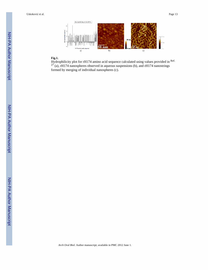
Fig.1.Hydrophilicity plot for rH174 amino acid sequence calculated using values provided in Ref.27 (a), rH174 nanospheres observed in aqueous suspensions (b), and rH174 nanostringsformed by merging of individual nanospheres (c).
Uskoković et al. Page 13
Arch Oral Biol. Author manuscript; available in PMC 2012 June 1.
NIH
-PA Author Manuscript
NIH
-PA Author Manuscript
NIH
-PA Author Manuscript

Fig.2.Hydrodynamic diameters and ζ-potentials of [rH174] = 0.2 mg/ml, 100 mM KCl and 30 mMTris/Bis-Tris/HCl at pH 9 as functions of [Ca2+] and [HxPO4
x-3] in the range of 0 - 8 mM.
Uskoković et al. Page 14
Arch Oral Biol. Author manuscript; available in PMC 2012 June 1.
NIH
-PA Author Manuscript
NIH
-PA Author Manuscript
NIH
-PA Author Manuscript

Fig.3.Hydrodynamic diameters and ζ-potentials of [rH174] = 0.2 mg/ml, 100 mM KCl and 30 mMTris/Bis-Tris/HCl at different pHs and as functions of [Ca2+] and [HxPO4
x-3] in the range of0 - 8 mM.
Uskoković et al. Page 15
Arch Oral Biol. Author manuscript; available in PMC 2012 June 1.
NIH
-PA Author Manuscript
NIH
-PA Author Manuscript
NIH
-PA Author Manuscript

Fig.4.Concentrations of free Ca2+ (●, ■) and HxPO4
x-3 (○) during aging of sols containing: 0.4mg/ml rH174, 1.6 mM CaCl2, 1.0 mM KH2PO4, and 150 mM KCl at 37 °C (●); 0.4 mg/mlrH174, 1.0 mM KH2PO4, and 150 mM KCl at 37 °C (○); and 0.4 mg/ml rH174, 1.6 mMCaCl2, and 150 mM KCl, at 37 °C (■).
Uskoković et al. Page 16
Arch Oral Biol. Author manuscript; available in PMC 2012 June 1.
NIH
-PA Author Manuscript
NIH
-PA Author Manuscript
NIH
-PA Author Manuscript

Fig.5.Hydrodynamic diameters of 0.2 mg/ml rH174 in 100 mM KCl and 30 mM Tris/Bis-Tris/HCl at various pHs and concentrations of calcium and phosphate. Ca2+ and HxPO4
x-3
concentrations higher than or equal to 1.5 mM are denoted with a star shape.
Uskoković et al. Page 17
Arch Oral Biol. Author manuscript; available in PMC 2012 June 1.
NIH
-PA Author Manuscript
NIH
-PA Author Manuscript
NIH
-PA Author Manuscript

Fig.6.Effect of adding CaCl2 and KH2PO4 to rH174 sols that initially contain differentconcentrations of KH2PO4 and CaCl2, respectively, on ζ-potential of rH174 nanospheres atpH 7.4.
Uskoković et al. Page 18
Arch Oral Biol. Author manuscript; available in PMC 2012 June 1.
NIH
-PA Author Manuscript
NIH
-PA Author Manuscript
NIH
-PA Author Manuscript

Fig.7.Change in zeta-potential of HAP (a) and rH174 (b) dispersed in water following the additionof CaCl2 (-●-) and KH2PO4 (-○-) up to 15 mM at 25 °C and at pH 7.40 +- 0.02.
Uskoković et al. Page 19
Arch Oral Biol. Author manuscript; available in PMC 2012 June 1.
NIH
-PA Author Manuscript
NIH
-PA Author Manuscript
NIH
-PA Author Manuscript

Fig.8.Average hydrodynamic diameter (a) and ζ-potential (b) of pure rH174 and the colloidalmixture of HAP and rH174 at [rH174] = 0 – 0.16 mM when rH174 was added in incrementsto HAP suspension comprising 150 mM KCl, 20 mM Tris/HCl, at 25 °C and pH 7.4.
Uskoković et al. Page 20
Arch Oral Biol. Author manuscript; available in PMC 2012 June 1.
NIH
-PA Author Manuscript
NIH
-PA Author Manuscript
NIH
-PA Author Manuscript

Fig.9.Average hydrodynamic diameter (a) and ζ-potential (b) of the colloidal mixture of HAP andrH174 at [rH174] = 0.16 mM when rH174 was abruptly added to the HAP sol comprising150 mM KCl, 20 mM Tris/HCl, at 25 °C and pH 7.40 +- 0.02, and continuously measuredover 18 h aging time.
Uskoković et al. Page 21
Arch Oral Biol. Author manuscript; available in PMC 2012 June 1.
NIH
-PA Author Manuscript
NIH
-PA Author Manuscript
NIH
-PA Author Manuscript

Fig.10.TEM micrographs of a pure HAP particle suspended in water (a) and HAP particles coatedwith rH174 upon their mixing and dispersion in aqueous medium (b-d).
Uskoković et al. Page 22
Arch Oral Biol. Author manuscript; available in PMC 2012 June 1.
NIH
-PA Author Manuscript
NIH
-PA Author Manuscript
NIH
-PA Author Manuscript
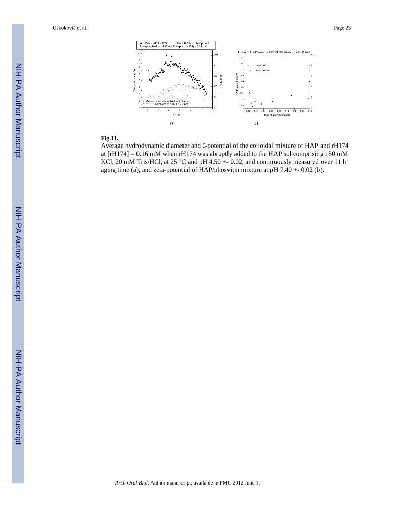
Fig.11.Average hydrodynamic diameter and ζ-potential of the colloidal mixture of HAP and rH174at [rH174] = 0.16 mM when rH174 was abruptly added to the HAP sol comprising 150 mMKCl, 20 mM Tris/HCl, at 25 °C and pH 4.50 +- 0.02, and continuously measured over 11 haging time (a), and zeta-potential of HAP/phosvitin mixture at pH 7.40 +- 0.02 (b).
Uskoković et al. Page 23
Arch Oral Biol. Author manuscript; available in PMC 2012 June 1.
NIH
-PA Author Manuscript
NIH
-PA Author Manuscript
NIH
-PA Author Manuscript