
Dynamic Imaging of the Hepatitis C Virus NS5A Protein during a ...
17
Dynamic Imaging of the Hepatitis C Virus NS5A Protein during a Productive Infection Nicholas S. Eyre, a,b Guillaume N. Fiches, a,b Amanda L. Aloia, a,b Karla J. Helbig, a,b Erin M. McCartney, a,b Christopher S. P. McErlean, c Kui Li, d Anupriya Aggarwal, e Stuart G. Turville, e Michael R. Beard a,b School of Molecular and Biomedical Science and Centre for Molecular Pathology, University of Adelaide, Adelaide, South Australia, Australia a ; Centre for Cancer Biology, SA Pathology, Adelaide, South Australia, Australia b ; School of Chemistry, The University of Sydney, Sydney, New South Wales, Australia c ; Department of Microbiology, Immunology and Biochemistry, University of Tennessee Health Science Center, Memphis, Tennessee, USA d ; Kirby Institute UNSW, Immunovirology and Pathogenesis Program, Darlinghurst, New South Wales, Australia e ABSTRACT Hepatitis C virus (HCV) NS5A is essential for viral genome replication within cytoplasmic replication complexes and virus as- sembly at the lipid droplet (LD) surface, although its definitive functions are poorly understood. We developed approaches to investigate NS5A dynamics during a productive infection. We report here that NS5A motility and efficient HCV RNA replication require the microtubule network and the cytoplasmic motor dynein and demonstrate that both motile and relatively static NS5A-positive foci are enriched with host factors VAP-A and Rab5A. Pulse-chase imaging revealed that newly synthesized NS5A foci are small and distinct from aged foci, while further studies using a unique dual fluorescently tagged infectious HCV chimera showed a relatively stable association of NS5A foci with core-capped LDs. These results reveal new details about the dynamics and maturation of NS5A and the nature of potential sites of convergence of HCV replication and assembly pathways. IMPORTANCE Hepatitis C virus (HCV) is a major cause of serious liver disease worldwide. An improved understanding of the HCV replication cycle will enable development of novel and improved antiviral strategies. Here we have developed complementary fluorescent labeling and imaging approaches to investigate the localization, traffic and interactions of the HCV NS5A protein in living, vi- rus-producing cells. These studies reveal new details as to the traffic, composition and biogenesis of NS5A foci and the nature of their association with putative sites of virus assembly. H epatitis C virus (HCV) is a major cause of serious liver disease worldwide and is the founding member of the Hepacivirus genus within the Flaviviridae family of positive sense RNA viruses. Following entry and uncoating, the 9.6-kb HCV genome is di- rectly translated at the rough endoplasmic reticulum (ER) and the encoded polyprotein is co- and posttranslationally cleaved by host and viral proteases to liberate the structural proteins (core, E1, and E2), the hydrophobic peptide p7 and the nonstructural (NS) pro- teins (NS2, NS3, NS4A, NS4B, NS5A, and NS5B) (reviewed in reference 1). Together with a growing list of essential host factors, the viral NS3-5B proteins are each essential components of cyto- plasmic replication complexes (RCs) that are responsible for rep- lication of the viral genome (2). In contrast, the remaining HCV proteins are dispensable for genome replication, but all play crit- ical roles in the assembly of infectious virus particles (reviewed in reference 3). Interestingly, most if not all of the NS proteins also play essential roles in virus particle assembly. Although NS5A is considered to be a critical regulator of both viral RNA replication in RCs and infectious virion assembly, little is known about the dynamics of RCs and their NS5A-dependent association with sites of virus assembly. Like all positive-strand RNA viruses, HCV induces cytoplas- mic membrane alterations that support and compartmentalize the replication of its genome (4). In this context the NS4B protein has been shown to induce the formation of a convoluted multive- siculated cytoplasmic structure known as the “membranous web” that is at least partly derived from the ER (5, 6). These structures are enriched with other NS proteins and HCV RNA and, at the ultrastructural level, contain numerous heterogeneous single- membrane vesicles, double-membrane vesicles, and multimem- brane vesicles (7–9). Recent studies have indicated that double- membrane vesicles are the likely sites of efficient HCV RNA replication and that NS5A is essential for their formation (7, 9). This function of NS5A may be at least partially attributable to its ability to recruit and activate the lipid kinase phosphatidylinosi- tol-kinase III alpha (PI4KIII) to stimulate the local production of phosphatidylinositol 4-phosphate and induce morphologically normal membranous webs (10, 11). Among a growing list of other host factors that have been described as important cofactors for HCV RNA replication are proteins involved in lipid transport (VAP-A and ANXA2) and early endosome regulation and traffic (Rab5 and EEA1) (12–14). Another essential property of RCs is their association with sites of virus particle assembly (4). In this context a function(s) of NS5A that maps to its C terminus (domain III) is thought to dictate the transfer of HCV RNA between RCs and core-coated LDs for encapsidation, potentially via a direct core-NS5A interac- Received 11 September 2013 Accepted 24 December 2013 Published ahead of print 15 January 2014 Editor: M. S. Diamond Address correspondence to Michael R. Beard, [email protected]. Supplemental material for this article may be found at http://dx.doi.org/10.1128 /JVI.02490-13. Copyright © 2014, American Society for Microbiology. All Rights Reserved. doi:10.1128/JVI.02490-13 3636 jvi.asm.org Journal of Virology p. 3636 –3652 April 2014 Volume 88 Number 7 on March 25, 2018 by guest http://jvi.asm.org/ Downloaded from
-
Upload
phungtuyen -
Category
Documents
-
view
215 -
download
0
Transcript of Dynamic Imaging of the Hepatitis C Virus NS5A Protein during a ...

Dynamic Imaging of the Hepatitis C Virus NS5A Protein during aProductive Infection
Nicholas S. Eyre,a,b Guillaume N. Fiches,a,b Amanda L. Aloia,a,b Karla J. Helbig,a,b Erin M. McCartney,a,b Christopher S. P. McErlean,c
Kui Li,d Anupriya Aggarwal,e Stuart G. Turville,e Michael R. Bearda,b
School of Molecular and Biomedical Science and Centre for Molecular Pathology, University of Adelaide, Adelaide, South Australia, Australiaa; Centre for Cancer Biology,SA Pathology, Adelaide, South Australia, Australiab; School of Chemistry, The University of Sydney, Sydney, New South Wales, Australiac; Department of Microbiology,Immunology and Biochemistry, University of Tennessee Health Science Center, Memphis, Tennessee, USAd; Kirby Institute UNSW, Immunovirology and PathogenesisProgram, Darlinghurst, New South Wales, Australiae
ABSTRACT
Hepatitis C virus (HCV) NS5A is essential for viral genome replication within cytoplasmic replication complexes and virus as-sembly at the lipid droplet (LD) surface, although its definitive functions are poorly understood. We developed approaches toinvestigate NS5A dynamics during a productive infection. We report here that NS5A motility and efficient HCV RNA replicationrequire the microtubule network and the cytoplasmic motor dynein and demonstrate that both motile and relatively staticNS5A-positive foci are enriched with host factors VAP-A and Rab5A. Pulse-chase imaging revealed that newly synthesized NS5Afoci are small and distinct from aged foci, while further studies using a unique dual fluorescently tagged infectious HCV chimerashowed a relatively stable association of NS5A foci with core-capped LDs. These results reveal new details about the dynamicsand maturation of NS5A and the nature of potential sites of convergence of HCV replication and assembly pathways.
IMPORTANCE
Hepatitis C virus (HCV) is a major cause of serious liver disease worldwide. An improved understanding of the HCV replicationcycle will enable development of novel and improved antiviral strategies. Here we have developed complementary fluorescentlabeling and imaging approaches to investigate the localization, traffic and interactions of the HCV NS5A protein in living, vi-rus-producing cells. These studies reveal new details as to the traffic, composition and biogenesis of NS5A foci and the nature oftheir association with putative sites of virus assembly.
Hepatitis C virus (HCV) is a major cause of serious liver diseaseworldwide and is the founding member of the Hepacivirus
genus within the Flaviviridae family of positive sense RNA viruses.Following entry and uncoating, the �9.6-kb HCV genome is di-rectly translated at the rough endoplasmic reticulum (ER) and theencoded polyprotein is co- and posttranslationally cleaved by hostand viral proteases to liberate the structural proteins (core, E1, andE2), the hydrophobic peptide p7 and the nonstructural (NS) pro-teins (NS2, NS3, NS4A, NS4B, NS5A, and NS5B) (reviewed inreference 1). Together with a growing list of essential host factors,the viral NS3-5B proteins are each essential components of cyto-plasmic replication complexes (RCs) that are responsible for rep-lication of the viral genome (2). In contrast, the remaining HCVproteins are dispensable for genome replication, but all play crit-ical roles in the assembly of infectious virus particles (reviewed inreference 3). Interestingly, most if not all of the NS proteins alsoplay essential roles in virus particle assembly. Although NS5A isconsidered to be a critical regulator of both viral RNA replicationin RCs and infectious virion assembly, little is known about thedynamics of RCs and their NS5A-dependent association with sitesof virus assembly.
Like all positive-strand RNA viruses, HCV induces cytoplas-mic membrane alterations that support and compartmentalizethe replication of its genome (4). In this context the NS4B proteinhas been shown to induce the formation of a convoluted multive-siculated cytoplasmic structure known as the “membranous web”that is at least partly derived from the ER (5, 6). These structuresare enriched with other NS proteins and HCV RNA and, at theultrastructural level, contain numerous heterogeneous single-
membrane vesicles, double-membrane vesicles, and multimem-brane vesicles (7–9). Recent studies have indicated that double-membrane vesicles are the likely sites of efficient HCV RNAreplication and that NS5A is essential for their formation (7, 9).This function of NS5A may be at least partially attributable to itsability to recruit and activate the lipid kinase phosphatidylinosi-tol-kinase III alpha (PI4KIII�) to stimulate the local productionof phosphatidylinositol 4-phosphate and induce morphologicallynormal membranous webs (10, 11). Among a growing list of otherhost factors that have been described as important cofactors forHCV RNA replication are proteins involved in lipid transport(VAP-A and ANXA2) and early endosome regulation and traffic(Rab5 and EEA1) (12–14).
Another essential property of RCs is their association with sitesof virus particle assembly (4). In this context a function(s) ofNS5A that maps to its C terminus (domain III) is thought todictate the transfer of HCV RNA between RCs and core-coatedLDs for encapsidation, potentially via a direct core-NS5A interac-
Received 11 September 2013 Accepted 24 December 2013
Published ahead of print 15 January 2014
Editor: M. S. Diamond
Address correspondence to Michael R. Beard, [email protected].
Supplemental material for this article may be found at http://dx.doi.org/10.1128/JVI.02490-13.
Copyright © 2014, American Society for Microbiology. All Rights Reserved.
doi:10.1128/JVI.02490-13
3636 jvi.asm.org Journal of Virology p. 3636 –3652 April 2014 Volume 88 Number 7
on March 25, 2018 by guest
http://jvi.asm.org/
Dow
nloaded from
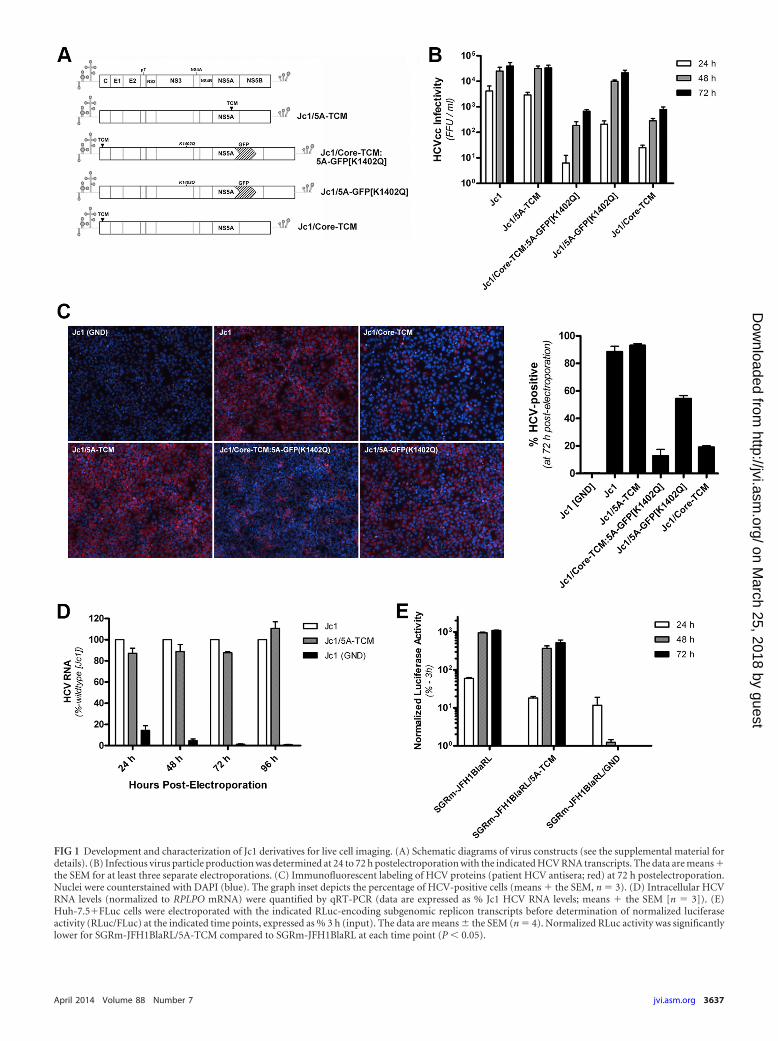
FIG 1 Development and characterization of Jc1 derivatives for live cell imaging. (A) Schematic diagrams of virus constructs (see the supplemental material fordetails). (B) Infectious virus particle production was determined at 24 to 72 h postelectroporation with the indicated HCV RNA transcripts. The data are means �the SEM for at least three separate electroporations. (C) Immunofluorescent labeling of HCV proteins (patient HCV antisera; red) at 72 h postelectroporation.Nuclei were counterstained with DAPI (blue). The graph inset depicts the percentage of HCV-positive cells (means � the SEM, n � 3). (D) Intracellular HCVRNA levels (normalized to RPLPO mRNA) were quantified by qRT-PCR (data are expressed as % Jc1 HCV RNA levels; means � the SEM [n � 3]). (E)Huh-7.5�FLuc cells were electroporated with the indicated RLuc-encoding subgenomic replicon transcripts before determination of normalized luciferaseactivity (RLuc/FLuc) at the indicated time points, expressed as % 3 h (input). The data are means � the SEM (n � 4). Normalized RLuc activity was significantlylower for SGRm-JFH1BlaRL/5A-TCM compared to SGRm-JFH1BlaRL at each time point (P � 0.05).
April 2014 Volume 88 Number 7 jvi.asm.org 3637
on March 25, 2018 by guest
http://jvi.asm.org/
Dow
nloaded from

Eyre et al.
3638 jvi.asm.org Journal of Virology
on March 25, 2018 by guest
http://jvi.asm.org/
Dow
nloaded from

tion (15–17). Although new details about the biogenesis of HCVRCs and their association with potential sites of virus assembly areemerging, there is a paucity of information regarding the dynam-ics of these events. In the present study we have developed ap-proaches to visualize the localization and traffic of fluorescentlytagged NS5A during a productive HCV replication cycle. We showthat long-range motility of NS5A-positive structures (putativeRCs) is dependent on host microtubules and cytoplasmic dyneinand that both relatively static and motile NS5A-positive structuresare enriched with previously identified co-opted host factors. Us-ing pulse-chase imaging, we show that newly synthesized NS5A islocalized to foci that are smaller than and distinct from those ofaged NS5A, although both classes display characteristics of micro-tubule-dependent transport. Finally, for the first time, we havevisualized the association of putative RCs with sites of virus assem-bly in living cells and suggest that weak and/or transient interac-tions of HCV core and NS5A proteins may link HCV replicationand assembly pathways. Collectively, these studies provide newdetails as to the dynamics, components, and biogenesis of NS5Afoci and their association with sites of virion assembly.
MATERIALS AND METHODSViruses, plasmids, siRNAs, and cells. Plasmids pJc1/GFP (18) and pJFH1(19) were generously provided by Ralf Bartenschlager (University ofHeidelberg, Heidelberg, Germany) and Takaji Wakita (National Institute ofInfectious Diseases, Tokyo, Japan), respectively. Subgenomic repliconSGRm-JFH1BlaRL and replication-defective derivative SGRm-JFH1BlaRL/GND have been described previously (20). Detailed descriptions of alltagged Jc1- and SGRm-JFH1BlaRL derivatives and lentiviral expression/shRNA vectors are provided in the supplemental material, as are detailsregarding in vitro HCV RNA transcription, electroporation/transfection,infections, and determination of HCV infectivity. CellLight Tubulin-RFP(Life Technologies) was used to visualize microtubules according to man-ufacturer’s instructions. Nontargeting and DYNC1H1-specific small in-terfering RNAs (siRNAs) were purchased from Life Technologies(Silencer Select; catalogue numbers s4202 and 4390846, respectively).
Huh-7.5 cells (21) were generously provided by Charles Rice (Rocke-feller University, New York, NY). Huh-7.5 and 293T cells were cultured incomplete Dulbecco modified Eagle medium (i.e., Dulbecco modified Ea-gle medium [DMEM] supplemented with 100 U of penicillin/ml, 100 mgof streptomycin/ml, and 10% fetal bovine serum [Life Technologies]).Details regarding the generation of stable Huh-7.5-derived cell lines aregiven in the supplemental material. For live cell imaging studies, cells werecultured in complete DMEM lacking phenol red.
Antibodies and chemical inhibitors. Primary antibodies againstNS5A (mouse monoclonal antibody [MAb] 9E10) and core (rabbit poly-clonal RR8) were generously provided by Charles Rice (Rockefeller Uni-versity) and Michinori Kohara (Tokyo Metropolitan Institute of MedicalScience, Tokyo, Japan), respectively. Mouse MAbs against core (C7-50;Thermo Scientific), FLAG (M2; Sigma), Myc (9E10; Roche Applied Sci-
ence), and �-actin (AC-15; Sigma) were purchased. Mouse IgG1() andIgG2a() isotype control MAbs (BD Pharmingen) and rabbit polyclonalantibodies against VAP-A (Novus Biologicals), Rab5 (Cell SignalingTechnology), DYNC1H1 (Santa Cruz; R-325), and GAPDH (glyceralde-hyde-3-phosphate dehydrogenase; Rockland Immunochemicals) werepurchased as indicated. Human anti-HCV antiserum was prepared asdescribed previously (22). Alexa Fluor-350-, -488-, -555-, and -594-con-jugated secondary antibodies (Life Technologies) and horseradish perox-idase-conjugated secondary antibodies (Thermo Scientific) were pur-chased as indicated. Chemical inhibitors are detailed in the supplementalmaterial.
Labeling of TCM-tagged proteins with biarsenical dyes. The biar-senical dyes FlAsH-EDT2 and ReAsH-EDT2 were synthesized as describedpreviously (23). Labeling of tetracysteine motif (TCM)-tagged HCV pro-teins was performed as described previously (24), with minor modifica-tions. Briefly, for each 35-mm dish of cells, 1 l of 1 mM FlAsH (orReAsH) solution (prepared in dimethyl sulfoxide [DMSO]) was mixedwith 1 l of 25 mM 1,2-ethanedithiol (EDT; Sigma) (diluted in DMSO)and then incubated for 10 min at room temperature. Meanwhile, cellswere washed twice with complete Hanks balanced salt solution (Life Tech-nologies) containing 5.6 mM glucose (HBSS/glucose) before the additionof 1 ml of HBSS/glucose. Next, 1 ml of HBSS/glucose was added to a newtube, and a 100-l aliquot of this was taken and added to the FlAsH/EDTsolution and mixed by pipetting. After incubation for 5 min at roomtemperature, this FlAsH/EDT/HBSS/glucose solution was mixed with theremaining 900 l of HBSS/glucose and added to the cell culture dish toachieve a final concentration of 500 nM FlAsH-EDT2. The cells were thenreturned to culture at 37°C for 30 min before being washed twice withHBSS/glucose and three times at 37°C for 15 min with HBSS/glucosecontaining 2.8 mM 2,3-dimercapto-1-propanol (Sigma). The cells werethen washed twice with HBSS/glucose and fixed for immunofluorescentlabeling or harvested by trypsinization and reseeded into 0.2% gelatin-coated 35-mm coverslip bottom dishes (MatTek) and returned to culturefor 24 h prior to live cell imaging.
Labeling of SNAP-tagged NS5A with fluorescent SNAP-tag sub-strates. Labeling of SNAP-tagged NS5A with fluorescent SNAP-tag sub-strates, SNAP-Cell 505 and SNAP-Cell TMR-Star, was performed accord-ing to the manufacturer’s instructions (New England BioLabs).
In situ PLAs. In situ proximity ligation assays (PLAs) were performedusing a Duolink II In Situ Red Starter Kit (Olink Bioscience) according tothe manufacturer’s instructions. Briefly, cells were first cultured on 0.2%gelatin-coated coverslips, fixed with ice-cold acetone-methanol (1:1) for10 min at 4°C, permeabilized, blocked, and incubated with primary anti-bodies (mouse anti-core [C7-50] � rabbit anti-core [RR8] or mouse anti-NS5A [9E10] � rabbit anti-core [RR8]; each diluted 1:200). Samples werethen washed three times with phosphate-buffered saline (PBS)–1% bo-vine serum albumin (BSA) and incubated with appropriately diluted PLAprobes (anti-Mouse MINUS and anti-Rabbit PLUS diluted in PBS–1%BSA [1:1:3]) for 1 h at 37°C in a humidity chamber. The samples were thenwashed and processed for probe ligation, signal amplification, andmounting according to manufacturer’s instructions.
FIG 2 Specific labeling of TCM-tagged HCV proteins with biarsenical dyes. (A) At 3 days postelectroporation with Jc1/5A-TCM RNA, Huh-7.5 cells were labeledwith FlAsH (green; left panel), fixed, and processed for immunofluorescence with anti-NS5A antibody (red; middle panel). Merged images (right panel) showcolocalization (yellow) and DAPI-stained nuclei (blue) (Pearson correlation � 0.95). (B) At 3 days postelectroporation with Jc1/Core-TCM:5A-GFP[K1402Q]RNA, Huh-7.5 cells were labeled with ReAsH (red; top left panel), fixed, and processed for immunofluorescent labeling of core (blue; top middle panel).NS5A-GFP epifluorescence was also captured (green; top right panel). Insets and merged images are depicted in the lower panels. Pearson correlations forcolocalization of ReAsH-labeling with antibody labeling and NS5A-GFP epifluorescence were 0.84 and 0.65, respectively. (C) Live cell imaging of NS5A-TCM.At 48 h postelectroporation with Jc1/5A-TCM RNA, Huh-7.5 cells were labeled with ReAsH and examined by live cell imaging at 72 h. Images were acquired every5.3 s for 2 min (see Movie S1 in the supplemental material). The velocities and path lengths of a number of discrete NS5A-positive are depicted in adjacent graphs.(D) Live cell imaging of NS5A-GFP at 4 days postinfection with Jc1/5A-GFP[K1402Q] (multiplicity of infection [MOI] of �0.05) (see Movie S2 in thesupplemental material). Images were acquired every 2 s for 3 min. The insets depict the traffic of a representative motile NS5A-GFP structure (arrows). Imagesharpening (using a Gaussian blur filter with a radius of 1.5 pixels), background subtraction, and contrast stretching were then applied (see the supplementalmaterial). For all micrographs, scale bars represent 10 and 5 m for main and inset images, respectively.
Dynamic Imaging of HCV NS5A Protein
April 2014 Volume 88 Number 7 jvi.asm.org 3639
on March 25, 2018 by guest
http://jvi.asm.org/
Dow
nloaded from

Fluorescence microscopy. Laser scanning confocal microscopy wasperformed using a Bio-Rad Radiance 2100 confocal system linked to anOlympus IX70 inverted microscope or a Zeiss LSM 700 confocal micro-scope system. For both systems, samples were visualized using a �60/1.4NA water immersion objective lens (with a �2 to �3 zoom), andimages were acquired sequentially for each fluorophore. Wide-field fluo-rescence microscopy was performed using a Nikon TiE inverted fluores-cence microscope using a Plan Apochromat �60 NA 1.4 oil immersionlens or a Super Plan Fluor �40 NA 0.4 or Plan Fluor �10 NA 0.3 objectivelens (Nikon), as appropriate. Illumination was provide by an IntensilightC-HGFIE precentered fiber illuminator mercury light source (Nikon),while BrightLine single-band filter sets (DAPI-5060C-NTE-ZERO, FITC-3540C-NTE-ZERO, and TxRed-4040C-NTE-ZERO) were from Semrock,and emitted light was collected with a monochrome 12-bit cooled charge-coupled device camera with a maximum resolution of 1,280�1,024 (DS-Qi1;Nikon). For live cell imaging, cells cultured on coverslip bottom 35-mmdishes (MatTek) were maintained at 37°C using a heated microscope stageinsert (Okolab) and visualized using the above �60 NA 1.4 oil immersionobjective lens. For some of these experiments, a Nikon perfect focus sys-tem was applied. Unless otherwise indicated (see movie legends in thesupplemental material), images were acquired (without binning) every 3 susing exposure times of 1 s and a maximum camera gain setting of 2.Image processing and analysis is described in the supplemental material.
Statistical analysis. Data are expressed as means � the standard errorsof the mean (SEM). Statistical analysis was performed by using the Stu-dent t test or the Mann-Whitney U-test using Prism 5 (GraphPad Soft-ware), with P � 0.05 considered to be statistically significant.
RESULTSDevelopment and characterization of infectious HCV deriva-tives for live cell imaging of NS5A and core. To track NS5A lo-calization with minimal disruption of the HCV life cycle, we usedcomplementary fluorescent tagging approaches. First, we insertedan optimized tetracysteine motif (TCM; amino acids FLNCCPGCCMEP) within domain III of NS5A in the infectious HCV chi-mera Jc1 (25), to generate Jc1/5A-TCM and enable covalent label-ing of encoded NS5A-TCM with fluorescent biarsenical dyes(FlAsH/ReAsH) for live cell imaging. Second, to complement ourfindings with Jc1/5A-TCM, we incorporated the adaptive muta-tion K1402Q (11) into Jc1/GFP (18), which encodes a GFP inser-tion in domain III of NS5A in Jc1, to generate Jc1/5A-GFP[K1402Q]. Similarly, to enable simultaneous visualizationof the localization and traffic of HCV NS5A and core proteins,we inserted a TCM within the HCV core coding sequence (26, 27) ofJc1/5A-GFP[K1402Q], to generate Jc1/Core-TCM:5A-GFP[K1402Q](Fig. 1A). Compared to parent Jc1, the incorporation of a TCMwithin NS5A had no appreciable effect on NS5A processing (notshown) and no significant impact on infectious virus production(Fig. 1B) or the spread of HCV infection through Huh-7.5 cellculture (Fig. 1C) or intracellular HCV RNA levels (Fig. 1D). Ac-cordingly, incorporation of a TCM within NS5A in a Renilla lu-ciferase (RLuc)-encoding JFH1-derived subgenomic replicon hadminimal impact on its replicative capacity (Fig. 1E). As reportedpreviously (11), we found that the incorporation of the K1402Qadaptive mutation into Jc1/GFP restored infectivity to levels ap-proaching those of untagged Jc1, despite delayed kinetics (Fig.1B). Jc1/Core-TCM:5A-GFP[K1402Q] displayed an �1.5-log im-pairment of infectious virus production compared to untaggedJc1 and delayed spread of infection through Huh-7.5 cell culture(Fig. 1B and C) that could be attributed predominantly to theTCM insertion in core (Fig. 1B). Importantly, neither TCM norGFP insertions in NS5A nor the TCM insertion in core apprecia-
bly altered the localization of these proteins or the degree of core/NS5A colocalization in Huh-7.5 cells (not shown).
We next confirmed specific labeling of TCM-tagged HCV pro-teins with fluorescent biarsenical dyes. Huh-7.5 cells were electro-porated with Jc1/5A-TCM RNA, cultured, and labeled withFlAsH, before fixation and immunofluorescent labeling of NS5A.This revealed near-complete overlap between the signals associ-ated with FlAsH- and antibody-mediated labeling of NS5A-TCM,with minimal labeling of uninfected cells (Pearson correlation �0.95) (Fig. 2A). The specificity of labeling of core-TCM encodedby Jc1/Core-TCM:5A-GFP[K1402Q] was similarly determined,except that cells were labeled with ReAsH and an anti-core anti-body. This revealed a close overlap between ReAsH (red)- andantibody (blue)-mediated labeling of core-TCM (Pearson corre-lation � 0.84) and a partial overlap of ReAsH signals with NS5A-GFP epifluorescence (Pearson correlation � 0.65) (Fig. 2B). As-sessment of the impact of biarsenical dye labeling on infectiousvirus production revealed a 50.6% � 3.3% decrease (means � theSEM; n � 3) in infectious virus production over a 24-h periodfollowing labeling (not shown).
Next, we examined the localization and traffic of NS5A in liv-ing cells during a productive HCV infection. Huh-7.5 cells wereelectroporated with Jc1/5A-TCM RNA, labeled with ReAsH andanalyzed by live cell imaging (Fig. 2C; see Movie S1 in the supple-mental material). This labeling strategy, with a 24-h culture periodbetween FlAsH/ReAsH labeling and live cell imaging, was used tominimize background labeling and limit any unanticipated effectsof labeling, such as the exposure of cells to reducing agents andserum deprivation. ReAsH-labeled NS5A-TCM was predomi-nantly found in large, relatively static structures and smallerhighly motile structures that shuttle throughout the cytoplasm ina sporadic manner (Fig. 2C; see Movie S1 in the supplementalmaterial). Manual tracking analysis demonstrated that thesestructures traffic at velocities in the order of 1 m s�1 and fre-quently pause and change direction, sometimes moving backalong what appear to be the same paths (e.g., “Object 3”). Consis-tent with this, in cells that had been infected with Jc1/5A-GFP[K1402Q], GFP-tagged NS5A displayed similar localizationand trafficking patterns (Fig. 2D and see Movie S2 in the supple-mental material). Further analysis of NS5A-GFP traffic at hightemporal resolution (16 frames s�1), which more readily distin-guishes velocity changes within bursts, revealed that typical motileNS5A-GFP foci display bidirectional traffic at peak velocities of�5 m s�1 for runs of �2 m (not shown). Collectively, theseresults show that in a productive infection TCM-tagged NS5A andcore proteins can be specifically labeled with fluorescent biarseni-cal dyes and that NS5A localizes to both relatively static structuresand highly motile structures that traffic sporadically throughoutthe cytoplasm. These results raise questions as to the mechanismsand purpose of NS5A motility and whether both motile and im-motile NS5A foci represent RCs.
NS5A traffic is dependent on the host microtubule networkand cytoplasmic dynein. In the context of a subgenomic HCVreplicon it has previously been shown that NS5A traffic is micro-tubule dependent (28). To confirm and extend this work, we ob-served the traffic of motile NS5A-positive structures in closeassociation with �-tubulin-RFP-positive microtubules during aproductive infection (Fig. 3A and see Movie S3 in the supplemen-tal material) and demonstrated that treatment of parallel cultureswith inhibitors of microtubule dynamics, nocodazole and vinblas-
Eyre et al.
3640 jvi.asm.org Journal of Virology
on March 25, 2018 by guest
http://jvi.asm.org/
Dow
nloaded from

tin, resulted in arrest of these structures and no apparent linearfoci movement (Fig. 3B). Furthermore, consistent with earlierstudies (29), we found that transient disruption of microtubuledynamics dramatically inhibited HCV RNA replication (Fig. 3C).
We next investigated the involvement of the motor proteindynein in the traffic of NS5A-positive foci, given the central role ofthis motor in microtubule-dependent vesicular/organellar traffic,
including that of early endosomes (see references 30 and 31 andreferences therein) which are co-opted by HCV (13). First, weinvestigated the impact of the selective cytoplasmic dynein inhib-itor ciliobrevin D (32) on HCV RNA replication. For this, Huh-7.5�FLuc cells were electroporated with mSGR-JFH1BlaRL RNAand treated 48 h later with ciliobrevin D at various concentrationsfor 24 h. Dual luciferase assays revealed a ca. 40% reduction in
FIG 3 Involvement of the microtubule network in NS5A-TCM traffic and HCV replication. (A) Huh-7.5 cells were electroporated with Jc1/5A-TCM RNA,labeled with FlAsH (green) at 48 h postelectroporation, transduced with CellLight Tubulin-RFP (red), and analyzed by live cell imaging at 72 h postelectropo-ration. Images were acquired every 7.5 s for 3 min. Image sharpening (using a Gaussian blur filter with a radius of 1.5 pixels), background subtraction, andcontrast stretching were then applied (see Materials and Methods). The traffic of a representative NS5A-TCM-positive structure (arrowhead) in close proximityto RFP-labeled microtubules is depicted in the inset images (see Movie S3 in the supplemental material). Scales bars represent 10 and 5 m for main and insetimages, respectively. (B) Huh-7.5 cells were electroporated, transduced, and labeled, as described above, before exposure to 20 M nocodazole, 20 Mvinblastine, or carrier alone (0.1% DMSO) for 1 h and live cell imaging. These treatments induced gross disruption of the microtubule network, as judged bydiffuse cytoplasmic localization of �-tubulin-RFP (not shown). The graph depicts the mean velocities of arbitrarily selected NS5A-TCM-positive structures overthe course of 3 min (n � 20 to 25 structures/group). Compared to the controls the mean velocities of NS5A-TCM-positive structures were significantly differentfor the nocodazole (***, P � 0.0001) and vinblastine (***, P � 0.0001) treatment groups (unpaired Student t tests). (C) Huh-7.5�FLuc cells were electroporatedwith SGRm-JFH1BlaRL or SGRm-JFH1BlaRL/GND transcripts and exposed to carrier (0.1% DMSO), 20 M nocodazole, or 20 M vinblastine for 1 h at 24 hpostelectroporation before being washed and returned to culture. Samples were collected at the indicated time points before determination of the normalizedluciferase activity (RLuc:FLuc, to normalize HCV replication levels to cell number/viability), and values are expressed as a percentage of normalized input valuesat 4 h. The data are means � the SEM (n � 4).
Dynamic Imaging of HCV NS5A Protein
April 2014 Volume 88 Number 7 jvi.asm.org 3641
on March 25, 2018 by guest
http://jvi.asm.org/
Dow
nloaded from

normalized replicon-encoded RLuc activity for cells exposed to100 M ciliobrevin D (Fig. 4A). Consistent with this, we foundthat siRNA knockdown of the heavy chain of cytoplasmic dynein1 (DYNC1H1) significantly inhibited normalized HCV replicon-
encoded RLuc activity (Fig. 4B, upper panel) to a degree that wassimilar to the degree of knockdown of DYNC1H1 protein, deter-mined by Western blotting of lysates from the same experiment(Fig. 4B, lower panel).
FIG 4 Involvement of cytoplasmic dynein in HCV RNA replication and NS5A motility. (A) Huh-7.5�FLuc cells were electroporated with SGRm-JFH1BlaRLRNA and cultured for 48 h before exposure to ciliobrevin D (or 1% DMSO) for 24 h and determination of normalized luciferase activity (RLuc/FLuc). The dataare expressed as the % control. (B) Huh-7.5�FLuc cells were cotransfected with SGRm-JFH1BlaRL RNA (1 g) and either nontargeting or DYNC1H1-specificsiRNAs (118 pmol) before determination of normalized luciferase activity at the indicated time points. The data are expressed as the % 3 h (input). For panelsA and B, data are means � the SEM (n � 4). **, P � 0.01; ***, P � 0.001 (Student t test). (C) Huh-7.5 cells were coelectroporated with Jc1/5A-GFP[K1402Q]RNA (5 g) and either nontargeting or DYNC1H1-specific siRNAs (1 nmol of each) and cultured for 48 h prior to live cell imaging. Images were acquired every3 s for 3 min. Images (taken from Movie S4 in the supplemental material) are color-coded projections of entire time-lapse acquisitions. (D) Manual tracking ofNS5A-GFP foci in control and DYNC1H1 siRNA-transfected cells. Motile structures (identified as being displaced in the first 6 s of each time-lapse acquisition)were subjected to manual tracking analysis, excluding structures that moved �10 m in 3 min. Path lengths for control (n � 92 foci) and DYNC1H1siRNA-transfected (n � 63 foci) cells are depicted in panel D. (E and F) The frequencies of structures that display the indicated path lengths are depicted in panelE, and total path lengths are depicted in panel F. Horizontal bars represent mean values for control and DYNC1H1 siRNA transfected cells (n � 65 and 48 foci,respectively). ***, P � 0.001, Mann-Whitney U-test.
Eyre et al.
3642 jvi.asm.org Journal of Virology
on March 25, 2018 by guest
http://jvi.asm.org/
Dow
nloaded from

To correlate these effects with changes in NS5A traffic, Huh-7.5 cells were coelectroporated with Jc1/5A-GFP[K1402Q] RNAand either control or DYNC1H1 siRNAs before live cell imaging(Fig. 4C; see Movie S4 in the supplemental material). Althoughmotile NS5A-GFP foci were still readily detectable in DYNC1H1siRNA-transfected cells, the frequency of long-range NS5A-GFPfoci movements ( 20 m/3 min) and the average path length ofmotile NS5A-GFP foci were significantly reduced in DYNC1H1siRNA-transfected cells compared to controls (Fig. 4D to F). To-gether, these results show that the microtubule network is essen-tial for NS5A foci motility and efficient HCV RNA replication andthat cytoplasmic dynein plays an important role in long-rangetraffic of NS5A foci and efficient HCV RNA replication.
NS5A colocalizes and cotraffics with proviral host factors.The identification of relatively static and motile subclasses ofNS5A foci led us to investigate their composition in regard to hostfactors that are known to be important for HCV RNA replication.One such factor is vesicle-associated membrane protein-associ-ated protein A (VAP-A); a SNARE-like protein that interacts withNS5A and NS5B and may help to anchor these proteins to ER-derived membranes that are sites of HCV replication (14, 33, 34).To examine the localization and traffic of NS5A with respect toVAP-A, Huh-7.5�mCherry-VAP-A cells were electroporatedwith Jc1/5A-TCM RNA transcripts, labeled with FlAsH, and visu-alized by live cell imaging (Fig. 5A and see Movie S5 in the sup-plemental material). Both relatively static and motile NS5A-posi-
FIG 5 Colocalization and cotraffic of NS5A with VAP-A. (A) Huh-7.5�mCherry-VAP-A cells were electroporated with Jc1/5A-TCM RNA, labeled with FlAsH(green) at 48 h and analyzed by live cell imaging at 72 h (see Movie S5 in the supplemental material). Inset 1 shows enrichment of mCherry-VAP-A in bothrelatively static and motile NS5A-TCM-positive structures (arrowheads and arrows, respectively), while inset 2 depicts the additional reticular localization ofmCherry-VAP-A and the movements of two representative motile NS5A-TCM foci. (B) Huh-7.5�mCherry-VAP-A cells were electroporated with Jc1/5A-GFP[K1402Q] RNA and analyzed by live cell imaging at 72 h postelectroporation (see Movie S6 in the supplemental material). A 16.2-m line-scan (3 pixelswide) was used to generate the inset kymograph. (C) Huh-7.5�mCherry-VAP-A cells were infected with Jc1/5A-GFP[K1402Q] (MOI of �0.05) and analyzedby live cell imaging at 3 days postinfection. Scale bars are 10 and 5 m for main and inset images, respectively.
Dynamic Imaging of HCV NS5A Protein
April 2014 Volume 88 Number 7 jvi.asm.org 3643
on March 25, 2018 by guest
http://jvi.asm.org/
Dow
nloaded from

Eyre et al.
3644 jvi.asm.org Journal of Virology
on March 25, 2018 by guest
http://jvi.asm.org/
Dow
nloaded from

tive structures were enriched with mCherry-VAP-A, indicatingthat the presence of VAP-A does not distinguish between staticand motile putative replication complexes (Fig. 5A, inset 1; seeMovie S5 in the supplemental material). Furthermore, motileNS5A-positive structures appeared to traffic along VAP-A-posi-tive ER-like membranes (Fig. 5A, inset 2; see Movie S5 in thesupplemental material). Similarly, in Huh-7.5�mCherry-VAP-Acells that were electroporated with Jc1/5A-GFP[K1402Q] tran-scripts NS5A-GFP and mCherry-VAP-A colocalized (Pearsoncorrelation � 0.83 � 0.02 [n � 9 cells]) and cotrafficked, in closeassociation with mCherry-VAP-A-positive ER tubules (Fig. 5Band see Movie S6 in the supplemental material). This cotraffickingwas also observed in an infection scenario, indicating that colocal-ization was not an artifact of HCV RNA electroporation (Fig. 5C).Although effective shRNA knockdown of VAP-A (Fig. 6A) inhib-ited HCV RNA replication (Fig. 6C), neither the subcellular local-ization of NS5A (Fig. 6B) nor NS5A-TCM traffic (not shown)were appreciably altered, suggesting that VAP-A is not a majordeterminant of NS5A localization and traffic. Furthermore, FRETanalysis revealed the interaction of mCherry-VAP-A with NS5A-GFP in cytoplasmic foci of varying size in Jc1/5A-GFP[K1402Q]-infected Huh-7.5�mCherry-VAP-A cells (Fig. 6D), suggestingthat the interaction of these proteins is not restricted to a partic-ular subset of NS5A-positive structures.
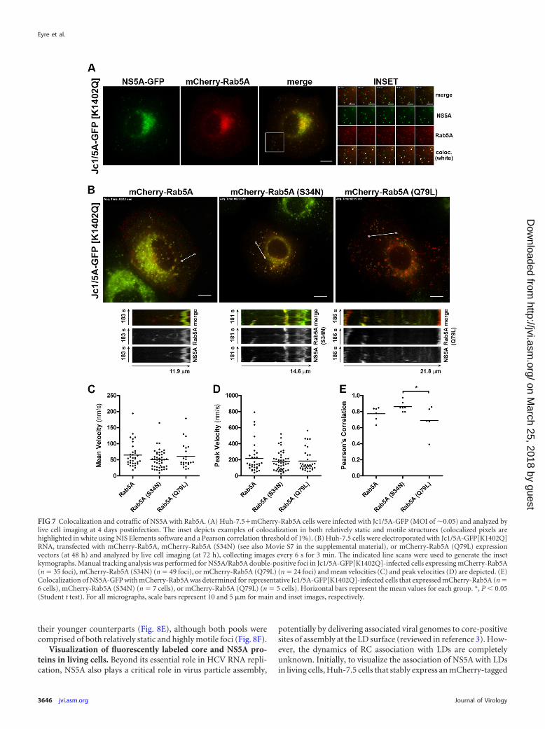
Another host factor that is reported to be important for HCVRNA replication is the early endosomal protein Rab5A (13, 35).Rab5A regulates tethering and homotypic fusion of early endo-somes and has been shown to interact with NS4B and influencethe formation of sites of HCV RNA replication (35, 36). First, weinvestigated the influence of Rab5A overexpression and GTPaseactivity on HCV RNA replication, given previous reports thatsiRNA knockdown of Rab5A inhibits HCV RNA replication (13,36). These experiments revealed minor inhibitory effects of induc-ible mCherry-Rab5A, mCherry-Rab5A (S34N), and mCherry-Rab5A (Q79L) overexpression on HCV RNA replication (Fig. 6E).Second, we examined the localization and traffic of NS5A withrespect to Rab5A by live cell imaging. Infection of Huh-7.5 cellsthat constitutively express low levels of mCherry-tagged Rab5Awith Jc1/5A-GFP[K1402Q] and live cell imaging revealed a closeoverlap in the localization and traffic of NS5A-GFP and mCherry-Rab5A in both small, motile structures and larger, relativelystatic structures (Fig. 7A). To complement these findings andinvestigate the influence of Rab5A GTPase activity, Jc1/5A-
GFP[K1402Q]-infected Huh-7.5 cells were transiently transfectedwith wild-type, GDP-locked (S34N) or GTP-locked (Q79L)mCherry-Rab5A expression constructs, before live cell imaging.While NS5A-GFP extensively colocalized and cotrafficked withwild-type, GDP-locked (S34N), and GTP-locked (Q79L)mCherry-Rab5A proteins in small mCherry-Rab5A-positivestructures, it was largely excluded from enlarged mCherry-Rab5A(Q79L)-positive endosomes (Fig. 7B and C), such that co-localization of NS5A-GFP with mCherry-Rab5A (Q79L) was sig-nificantly lower than that of mCherry-Rab5A (S34N) (extensivecolocalization and cotraffic of NS5A-GFP with mCherry-Rab5A[S34N] is depicted in Movie S7 in the supplemental mate-rial). However, manual tracking analysis revealed that, comparedto the wild type, neither average nor peak velocities of NS5A-positive structures were significantly altered by the presence ofGDP-locked or GTP-locked mCherry-Rab5A (Fig. 7D). Althoughmore complex aspects of NS5A traffic, such as directionality, maybe altered by expression of GDP-/GTP-locked Rab5A, these re-sults indicate that the GTPase activity of Rab5A does not alter thevelocities at which putative RCs travel. Taken together, these re-sults indicate that Rab5A is enriched in both relatively static andmotile NS5A-positive structures and that this colocalization atleast is partly dependent on the GTPase activity of Rab5A.
Imaging the biogenesis of replication complexes usingSNAP-tagged NS5A. Although both TCM- and GFP-taggedNS5A localized to cytoplasmic foci of various sizes and motilities,it was not clear whether NS5A-positive foci interacted and ex-changed components with one another and whether time influ-enced their appearance and motility. To further investigate this,we replaced the GFP-coding sequence of Jc1/5A-GFP[K1402Q]with that of a SNAP tag to generate Jc1/5A-SNAP. This insertionminimally altered infectious HCV production (Fig. 8A), had nounexpected effects on NS5A processing (Fig. 8C), and importantlyenabled specific labeling of SNAP-tagged NS5A with fluorescentSNAP ligands (Fig. 8B; Pearson correlation � 0.98). Furthermore,this virus enabled reliable pulse-chase labeling of NS5A-SNAPwith spectrally distinct fluorescent SNAP ligands (Fig. 8D). Inter-estingly, aged NS5A-SNAP foci (synthesized in the period from 0to 48 h postelectroporation) minimally colocalized with newlysynthesized NS5A (synthesized in the period from 48 to 72 h post-electroporation) (Pearson correlation � 0.36 � 0.04 [n � 11fields]). Furthermore, aged NS5A foci were �2-fold larger than
FIG 6 Involvement of VAP-A and Rab5A in HCV replication. (A) Stable knockdown of VAP-A in Huh-7.5 cells. After transduction with lentiviral shRNAvectors, antibiotic selection, and enrichment by FACS, the VAP-A protein levels were assessed by Western blotting. The graph inset depicts densitometric analysisof VAP-A proteins levels, normalized to the loading control �-actin (means � the SEM, n � 3). (B) NS5A localization in control shRNA- and VAP-AshRNA-expressing cell lines. Cells were infected with Jc1/5A-FLAG (MOI of �0.05), fixed at 72 h postinfection, and processed for indirect immunofluorescencelabeling using anti-FLAG antibody. Samples were analyzed by confocal microscopy (using identical settings), collecting serial 0.5-m z-sections. (C) HCVreplication is impaired by VAP-A knockdown. Huh-7.5 cells expressing control- or VAP-A-specific shRNAs were transfected with SGRm-JFH1BlaRL transcriptsbefore determination of RLuc activity at the indicated time points, expressed as a percentage of input values at 3 h. The data are means � the SEM (n � 4). *, P �0.05, unpaired Student t test. (D) NS5A-GFP interacts with mCherry-VAP-A as determined by FRET by acceptor photobleaching. Huh-7.5 cells or Huh-7.5 cellsstably expressing mCherry-VAP-A were electroporated with Jc1/5A-GFP[K1402Q] transcripts and fixed at 72 h postelectroporation before FRET analysis.NS5A-GFP-positive Huh-7.5 cells serve as “donor only” controls. The difference in fluorescence (DIF) between NS5A-GFP fluorescence pre- and postbleachingof the mCherry channel is depicted. The “Fire” look-up table for ImageJ has been applied to indicate the intensity of DIF signals. The graph inset displays the meanDIF�SEM for individual NS5A-GFP-positive structures from control (n � 30) and NS5A/VAP-A (n � 33) groups (*, P � 0.05 [unpaired Student t test]). (E)Huh-7.5�FLuc cells were transduced with lentiviral vectors enabling tetracycline-inducible expression of mCherry-Rab5A or mutant derivatives (S34N orQ79L). After selection and enriched by FACS, stable cell lines were electroporated with SGRm-JFH1BlaRL transcripts and cultured in the presence of doxycyclineat the indicated concentrations before determination of normalized luciferase activity (RLuc/FLuc) at 48 h postelectroporation. The data are expressed as % 3 h(input) (means � the SEM [n � 3]). *, P � 0.05; **, P � 0.01 (Student t test). (F) For each group in panel E, samples were pooled and processed for Westernanalysis of doxycycline-induced mCherry-Rab5A and mutant derivatives (S34N and Q79L), endogenous Rab5A, and �-actin (loading control).
Dynamic Imaging of HCV NS5A Protein
April 2014 Volume 88 Number 7 jvi.asm.org 3645
on March 25, 2018 by guest
http://jvi.asm.org/
Dow
nloaded from
their younger counterparts (Fig. 8E), although both pools werecomprised of both relatively static and highly motile foci (Fig. 8F).
Visualization of fluorescently labeled core and NS5A pro-teins in living cells. Beyond its essential role in HCV RNA repli-cation, NS5A also plays a critical role in virus particle assembly,
potentially by delivering associated viral genomes to core-positivesites of assembly at the LD surface (reviewed in reference 3). How-ever, the dynamics of RC association with LDs are completelyunknown. Initially, to visualize the association of NS5A with LDsin living cells, Huh-7.5 cells that stably express an mCherry-tagged
FIG 7 Colocalization and cotraffic of NS5A with Rab5A. (A) Huh-7.5�mCherry-Rab5A cells were infected with Jc1/5A-GFP (MOI of �0.05) and analyzed bylive cell imaging at 4 days postinfection. The inset depicts examples of colocalization in both relatively static and motile structures (colocalized pixels arehighlighted in white using NIS Elements software and a Pearson correlation threshold of 1%). (B) Huh-7.5 cells were electroporated with Jc1/5A-GFP[K1402Q]RNA, transfected with mCherry-Rab5A, mCherry-Rab5A (S34N) (see also Movie S7 in the supplemental material), or mCherry-Rab5A (Q79L) expressionvectors (at 48 h) and analyzed by live cell imaging (at 72 h), collecting images every 6 s for 3 min. The indicated line scans were used to generate the insetkymographs. Manual tracking analysis was performed for NS5A/Rab5A double-positive foci in Jc1/5A-GFP[K1402Q]-infected cells expressing mCherry-Rab5A(n � 35 foci), mCherry-Rab5A (S34N) (n � 49 foci), or mCherry-Rab5A (Q79L) (n � 24 foci) and mean velocities (C) and peak velocities (D) are depicted. (E)Colocalization of NS5A-GFP with mCherry-Rab5A was determined for representative Jc1/5A-GFP[K1402Q]-infected cells that expressed mCherry-Rab5A (n �6 cells), mCherry-Rab5A (S34N) (n � 7 cells), or mCherry-Rab5A (Q79L) (n � 5 cells). Horizontal bars represent the mean values for each group. *, P � 0.05(Student t test). For all micrographs, scale bars represent 10 and 5 m for main and inset images, respectively.
Eyre et al.
3646 jvi.asm.org Journal of Virology
on March 25, 2018 by guest
http://jvi.asm.org/
Dow
nloaded from

derivative of the LD structural protein ADRP were electroporatedwith Jc1/5A-TCM transcripts before labeling with FlAsH and livecell imaging. Consistent with previous studies in fixed cells (9, 37),we observed only moderate colocalization between NS5A-TCMand ADRP-coated LDs. However, both motile and relatively staticNS5A-TCM-positive foci could frequently be observed in closeproximity to ADRP-positive LDs, with motile NS5A-TCM-posi-tive foci even observed to traverse the surface of LDs (see Movie S8in the supplemental material). Given that ADRP is actively dis-placed from the LD surface by core (27, 38), we next investigatedthe localization and short-term traffic of core-TCM and NS5A-GFP in living Jc1/Core-TCM:5A-GFP[K1402Q]-infected Huh-7.5 cells (see Movie S9 in the supplemental material). As for singlyNS5A-tagged viruses, NS5A-GFP encoded by Jc1/Core-TCM:5A-GFP[K1402Q] localized to bright cytoplasmic foci of various sizesand motilities that did not extensively colocalize with core-TCM(Pearson correlation � 0.31), which was predominantly localizedto “caps” on LD-like structures (Fig. 9). Where NS5A-GFP andcore-TCM were found in close proximity on core-TCM-cappedLD-like structures, NS5A-GFP predominantly remained in dis-tinct foci in contrast to core-TCM, which was more homog-enously distributed across the LD surface. Furthermore, at least inthe short-term, we found that these core-proximal NS5A-GFP-positive foci were relatively immotile (see Movie S9 in the supple-mental material).
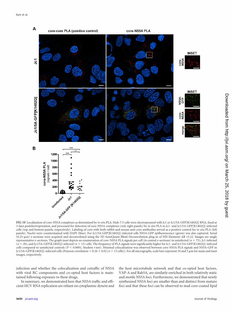
It has been previously reported that HCV core interacts withNS5A (16). In contrast, Appel et al. reported a failure to detectcore-NS5A interactions in the context of the complete HCV lifecycle, suggesting that core-NS5A interactions may be unstable,transient, and/or indirect (15). We therefore investigated the pres-ence and localization of core-NS5A complexes by in situ proxim-ity ligation assays (PLAs) that allow for the detection of weak ortransient protein-protein interactions. These studies revealed theready and specific detection of core-NS5A complexes in both Jc1-and Jc1/5A-GFP[K1402Q]-infected cells in cytoplasmic foci (Fig.10). Surprisingly, core-NS5A complexes minimally colocalizedwith bright NS5A-positive foci (Pearson correlation � 0.26 �0.02 [n � 13 cells]), suggesting that few copies of NS5A are com-plexed with core at any given time. Taken together, these resultsconfirm the interaction (�40-nm proximity) of NS5A with corein the context of an authentic HCV infection system and revealnew details about the localization of these complexes.
DISCUSSION
The factors that dictate segregation of NS5A into both relativelystatic and highly motile cytoplasmic structures and the possiblerole(s) of NS5A (putative RC) motility in the HCV life cycle areunknown. Consistent with a previous study of NS5A dynamics inthe context of a subgenomic replicon (28), we found that the mo-tility of NS5A-positive structures were dramatically impaired byinhibitors of microtubule polymerization, as was HCV RNA rep-lication. It is not clear how microtubule dynamics and/or micro-tubule-dependent traffic of NS5A foci may contribute to efficientHCV replication. Given that the localization and microtubule-dependent trafficking patterns of NS5A foci bear a striking resem-blance to putative RCs of the unrelated plus-strand virus mousehepatitis coronavirus (39), it is appealing to speculate that com-mon mechanisms in RC traffic and function exist. However, incontrast to our findings with HCV, a functional microtubule net-work was dispensable for efficient mouse hepatitis coronavirus
replication and virus production (39). Interestingly, it has beenreported that both HCV NS3 and NS5A proteins directly interactwith microtubules and actin filaments (40), and this may contrib-ute to RC architecture, motility and/or functionality.
Although we observed a close overlap of NS5A and VAP-A inmotile and relatively static punctae, mCherry-VAP-A also dis-played an extensive ER-like localization, suggesting that it may beincorporated into RCs during their biogenesis at the ER mem-brane. In contrast, NS5A was strongly localized to cytoplasmicfoci that were also enriched with the early endosome proteinRab5A. Furthermore, a reliance on endocytic trafficking machin-ery for NS5A traffic was also suggested by the observation that,expressed alone, fluorescently tagged Rab5A displays localizationand trafficking profiles that bear striking similarities to those ofNS5A during active HCV replication (31; data not shown). Wetherefore speculate that the interaction of NS4B with Rab5 (35,36) strongly influences the localization and motility of RCs.
The complex movements of endosomes along microtubulesare dictated by interactions of endosomes with motor proteins,including the minus-end motor dynein, minus-end kinesins(such as KIFC1 and KIFC2), plus-end kinesins (such as kinesin-1), and the kinesin-3 family member KIF16B (reviewed in refer-ences 31, 41, and 42). Consistent with this, we found that dyneinknockdown significantly inhibited the proportion of NS5A focithat displayed long-range motility and, like the chemical inhibi-tion of dynein, significantly inhibited HCV RNA replication.However, it is not clear whether NS5A motility directly contrib-utes to efficient HCV RNA replication or whether disruption ofdynein-dependent organellar/vesicular traffic indirectly limitsHCV RNA replication, for example, by depriving RCs of access toessential host factors or lipids. Future endeavors to identify theHCV protein(s) and residues that directly or indirectly coupleNS5A foci to motor proteins may enable definitive determinationof the contribution of RC motility to HCV replication and accord-ingly may reveal future targets of antiviral therapy.
Along with playing essential roles in viral RNA replication andtranslation, viral RCs must also closely associate with sites of virusparticle assembly, at least transiently, to enable transfer of progenyviral genomes to virus particle assembly complexes (4). For HCV,newly synthesized viral RNA is thought to be transferred from RCsto LD-associated core for encapsidation in a process that dependson core-NS5A interaction (3). Our studies involving near-simul-taneous visualization of core and NS5A localization and traffic invirus-producing cells revealed that the colocalization of these pro-teins was relatively infrequent and that, where NS5A-GFP wasfound in close proximity to core-TCM at the surface of LD-likestructures, NS5A-GFP remained largely in discrete foci. Our ob-servation that core-NS5A complexes minimally overlap in local-ization with NS5A-enriched foci suggest that a minor proportionof NS5A interacts with core at any given time and that these inter-actions may not occur in the context of putative RCs. The local-ization of NS5A-GFP in LD-proximal foci was in contrast to thatof core-TCM, which more uniformly coated the LD surface. Thisobservation is consistent with immunoelectron microscopic anal-ysis of the localization of core and NS5A in JFH-1-infected Huh-7cells, whereby NS5A was detected in clusters at LD-proximal sitesand core was more dispersedly embedded in the LD surface (43). Itis interesting that more uniform localization of NS5A to the LDsurface is particularly prominent when it is expressed alone orwhen its localization is altered by exposure of HCV replicon-har-
Dynamic Imaging of HCV NS5A Protein
April 2014 Volume 88 Number 7 jvi.asm.org 3647
on March 25, 2018 by guest
http://jvi.asm.org/
Dow
nloaded from

Eyre et al.
3648 jvi.asm.org Journal of Virology
on March 25, 2018 by guest
http://jvi.asm.org/
Dow
nloaded from

boring cells to potent NS5A-targeted antiviral drugs (37). Wetherefore speculate that uniform localization of NS5A to LDs is theresult of its dissociation from RCs as a result of changes in itsconformation and interaction with viral RC components (such as
NS3 and/or NS5B) and/or co-opted host factors (such as VAP-Aand/or PI4KIII�) during virus particle assembly. It will thereforebe interesting to examine how NS5A-targeted antiviral drugs alterthe localization and traffic of NS5A in the context of a productive
FIG 8 Pulse-chase imaging of SNAP-tagged NS5A. (A) Infectious virus production by Huh-7.5 cells electroporated with Jc1 or Jc1/5A-SNAP transcripts. (B)Specific labeling of NS5A-SNAP with fluorescent SNAP-tag ligands. Huh-7.5 cells were mock electroporated or electroporated with Jc1 or Jc1/5A-SNAPtranscripts and labeled with SNAP-Cell TMR-Star (red) at 3 days, immediately prior to fixation and immunofluorescent labeling of NS5A (green). Mergedimages (with DAPI-stained nuclei; blue) are depicted in the right panels. Scale bars, 25 m. (C) Western analysis of NS5A protein encoded by wild-type Jc1 andJc1/5A-SNAP at 72 h postelectroporation. �-Actin served as a loading control. (D) Huh-7.5 cells were electroporated with Jc1/5A-SNAP transcripts and labeledwith SNAP-Cell 505 at 48 h, blocked (with SNAP-Cell Block), and returned to culture before labeling with SNAP-Cell TMR-Star at 72 h. Cells were then fixed,counterstained with DAPI, and analyzed by deconvolution fluorescence microscopy. A maximum intensity projection of a representative z-stack is shown. (E)Quantification of the size of “aged” (24- to 72-h-old) and “young” (0- to 24-h-old) NS5A foci. The areas of SNAP-Cell 505-labeled foci (“aged”; n � 5998) andSNAP-Cell TMR-Star-labeled foci (“young”; n � 7682) foci were quantified (from 14 representative cells) from middle z-sections of Jc1/5A-SNAP-infectedHuh-7.5 cells. ***, P � 0.0001 (Student t test). (F) Live cell imaging of Huh-7.5 cells that were electroporated with Jc1/5A-SNAP transcripts and labeled withfluorescent SNAP-tag ligands at the indicated time points. The 8.4-m line scan was used to generate the inset kymograph. Scale bars, 10 m.
FIG 9 Live cell imaging of HCV core and NS5A proteins in the context of a productive infection. Huh-7.5 cells were electroporated with Jc1/Core-TCM:5A-GFP[K1402Q] transcripts and labeled with ReAsH at 48 h postelectroporation, before live cell imaging of ReAsH-labeled core-TCM (red) and NS5A-GFPepifluorescence (green) at 72 h postelectroporation. Insets 1 and 2 (see Movie S9 in the supplemental material) highlight the relatively stable association ofNS5A-GFP-positive foci with core-TCM-coated LDs (arrowheads). Note that brightness and contrast settings have been linearly increased for the “zoom insets”to enhance the visibility of dim structures. For all micrographs, scale bars represent 10 and 5 m for main and inset images, respectively.
Dynamic Imaging of HCV NS5A Protein
April 2014 Volume 88 Number 7 jvi.asm.org 3649
on March 25, 2018 by guest
http://jvi.asm.org/
Dow
nloaded from
infection and whether the colocalization and cotraffic of NS5Awith viral RC components and co-opted host factors is main-tained following exposure to these drugs.
In summary, we demonstrated here that NS5A traffic and effi-cient HCV RNA replication are reliant on cytoplasmic dynein and
the host microtubule network and that co-opted host factors,VAP-A and Rab5A, are similarly enriched in both relatively staticand motile NS5A foci. Furthermore, we demonstrated that newlysynthesized NS5A foci are smaller than and distinct from maturefoci and that these foci can be observed to stud core-coated lipid
FIG 10 Localization of core-NS5A complexes as determined by in situ PLA. Huh-7.5 cells were electroporated with Jc1 or Jc1/5A-GFP[K1402Q] RNA, fixed at3 days postelectroporation, and processed for detection of core-NS5A complexes (red; right panels) by in situ PLA in Jc1- and Jc1/5A-GFP[K1402Q]-infectedcells (top and bottom panels, respectively). Labeling of core with both rabbit and mouse anti-core antibodies served as a positive control for in situ PLA (leftpanels). Nuclei were counterstained with DAPI (blue). For Jc1/5A-GFP[K1402Q]-infected cells NS5A-GFP epifluorescence (green) was also captured. Serial(0.25-m) z-sections were acquired and deconvoluted using the 3D AutoQuant Blind Deconvolution plug-in of NIS Elements AR v3.22. Images are singlerepresentative z-sections. The graph inset depicts an enumeration of core-NS5A PLA signals per cell (in central z-sections) in uninfected (n � 73), Jc1-infected(n � 20), and Jc1/5A-GFP[K1402Q]-infected (n � 13) cells. The frequency of PLA signals were significantly higher for Jc1- and Jc1/5A-GFP[K1402Q]-infectedcells compared to uninfected controls (P � 0.0001, Student t test). Minimal colocalization was observed between core-NS5A PLA signals and NS5A-GFP inJc1/5A-GFP[K1402Q]-infected cells (Pearson correlation � 0.26 � 0.02 [n � 13 cells]). For all micrographs, scale bars represent 10 and 5 m for main and insetimages, respectively.
Eyre et al.
3650 jvi.asm.org Journal of Virology
on March 25, 2018 by guest
http://jvi.asm.org/
Dow
nloaded from

droplets in living, virus-producing cells. These results uncoverpreviously undescribed dynamics of NS5A in a productive HCVinfection and provide a foundation to further dissect the mecha-nisms involved in RC biogenesis, motility, and association withsites of virus assembly for genome transfer and encapsidation.
ACKNOWLEDGMENTS
We are grateful to Ralf Bartenschlager, Michinori Kohara, JohnMcLauchlan, Charles Rice, Paul Targett-Adams, and Takaji Wakita forgenerously providing reagents. Assistance with confocal microscopy andfluorescence-activated cell sorting (FACS) were kindly provided by Gha-far Sarvestani and Katherine Pilkington, respectively (SA Pathology, Ad-elaide, Australia). We thank all members of our laboratory for helpfuldiscussions.
This study was supported by grants from the NHMRC of Australia(1027641 and 510448), an NHMRC Career Development Award (toS.G.T.), and an NHMRC Senior Research Fellowship (to M.R.B.).
REFERENCES1. Moradpour D, Penin F, Rice CM. 2007. Replication of hepatitis C virus.
Nat. Rev. Microbiol. 5:453– 463. http://dx.doi.org/10.1038/nrmicro1645.2. Lohmann V, Korner F, Koch J, Herian U, Theilmann L, Barten-
schlager R. 1999. Replication of subgenomic hepatitis C virus RNAs ina hepatoma cell line. Science 285:110 –113. http://dx.doi.org/10.1126/science.285.5424.110.
3. Bartenschlager R, Penin F, Lohmann V, Andre P. 2011. Assembly ofinfectious hepatitis C virus particles. Trends Microbiol. 19:95–103. http://dx.doi.org/10.1016/j.tim.2010.11.005.
4. den Boon JA, Diaz A, Ahlquist P. 2010. Cytoplasmic viral replicationcomplexes. Cell Host Microbe 8:77– 85. http://dx.doi.org/10.1016/j.chom.2010.06.010.
5. Egger D, Wolk B, Gosert R, Bianchi L, Blum HE, Moradpour D, BienzK. 2002. Expression of hepatitis C virus proteins induces distinct mem-brane alterations including a candidate viral replication complex. J. Virol.76:5974 –5984. http://dx.doi.org/10.1128/JVI.76.12.5974-5984.2002.
6. Gosert R, Egger D, Lohmann V, Bartenschlager R, Blum HE, Bienz K,Moradpour D. 2003. Identification of the hepatitis C virus RNA replica-tion complex in Huh-7 cells harboring subgenomic replicons. J. Virol.77:5487–5492. http://dx.doi.org/10.1128/JVI.77.9.5487-5492.2003.
7. Ferraris P, Beaumont E, Uzbekov R, Brand D, Gaillard J, Blanchard E,Roingeard P. 2013. Sequential biogenesis of host cell membrane rear-rangements induced by hepatitis C virus infection. Cell. Mol. Life Sci.70:1297–1306. http://dx.doi.org/10.1007/s00018-012-1213-0.
8. Ferraris P, Blanchard E, Roingeard P. 2010. Ultrastructural and bio-chemical analyses of hepatitis C virus-associated host cell membranes. J.Gen. Virol. 91:2230 –2237. http://dx.doi.org/10.1099/vir.0.022186-0.
9. Romero-Brey I, Merz A, Chiramel A, Lee JY, Chlanda P, Haselman U,Santarella-Mellwig R, Habermann A, Hoppe S, Kallis S, Walther P,Antony C, Krijnse-Locker J, Bartenschlager R. 2012. Three-dimensionalarchitecture and biogenesis of membrane structures associated with hep-atitis C virus replication. PLoS Pathog. 8:e1003056. http://dx.doi.org/10.1371/journal.ppat.1003056.
10. Berger KL, Kelly SM, Jordan TX, Tartell MA, Randall G. 2011. HepatitisC virus stimulates the phosphatidylinositol 4-kinase III alpha-dependentphosphatidylinositol 4-phosphate production that is essential for its rep-lication. J. Virol. 85:8870 – 8883. http://dx.doi.org/10.1128/JVI.00059-11.
11. Reiss S, Rebhan I, Backes P, Romero-Brey I, Erfle H, Matula P, KaderaliL, Poenisch M, Blankenburg H, Hiet MS, Longerich T, Diehl S, RamirezF, Balla T, Rohr K, Kaul A, Buhler S, Pepperkok R, Lengauer T,Albrecht M, Eils R, Schirmacher P, Lohmann V, Bartenschlager R.2011. Recruitment and activation of a lipid kinase by hepatitis C virusNS5A is essential for integrity of the membranous replication compart-ment. Cell Host Microbe 9:32– 45. http://dx.doi.org/10.1016/j.chom.2010.12.002.
12. Backes P, Quinkert D, Reiss S, Binder M, Zayas M, Rescher U, Gerke V,Bartenschlager R, Lohmann V. 2010. Role of annexin A2 in the produc-tion of infectious hepatitis C virus particles. J. Virol. 84:5775–5789. http://dx.doi.org/10.1128/JVI.02343-09.
13. Berger KL, Cooper JD, Heaton NS, Yoon R, Oakland TE, Jordan TX,Mateu G, Grakoui A, Randall G. 2009. Roles for endocytic trafficking and
phosphatidylinositol 4-kinase III alpha in hepatitis C virus replication.Proc. Natl. Acad. Sci. U. S. A. 106:7577–7582. http://dx.doi.org/10.1073/pnas.0902693106.
14. Gao L, Aizaki H, He JW, Lai MM. 2004. Interactions between viralnonstructural proteins and host protein hVAP-33 mediate the formationof hepatitis C virus RNA replication complex on lipid raft. J. Virol. 78:3480 –3488. http://dx.doi.org/10.1128/JVI.78.7.3480-3488.2004.
15. Appel N, Zayas M, Miller S, Krijnse-Locker J, Schaller T, Friebe P, Kallis S,Engel U, Bartenschlager R. 2008. Essential role of domain III of nonstruc-tural protein 5A for hepatitis C virus infectious particle assembly. PLoSPathog. 4:e1000035. http://dx.doi.org/10.1371/journal.ppat.1000035.
16. Masaki T, Suzuki R, Murakami K, Aizaki H, Ishii K, Murayama A, DateT, Matsuura Y, Miyamura T, Wakita T, Suzuki T. 2008. Interaction ofhepatitis C virus nonstructural protein 5A with core protein is critical forthe production of infectious virus particles. J. Virol. 82:7964 –7976. http://dx.doi.org/10.1128/JVI.00826-08.
17. Tellinghuisen TL, Foss KL, Treadaway J. 2008. Regulation of hepatitis Cvirion production via phosphorylation of the NS5A protein. PLoS Pathog.4:e1000032. http://dx.doi.org/10.1371/journal.ppat.1000032.
18. Schaller T, Appel N, Koutsoudakis G, Kallis S, Lohmann V, Piet-schmann T, Bartenschlager R. 2007. Analysis of hepatitis C virus super-infection exclusion by using novel fluorochrome gene-tagged viral ge-nomes. J. Virol. 81:4591– 4603. http://dx.doi.org/10.1128/JVI.02144-06.
19. Wakita T, Pietschmann T, Kato T, Date T, Miyamoto M, Zhao Z,Murthy K, Habermann A, Krausslich HG, Mizokami M, BartenschlagerR, Liang TJ. 2005. Production of infectious hepatitis C virus in tissueculture from a cloned viral genome. Nat. Med. 11:791–796. http://dx.doi.org/10.1038/nm1268.
20. Zhou Z, Wang N, Woodson SE, Dong Q, Wang J, Liang Y, RijnbrandR, Wei L, Nichols JE, Guo JT, Holbrook MR, Lemon SM, Li K. 2011.Antiviral activities of ISG20 in positive-strand RNA virus infections.Virology 409:175–188. http://dx.doi.org/10.1016/j.virol.2010.10.008.
21. Blight KJ, McKeating JA, Rice CM. 2002. Highly permissive cell lines forsubgenomic and genomic hepatitis C virus RNA replication. J. Virol. 76:13001–13014. http://dx.doi.org/10.1128/JVI.76.24.13001-13014.2002.
22. Eyre NS, Phillips RJ, Bowden S, Yip E, Dewar B, Locarnini SA, BeardMR. 2009. Hepatitis B virus and hepatitis C virus interaction in Huh-7cells. J. Hepatol. 51:446 – 457. http://dx.doi.org/10.1016/j.jhep.2009.04.025.
23. Adams SR, Tsien RY. 2008. Preparation of the membrane-permeantbiarsenicals FlAsH-EDT2 and ReAsH-EDT2 for fluorescent labeling oftetracysteine-tagged proteins. Nat. Protoc. 3:1527–1534. http://dx.doi.org/10.1038/nprot.2008.144.
24. Hoffmann C, Gaietta G, Zurn A, Adams SR, Terrillon S, Ellisman MH,Tsien RY, Lohse MJ. 2010. Fluorescent labeling of tetracysteine-taggedproteins in intact cells. Nat. Protoc. 5:1666 –1677. http://dx.doi.org/10.1038/nprot.2010.129.
25. Pietschmann T, Kaul A, Koutsoudakis G, Shavinskaya A, Kallis S,Steinmann E, Abid K, Negro F, Dreux M, Cosset FL, Bartenschlager R.2006. Construction and characterization of infectious intragenotypic andintergenotypic hepatitis C virus chimeras. Proc. Natl. Acad. Sci. U. S. A.103:7408 –7413. http://dx.doi.org/10.1073/pnas.0504877103.
26. Coller KE, Heaton NS, Berger KL, Cooper JD, Saunders JL, RandallG. 2012. Molecular determinants and dynamics of hepatitis C virussecretion. PLoS Pathog. 8:e1002466. http://dx.doi.org/10.1371/journal.ppat.1002466.
27. Counihan NA, Rawlinson SM, Lindenbach BD. 2011. Trafficking ofhepatitis C virus core protein during virus particle assembly. PLoS Pathog.7:e1002302. http://dx.doi.org/10.1371/journal.ppat.1002302.
28. Wolk B, Buchele B, Moradpour D, Rice CM. 2008. A dynamic view ofhepatitis C virus replication complexes. J. Virol. 82:10519 –10531. http://dx.doi.org/10.1128/JVI.00640-08.
29. Bost AG, Venable D, Liu L, Heinz BA. 2003. Cytoskeletal requirementsfor hepatitis C virus (HCV) RNA synthesis in the HCV replicon cell cul-ture system. J. Virol. 77:4401– 4408. http://dx.doi.org/10.1128/JVI.77.7.4401-4408.2003.
30. Driskell OJ, Mironov A, Allan VJ, Woodman PG. 2007. Dynein isrequired for receptor sorting and the morphogenesis of early endosomes.Nat. Cell Biol. 9:113–120. http://dx.doi.org/10.1038/ncb1525.
31. Flores-Rodriguez N, Rogers SS, Kenwright DA, Waigh TA, WoodmanPG, Allan VJ. 2011. Roles of dynein and dynactin in early endosomedynamics revealed using automated tracking and global analysis. PLoSOne 6:e24479. http://dx.doi.org/10.1371/journal.pone.0024479.
Dynamic Imaging of HCV NS5A Protein
April 2014 Volume 88 Number 7 jvi.asm.org 3651
on March 25, 2018 by guest
http://jvi.asm.org/
Dow
nloaded from
32. Firestone AJ, Weinger JS, Maldonado M, Barlan K, Langston LD,O’Donnell M, Gelfand VI, Kapoor TM, Chen JK. 2012. Small-moleculeinhibitors of the AAA� ATPase motor cytoplasmic dynein. Nature 484:125–129. http://dx.doi.org/10.1038/nature10936.
33. Evans MJ, Rice CM, Goff SP. 2004. Phosphorylation of hepatitis C virusnonstructural protein 5A modulates its protein interactions and viral RNAreplication. Proc. Natl. Acad. Sci. U. S. A. 101:13038 –13043. http://dx.doi.org/10.1073/pnas.0405152101.
34. Tu H, Gao L, Shi ST, Taylor DR, Yang T, Mircheff AK, Wen Y,Gorbalenya AE, Hwang SB, Lai MM. 1999. Hepatitis C virus RNApolymerase and NS5A complex with a SNARE-like protein. Virology 263:30 – 41. http://dx.doi.org/10.1006/viro.1999.9893.
35. Stone M, Jia S, Heo WD, Meyer T, Konan KV. 2007. Participation ofrab5, an early endosome protein, in hepatitis C virus RNA replicationmachinery. J. Virol. 81:4551– 4563. http://dx.doi.org/10.1128/JVI.01366-06.
36. Manna D, Aligo J, Xu C, Park WS, Koc H, Heo WD, Konan KV. 2010.Endocytic Rab proteins are required for hepatitis C virus replication com-plex formation. Virology 398:21–37. http://dx.doi.org/10.1016/j.virol.2009.11.034.
37. Targett-Adams P, Graham EJ, Middleton J, Palmer A, Shaw SM, Lav-ender H, Brain P, Tran TD, Jones LH, Wakenhut F, Stammen B, PrydeD, Pickford C, Westby M. 2011. Small molecules targeting hepatitis Cvirus-encoded NS5A cause subcellular redistribution of their target: in-
sights into compound modes of action. J. Virol. 85:6353– 6368. http://dx.doi.org/10.1128/JVI.00215-11.
38. Boulant S, Douglas MW, Moody L, Budkowska A, Targett-Adams P,McLauchlan J. 2008. Hepatitis C virus core protein induces lipid dropletredistribution in a microtubule- and dynein-dependent manner. Traffic9:1268 –1282. http://dx.doi.org/10.1111/j.1600-0854.2008.00767.x.
39. Hagemeijer MC, Verheije MH, Ulasli M, Shaltiel IA, de Vries LA,Reggiori F, Rottier PJ, de Haan CA. 2010. Dynamics of coronavirusreplication-transcription complexes. J. Virol. 84:2134 –2149. http://dx.doi.org/10.1128/JVI.01716-09.
40. Lai CK, Jeng KS, Machida K, Lai MM. 2008. Association of hepatitis Cvirus replication complexes with microtubules and actin filaments is de-pendent on the interaction of NS3 and NS5A. J. Virol. 82:8838 – 8848.http://dx.doi.org/10.1128/JVI.00398-08.
41. Hunt SD, Stephens DJ. 2011. The role of motor proteins in endosomalsorting. Biochem. Soc. Trans. 39:1179 –1184. http://dx.doi.org/10.1042/BST0391179.
42. Soldati T, Schliwa M. 2006. Powering membrane traffic in endocytosisand recycling. Nat. Rev. Mol. Cell. Biol. 7:897–908. http://dx.doi.org/10.1038/nrm2060.
43. Miyanari Y, Atsuzawa K, Usuda N, Watashi K, Hishiki T, Zayas M,Bartenschlager R, Wakita T, Hijikata M, Shimotohno K. 2007. The lipiddroplet is an important organelle for hepatitis C virus production. Nat.Cell Biol. 9:1089 –1097. http://dx.doi.org/10.1038/ncb1631.
Eyre et al.
3652 jvi.asm.org Journal of Virology
on March 25, 2018 by guest
http://jvi.asm.org/
Dow
nloaded from


![Hepatitis B virus and hepatitis C virus play different ... · alcoholic cirrhosis, hepatitis viruses, tobacco and metabolic diseases[4]. Hepatitis viruses, including hepatitis B virus](https://static.fdocuments.in/doc/165x107/60e46cab5bd9101a6f539e91/hepatitis-b-virus-and-hepatitis-c-virus-play-different-alcoholic-cirrhosis.jpg)