Dynamic and Conventional Spin-Echo MR of Pituitary ... · Dynamic and Conventional Spin-Echo MR of...
8
Dynamic and Conventional Spin-Echo MR of Pituitary Microlesions Walter S. Bartynski and Luke Lin PURPOSE: To determine whether dynamic traditional spin-echo MR imaging, with the use of routine T1 parameters during contrast infusion, is superior to standard MR imaging after contrast administration for detecting microlesions of the pituitary gland. METHODS: Sixty-four patients with pituitary microlesions 3 to 10 mm in diameter were examined with a dynamic traditional spin-echo technique; that is, a typical T1 spin-echo sequence of 500 – 600/20 –25/2 (repetition time/echo time/excitations), 3-mm-thick sections, 16-cm field of view, 256 3 128 matrix, and a scan time ranging from 2 minutes to 2 minutes 40 seconds during contrast infusion. In addition, standard imaging with unenhanced and contrast-enhanced spin-echo sequences were obtained. The three sequences were evaluated retrospectively and graded for gland-lesion contrast conspicuity, lesion homogeneity, and delineation of lesion margin. RESULTS: The dynamic sequence was judged to be better than the standard enhanced sequence for depicting microlesions in 42% to 47% of patients. Lesions were identified only on the dynamic study in an additional 11% to 14% of patients. Lesions were seen equally well on the standard and dynamic sequences only in 16% to 23% of cases. The standard postcontrast sequence was judged better in 12.5% to 17% of cases, with lesions identified only on the standard sequence in an additional 8% to 9%. CONCLUSION: Dynamic traditional spin-echo MR imaging improved lesion detection and provided increased clarity over standard sequences after contrast infusion. Both sequences are important, since lesions were detected only on the dynamic sequence in 11% to 14% of patients and only on the standard sequence in 8% to 9% of patients. Index terms: Pituitary gland, magnetic resonance; Magnetic resonance, technique AJNR Am J Neuroradiol 18:965–972, May 1997 In the early 1980s, Hemminghytt et al (1) noted that detection of pituitary microadenoma with computed tomography (CT) was time de- pendent after contrast administration. This was postulated to be due to a difference in the time course of enhancement of the microadenoma relative to the adjacent normal gland. Bonne- ville et al (2) described the CT pattern of pitu- itary enhancement early after contrast infusion, and encouraged the use of dynamic scanning in the evaluation of microadenomas. A similar time dependence in gland enhance- ment and adenoma-gland contrast difference has been documented with magnetic resonance (MR) imaging (3–7). MR imaging studies have generally been obtained with either signal-lim- ited rapid T1 techniques or unique sequences not available on most MR imaging systems. The purpose of our investigation was to determine whether dynamic traditional spin-echo imaging, with the use of a traditional spin-echo technique but during contrast infusion, resulted in im- proved lesion detection relative to routine imag- ing after contrast administration. Materials and Methods Four hundred forty-four MR imaging examinations of the pituitary gland were performed at our institution during a 7-year period. In 187 studies, a macroadenoma was present, prior surgery had been performed, or a pituitary lesion previously studied by MR imaging was present. These cases were excluded from the study. Two hundred fifty-seven examinations were therefore available in which the gland was of normal size and there was no previously known lesion. In 64 (25%) of these 257 patients, a new microadenoma or microlesion was identified in the pitu- Received June 13, 1996; accepted after revision November 22. From the Department of Radiology, Western Pennsylvania Hospital, 4800 Friendship Ave, Pittsburgh, PA 15224. Address reprint requests to W. S. Bartynski, MD. AJNR 18:965–972, May 1997 0195-6108/97/1805–0965 © American Society of Neuroradiology 965
Transcript of Dynamic and Conventional Spin-Echo MR of Pituitary ... · Dynamic and Conventional Spin-Echo MR of...
Dynamic and Conventional Spin-Echo MR of Pituitary Microlesions
Walter S. Bartynski and Luke Lin
PURPOSE: To determine whether dynamic traditional spin-echo MR imaging, with the use ofroutine T1 parameters during contrast infusion, is superior to standard MR imaging after contrastadministration for detecting microlesions of the pituitary gland. METHODS: Sixty-four patients withpituitary microlesions 3 to 10 mm in diameter were examined with a dynamic traditional spin-echotechnique; that is, a typical T1 spin-echo sequence of 500–600/20–25/2 (repetition time/echotime/excitations), 3-mm-thick sections, 16-cm field of view, 256 3 128 matrix, and a scan timeranging from 2 minutes to 2 minutes 40 seconds during contrast infusion. In addition, standardimaging with unenhanced and contrast-enhanced spin-echo sequences were obtained. The threesequences were evaluated retrospectively and graded for gland-lesion contrast conspicuity, lesionhomogeneity, and delineation of lesion margin. RESULTS: The dynamic sequence was judged tobe better than the standard enhanced sequence for depicting microlesions in 42% to 47% ofpatients. Lesions were identified only on the dynamic study in an additional 11% to 14% of patients.Lesions were seen equally well on the standard and dynamic sequences only in 16% to 23% ofcases. The standard postcontrast sequence was judged better in 12.5% to 17% of cases, withlesions identified only on the standard sequence in an additional 8% to 9%. CONCLUSION:Dynamic traditional spin-echo MR imaging improved lesion detection and provided increasedclarity over standard sequences after contrast infusion. Both sequences are important, sincelesions were detected only on the dynamic sequence in 11% to 14% of patients and only on thestandard sequence in 8% to 9% of patients.
Index terms: Pituitary gland, magnetic resonance; Magnetic resonance, technique
AJNR Am J Neuroradiol 18:965–972, May 1997
In the early 1980s, Hemminghytt et al (1)noted that detection of pituitary microadenomawith computed tomography (CT) was time de-pendent after contrast administration. This waspostulated to be due to a difference in the timecourse of enhancement of the microadenomarelative to the adjacent normal gland. Bonne-ville et al (2) described the CT pattern of pitu-itary enhancement early after contrast infusion,and encouraged the use of dynamic scanning inthe evaluation of microadenomas.
A similar time dependence in gland enhance-ment and adenoma-gland contrast differencehas been documented with magnetic resonance
Received June 13, 1996; accepted after revision November 22.From the Department of Radiology, Western Pennsylvania Hospital,
4800 Friendship Ave, Pittsburgh, PA 15224. Address reprint requests toW. S. Bartynski, MD.
AJNR 18:965–972, May 1997 0195-6108/97/1805–0965
© American Society of Neuroradiology
96
(MR) imaging (3–7). MR imaging studies havegenerally been obtained with either signal-lim-ited rapid T1 techniques or unique sequencesnot available on most MR imaging systems. Thepurpose of our investigation was to determinewhether dynamic traditional spin-echo imaging,with the use of a traditional spin-echo techniquebut during contrast infusion, resulted in im-proved lesion detection relative to routine imag-ing after contrast administration.
Materials and MethodsFour hundred forty-four MR imaging examinations of
the pituitary gland were performed at our institution duringa 7-year period. In 187 studies, a macroadenoma waspresent, prior surgery had been performed, or a pituitarylesion previously studied by MR imaging was present.These cases were excluded from the study. Two hundredfifty-seven examinations were therefore available in whichthe gland was of normal size and there was no previouslyknown lesion. In 64 (25%) of these 257 patients, a newmicroadenoma or microlesion was identified in the pitu-
5
TABLE 1: Dynamic and conventional pituitary imaging protocol
Type of SequenceSection Thickness,
mmRepetition Time,
msEcho Time,
msNo. of
ExcitationsMatrix Size
Field ofView, cm
Time, min
Sagittal scout 3 300 20 1 256 3 128 24 0:47Coronal unenhanced 3 500–600 20–25 2 256 3 192 16 3:50Coronal dynamic contrast enhanced* 3 500–600 20–25 2 256 3 128 16 2:41Coronal standard contrast enhanced 3 500–600 20–25 4 256 3 128 16 5:10
* This sequence included prescanning, insertion of intravenous tubing, repositioning of patient, injection of one third contrast bolus, andscanning during injection of remaining two thirds of contrast bolus.
966 BARTYNSKI AJNR: 18, May 1997
itary gland. These 64 lesions form the basis of this report.All studies were obtained on a 1.5-T MR imaging unit.Examinations were performed with a dynamic traditionalspin-echo enhancement technique acquired during con-trast infusion in addition to a standard postcontrast tech-nique.
Dynamic Traditional Spin-Echo Technique
The dynamic traditional spin-echo technique is a rou-tine spin-echo sequence obtained during contrast infusion.Imaging sequences and parameters are reviewed in Table1. A sagittal scout view (300/20/1 [repetition time/echotime/excitation], 3-mm-thick sections, 24-cm field of view[FOV], 256 3 128 matrix, 47-second scan time) was ob-tained, followed by a routine unenhanced coronal T1-weighted sequence of the pituitary gland (500–600/20–25/2, 3-mm-thick sections, 16-cm FOV, 256 3 192matrix, 3-minute 50-second scan time).
Manual or automatic prescanning was then performedto tune the MR imaging system before contrast infusionand before beginning data acquisition for the dynamicspin-echo sequence. When the prescan was completed,the patient was removed from the magnet bore and abutterfly needle was inserted into an antecubital vein. Thecontrast syringe, with extension tubing, was then con-nected and the patient was moved back to the center of themagnet.
The dynamic spin-echo sequence was a routine coronalT1 spin-echo sequence obtained during contrast infusion(500–600/20–25/2, 3-mm-thick sections, 16-cm FOV,256 3 128 matrix, scan time of 2 minutes to 2 minutes 41seconds). One third of a routine 0.1 mM/kg dose of gado-pentetate dimeglumine was infused over 30 seconds be-fore the start of data acquisition for the dynamic spin-echoscan sequence. After one third of the contrast material wasinfused, the technologist began image acquisition. Theremaining two thirds of the contrast material was infusedover 1 minute 30 seconds during the dynamic spin-echosequence image acquisition. At the end of contrast infu-sion, a saline flush was injected to clear the tube and armveins of contrast agent. The dynamic spin-echo scan wasobtained in approximately 2 minutes 40 seconds. Imme-diately after the dynamic spin-echo scan was completed,without prescanning, a standard postcontrast sequencewas obtained (500–600/20–25/4, 3-mm-thick sections,16-cm FOV, 256 3 128 matrix, 5-minute 10-second scan
time). Data acquisition for the standard postcontrast se-quence began immediately after the dynamic spin-echosequence, approximately 1 minute after the end of thecontrast injection.
Study PopulationThe study population comprised the 64 patients in
whom a new pituitary microadenoma or microlesion (lessthan 1 cm in diameter) was confidently identified. Fivepatients were men and 59 were women; ages ranged from18 to 44 years (average, 35 years). The most frequentclinical reason for referral was menstrual irregularity or anabnormal hormone level. Pituitary hormone data wereavailable in 34 patients. Twenty-six patients had an ele-vated prolactin level. Two patients had known Cushingsyndrome and the adenomas identified in these patientswere surgically removed. Several patients were referred fornonspecific problems, mainly headache. In six of thesepatients, pituitary hormone levels were normal. Most of thepatients with hyperprolactinemia were being managedwith bromocriptine at the time of the MR imaging exami-nation.
Lesion Detection
Lesions were typically identified as a focal area of hy-pointensity within the pituitary gland. A patient was notincluded in the study unless the lesion was confidentlydetermined to be 3 mm or greater in diameter. A narrowwindow display of the pituitary gland was used to improvelesion conspicuity, as has been previously suggested (8,9). In addition, we required that two neuroradiologistsidentify each lesion on at least one of the enhanced se-quences. Lesion size and morphologic characteristics werealso noted. Focal regions of signal abnormality at the sellafloor, caused by local magnetic susceptibility artifacts,were carefully identified and not included (10, 11). Threetypes of lesion were recognized: focal round or oval withrelatively sharp margination, focal with irregular marginsand borders, and focal but with indistinct or unsharp bor-ders.
Lesion Grading
The 64 studies were independently and retrospectivelyreviewed by two neuroradiologists who were blinded to the
TABLE 2: Lesion grading and reader correlation
Reader 1 Reader 2 Percentage ofAgreement
95% ConfidenceIntervaln % n %
Equal 10 16 15 23 .3889 .1730–.6425Dynamic better than standard 30 47 27 42 .7813 .6003–.9072Standard better than dynamic 11 17 8 12.5 .5833 .2767–.8483Dynamic only 7 11 9 14 .7778 .3999–.9719Standard only 6 9 5 8 .8333 .3588–.9958Total 64 100 64 100
Note.—Percentages have been rounded off.
AJNR: 18, May 1997 PITUITARY MICROLESIONS 967
specific clinical indications for the study. All three se-quences—unenhanced, dynamic spin-echo, and standardpostcontrast—were reviewed side by side for this retro-spective evaluation. Each sequence was independentlyevaluated for whether a lesion was or was not identified.The dynamic spin-echo and standard postcontrast se-quences were further graded by a subjective method forgland-lesion contrast conspicuity, lesion homogeneity,and discrimination of lesion margin. These three featureswere considered together because lesions often changedbetween sequences. In many lesions, either partial fill-ing-in of the hypointense region was seen or partial fill-ing-in of the peripheral margin occurred. Several lesionsshowed partial enhancement greater than the adjacentgland on one of the sequences. These three features weretherefore considered together by each neuroradiologist ingrading lesion visibility. Sequences were graded as towhether the lesion was better seen, less well seen, orequally well seen on the two sequences.
Side-by-side assessment was chosen to emphasizeconfidence in lesion presence and detectability. This is asignificant problem in the pituitary, where surgical proof ofa lesion is frequently lacking owing to long-term medicalmanagement with hormone-suppression therapy. The fo-cus of this study was on changes in confidently recognizedlesions. Side-by-side evaluation allowed sequence com-parison while sacrificing blinded lesion detection statisticsfor any single sequence. Because of the side-by-side re-view, k statistical evaluation of the data was not per-formed. Paired agreement statistics were used to reinforcethat the trend in lesion grading was statistically consistentbetween the two observers.
Lesion grading established five categories of lesion vi-sualization between the two enhanced sequences: equal,dynamic better, standard better, dynamic only, and stan-dard only. The frequency of lesion detection within thesefive categories was tabulated. The percentage of pairedagreement was tabulated and a 95% confidence intervalwas calculated for each category.
Results
Thirty-three lesions (51.5%) were focal roundor focal oval in appearance with relatively sharp
margins. Eighteen lesions (28%) were focal withirregular margins but with sharply defined bor-ders. Thirteen lesions (20%) were focal but witha diffuse peripheral border. Lesion size variedfrom 3 mm to 10 mm (average, 5.3 mm).
Lesion grading and paired agreement resultsbetween observers are summarized in Table 2.Lesions were identified on one of the two en-hanced sequences in all patients. This was acomponent of the inclusion criteria. In 45% to50% of patients, the lesion, or a focal region ofsignal abnormality, was noted on the unen-hanced sequence. These were typically regionsof hypointensity. In six cases, small focal areasof hyperintensity, most likely the result of slightlesion hemorrhage, were also noted. Lesionswere judged to be equally well seen on the dy-namic and standard sequences in only 16%of cases by reader 1 and in 23% of cases byreader 2.
The dynamic spin-echo sequence was judgedbetter than the standard postcontrast sequencefor lesion detection in 42% to 47% of cases (Fig1). Reduced lesion visibility on the standardpostcontrast sequence was usually due to pro-gressive lesion enhancement with diminishedcontrast discrimination between the lesion andthe adjacent normal gland. In eight patients,reduced lesion visibility was due to a reductionin lesion size caused by a partial filling-in orenhancement of the lesion at its border (Fig 1).In eight patients, lesions became nearly isoin-tense with the adjacent normal gland with onlyfaint lesion detection. This was due to progres-sive enhancement of the lesion in six of eightpatients (Fig 2) and to early reduction in en-hancement of the adjacent normal gland ineight of eight patients (Fig 3).
The standard postcontrast sequence wasjudged better than the dynamic spin-echo se-
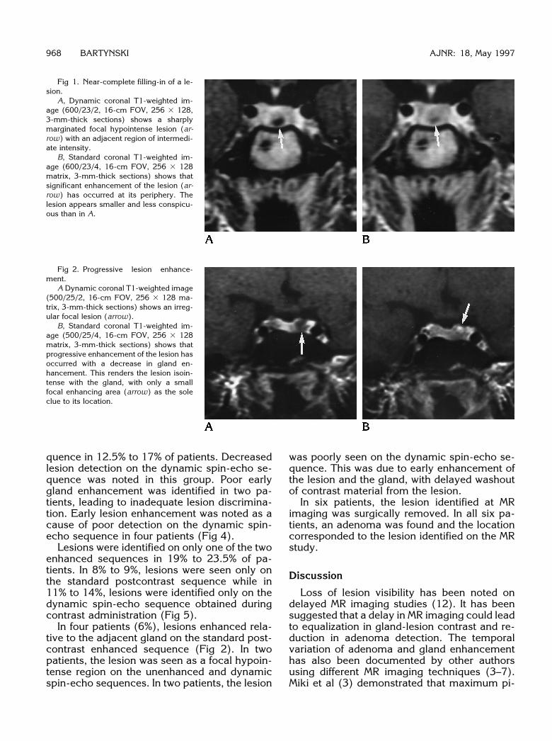
Fig 2. Progressive lesion enhance-ment.
A Dynamic coronal T1-weighted image(500/25/2, 16-cm FOV, 256 3 128 ma-trix, 3-mm-thick sections) shows an irreg-ular focal lesion (arrow).
B, Standard coronal T1-weighted im-age (500/25/4, 16-cm FOV, 256 3 128matrix, 3-mm-thick sections) shows thatprogressive enhancement of the lesion hasoccurred with a decrease in gland en-hancement. This renders the lesion isoin-tense with the gland, with only a smallfocal enhancing area (arrow) as the soleclue to its location.
Fig 1. Near-complete filling-in of a le-sion.
A, Dynamic coronal T1-weighted im-age (600/23/2, 16-cm FOV, 256 3 128,3-mm-thick sections) shows a sharplymarginated focal hypointense lesion (ar-row) with an adjacent region of intermedi-ate intensity.
B, Standard coronal T1-weighted im-age (600/23/4, 16-cm FOV, 256 3 128matrix, 3-mm-thick sections) shows thatsignificant enhancement of the lesion (ar-row) has occurred at its periphery. Thelesion appears smaller and less conspicu-ous than in A.
968 BARTYNSKI AJNR: 18, May 1997
quence in 12.5% to 17% of patients. Decreasedlesion detection on the dynamic spin-echo se-quence was noted in this group. Poor earlygland enhancement was identified in two pa-tients, leading to inadequate lesion discrimina-tion. Early lesion enhancement was noted as acause of poor detection on the dynamic spin-echo sequence in four patients (Fig 4).
Lesions were identified on only one of the twoenhanced sequences in 19% to 23.5% of pa-tients. In 8% to 9%, lesions were seen only onthe standard postcontrast sequence while in11% to 14%, lesions were identified only on thedynamic spin-echo sequence obtained duringcontrast administration (Fig 5).
In four patients (6%), lesions enhanced rela-tive to the adjacent gland on the standard post-contrast enhanced sequence (Fig 2). In twopatients, the lesion was seen as a focal hypoin-tense region on the unenhanced and dynamicspin-echo sequences. In two patients, the lesion
was poorly seen on the dynamic spin-echo se-quence. This was due to early enhancement ofthe lesion and the gland, with delayed washoutof contrast material from the lesion.
In six patients, the lesion identified at MRimaging was surgically removed. In all six pa-tients, an adenoma was found and the locationcorresponded to the lesion identified on the MRstudy.
Discussion
Loss of lesion visibility has been noted ondelayed MR imaging studies (12). It has beensuggested that a delay in MR imaging could leadto equalization in gland-lesion contrast and re-duction in adenoma detection. The temporalvariation of adenoma and gland enhancementhas also been documented by other authorsusing different MR imaging techniques (3–7).Miki et al (3) demonstrated that maximum pi-
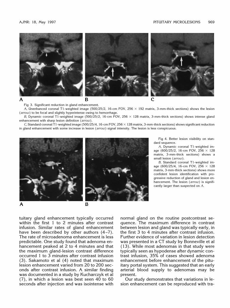
Fig 4. Better lesion visibility on stan-dard sequence.
A, Dynamic coronal T1-weighted im-age (600/25/2, 16-cm FOV, 256 3 128matrix, 3-mm-thick sections) shows asmall lesion (arrow).
B, Standard coronal T1-weighted im-age (600/25/4, 16-cm FOV, 256 3 128matrix, 3-mm-thick sections) shows moreconfident lesion identification with pro-gressive reduction of gland and lesion en-hancement. The lesion (arrow) is signifi-cantly larger than suspected on A.
Fig 3. Significant reduction in gland enhancement.A, Unenhanced coronal T1-weighted image (500/25/2, 16-cm FOV, 256 3 192 matrix, 3-mm-thick sections) shows the lesion
(arrow) to be focal and slightly hyperintense owing to hemorrhage.B, Dynamic coronal T1-weighted image (500/25/2, 16-cm FOV, 256 3 128 matrix, 3-mm-thick sections) shows intense gland
enhancement with sharp lesion definition (arrow).C, Standard coronal T1-weighted image (500/25/4, 16-cm FOV, 256 3 128 matrix, 3-mm-thick sections) shows significant reduction
in gland enhancement with some increase in lesion (arrow) signal intensity. The lesion is less conspicuous.
AJNR: 18, May 1997 PITUITARY MICROLESIONS 969
tuitary gland enhancement typically occurredwithin the first 1 to 2 minutes after contrastinfusion. Similar rates of gland enhancementhave been described by other authors (4–7).The rate of microadenoma enhancement is lesspredictable. One study found that adenoma en-hancement peaked at 2 to 4 minutes and thatthe maximum gland-lesion contrast differenceoccurred 1 to 3 minutes after contrast infusion(3). Sakamoto et al (4) noted that maximumlesion enhancement varied from 20 to 200 sec-onds after contrast infusion. A similar findingwas documented in a study by Kucharczyk et al(7), in which a lesion was best seen 40 to 60seconds after injection and was isointense with
normal gland on the routine postcontrast se-quence. The maximum difference in contrastbetween lesion and gland was typically early, inthe first 3 to 4 minutes after contrast infusion.Further evidence of variation in lesion detectionwas presented in a CT study by Bonneville et al(13). While most adenomas in that study weretypically seen as hypodense after dynamic con-trast infusion, 35% of cases showed adenomaenhancement before enhancement of the pitu-itary portal system. This suggested that an earlyarterial blood supply to adenomas may bepresent.
Our study demonstrates that variations in le-sion enhancement can be reproduced with tra-
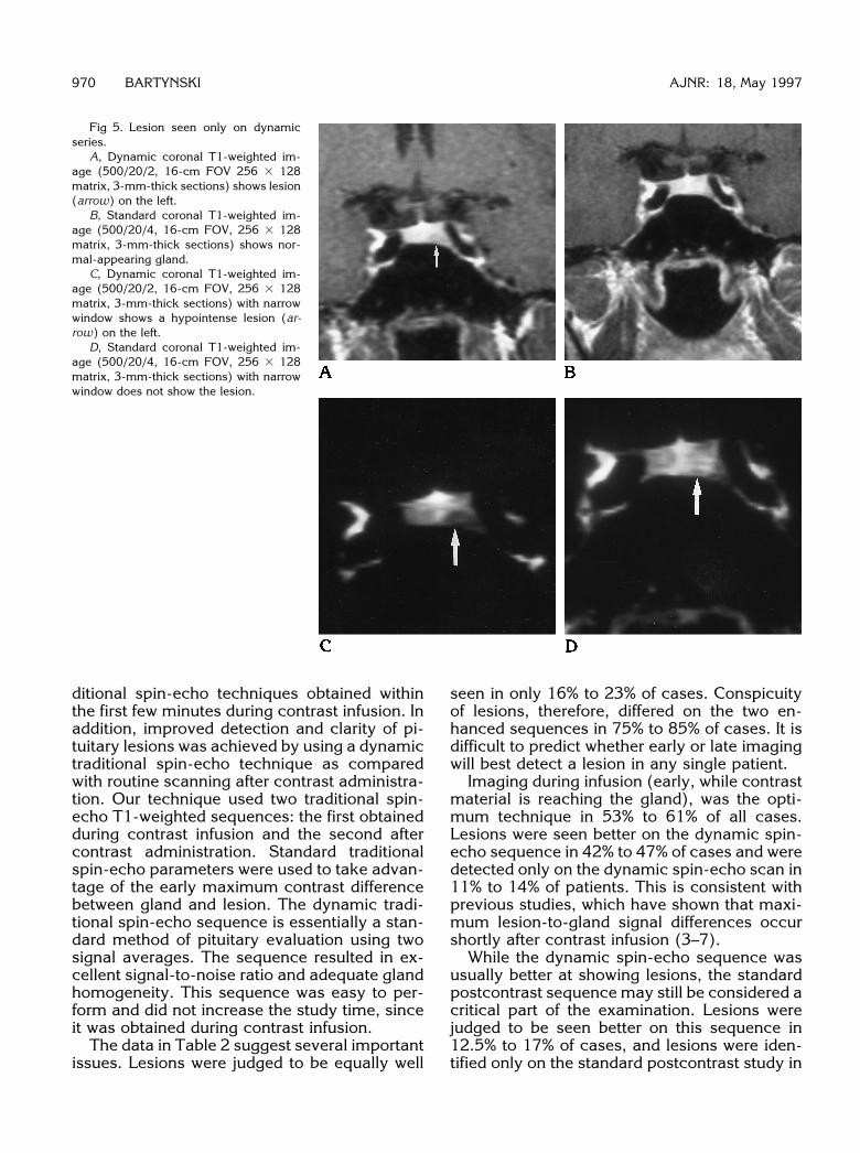
Fig 5. Lesion seen only on dynamicseries.
A, Dynamic coronal T1-weighted im-age (500/20/2, 16-cm FOV 256 3 128matrix, 3-mm-thick sections) shows lesion(arrow) on the left.
B, Standard coronal T1-weighted im-age (500/20/4, 16-cm FOV, 256 3 128matrix, 3-mm-thick sections) shows nor-mal-appearing gland.
C, Dynamic coronal T1-weighted im-age (500/20/2, 16-cm FOV, 256 3 128matrix, 3-mm-thick sections) with narrowwindow shows a hypointense lesion (ar-row) on the left.
D, Standard coronal T1-weighted im-age (500/20/4, 16-cm FOV, 256 3 128matrix, 3-mm-thick sections) with narrowwindow does not show the lesion.
970 BARTYNSKI AJNR: 18, May 1997
ditional spin-echo techniques obtained withinthe first few minutes during contrast infusion. Inaddition, improved detection and clarity of pi-tuitary lesions was achieved by using a dynamictraditional spin-echo technique as comparedwith routine scanning after contrast administra-tion. Our technique used two traditional spin-echo T1-weighted sequences: the first obtainedduring contrast infusion and the second aftercontrast administration. Standard traditionalspin-echo parameters were used to take advan-tage of the early maximum contrast differencebetween gland and lesion. The dynamic tradi-tional spin-echo sequence is essentially a stan-dard method of pituitary evaluation using twosignal averages. The sequence resulted in ex-cellent signal-to-noise ratio and adequate glandhomogeneity. This sequence was easy to per-form and did not increase the study time, sinceit was obtained during contrast infusion.
The data in Table 2 suggest several importantissues. Lesions were judged to be equally well
seen in only 16% to 23% of cases. Conspicuityof lesions, therefore, differed on the two en-hanced sequences in 75% to 85% of cases. It isdifficult to predict whether early or late imagingwill best detect a lesion in any single patient.
Imaging during infusion (early, while contrastmaterial is reaching the gland), was the opti-mum technique in 53% to 61% of all cases.Lesions were seen better on the dynamic spin-echo sequence in 42% to 47% of cases and weredetected only on the dynamic spin-echo scan in11% to 14% of patients. This is consistent withprevious studies, which have shown that maxi-mum lesion-to-gland signal differences occurshortly after contrast infusion (3–7).
While the dynamic spin-echo sequence wasusually better at showing lesions, the standardpostcontrast sequence may still be considered acritical part of the examination. Lesions werejudged to be seen better on this sequence in12.5% to 17% of cases, and lesions were iden-tified only on the standard postcontrast study in
AJNR: 18, May 1997 PITUITARY MICROLESIONS 971
8% to 9.5% of cases. While the dynamic spin-echo sequence was better at lesion detection inmost instances, in a moderate number of cases(20% to 27%), the lesions were detected betteron the standard postcontrast scan. Lesions wereseen on only one enhanced sequence in ap-proximately 20% of patients. This implies thatlesions may be missed if either enhanced se-quence is eliminated.
The results of our study are not surprising.Variation in lesion detection is an expected con-sequence of the rate of time-dependent en-hancement of two structures (such as glandversus lesion) over time. Our results reinforcethe findings of previous studies, which haveshown that gland and lesion contrast enhance-ment change rapidly in the first 5 minutes aftercontrast infusion (3–7). The rate of gland andlesion enhancement is unpredictable. Imagingat two points in time optimizes lesion detection.
Lesion enhancement relative to adjacentgland was identified in four (6%) of our patients.Two of these lesions that enhanced relative tothe gland on the standard postcontrast se-quence were seen on the dynamic spin-echosequence. This was most likely due to earlyenhancement of the lesion with a similar gland-to-lesion signal intensity. Early lesion enhance-ment, before pituitary portal enhancement, wasdescribed by Bonneville et al (13), who used adynamic CT technique. Several reports haveevaluated the time course of hypothalamic pi-tuitary portal flow, and early microadenoma en-hancement by the arterial blood supply hasbeen suggested. In two other of our patients,while enhancement of the lesion was seen onthe standard postcontrast sequence, the lesionwas hypointense and clearly identified on thedynamic spin-echo sequence. This was mostlikely due to slower lesion enhancement withprogressive washout of contrast within the nor-mal gland and delayed washout of contrast fromthe lesion. Previous authors have noted this“flip-flop” effect (14–16).
The injection technique used in our study wasdesigned to accomplish two goals: to obtain asequence during maximum gland-to-lesionenhancement differences and to obtain a rou-tine postcontrast sequence at a set time aftercontrast infusion. Administering the bolus overthe first two thirds of the dynamic sequence waschosen to create uniform delivery during earlyscanning. The recommended slow rate ofcontrast infusion was one of the factors that
initiated the use of our dynamic approach. Thetiming of the standard postcontrast sequencewas similar to a routine enhanced sequenceobtained after contrast infusion when onefactors in patient repositioning and sequenceprescanning.
Side-by-side assessment of lesions was cho-sen to emphasize changes in confidently de-tected lesions. This is a significant problem inpituitary studies, in which surgical proof of alesion is frequently lacking owing to long-termmedical management with hormone-suppres-sion therapy. Side-by-side evaluation allowedsequence comparison while sacrificing blindedlesion detection statistics for any single se-quence. In spite of the side-by-side review, 19%to 23.5% of confidently identified lesions werenot visible on one of the two enhanced se-quences even when lesion location was pro-vided by the other two sequences. This rein-forces the idea that lesions can become invisibleon any single enhanced sequence.
Imaging sequences were graded for threespecific characteristics: contrast conspicuitybetween the lesion and adjacent normal gland,homogeneity of the hypointense lesion, andcrispness or clarity of the lesion margin. Mea-surement of signal differences between the le-sion and the adjacent normal gland was notused in our study. This approach is justified forseveral reasons. We observed that lesions fre-quently changed between sequences. Alterationin gland signal intensity was often seen withprogressive enhancement or filling-in of the hy-pointense lesion. Many lesions were nonhomo-geneous on one or both sequences, and an av-erage region of interest would not, in theseinstances, truly reflect lesion conspicuity. In ad-dition, lesion enhancement was seen on onesequence, with several lesions developinghigher signal relative to gland. Our subjectiveassessment allowed all of these characteristicsto be included into a simple grading system:equal, better, or reduced lesion visibility.
The time variation in lesion visibility aftercontrast injection is a factor to consider whencomparing any two techniques for evaluation ofpituitary lesions. The difference in lesions de-tected with the dynamic traditional spin-echotechnique in our study will most likely occurwhen any two sequences are compared aftercontrast enhancement of the pituitary gland.The choice of which technique to perform firstcould have a dramatic impact on lesion detec-
tion and/or on the statistical results of techniquecomparison after contrast administration.
Some investigators have suggested that a re-duced dose of MR imaging contrast is equiva-lent to a standard dose of contrast for microad-enoma detection (17, 18). Because dataacquisition for a standard enhanced sequencetypically begins after realignment of the patientin the magnet and prescanning has occurred,image acquisition is likely to begin at the pointof maximum gland enhancement, and mostdata are obtained during the washout of con-trast from both lesion and gland. In one study,normalized infundibular and posterior pituitarysignal were actually lower on the full-dose se-quence than on the half-dose sequence (18).
This report focuses on our experience withthe dynamic traditional spin-echo technique at1.5 T. We have had less favorable results withthis technique on a 0.5-T system, in which thedynamic spin-echo sequence (with 3-mm-thicksections, 15- to 20-cm FOV, and two data av-erages) tends to have poor signal.
In conclusion, dynamic traditional spin-echoimaging of the pituitary gland provides im-proved visibility and clarity of lesions as com-pared with only routine scanning after contrastadministration. Our dynamic technique waseasy to perform and did not add a time penaltyto the pituitary study. The practice of examiningthe gland twice, once during and once aftercontrast infusion, was effective.
AcknowledgmentsWe thank Richard Day for his assistance with statistical
analysis and Dawn Scheib for her assistance with manu-script preparation.
References1. Hemminghytt S, Kalkhoff RK, Daniels DL, Williams AL, Grogan
JP, Haughton VM. Computed tomographic study of hormone-secreting microadenomas. Radiology 1983;46:65–69
972 BARTYNSKI
2. Bonneville JF, Cattin F, Moussa-Bacha K, Portha C. Dynamiccomputed tomography of the pituitary gland: the “tuft sign.” Ra-diology 1983;149:145–148
3. Miki Y, Michimasa M, Nishizawa S, et al. Pituitary adenomas andnormal pituitary tissue: enhancement patterns on gadopentetate-enhanced MR imaging. Radiology 1990;177:35–38
4. Sakamoto Y, Takahashi M, Korogi Y, Bussaka H, Ushio Y. Normaland abnormal pituitary glands: gadopentetate dimeglumine-en-hanced MR imaging. Radiology 1991;178:441–445
5. Finelli DA, Kaufman B. Varied microcirculation of pituitary ade-nomas at rapid, dynamic, contrast-enhanced MR imaging. Radi-ology 1993;189:205–210
6. Yuh WTC, Fisher DJ, Nguyen HD, et al. Sequential MR enhance-ment pattern in normal pituitary gland and in pituitary adenoma.AJNR Am J Neuroradiol 1994;15:101–108
7. Kucharczyk W, Bishop JE, Plewers DB, Keller MA, George S.Detection of pituitary microadenomas: comparison of dynamickeyhole fast spin-echo, unenhanced and conventional contrast-enhanced MR imaging. AJR Am J Roentgenol 1994;163:671–679
8. Kucharczyk W, Davis DO, Kelly WM, Sze G, Norman D, NewtonTH. Pituitary adenomas: high resolution MR imaging at 1.5T.Radiology 1986;161:761–765
9. Kulkarni MV, Lee KF, McArdle CB, Yeakley JW, Haar FL. 1.5-TMR imaging of pituitary microadenomas: technical considerationsand CT correlation. AJNR Am J Neuroradiol 1988;9:5–11
10. Sakurai K, Fujita N, Harada K, Kim SW, Nakanishi K, Kozuka T.Magnetic susceptibility artifact in spin-echo MR imaging of thepituitary gland. AJNR Am J Neuroradiol 1993;13:1301–1308
11. Elster AD. Sellar susceptibility artifacts: theory and implications.AJNR Am J Neuroradiol 1993;14:129–136
12. Davis PC, Hoffman JC, Malko JA, et al. Gadolinium-DTPA andMR imaging of pituitary adenoma: a preliminary report. AJNRAm J Neuroradiol 1987;8:817–823
13. Bonneville JF, Cattin F, Gorczyca W, Hardy J. Pituitary microad-enomas: early enhancement with dynamic CT–implications ofarterial blood supply and potential importance. Radiology 1993;187:857–861
14. Dwyer AJ, Frank JA, Doppman JL, et al. Pituitary adenomas inpatients with Cushing disease: initial experience with Gd-DTPA-enhanced MR imaging. Radiology 1987;163:421–426
15. Doppman JL, Frank JA, Dwyer AJ, et al. Gadolinium DTPAenhanced MR imaging of ACTH-secreting microadenomas of thepituitary gland. J Comput Assist Tomogr 1988;12:728–735
16. Newton DR, Dillon WP, Norman D, Newton TH, Wilson CB. Gd-DTPA-enhanced imaging of pituitary adenomas. AJNR Am JNeuroradiol 1989;10:949–954
17. Davis PC, Gokhale KA, Joseph GJ, et al. Pituitary adenoma:correlation of half-dose gadolinium-enhanced MR imagingwith surgical findings in 26 patients. Radiology 1991;180:779–784
18. Giacometti AR, Joseph GJ, Peterson JE, Davis PC. Comparisonof full- and half-dose gadolinium-DTPA: MR imaging of the nor-mal sella. AJNR Am J Neuroradiol 1993;14:123–127
AJNR: 18, May 1997


![Concepts and Engineering Aspects of a Neutron Resonance Spin … · 2014. 4. 23. · Neutron Resonance Spin-Echo (NRSE) [1,2] is an alternative to the conventional Neutron Spin-Echo](https://static.fdocuments.in/doc/165x107/610964ceb9a53a05954102e6/concepts-and-engineering-aspects-of-a-neutron-resonance-spin-2014-4-23-neutron.jpg)