Dual role of methionyl-tRNA synthetase in the regulation ... ·...
6
Dual role of methionyl-tRNA synthetase in the regulation of translation and tumor suppressor activity of aminoacyl- tRNA synthetase-interacting multifunctional protein-3 Nam Hoon Kwon a,1 , Taehee Kang a,1 , Jin Young Lee a , Hyo Hyun Kim a , Hye Rim Kim a , Jeena Hong a , Young Sun Oh a , Jung Min Han a , Min Jeong Ku b , Sang Yeol Lee b , and Sunghoon Kim a,c,2 a Medicinal Bioconvergence Research Center, Seoul National University, Seoul 151-742, Korea; b Department of Life Sciences, College of Natural Science, Kyungwon University, Seongnam-si, Gyeonggi-do 461-701 Korea; and c World Class University Department of Molecular Medicine and Biopharmaceutical Sciences, Graduate School of Convergence Science and Technology, Seoul National University, Suwon 443-270, Korea Edited by Mats Ljungman, University of Michigan Comprehensive Cancer Center, Ann Arbor, MI, and accepted by the Editorial Board October 27, 2011 (received for review March 10, 2011) Mammalian methionyl-tRNA synthetase (MRS) plays an essential role in initiating translation by transferring Met to initiator tRNA (tRNA i Met ). MRS also provides a cytosolic anchoring site for amino- acyl-tRNA synthetase-interacting multifunctional protein-3 (AIMP3)/ p18, a potent tumor suppressor that is translocated to the nucleus for DNA repair upon DNA damage. However, the mechanism by which this enzyme mediates these two seemingly unrelated functions is unknown. Here we demonstrate that AIMP3 is released from MRS by UV irradiation-induced stress. Dissociation was induced by phos- phorylation of MRS at Ser662 by general control nonrepressed-2 (GCN2) following UV irradiation. Substitution of Ser662 to Asp (S662D) induced a conformational change in MRS and significantly reduced its interaction with AIMP3. This mutant possessed signifi- cantly reduced MRS catalytic activity because of loss of tRNA Met bind- ing, resulting in down-regulation of global translation. According to the Met incorporation assay using stable HeLa cells expressing MRS S662A or eukaryotic initiation factor-2 subunit-α (eIF2α) S51A, inac- tivation of GCN2-induced phosphorylation at eIF2α or MRS aug- mented the role of the other, suggesting a cross-talk between MRS and eIF2α for efficient translational inhibition. This work reveals a unique mode of regulation of global translation as mediated by aminoacyl-tRNA synthetase, specifically MRS, which we herein iden- tified as a previously unidentified GCN2 substrate. In addition, our research suggests a dual role for MRS: (i ) as a coregulator with eIF2α for GCN2-mediated translational inhibition; and (ii ) as a coupler of translational inhibition and DNA repair following DNA damage by releasing bound tumor suppressor AIMP3 for its nuclear translocation. T ranslational regulation is a mechanism by which genetic ex- pression can be modulated to cope with various biological conditions. In diseases such as cancer, dysregulation of protein synthesis is frequently observed; therefore, accurate translational control appears to be important for the maintenance of normal growth and proliferation (1, 2). Under stress conditions, global translational control mainly occurs at the point of translational initiation through modification of eukaryotic initiation factors (eIFs). A key regulatory mechanism of this process is phos- phorylation of eIF2 subunit-α (eIF2α), which prevents formation of a ternary complex (TC) comprising eIF2, GTP, and Met- charged initiator tRNA (Met-tRNA i Met ), thereby inhibiting fur- ther rounds of translation initiation (3). Aminoacyl-tRNA synthetases (ARSs) are essential enzymes for protein synthesis, linking codons to their corresponding amino acids (4, 5). A key factor in translation initiation, methionyl- tRNA synthetase (MRS) produces Met-tRNA i Met , which is in- dispensable for TC formation. MRS has been also found in the nucleus, where it may play a role in the biogenesis of rRNA (6). Under oxidative stress, MRS charges Met to noncognate tRNAs at a high frequency, resulting in reduced translational fidelity (7). Stable overexpression of the MRS substrate tRNA i Met can cause oncogenic transformation (8). These reports indicate the unique significance of MRS in translational control; however, little is known regarding the regulation of its catalytic activity and non- canonical activities. In mammalian systems, nine different ARSs including MRS form an intriguing macromolecular complex, the multisynthetase complex (MSC), which contains three nonenzymatic factors des- ignated ARS-interacting multifunctional protein-1 (AIMP1)/p43, AIMP2/p38, and AIMP3/p18 (4, 5, 9). Within this complex, MRS forms a strong association with the tumor suppressor AIMP3 (10– 12). Although AIMP3 is mainly anchored to MRS in the cytosol, it also activates ATM/ATR in the nucleus upon DNA damage or oncogenic stress (11, 13, 14). However, it is unclear whether nu- clear AIMP3 is actually translocated from the cytosolic MSC. Reasoning that translational regulation and the DNA damage response should be closely coupled for the precise control of cell survival, we hypothesized cross-talk between nuclear trans- location of AIMP3 and translational control following UV irra- diation. This study was designed to address the roles of MRS in the regulation of translation and cellular localization of the tumor suppressor AIMP3 and its underlying molecular mechanism. Results AIMP3 and MRS Interact Through GST-Homology Domains. The struc- tural organization of MRS varies significantly between species (15). Human MRS contains eukaryote-specific noncatalytic exten- sions, such as GST-homology and WHEP domains (16) (Fig. 1). WHEP domain is present in tryptophanyl-tRNA synthetase (WRS), histidyl-tRNA synthetase (HRS), and glutamyl-prolyl- tRNA synthetase (EPRS), for which the domain is named. AIMP3 also contains a C-terminal GST-homology domain. Although in- teraction between MRS and AIMP3 has been reported previously (10–12), it is unclear which domains mediate the interaction. To map the interaction between MRS and AIMP3, MRS deletion fragments, D1, D2, D3, and the N- and C-terminal domains of AIMP3 were attested for binding in a yeast two-hybrid assay. Among all of the pairs of LexA-AIMP3 and B42-MRS deriv- atives, AIMP3 strongly interacted with MRS D1 (Fig. S1A). We also tested the interaction of MRS with the AIMP3 C terminus in a pairwise test of LexA-MRS and B42-AIMP3 fragments (Fig. S1B). Pull-down assays revealed that AIMP3 also showed strong interaction with MRS D1 and MRS showed interaction with the AIMP3 C terminus (Fig. S1 C and D). Together, these results demonstrate that MRS and AIMP3 interact via their GST-ho- mology domains (Fig. 1). Author contributions: N.H.K. and S.K. designed research; N.H.K., T.K., J.Y.L., H.H.K., H.R.K., J.H., Y.S.O., J.M.H., and M.J.K. performed research; N.H.K., T.K., S.Y.L., and S.K. analyzed data; and N.H.K., T.K., and S.K. wrote the paper. The authors declare no conflict of interest. This article is a PNAS Direct Submission. M.L. is a guest editor invited by the Editorial Board. 1 N.H.K. and T.K. contributed equally to this work. 2 To whom correspondence should be addressed. E-mail: [email protected]. This article contains supporting information online at www.pnas.org/lookup/suppl/doi:10. 1073/pnas.1103922108/-/DCSupplemental. www.pnas.org/cgi/doi/10.1073/pnas.1103922108 PNAS | December 6, 2011 | vol. 108 | no. 49 | 19635–19640 CELL BIOLOGY
Transcript of Dual role of methionyl-tRNA synthetase in the regulation ... ·...
Dual role of methionyl-tRNA synthetase in the regulationof translation and tumor suppressor activity of aminoacyl-tRNA synthetase-interacting multifunctional protein-3Nam Hoon Kwona,1, Taehee Kanga,1, Jin Young Leea, Hyo Hyun Kima, Hye Rim Kima, Jeena Honga, Young Sun Oha,Jung Min Hana, Min Jeong Kub, Sang Yeol Leeb, and Sunghoon Kima,c,2
aMedicinal Bioconvergence Research Center, Seoul National University, Seoul 151-742, Korea; bDepartment of Life Sciences, College of Natural Science,Kyungwon University, Seongnam-si, Gyeonggi-do 461-701 Korea; and cWorld Class University Department of Molecular Medicine and BiopharmaceuticalSciences, Graduate School of Convergence Science and Technology, Seoul National University, Suwon 443-270, Korea
Edited by Mats Ljungman, University of Michigan Comprehensive Cancer Center, Ann Arbor, MI, and accepted by the Editorial Board October 27, 2011(received for review March 10, 2011)
Mammalianmethionyl-tRNA synthetase (MRS) plays anessential rolein initiating translation by transferring Met to initiator tRNA(tRNAi
Met). MRS also provides a cytosolic anchoring site for amino-acyl-tRNA synthetase-interacting multifunctional protein-3 (AIMP3)/p18, a potent tumor suppressor that is translocated to thenucleus forDNA repair upon DNA damage. However, the mechanism by whichthis enzyme mediates these two seemingly unrelated functions isunknown. Here we demonstrate that AIMP3 is released from MRSby UV irradiation-induced stress. Dissociation was induced by phos-phorylation of MRS at Ser662 by general control nonrepressed-2(GCN2) following UV irradiation. Substitution of Ser662 to Asp(S662D) induced a conformational change in MRS and significantlyreduced its interaction with AIMP3. This mutant possessed signifi-cantly reducedMRS catalytic activity because of loss of tRNAMet bind-ing, resulting in down-regulation of global translation. According tothe Met incorporation assay using stable HeLa cells expressing MRSS662A or eukaryotic initiation factor-2 subunit-α (eIF2α) S51A, inac-tivation of GCN2-induced phosphorylation at eIF2α or MRS aug-mented the role of the other, suggesting a cross-talk between MRSand eIF2α for efficient translational inhibition. This work revealsa unique mode of regulation of global translation as mediated byaminoacyl-tRNA synthetase, specifically MRS, which we herein iden-tified as a previously unidentified GCN2 substrate. In addition, ourresearch suggests a dual role for MRS: (i) as a coregulator with eIF2αfor GCN2-mediated translational inhibition; and (ii) as a coupler oftranslational inhibition and DNA repair following DNA damage byreleasingboundtumor suppressorAIMP3 for its nuclear translocation.
Translational regulation is a mechanism by which genetic ex-pression can be modulated to cope with various biological
conditions. In diseases such as cancer, dysregulation of proteinsynthesis is frequently observed; therefore, accurate translationalcontrol appears to be important for the maintenance of normalgrowth and proliferation (1, 2). Under stress conditions, globaltranslational control mainly occurs at the point of translationalinitiation through modification of eukaryotic initiation factors(eIFs). A key regulatory mechanism of this process is phos-phorylation of eIF2 subunit-α (eIF2α), which prevents formationof a ternary complex (TC) comprising eIF2, GTP, and Met-charged initiator tRNA (Met-tRNAi
Met), thereby inhibiting fur-ther rounds of translation initiation (3).Aminoacyl-tRNA synthetases (ARSs) are essential enzymes
for protein synthesis, linking codons to their corresponding aminoacids (4, 5). A key factor in translation initiation, methionyl-tRNA synthetase (MRS) produces Met-tRNAi
Met, which is in-dispensable for TC formation. MRS has been also found in thenucleus, where it may play a role in the biogenesis of rRNA (6).Under oxidative stress, MRS charges Met to noncognate tRNAsat a high frequency, resulting in reduced translational fidelity (7).Stable overexpression of the MRS substrate tRNAi
Met can causeoncogenic transformation (8). These reports indicate the uniquesignificance of MRS in translational control; however, little is
known regarding the regulation of its catalytic activity and non-canonical activities.In mammalian systems, nine different ARSs including MRS
form an intriguing macromolecular complex, the multisynthetasecomplex (MSC), which contains three nonenzymatic factors des-ignated ARS-interacting multifunctional protein-1 (AIMP1)/p43,AIMP2/p38, and AIMP3/p18 (4, 5, 9). Within this complex, MRSforms a strong association with the tumor suppressor AIMP3 (10–12). AlthoughAIMP3 is mainly anchored toMRS in the cytosol, italso activates ATM/ATR in the nucleus upon DNA damage oroncogenic stress (11, 13, 14). However, it is unclear whether nu-clear AIMP3 is actually translocated from the cytosolic MSC.Reasoning that translational regulation and the DNA damageresponse should be closely coupled for the precise control of cellsurvival, we hypothesized cross-talk between nuclear trans-location of AIMP3 and translational control following UV irra-diation. This study was designed to address the roles ofMRS in theregulation of translation and cellular localization of the tumorsuppressor AIMP3 and its underlying molecular mechanism.
ResultsAIMP3 and MRS Interact Through GST-Homology Domains. The struc-tural organization of MRS varies significantly between species(15). HumanMRS contains eukaryote-specific noncatalytic exten-sions, such as GST-homology and WHEP domains (16) (Fig. 1).WHEP domain is present in tryptophanyl-tRNA synthetase(WRS), histidyl-tRNA synthetase (HRS), and glutamyl-prolyl-tRNA synthetase (EPRS), for which the domain is named. AIMP3also contains a C-terminal GST-homology domain. Although in-teraction betweenMRS and AIMP3 has been reported previously(10–12), it is unclear which domains mediate the interaction. Tomap the interaction between MRS and AIMP3, MRS deletionfragments, D1, D2, D3, and the N- and C-terminal domains ofAIMP3 were attested for binding in a yeast two-hybrid assay.Among all of the pairs of LexA-AIMP3 and B42-MRS deriv-atives, AIMP3 strongly interacted with MRS D1 (Fig. S1A). Wealso tested the interaction of MRS with the AIMP3 C terminusin a pairwise test of LexA-MRS and B42-AIMP3 fragments (Fig.S1B). Pull-down assays revealed that AIMP3 also showed stronginteraction with MRS D1 and MRS showed interaction with theAIMP3 C terminus (Fig. S1 C and D). Together, these resultsdemonstrate that MRS and AIMP3 interact via their GST-ho-mology domains (Fig. 1).
Author contributions: N.H.K. and S.K. designed research; N.H.K., T.K., J.Y.L., H.H.K., H.R.K.,J.H., Y.S.O., J.M.H., and M.J.K. performed research; N.H.K., T.K., S.Y.L., and S.K. analyzeddata; and N.H.K., T.K., and S.K. wrote the paper.
The authors declare no conflict of interest.
This article is a PNAS Direct Submission. M.L. is a guest editor invited by the EditorialBoard.1N.H.K. and T.K. contributed equally to this work.2To whom correspondence should be addressed. E-mail: [email protected].
This article contains supporting information online at www.pnas.org/lookup/suppl/doi:10.1073/pnas.1103922108/-/DCSupplemental.
www.pnas.org/cgi/doi/10.1073/pnas.1103922108 PNAS | December 6, 2011 | vol. 108 | no. 49 | 19635–19640
CELL
BIOLO
GY

AIMP3 Is Released from MRS and Translocated to Nucleus upon UVStress.AIMP3 is localized to the nucleus following UV irradiation(14), prompting us to investigate whether UV irradiation affectsthe interaction between MRS and AIMP3. In immunoprecipita-tion (IP) assays using HeLa cells, we found that although the in-teraction between MRS and AIMP3 was maintained without UVirradiation, levels of MRS-bound AIMP3 gradually decreasedafter UV irradiation (Fig. 2A). MRS binding by EPRS, anothercomponent of MSC, was unaffected by UV, indicating thatAIMP3 was specifically released from MSC (Fig. 2A). The dis-sociation of MRS and AIMP3 was coincident with induction ofphosphorylated eIF2α (p-eIF2α), a known marker for UV-de-pendent translational inhibition (17).To visualize the direct interaction between MRS and AIMP3,
we used bimolecular fluorescence complement (BiFC) analysis byfusing the Venus N-terminal (VN) and C-terminal (VC) frag-ments to AIMP3 and MRS, respectively (18). VN and VC arenonfluorescent fragments; however, their close proximity, medi-ated by the interaction of the fused binding proteins, reconstitutedVenus fluorescence. Using alternate constructs, we confirmedthat cotransfection of Flag–AIMP3-VN and HA–MRS-VC pro-duced green fluorescence, as mediated by MRS and AIMP3 in-teraction (Fig. S2A).To test whether UV irradiation would induce dissociation of
MRS and AIMP3, we transfected HCT116 cells with both fusionproteins and exposed the cells to UV. We found the number of
green-fluorescent cells decreased, indicating UV-induced disso-ciation of MRS and AIMP3 (Fig. 2B). Red fluorescence fromAIMP3 was unchanged, indicating that AIMP3 levels and cellviability were not affected (Fig. 2B). Counts of green versus redfluorescence indicate that dissociation of MRS and AIMP3 is agradual process (Fig. 2C).When BiFC was monitored at higher magnification, we ob-
served UV-dependent nuclear AIMP3 foci formation (Fig. 2D),suggesting that AIMP3 dissociation fromMRS is accompanied bynuclear localization. UV-induced nuclear foci formation was alsoobserved when GFP-AIMP3 was overexpressed in HeLa cells(Fig. S2B). UV-induced nuclear translocation of AIMP3 wasconfirmed by cell fractionation and immunoblotting (Fig. 2E).Although AIMP3 did not appear to be significantly changed in thecytosol, an increase in AIMP3 in the nucleus following UV irra-diation was clearly demonstrated. A portion of MRS was alsofound in the nuclear fraction, as previously reported (6); however,this localization occurred regardless ofUV irradiation.We did notobserve dissociation of MRS and AIMP3 by cycloheximidetreatment (Fig. S2C), suggesting the dissociation is not induced byblockage of de novo protein synthesis.
Phosphorylation of MRS by UV-Activated General Control Non-repressed-2 Is Responsible for Dissociation of AIMP3. For UV-in-duced dissociation of MRS and AIMP3, we hypothesized the in-volvement of general control nonrepressed-2 (GCN2), as it isknown to down-regulate translation via UV-dependent phosphor-ylation of eIF2α (17, 19). To test this hypothesis, we used GCN2-specific siRNA. Following UV irradiation, MRS and AIMP3 dis-associated in HeLa cells transfected with control siRNA; however,they did not disassociate in cells with knockdown of GCN2 (Fig.3A). Similar experiments were conducted using GCN2 mouse em-bryonic fibroblast (MEF) cells. Following UV irradiation, MRSand AIMP3 did not disassociate inGCN2−/− cells in contrast to intheWTcells (Fig. 3B). These data suggest thatGCN2 is involved incontrolling the interaction between MRS and AIMP3.To investigate posttranslational modifications (PTMs) of MRS
and AIMP3 following UV irradiation, 2D-PAGE was used tocompare the protein mobility. UV irradiation generated additionalspots of MRS that shifted to the acidic side, which disappearedwhen lysates were treated with alkaline phosphatase (Fig. 3C). Incontrast, no obvious UV-induced acidic protein shifts were ob-served for AIMP3.To determine which amino acid residues were phosphorylated
upon UV stress, MRS was immunoblotted with phospho-specific
Fig. 1. Schematic representation of functional domain arrangements inhuman MRS and AIMP3. Human MRS contains an N-terminal extension thathas homology to GST (gray). The central catalytic domain (white) containsclass I signature motifs, such as HIGH and KMSKS. The C-terminal domain(pink for anticodon binding and green for WHEP domains) interacts withtRNAMet. The GST-homology domain of MRS specifically interacts with an-other GST-homology domain (gray) of AIMP3. Ser662 is phosphorylated byUV-activated GCN2. D1 (1–266 residues), D2 (267–597), and D3 (598–900) arethe deletion fragments of MRS that were used for further assays.
Fig. 2. AIMP3 dissociates from MRS and translocates to the nucleus following UV irradiation. (A) MRS-interacting proteins in UV-irradiated HeLa cells (60 J/m2)were determined by IP. Levels of proteins in whole cell lysates (WCL) were also detected by immunoblotting. (B) HCT116 cells cotransfected with Flag–AIMP3-VN and HA–MRS-VC were UV-irradiated. Reconstitution of Venus fluorescence by the interaction of MRS and AIMP3 is shown by green fluorescence.AIMP3 alone was observed by red fluorescence of the Alexa Fluor 555-conjugated anti-Flag Ab (200x). (C) The graph presents the relative ratios of green (MRS:AIMP3 complex) versus red (AIMP3) spot counts. Data are represented as mean ± SD (n = 3). (D) The dissociation of AIMP3 from MRS and nuclear localizationwas monitored at higher magnification (600x) using HeLa cells. The red fluorescence from AIMP3 merged with DAPI-stained nucleus, forming foci. (E) After UVirradiation, cytosolic and nuclear AIMP3 was analyzed by immunoblotting. YY1 and tubulin were used as controls.
19636 | www.pnas.org/cgi/doi/10.1073/pnas.1103922108 Kwon et al.

Abs in control and GCN2-knockdown cells. Phosphorylation ofMRS at serine residues was observed in MRS extracted from thecontrol cells and was significantly reduced in GCN2-knockdowncells (Fig. 3D, Upper). Under the same conditions, UV-dependentactivation of GCN2 was confirmed by immunoblotting as previouslyreported (20) (Fig. 3D, Lower). Importantly, MRS, but not AIMP3,was coimmunoprecipitated with GCN2 following UV irradiation(Fig. 3E), suggesting that MRS is a direct substrate of GCN2.We introduced both initiator and elongator tRNAMet (tRNAe
Met)into HeLa cells and determined their effects on the interactionbetween MRS and AIMP3, as GCN2 is activated by unchargedtRNA (21). We confirmed the enhanced expression of the trans-fected tRNAs by Northern blotting (Fig. S3A). Increased expres-sion of both tRNAs triggered the dissociation of AIMP3 fromMRS and phosphorylation of MRS (Fig. S3B) and also enhancedp-GCN2 and p-eIF2α, as previously reported (22). A previousreport shows that deprivation of amino acids also activates GCN2by increasing free tRNAs (17), prompting us to test whether Metdeprivation would also induce separation of AIMP3 from MRS;this was indeed the case, and the process was reversed by Metaddition (Fig. S3C). As expected, this Met-sensitive separationwas abolished when GCN2 was knocked (Fig. S3D). Taken
together, these results suggest that GCN2 is responsible for MRSphosphorylation and causes release of AIMP3 from MRS.To identify the phosphorylation sites induced by UV stress,
two different MRS proteins were prepared. First, endogenousMRS was immunoprecipitated from UV-treated HeLa cells.Second, MRS was subjected to an in vitro GCN2 kinase assay.MRS bands extracted from gels after SDS/PAGE were digestedand processed for nano-LC-MS/MS analysis. Based on theMascot search results, phosphorylation at Ser229, Ser472, andSer662 was detected in both cases (Fig. S4A). We then con-ducted the GCN2 kinase assay with maltose-binding protein(MBP)-MRS domains, D1, D2, and D3. Relatively strong ra-dioactivity was detected in the D3 harboring Ser662 (Fig. 3F),and the signal was not observed in D3 WT incubated with cat-alytically inactive GCN2 or in the D3 S662A mutant (Fig. S4B).To validate the phosphorylation site mapping, we synthesized
a biotinylated MRS peptide spanning Ser662 and the same pep-tide with a S662A substitution. Although GCN2-induced phos-phorylation was apparent in the WT MRS peptide, it was notobserved in the S662A peptide (Fig. 3G). Results of two positivecontrol peptides derived from GCN2 and eIF2α (19, 20, 23) anda negative control, the GCN2 K618R inactive mutant, validatedthe GCN2 kinase assay. We next detected MRS phosphoryla-tion using p-Ser662–specific Ab and found that UV-dependentp-Ser662 signal was specific to GCN2 and Ser662 residue (Fig. S4C andD). This result suggests that under conditions of UV stress,GCN2 mediates phosphorylation of MRS at Ser662.
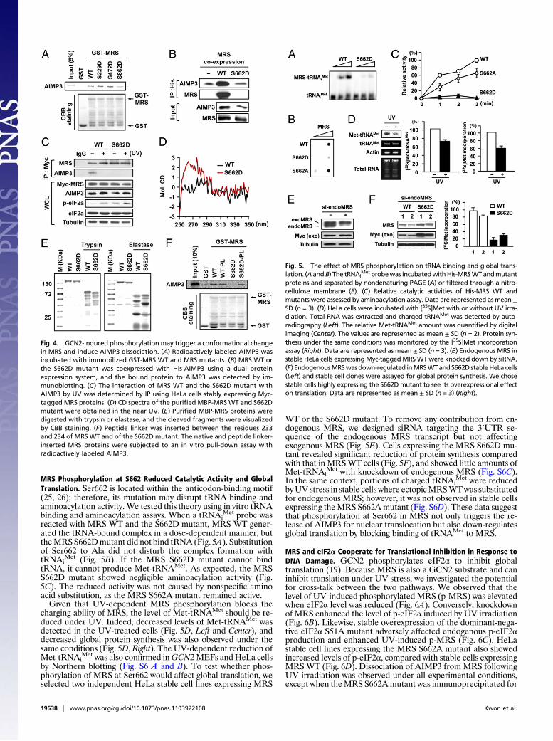
Phosphorylation of MRS at Ser662 Induces a Conformational Changeto Release AIMP3. To determine the effect of GCN2-inducedphosphorylation on the interaction betweenMRS andAIMP3, weintroduced S229D, S472D, and S662D mutations into MRS, andexamined how these mutations would affect the interaction ofMRS with AIMP3. Using a GST pull-down assay, we found thebinding ofMRSS662D toAIMP3was reduced in comparisonwiththe WT and other mutants (Fig. 4A). We then prepared DNAconstructs whereby His-AIMP3 is coexpressed with WT MRS orthe S662D mutant. When His-AIMP3 was purified from cellscoexpressing the constructs, the bound S662D mutant was sig-nificantly reduced compared with WT MRS (Fig. 4B). Althoughthe dissociation of MRS S662D and AIMP3 was confirmed by IPassay using HeLa cells stably expressing MRS WT or the S662D(Fig. 4C), the MRS S662A mutant interacted stably with AIMP3regardless of UV stress (Fig. S5A). These results suggest thatphosphorylation of MRS at Ser662 may decrease the binding af-finity of MRS to AIMP3.Because Ser662 is distal to the N-terminal extension of MRS
(Fig. 1), phosphorylation at Ser662 is unlikely to directly affect theinteraction with AIMP3; instead, it may induce a conformationalchange that leads to the dissociation of AIMP3. We tested thishypothesis by comparing circular dichroism (CD) spectra of MRSWT and the S662D mutant. The MRS S662D mutant had a dif-ferent CD spectrum in the near UV range (Fig. 4D), suggestinga difference in its tertiary structure. The two proteins were alsosubjected to trypsin and elastase digestion. With both enzymes,the MRS S662D mutant showed a slightly different digestionpattern (Fig. 4E), further suggesting a conformational difference.Next, we inserted a Gly-Gly-Gly-Gly-Ser peptide linker into theregion between the N-terminal AIMP3 binding and catalyticdomains of MRS. We expected the insertion of this flexible pep-tide to loosen the conformational linkage between the AIMP3-binding region and Ser662-harboring domain and thus provideconformational flexibility. We did not expect the selected positionfor the peptide-linker insertion to alter MRS activity or protein–protein interactions (12, 24). Indeed, we found that in the WTbackground, the aminoacylation activity of MRS was not influ-enced by peptide-linker insertion (Fig. S5B), andAIMP3 bound tothe normal and peptide linker-inserted MRS with similar affinity(Fig. 4F). In contrast, the insertion of the peptide linker intoMRSS662D restored its ability to bind AIMP3 (Fig. 4F). These datasuggest that Ser662 phosphorylationmay induce a conformationalchange that is propagated to the N-terminal extension, therebyreleasing the bound AIMP3.
Fig. 3. GCN2-induced phosphorylation is responsible for the dissociation ofMRSand AIMP3. (A and B) HeLa cells treated with si-control or si-GCN2 for 72 h (A) orGCN2+/+ andGCN2−/−MEFs (B) were UV-irradiated. Interaction betweenMRS andAIMP3wasdetectedby IPand immunoblotting.The relative ratioofboundAIMP3to MRS was quantified and presented to show the extent of AIMP3 dissociation.(C) Proteins fromHeLa cellswere separatedby2D-PAGE.Half of theUV-irradiatedsamples were treated with alkaline phosphatase (AP) for dephosphorylation. (D)The effect of GCN2 knockdown on MRS phosphorylation was detected (Upper).UV-dependentphosphorylationofGCN2wasvalidatedusingp-ThrAb. (E)CellularinteractionofGCN2withMRSwasobserved inUV-irradiatedHeLa cells. (F) Kinaseassaywasdoneby incubationof Flag-GCN2, [γ-32P]ATP, andMBP-MRSD1,D2, andD3. After stained with Coomassie brilliant blue (CBB), gel was dried and exposedfor autoradiography. (G) The immobilized GST-GCN2 kinase domain (KD) wasmixedwith biotinylated synthetic peptides containing known sequences of GCN2substrates (GCN2 Thr899 and eIF2α Ser51) and [γ-32P]ATP to detect phosphoryla-tion. An inactive form of GCN2 KD K618R mutant was used as a control.
Kwon et al. PNAS | December 6, 2011 | vol. 108 | no. 49 | 19637
CELL
BIOLO
GY
MRS Phosphorylation at S662 Reduced Catalytic Activity and GlobalTranslation. Ser662 is located within the anticodon-binding motif(25, 26); therefore, its mutation may disrupt tRNA binding andaminoacylation activity. We tested this theory using in vitro tRNAbinding and aminoacylation assays. When a tRNAi
Met probe wasreacted with MRS WT and the S662D mutant, MRS WT gener-ated the tRNA-bound complex in a dose-dependent manner, buttheMRSS662Dmutant did not bind tRNA (Fig. 5A). Substitutionof Ser662 to Ala did not disturb the complex formation withtRNAi
Met (Fig. 5B). If the MRS S662D mutant cannot bindtRNA, it cannot produce Met-tRNAMet. As expected, the MRSS662D mutant showed negligible aminoacylation activity (Fig.5C). The reduced activity was not caused by nonspecific aminoacid substitution, as the MRS S662A mutant remained active.Given that UV-dependent MRS phosphorylation blocks the
charging ability of MRS, the level of Met-tRNAMet should be re-duced under UV. Indeed, decreased levels of Met-tRNAMet wasdetected in the UV-treated cells (Fig. 5D, Left and Center), anddecreased global protein synthesis was also observed under thesame conditions (Fig. 5D, Right). The UV-dependent reduction ofMet-tRNAi
Met was also confirmed inGCN2MEFs andHeLa cellsby Northern blotting (Fig. S6 A and B). To test whether phos-phorylation of MRS at Ser662 would affect global translation, weselected two independent HeLa stable cell lines expressing MRS
WT or the S662D mutant. To remove any contribution from en-dogenous MRS, we designed siRNA targeting the 3′UTR se-quence of the endogenous MRS transcript but not affectingexogenous MRS (Fig. 5E). Cells expressing the MRS S662D mu-tant revealed significant reduction of protein synthesis comparedwith that in MRSWT cells (Fig. 5F), and showed little amounts ofMet-tRNAi
Met with knockdown of endogenous MRS (Fig. S6C).In the same context, portions of charged tRNAi
Met were reducedbyUV stress in stable cells where ectopicMRSWTwas substitutedfor endogenous MRS; however, it was not observed in stable cellsexpressing theMRS S662Amutant (Fig. S6D). These data suggestthat phosphorylation at Ser662 in MRS not only triggers the re-lease of AIMP3 for nuclear translocation but also down-regulatesglobal translation by blocking binding of tRNAMet to MRS.
MRS and eIF2α Cooperate for Translational Inhibition in Response toDNA Damage. GCN2 phosphorylates eIF2α to inhibit globaltranslation (19). Because MRS is also a GCN2 substrate and caninhibit translation under UV stress, we investigated the potentialfor cross-talk between the two pathways. We observed that thelevel of UV-induced phosphorylated MRS (p-MRS) was elevatedwhen eIF2α level was reduced (Fig. 6A). Conversely, knockdownof MRS enhanced the level of p-eIF2α induced by UV irradiation(Fig. 6B). Likewise, stable overexpression of the dominant-nega-tive eIF2α S51A mutant adversely affected endogenous p-eIF2αproduction and enhanced UV-induced p-MRS (Fig. 6C). HeLastable cell lines expressing the MRS S662A mutant also showedincreased levels of p-eIF2α, compared with stable cells expressingMRSWT (Fig. 6D). Dissociation of AIMP3 from MRS followingUV irradiation was observed under all experimental conditions,except when theMRS S662Amutant was immunoprecipitated for
Fig. 4. GCN2-induced phosphorylation may trigger a conformational changein MRS and induce AIMP3 dissociation. (A) Radioactively labeled AIMP3 wasincubated with immobilized GST-MRS WT and MRS mutants. (B) MRS WT orthe S662D mutant was coexpressed with His-AIMP3 using a dual proteinexpression system, and the bound protein to AIMP3 was detected by im-munoblotting. (C) The interaction of MRS WT and the S662D mutant withAIMP3 by UV was determined by IP using HeLa cells stably expressing Myc-tagged MRS proteins. (D) CD spectra of the purified MBP-MRSWT and S662Dmutant were obtained in the near UV. (E) Purified MBP-MRS proteins weredigested with trypsin or elastase, and the cleaved fragments were visualizedby CBB staining. (F) Peptide linker was inserted between the residues 233and 234 of MRS WT and of the S662D mutant. The native and peptide linker-inserted MRS proteins were subjected to an in vitro pull-down assay withradioactively labeled AIMP3.
Fig. 5. The effect of MRS phosphorylation on tRNA binding and global trans-lation. (A andB) The tRNAi
Met probewas incubatedwithHis-MRSWTandmutantproteins and separated by nondenaturing PAGE (A) or filtered through a nitro-cellulose membrane (B). (C) Relative catalytic activities of His-MRS WT andmutants were assessed by aminoacylation assay. Data are represented asmean±SD (n = 3). (D) HeLa cells were incubated with [35S]Met with or without UV irra-diation. Total RNA was extracted and charged tRNAMet was detected by auto-radiography (Left). The relative Met-tRNAMet amount was quantified by digitalimaging (Center). The values are represented as mean ± SD (n = 2). Protein syn-thesis under the same conditions was monitored by the [35S]Met incorporationassay (Right). Data are represented as mean ± SD (n = 3). (E) Endogenous MRS instable HeLa cells expressing Myc-taggedMRSWTwere knocked down by siRNA.(F) EndogenousMRSwas down-regulated inMRSWTand S662D stableHeLa cells(Left) and stable cell clones were assayed for global protein synthesis. We chosestable cells highly expressing the S662Dmutant to see its overexpressional effecton translation. Data are represented as mean ± SD (n = 3) (Right).
19638 | www.pnas.org/cgi/doi/10.1073/pnas.1103922108 Kwon et al.
the analysis of bound AIMP3. This result indicates that phos-phorylation of MRS at Ser662 is the prerequisite step for AIMP3release. Knockdown or overexpression of MRS or eIF2α did notaffect the total protein level of its counterpart. These data suggestthat the levels of p-eIF2α and p-MRS can be controlled in acomplementary fashion.To observe how both proteins contribute to global translational
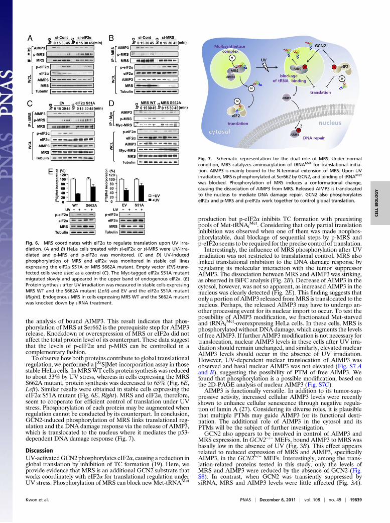
regulation, we performed a [35S]Met-incorporation assay in thosestable HeLa cells. InMRSWT cells protein synthesis was reducedto about 33% by UV stress, whereas in cells expressing the MRSS662A mutant, protein synthesis was decreased to 65% (Fig. 6E,Left). Similar results were obtained in stable cells expressing theeIF2α S51A mutant (Fig. 6E, Right). MRS and eIF2α, therefore,seem to cooperate for efficient control of translation under UVstress. Phosphorylation of each protein may be augmented whenregulation cannot be conducted by its counterpart. In conclusion,GCN2-induced phosphorylation of MRS links translational reg-ulation and the DNA damage response via the release of AIMP3,which is translocated to the nucleus where it mediates the p53-dependent DNA damage response (Fig. 7).
DiscussionUV-activatedGCN2 phosphorylates eIF2α, causing a reduction inglobal translation by inhibition of TC formation (19). Here, weprovide evidence that MRS is an additional GCN2 substrate thatworks coordinately with eIF2α for translational regulation underUV stress. Phosphorylation of MRS can block newMet-tRNAMet
production but p-eIF2α inhibits TC formation with preexistingpools of Met-tRNAi
Met. Considering that only partial translationinhibition was observed when one of them was made nonphos-phorylatable, dual blockage of sequential steps by p-MRS andp-eIF2α seems to be required for the precise control of translation.Interestingly, the influence of MRS phosphorylation after UV
irradiation was not restricted to translational control. MRS alsolinked translational inhibition to the DNA damage response byregulating its molecular interaction with the tumor suppressorAIMP3. The dissociation between MRS and AIMP3 was striking,as observed in BiFC analysis (Fig. 2B). Decrease of AIMP3 in thecytosol, however, was not so apparent, as increased AIMP3 in thenucleus was clearly detected (Fig. 2E). This finding suggests thatonly a portion of AIMP3 released fromMRS is translocated to thenucleus. Perhaps, the released AIMP3 may have to undergo an-other processing event for its nuclear import to occur. To test thepossibility of AIMP3 modification, we fractionated Met-starvedand tRNAi
Met-overexpressing HeLa cells. In these cells, MRS isphosphorylated without DNA damage, which augments the levelsof free AIMP3. If further AIMP3modification is not necessary fortranslocation, nuclear AIMP3 levels in these cells after UV irra-diation should remain unchanged, and similarly, elevated nuclearAIMP3 levels should occur in the absence of UV irradiation.However, UV-dependent nuclear translocation of AIMP3 wasobserved and basal nuclear AIMP3 was not elevated (Fig. S7 Aand B), suggesting the possibility of PTM of free AIMP3. Wefound that phosphorylation is a possible modification, based onthe 2D-PAGE analysis of nuclear AIMP3 (Fig. S7C).AIMP3 is functionally versatile. In addition to its tumor-sup-
pressive activity, increased cellular AIMP3 levels were recentlyshown to enhance cellular senescence through negative regula-tion of lamin A (27). Considering its diverse roles, it is plausiblethat multiple PTMs may guide AIMP3 for its functional desti-nation. The additional role of AIMP3 in the cytosol and itsPTMs will be the subject of further investigation.GCN2 also appears to be involved in control of AIMP3 and
MRS expression. In GCN2−/− MEFs, bound AIMP3 to MRS wasbasally low in the absence of UV (Fig. 3B). This effect appearsrelated to reduced expression of MRS and AIMP3, specificallyAIMP3, in the GCN2−/− MEFs. Interestingly, among the trans-lation-related proteins tested in this study, only the levels ofMRS and AIMP3 were reduced by the absence of GCN2 (Fig.S8). In contrast, when GCN2 was transiently suppressed bysiRNA, MRS and AIMP3 levels were little affected (Fig. 3A).
Fig. 6. MRS coordinates with eIF2α to regulate translation upon UV irra-diation. (A and B) HeLa cells treated with si-eIF2α or si-MRS were UV-irra-diated and p-MRS and p-eIF2α was monitored. (C and D) UV-inducedphosphorylation of MRS and eIF2α was monitored in stable cell linesexpressing the eIF2α S51A or MRS S662A mutant. Empty vector (EV)-trans-fected cells were used as a control (C). The Myc-tagged eIF2α S51A mutantmigrated slowly and appeared in the upper band of endogenous eIF2α. (E)Protein synthesis after UV irradiation was measured in stable cells expressingMRS WT and the S662A mutant (Left) and EV and the eIF2α S51A mutant(Right). Endogenous MRS in cells expressing MRS WT and the S662A mutantwas knocked down by siRNA treatment.
Fig. 7. Schematic representation for the dual role of MRS. Under normalcondition, MRS catalyzes aminoacylation of tRNAMet for translational initia-tion. AIMP3 is mainly bound to the N-terminal extension of MRS. Upon UVirradiation, MRS is phosphorylated at Ser662 by GCN2, and binding of tRNAMet
was blocked. Phosphorylation of MRS induces a conformational change,causing the dissociation of AIMP3 from MRS. Released AIMP3 is translocatedto the nucleus to mediate DNA damage repair. GCN2 also phosphorylateseIF2α and p-MRS and p-eIF2α work together to control global translation.
Kwon et al. PNAS | December 6, 2011 | vol. 108 | no. 49 | 19639
CELL
BIOLO
GY

Perhaps, in GCN2−/− cells constitutively lacking the GCN2 ac-tivity, a risk of uncontrolled translation may be compensated byreducing cellular levels of MRS and AIMP3, although the pre-cise role of GCN2 in this connection is currently unknown.Previous reports provide several examples of how the dynamic
relationship of MSC components is controlled by phosphoryla-tion. For example, in IFN-γ–activated U937 cells, EPRS dissoci-ates and forms new complexes called gamma-interferon activatedinhibitor of translation (GAIT) with the 3′UTR of target tran-scripts, which mediate translational gene silencing (28). In addi-tion, in mast cells, lysyl-tRNA synthetase is phosphorylated andtranslocates to the nucleus, where it controls the activity of themicrophthalmia transcription factor (29). Among the three non-enzymatic components of MSC, AIMP2 is phosphorylated andtranslocated to the nucleus, where it binds p53 uponDNA damage(30). The effects of p-MRS can be distinguished from these earlierreported modes of regulation. Specifically, p-MRS is not disasso-ciated from MSC, and in its associated state releases boundAIMP3. Moreover, phosphorylation of MSC components has notpreviously been shown to affect global translation. Here, we foundthat phosphorylation significantly suppresses the catalytic activityof MRS, leading to the down-regulation of global translation. Thisfinding provides evidence for a unique role of MRS as a co-ordinator of cytosolic translation and nuclear DNA repair.MRS phosphorylation links UV stress to the DNA damage re-
sponse through AIMP3 translocation, whereas eIF2α phosphoryla-tion increases the NF-κB response upon UV irradiation (23).AlthoughAIMP3 release fromMRS and nuclear translocation wereobserved within 1 h after UV irradiation, NF-κB activation by p-eIF2α is only observed starting 1 h after UV irradiation. Thus,GCN2 may use MRS-AIMP3 as an early response to UV-inducedDNA damage and eIF2α for the transcriptional control of NF-κB–dependent genes in the later stages. GCN2-mediated delay of cel-lular entry into the S phase byUV stress has been reported; however,the underlyingmechanism and cross-talk betweenGCN2 andATM/ATR in cell-cycle regulation and DNA repair are not clearly un-derstood (31). Our work suggests that the activation of GCN2 isfunctionally linked to the activation of ATM/ATR via AIMP3
released from p-MRS, although the actual relationship betweenATM/ATR, NF-κB, p-eIF2α, and AIMP3 needs further in-vestigation. In conclusion, we have demonstrated a dual role forhumanMRS in the regulation of cytosolic translation andmolecularinteraction with AIMP3, thus elucidating a mechanism for couplingthe control of protein synthesis to the DNA damage response. Thiswork is also significant in that MRS represents a previously un-detected gate for translational control, suggesting the possibility thatother ARSs may play unique roles in translational control andcell signaling.
Materials and MethodsProtocols for well established procedures (yeast two-hybrid assay, pull-downassay, BiFC, cell fractionation, 2D-PAGE, kinase assay, CD spectrum, amino-acylation assay, gel shift assay, Northern blotting, and Met incorporationassay) can be found in SI Materials and Methods.
Cell Culture. GCN2MEFs andHeLawere grown in DMEM supplementedwith 10%(vol/vol) FBS and 1% (vol/vol) penicillin/streptomycin in 5% (vol/vol) CO2 at 37 °C.For the selection of stable cell line, plasmids were transfected with FuGENE HD(Roche) and transfected cells were selected and maintained with 1 mg/mL genet-icin (G418, Duchefa Biochemie). For IP or BiFC, cells were UV-irradiated (60 J/m2),recovered with serum-free medium, and harvested at the indicated time points.
IP Assay. Cells were lysed with IP buffer (50 mM Tris-HCl pH 7.4, 150 mM NaCl,0.5%Triton X-100, 5mMEDTA, and 10% (vol/vol) glycerol) containing proteaseinhibitor and phosphatase inhibitor. Cell lysates were centrifuged and in-cubated overnight with IgG or specific Abs and then with Protein A/G agarose.IP proteins were separated by SDS/PAGE and detected by immunoblotting.
ACKNOWLEDGMENTS. We thank Dr. D. Ron for providing GCN2 mouse em-bryonic fibroblast cells and pCDNA3-GCN2, and Dr. C.-D. Hu for providingpBiFC-VN173 and pBiFC-VC155. This research was supported by the WorldClass University Project (R31-2008-000-10103-0) of Korea Science and Engi-neering Foundation, Global Frontier Research Grant NRF-M1AXA002-2010-0029785, and the Basic Science Research Program (2010-0023292) of theNational Research Foundation funded by the Ministry of Education, Scienceand Technology of Korea.
1. Gebauer F, Hentze MW (2004) Molecular mechanisms of translational control. Nat RevMol Cell Biol 5:827–835.
2. Pavon-Eternod M, et al. (2009) tRNA over-expression in breast cancer and functionalconsequences. Nucleic Acids Res 37:7268–7280.
3. Sonenberg N, Hinnebusch AG (2009) Regulation of translation initiation in eukar-yotes: mechanisms and biological targets. Cell 136:731–745.
4. Lee SW, Cho BH, Park SG, Kim S (2004) Aminoacyl-tRNA synthetase complexes: Be-yond translation. J Cell Sci 117:3725–3734.
5. Park SG, Ewalt KL, Kim S (2005) Functional expansion of aminoacyl-tRNA synthetasesand their interacting factors: new perspectives on housekeepers. Trends Biochem Sci30:569–574.
6. Ko YG, Kang YS, Kim EK, Park SG, Kim S (2000) Nucleolar localization of human me-thionyl-tRNA synthetase and its role in ribosomal RNA synthesis. J Cell Biol 149:567–574.
7. Netzer N, et al. (2009) Innate immune and chemically triggered oxidative stressmodifies translational fidelity. Nature 462:522–526.
8. Marshall L, Kenneth NS, White RJ (2008) Elevated tRNA(iMet) synthesis can drive cellproliferation and oncogenic transformation. Cell 133:78–89.
9. Park SG, Choi EC, Kim S (2010) Aminoacyl-tRNA synthetase-interacting multifunc-tional proteins (AIMPs): A triad for cellular homeostasis. IUBMB Life 62:296–302.
10. Han JM, et al. (2006) Hierarchical network between the components of the multi-tRNAsynthetase complex: Implications for complex formation. J Biol Chem 281:38663–38667.
11. Park BJ, et al. (2005) The haploinsufficient tumor suppressor p18 upregulates p53 viainteractions with ATM/ATR. Cell 120:209–221.
12. Rho SB, et al. (1999) Genetic dissection of protein-protein interactions in multi-tRNAsynthetase complex. Proc Natl Acad Sci USA 96:4488–4493.
13. Kim KJ, et al. (2008) Determination of three-dimensional structure and residues of thenovel tumor suppressor AIMP3/p18 required for the interaction with ATM. J BiolChem 283:14032–14040.
14. Park BJ, et al. (2006) AIMP3 haploinsufficiency disrupts oncogene-induced p53 acti-vation and genomic stability. Cancer Res 66:6913–6918.
15. Kaminska M, Deniziak M, Kerjan P, Barciszewski J, Mirande M (2000) A recurrentgeneral RNA binding domain appended to plant methionyl-tRNA synthetase acts asa cis-acting cofactor for aminoacylation. EMBO J 19:6908–6917.
16. Guo M, Yang XL, Schimmel P (2010) New functions of aminoacyl-tRNA synthetasesbeyond translation. Nat Rev Mol Cell Biol 11:668–674.
17. Wek RC, Jiang HY, Anthony TG (2006) Coping with stress: eIF2 kinases and trans-lational control. Biochem Soc Trans 34:7–11.
18. Shyu YJ, Suarez CD, Hu C-D (2008) Visualization of ternary complexes in living cells byusing a BiFC-based FRET assay. Nat Protoc 3:1693–1702.
19. Deng J, et al. (2002) Activation of GCN2 in UV-irradiated cells inhibits translation. CurrBiol 12:1279–1286.
20. Berlanga JJ, Santoyo J, De Haro C (1999) Characterization of a mammalian homologof the GCN2 eukaryotic initiation factor 2alpha kinase. Eur J Biochem 265:754–762.
21. Dong J, Qiu H, Garcia-Barrio M, Anderson J, Hinnebusch AG (2000) Uncharged tRNAactivates GCN2 by displacing the protein kinase moiety from a bipartite tRNA-bindingdomain. Mol Cell 6:269–279.
22. Wek SA, Zhu S, Wek RC (1995) The histidyl-tRNA synthetase-related sequence inthe eIF-2 alpha protein kinase GCN2 interacts with tRNA and is required for acti-vation in response to starvation for different amino acids. Mol Cell Biol 15:4497–4506.
23. Jiang HY, Wek RC (2005) GCN2 phosphorylation of eIF2alpha activates NF-kappaB inresponse to UV irradiation. Biochem J 385:371–380.
24. HeR,ZuLD,YaoP,ChenX,WangED(2009)Twonon-redundantfragments intheN-terminalpeptide of human cytosolic methionyl-tRNA synthetase were indispensable for the multi-synthetasecomplex incorporationandenzymeactivity.BiochimBiophysActa1794:347–354.
25. Nakanishi K, Ogiso Y, Nakama T, Fukai S, Nureki O (2005) Structural basis for anti-codon recognition by methionyl-tRNA synthetase. Nat Struct Mol Biol 12:931–932.
26. Ghosh A, Vishveshwara S (2007) A study of communication pathways in methionyl-tRNA synthetase by molecular dynamics simulations and structure network analysis.Proc Natl Acad Sci USA 104:15711–15716.
27. Oh YS, et al. (2010) Downregulation of lamin A by tumor suppressor AIMP3/p18 leadsto a progeroid phenotype in mice. Aging Cell 9:810–822.
28. Arif A, et al. (2009) Two-site phosphorylation of EPRS coordinates multimodal regu-lation of noncanonical translational control activity. Mol Cell 35:164–180.
29. Lee YN, Nechushtan H, Figov N, Razin E (2004) The function of lysyl-tRNA synthetaseand Ap4A as signaling regulators of MITF activity in FcepsilonRI-activated mast cells.Immunity 20:145–151.
30. Han JM, et al. (2008) AIMP2/p38, the scaffold for the multi-tRNA synthetase complex,responds to genotoxic stresses via p53. Proc Natl Acad Sci USA 105:11206–11211.
31. Grallert B, Boye E (2007) The Gcn2 kinase as a cell cycle regulator. Cell Cycle 6:2768–2772.
19640 | www.pnas.org/cgi/doi/10.1073/pnas.1103922108 Kwon et al.