Dual Inhibition of Bruton s Tyrosine Kinase and ... J 2017 BTK... · kinases downstream of the BCR...
29
1521-0103/361/2/312–321$25.00 https://doi.org/10.1124/jpet.116.238022 THE JOURNAL OF PHARMACOLOGY AND EXPERIMENTAL THERAPEUTICS J Pharmacol Exp Ther 361:312–321, May 2017 Copyright ª 2017 by The American Society for Pharmacology and Experimental Therapeutics Dual Inhibition of Bruton’s Tyrosine Kinase and Phosphoinositide-3-Kinase p110d as a Therapeutic Approach to Treat Non-Hodgkin’s B Cell Malignancies s Jennifer Alfaro, 1 Felipe Pérez de Arce, Sebastián Belmar, Glenda Fuentealba, Patricio Avila, Gonzalo Ureta, Camila Flores, Claudia Acuña, Luz Delgado, Diana Gaete, Brahmam Pujala, Anup Barde, Anjan K. Nayak, T. V. R. Upendra, Dhananjay Patel, Shailender Chauhan, Vijay K. Sharma, Stacy Kanno, Ramona G. Almirez, David T. Hung, Sarvajit Chakravarty, Roopa Rai, Sebastián Bernales, Kevin P. Quinn, Son M. Pham, and Emma McCullagh 1 Translational Research Group, Fundación Ciencia y Vida, Santiago, Chile (J.A., F.P.d.A., S.Bel., G.F., P.A., G.U., C.F., C.A., L.D., D.G.); Biological Sciences Department, Facultad de Ciencias Biológicas, Universidad Andrés Bello, Región de Valparaíso, Chile (F.P.d.A., S.Bel.); Chemistry Group, Integral BioSciences, Pvt. Ltd., Noida, India (B.P., A.B., A.K.N., T.V.R.U., D.P., S.C., V.K.S.); and Discovery Research, Medivation, Inc., now Pfizer, San Francisco, California (S.K., R.G.A., D.T.H., S.C., R.R., S.Ber., K.P.Q., S.M.P., E.M.) Received September 28, 2016; accepted March 10, 2017 ABSTRACT Although new targeted therapies, such as ibrutinib and idelalisib, have made a large impact on non-Hodgkin’s lymphoma (NHL) patients, the disease is often fatal because patients are initially resistant to these targeted therapies, or because they eventually develop resistance. New drugs and treatments are necessary for these patients. One attractive approach is to inhibit multiple parallel pathways that drive the growth of these hematologic tumors, possibly prolonging the duration of the response and reducing resistance. Early clinical trials have tested this approach by dosing two drugs in combination in NHL patients. We discovered a single molecule, MDVN1003 (1-(5-amino-2,3-dihydro-1H-inden-2-yl)-3-(8- fluoro-3,4-dihydro-2H-benzo[b][1,4]oxazin-6-yl)-1H-pyrazolo[3,4- d]pyrimidin-4-amine), that inhibits Bruton’s tyrosine kinase and phosphatidylinositol-3-kinase d, two proteins regulated by the B cell receptor that drive the growth of many NHLs. In this report, we show that this dual inhibitor prevents the activation of B cells and inhibits the phosphorylation of protein kinase B and extracel- lular signal-regulated kinase 1/2, two downstream mediators that are important for this process. Additionally, MDVN1003 induces cell death in a B cell lymphoma cell line but not in an irrelevant erythroblast cell line. Importantly, we found that this orally bioavail- able dual inhibitor reduced tumor growth in a B cell lymphoma xenograft model more effectively than either ibrutinib or idelalisib. Taken together, these results suggest that dual inhibition of these two key pathways by a single molecule could be a viable approach for treatment of NHL patients. Introduction Non-Hodgkin’s lymphomas (NHLs) are among the most com- mon of human cancers, and despite advancements of medical treatments and improvements in patient outcomes, the disease has a 30% mortality rate within the first five years after diagnosis (Howlader et al., 2014; Swerdlow et al., 2016). Activation of B cell receptor (BCR) signaling is a significant mechanistic driver of the development and growth of B cell–derived lymphoid tumors (Buchner and Muschen, 2014; Koehrer and Burger, 2016). Basal BCR signaling is necessary for the survival of B cells (Verkoczy et al., 2007; Wang et al., 2013), and signaling through the complex pathway is amplified during B cell activation (Woyach et al., 2012). The BCR is comprised of a membrane immunoglobulin complex, the ligation of which results in the phosphorylation of the cytoplasmic immunor- eceptor tyrosine-based activation motif of BCR coreceptors by two kinases, Lyn (Lck/Yes novel tyrosine kinase) and Syk (spleen tyrosine kinase). Propagation of the signal occurs via several parallel and interconnected pathways. Two important Financial support for this research was provided by Medivation, Inc., now Pfizer. Some authors are employees of Medivation, now Pfizer. Some authors are inventors on patents related to the subject matter. 1 J.A. and E.M. contributed equally to this work. https://doi.org/10.1124/jpet.116.238022. s This article has supplemental material available at jpet.aspetjournals.org. ABBREVIATIONS: AKT, protein kinase B; BCR, B cell receptor; BTK, Bruton’s tyrosine kinase; CLL, chronic lymphocytic leukemia; ERK 1/2, extracellular signal-regulated kinase 1/2; MCL, mantle cell lymphoma; MDVN1001, 5-(8-fluoro-3,4-dihydro-2H-benzo[b][1,4]oxazin-6-yl)-7-(5-(piperidin-4- yl)-2,3-dihydro-1H-inden-2-yl)-7H-pyrrolo[2,3-d]pyrimidin-4-amine; MDVN1002, 1-(5-amino-2,3-dihydro-1H-inden-2-yl)-3-(3-fluoro-4-isopropoxyphenyl)- 1H-pyrazolo[3,4-d]pyrimidin-4-amine; MDVN1003, 1-(5-amino-2,3-dihydro-1H-inden-2-yl)-3-(8-fluoro-3,4-dihydro-2H-benzo[b][1,4]oxazin-6-yl)-1H-pyrazolo [3,4-d]pyrimidin-4-amine; NHL, non-Hodgkin’ s lymphoma; pAKT, phosphorylated AKT; PCI-29732, 1-Cyclopentyl-3-(4-phenoxyphenyl)-1H-pyrazolo[3,4-d] pyrimidin-4-amine; pErk, phosphorylated Erk; PI3Kd, phosphatidylinositol-3-kinase d; PK, pharmacokinetic; SW13, 2-((4-amino-3-(3-fluoro-5-hydroxyphenyl)- 1H-pyrazolo[3,4-d]pyrimidin-1-yl)methyl)-5-methyl-3-(o-tolyl)quinazolin-4(3H)-one. 312 http://jpet.aspetjournals.org/content/suppl/2017/03/15/jpet.116.238022.DC1 Supplemental material to this article can be found at: at ASPET Journals on May 3, 2017 jpet.aspetjournals.org Downloaded from at ASPET Journals on May 3, 2017 jpet.aspetjournals.org Downloaded from at ASPET Journals on May 3, 2017 jpet.aspetjournals.org Downloaded from at ASPET Journals on May 3, 2017 jpet.aspetjournals.org Downloaded from at ASPET Journals on May 3, 2017 jpet.aspetjournals.org Downloaded from at ASPET Journals on May 3, 2017 jpet.aspetjournals.org Downloaded from at ASPET Journals on May 3, 2017 jpet.aspetjournals.org Downloaded from at ASPET Journals on May 3, 2017 jpet.aspetjournals.org Downloaded from at ASPET Journals on May 3, 2017 jpet.aspetjournals.org Downloaded from at ASPET Journals on May 3, 2017 jpet.aspetjournals.org Downloaded from at ASPET Journals on May 3, 2017 jpet.aspetjournals.org Downloaded from at ASPET Journals on May 3, 2017 jpet.aspetjournals.org Downloaded from at ASPET Journals on May 3, 2017 jpet.aspetjournals.org Downloaded from at ASPET Journals on May 3, 2017 jpet.aspetjournals.org Downloaded from at ASPET Journals on May 3, 2017 jpet.aspetjournals.org Downloaded from at ASPET Journals on May 3, 2017 jpet.aspetjournals.org Downloaded from at ASPET Journals on May 3, 2017 jpet.aspetjournals.org Downloaded from at ASPET Journals on May 3, 2017 jpet.aspetjournals.org Downloaded from at ASPET Journals on May 3, 2017 jpet.aspetjournals.org Downloaded from at ASPET Journals on May 3, 2017 jpet.aspetjournals.org Downloaded from at ASPET Journals on May 3, 2017 jpet.aspetjournals.org Downloaded from
Transcript of Dual Inhibition of Bruton s Tyrosine Kinase and ... J 2017 BTK... · kinases downstream of the BCR...

1521-0103/361/2/312–321$25.00 https://doi.org/10.1124/jpet.116.238022THE JOURNAL OF PHARMACOLOGY AND EXPERIMENTAL THERAPEUTICS J Pharmacol Exp Ther 361:312–321, May 2017Copyright ª 2017 by The American Society for Pharmacology and Experimental Therapeutics
Dual Inhibition of Bruton’s Tyrosine Kinase andPhosphoinositide-3-Kinase p110d as a TherapeuticApproach to Treat Non-Hodgkin’s B Cell Malignancies s
Jennifer Alfaro,1 Felipe Pérez de Arce, Sebastián Belmar, Glenda Fuentealba, Patricio Avila,Gonzalo Ureta, Camila Flores, Claudia Acuña, Luz Delgado, Diana Gaete, Brahmam Pujala,Anup Barde, Anjan K. Nayak, T. V. R. Upendra, Dhananjay Patel, Shailender Chauhan,Vijay K. Sharma, Stacy Kanno, Ramona G. Almirez, David T. Hung, Sarvajit Chakravarty,Roopa Rai, Sebastián Bernales, Kevin P. Quinn, Son M. Pham, and Emma McCullagh1
Translational Research Group, Fundación Ciencia y Vida, Santiago, Chile (J.A., F.P.d.A., S.Bel., G.F., P.A., G.U., C.F., C.A., L.D.,D.G.); Biological Sciences Department, Facultad de Ciencias Biológicas, Universidad Andrés Bello, Región de Valparaíso, Chile(F.P.d.A., S.Bel.); Chemistry Group, Integral BioSciences, Pvt. Ltd., Noida, India (B.P., A.B., A.K.N., T.V.R.U., D.P., S.C., V.K.S.);and Discovery Research, Medivation, Inc., now Pfizer, San Francisco, California (S.K., R.G.A., D.T.H., S.C., R.R., S.Ber., K.P.Q.,S.M.P., E.M.)
Received September 28, 2016; accepted March 10, 2017
ABSTRACTAlthough new targeted therapies, such as ibrutinib and idelalisib,have made a large impact on non-Hodgkin’s lymphoma (NHL)patients, the disease is often fatal because patients are initiallyresistant to these targeted therapies, or because they eventuallydevelop resistance. New drugs and treatments are necessary forthese patients. One attractive approach is to inhibit multiple parallelpathways that drive the growth of these hematologic tumors,possibly prolonging the duration of the response and reducingresistance. Early clinical trials have tested this approach by dosingtwo drugs in combination in NHL patients. We discovered a singlemolecule, MDVN1003 (1-(5-amino-2,3-dihydro-1H-inden-2-yl)-3-(8-fluoro-3,4-dihydro-2H-benzo[b][1,4]oxazin-6-yl)-1H-pyrazolo[3,4-d]pyrimidin-4-amine), that inhibits Bruton’s tyrosine kinase and
phosphatidylinositol-3-kinase d, two proteins regulated by theB cell receptor that drive the growth of many NHLs. In this report,we show that this dual inhibitor prevents the activation of B cellsand inhibits the phosphorylation of protein kinase B and extracel-lular signal-regulated kinase 1/2, two downstream mediators thatare important for this process. Additionally, MDVN1003 inducescell death in a B cell lymphoma cell line but not in an irrelevanterythroblast cell line. Importantly, we found that this orally bioavail-able dual inhibitor reduced tumor growth in a B cell lymphomaxenograft model more effectively than either ibrutinib or idelalisib.Taken together, these results suggest that dual inhibition of thesetwo key pathways by a single molecule could be a viable approachfor treatment of NHL patients.
IntroductionNon-Hodgkin’s lymphomas (NHLs) are among the most com-
mon of human cancers, and despite advancements of medicaltreatments and improvements in patient outcomes, the diseasehasa 30%mortality ratewithin the first five years after diagnosis(Howlader et al., 2014; Swerdlow et al., 2016). Activation of B cell
receptor (BCR) signaling is a significant mechanistic driverof the development and growth of B cell–derived lymphoidtumors (Buchner and Muschen, 2014; Koehrer and Burger,2016). Basal BCR signaling is necessary for the survival ofB cells (Verkoczy et al., 2007; Wang et al., 2013), and signalingthrough the complex pathway is amplified during B cellactivation (Woyach et al., 2012). The BCR is comprised of amembrane immunoglobulin complex, the ligation of whichresults in the phosphorylation of the cytoplasmic immunor-eceptor tyrosine-based activation motif of BCR coreceptors bytwo kinases, Lyn (Lck/Yes novel tyrosine kinase) and Syk(spleen tyrosine kinase). Propagation of the signal occurs viaseveral parallel and interconnected pathways. Two important
Financial support for this research was provided by Medivation, Inc., nowPfizer. Some authors are employees of Medivation, now Pfizer. Some authorsare inventors on patents related to the subject matter.
1J.A. and E.M. contributed equally to this work.https://doi.org/10.1124/jpet.116.238022.s This article has supplemental material available at jpet.aspetjournals.org.
ABBREVIATIONS: AKT, protein kinase B; BCR, B cell receptor; BTK, Bruton’s tyrosine kinase; CLL, chronic lymphocytic leukemia; ERK 1/2,extracellular signal-regulated kinase 1/2; MCL, mantle cell lymphoma; MDVN1001, 5-(8-fluoro-3,4-dihydro-2H-benzo[b][1,4]oxazin-6-yl)-7-(5-(piperidin-4-yl)-2,3-dihydro-1H-inden-2-yl)-7H-pyrrolo[2,3-d]pyrimidin-4-amine; MDVN1002, 1-(5-amino-2,3-dihydro-1H-inden-2-yl)-3-(3-fluoro-4-isopropoxyphenyl)-1H-pyrazolo[3,4-d]pyrimidin-4-amine; MDVN1003, 1-(5-amino-2,3-dihydro-1H-inden-2-yl)-3-(8-fluoro-3,4-dihydro-2H-benzo[b][1,4]oxazin-6-yl)-1H-pyrazolo[3,4-d]pyrimidin-4-amine; NHL, non-Hodgkin’s lymphoma; pAKT, phosphorylated AKT; PCI-29732, 1-Cyclopentyl-3-(4-phenoxyphenyl)-1H-pyrazolo[3,4-d]pyrimidin-4-amine; pErk, phosphorylated Erk; PI3Kd, phosphatidylinositol-3-kinase d; PK, pharmacokinetic; SW13, 2-((4-amino-3-(3-fluoro-5-hydroxyphenyl)-1H-pyrazolo[3,4-d]pyrimidin-1-yl)methyl)-5-methyl-3-(o-tolyl)quinazolin-4(3H)-one.
312
http://jpet.aspetjournals.org/content/suppl/2017/03/15/jpet.116.238022.DC1Supplemental material to this article can be found at:
at ASPE
T Journals on M
ay 3, 2017jpet.aspetjournals.org
Dow
nloaded from
at ASPE
T Journals on M
ay 3, 2017jpet.aspetjournals.org
Dow
nloaded from
at ASPE
T Journals on M
ay 3, 2017jpet.aspetjournals.org
Dow
nloaded from
at ASPE
T Journals on M
ay 3, 2017jpet.aspetjournals.org
Dow
nloaded from
at ASPE
T Journals on M
ay 3, 2017jpet.aspetjournals.org
Dow
nloaded from
at ASPE
T Journals on M
ay 3, 2017jpet.aspetjournals.org
Dow
nloaded from
at ASPE
T Journals on M
ay 3, 2017jpet.aspetjournals.org
Dow
nloaded from
at ASPE
T Journals on M
ay 3, 2017jpet.aspetjournals.org
Dow
nloaded from
at ASPE
T Journals on M
ay 3, 2017jpet.aspetjournals.org
Dow
nloaded from
at ASPE
T Journals on M
ay 3, 2017jpet.aspetjournals.org
Dow
nloaded from
at ASPE
T Journals on M
ay 3, 2017jpet.aspetjournals.org
Dow
nloaded from
at ASPE
T Journals on M
ay 3, 2017jpet.aspetjournals.org
Dow
nloaded from
at ASPE
T Journals on M
ay 3, 2017jpet.aspetjournals.org
Dow
nloaded from
at ASPE
T Journals on M
ay 3, 2017jpet.aspetjournals.org
Dow
nloaded from
at ASPE
T Journals on M
ay 3, 2017jpet.aspetjournals.org
Dow
nloaded from
at ASPE
T Journals on M
ay 3, 2017jpet.aspetjournals.org
Dow
nloaded from
at ASPE
T Journals on M
ay 3, 2017jpet.aspetjournals.org
Dow
nloaded from
at ASPE
T Journals on M
ay 3, 2017jpet.aspetjournals.org
Dow
nloaded from
at ASPE
T Journals on M
ay 3, 2017jpet.aspetjournals.org
Dow
nloaded from
at ASPE
T Journals on M
ay 3, 2017jpet.aspetjournals.org
Dow
nloaded from
at ASPE
T Journals on M
ay 3, 2017jpet.aspetjournals.org
Dow
nloaded from

kinases downstream of the BCR are Bruton’s tyrosine kinase(BTK) and phosphatidylinositol-3-kinase d (PI3Kd) (Seda andMraz, 2015).BTK is amember of the Tec family of tyrosine kinases and is
recruited to the cell membrane after the activation of the BCR.Together with Syk, BTK phosphorylates phospholipase C-g2,producing the classic second messengers diacylglycerol andinositol-1,4,5-triphosphate from phosphatidylinositol-4,5-bisphosphate. Diacylglycerol activates protein kinase C, andinositol-1,4,5-triphosphate triggers the release of intracellularcalcium, resulting in the activation of several downstreamsignaling molecules, including extracellular signal-regulatedkinase 1/2 (ERK 1/2) (Tomlinson et al., 2001). PI3Kd is alsorecruited to the cell membrane and activated in response toBCR signaling. PI3K is a heterodimer composed of the p85regulatory subunit and the p110 catalytic subunit, which inB cells is the d isoform. PI3Kd is involved in recruiting BTK tothe cell membrane and phosphorylates and activates severaldownstream signaling molecules, including protein kinase B(AKT) (Fruman and Rommel, 2014). The BTK and PI3Kdsignaling pathways are not insulated from one another, andthere is evidence of cross-talk between them (Puri et al., 2013).Both BTK and PI3Kd are involved in normal B cell signalpropagation but have been identified as being aberrantlyactivated in many B cell malignancies (Seda and Mraz, 2015).These proteins are the targets of the approved therapies:ibrutinib, an irreversible BTK inhibitor, and idelalisib, areversible PI3Kd inhibitor.Ibrutinib blocks the enzymatic activity of BTK through
covalent binding to a conserved cysteine residue (Cys481) inthe active site (Honigberg et al., 2010). Ibrutinib was approvedby the Food and Drug Administration for use in mantle celllymphoma (MCL), chronic lymphocytic leukemia (CLL), andWaldenström’s macroglobulinemia patients. Idelalisib is ahighly selective ATP competitive inhibitor of PI3Kd (Lannuttiet al., 2011; Marini et al., 2016) and has been approved for usein CLL, follicular cell NHL, and relapsed small lymphocyticlymphoma patients (Do et al., 2016).Although the approval of these targeted therapies by the
Food and Drug Administration was a significant advance, ithas been reported that about 30% of MCL and CLL patientsshow primary resistance to ibrutinib (Tucker and Rule, 2015).Furthermore, a subset of patients who initially respond to andreceive prolonged treatment of ibrutinib eventually relapsedue to the development of mutations in BTK that prevent thecovalent binding of ibrutinib or gain-of-function mutationsin phospholipase C-g2 (Byrd et al., 2013; Wang et al., 2013;Smith, 2015). The resistance to idelalisib is not as wellunderstood. Several reports have suggested that combinationdosing of ibrutinib and idelalisib could be advantageous sincetargeting parallel pathways downstream of the BCR couldreduce the risk of signal switching, leading to escape fromtumor growth inhibition (Jones and Byrd, 2014; MathewsGriner et al., 2014; Zhang et al., 2014; de Rooij et al., 2015;Koehrer and Burger, 2016).In this report, we describe proof-of-concept preclinical studies
with tool compoundMDVN1003 (1-(5-amino-2,3-dihydro-1H-inden-2-yl)-3-(8-fluoro-3,4-dihydro-2H-benzo[b][1,4]oxazin-6-yl)-1H-pyrazolo[3,4-d]pyrimidin-4-amine), a first-in-class dualinhibitor of BTK and PI3Kd kinases (Pujala et al., 2016). Wefound that MDVN1003 inhibits B cell activation upon BCRcross-linking ex vivo and in vivo, a phenomenon dependent
on BTK and PI3Kd. Additionally, MDVN1003 induces apo-ptosis and decreases viability of DOHH-2 cells, an NHL cellline. Finally, MDVN1003, an orally bioavailable molecule inmice, rats, and dogs, showed significant antitumor effects inan NHL xenograft model in mice. This effect was similar tothat seen with combination dosing of ibrutinib and idelalisibin the samemodel and greater than each of these drugs dosedas single agents. Our results suggest that a dual inhibitor ofBTK and PI3Kd could be an effective treatment strategy forB cell lymphoma patients.
Materials and MethodsReagents and Tumor Cell Lines. Ibrutinib (CAS 936563-96-1)
and idelalisib (CAS 870281-82-6) were purchased from ChemShuttle(Union City, CA). Compounds MDVN1003, MDVN1001 (5-(8-fluoro-3,4-dihydro-2H-benzo[b][1,4]oxazin-6-yl)-7-(5-(piperidin-4-yl)-2,3-dihydro-1H-inden-2-yl)-7H-pyrrolo[2,3-d]pyrimidin-4-amine), and MDVN1002(1-(5-amino-2,3-dihydro-1H-inden-2-yl)-3-(3-fluoro-4-isopropoxyphenyl)-1H-pyrazolo[3,4-d]pyrimidin-4-amine) were synthesized at Integral Bio-Sciences, Pvt. Ltd. (Noida, India). All tumor cell lines were purchasedfrom the American Type Culture Collection (Manassas, VA) and weretested for mycoplasma by polymerase chain reaction. All revived cellswere used within 20 passages and cultured for less than 6 months.Ramos and DOHH-2 cells were maintained in RPMI 1640 mediumcontaining 10% heat-inactivated fetal bovine serum supplemented withpenicillin and streptomycin.
Enzymatic Kinase Assays. In vitro kinase activity assays wereperformed by Reaction Biology Corporation (www.reactionbiology.com) as described on the Web site and as described previously (Pujalaet al., 2016).
Assessment of BCR-Dependent Signaling Levels. Ramoscells were pretreated for 30 minutes with compounds (0.1 or 1 mMfinal concentration) and then stimulated with a-human IgM(1.3 mg/ml) (#109-006-129; Jackson ImmunoResearch Laboratories,Inc.,Westgrove, PA) for 5minutes. Levels of phosphorylatedAKT (pAKT)(#4060; Cell Signaling Technology, Danvers, MA), AKT (#9272; CellSignalingTechnology), phosphorylated Erk (pErk; T202, Y204) (#4377;Cell Signaling Technology), and ERK (#4396; Cell Signaling Technology)were detected by western blot using the ChemiDoc Imaging System(Bio-Rad, Hercules, CA).
Measuring B Cell Activation by CD69 Expression. Spleno-cytes (1� 106 cells/well) were seeded in a 24-well plate and pretreatedfor 30 minutes with compounds at indicated concentrations and thenactivated for 4 hours with a-mouse IgD (3 mg/ml) (YULLOMD6-05;Accurate Chemical, Westbury, NY). Cells were stained with a-B220PE (#553090; BD Biosciences, San Jose, CA), a-CD69 APC (#130-103-947; Miltenyi Biotec, Gladbach, Germany), and with a live/deadfixable aqua dead cell kit (#L34957; ThermoFisher Scientific, GrandIsland, NY) and analyzed by flow cytometry using a MACSQuantAnalyzer 10 flow cytometer (Miltenyi Biotech, Gladbach, Germany).At each concentration of compound tested, the inhibition of B cellactivation was calculated as the percentage of live B cells expressingCD69 in the presence of the compound divided by the percentage oflive B cells expressing CD69 in the vehicle control. Splenocytes fromthree individual mice were treated independently per condition tested.The IC50 was calculated from the average of the three independentexperiments, and curve fitting was done by nonlinear regressionusing GraphPad Prism (GraphPad Software, La Jolla, CA).
Viability Assay. DOHH-2 or HEL 92.1.7 cells were seeded in a96-well white plate overnight at a density of 5000 cells/well. Cells weretreated with compounds at the indicated concentrations for 72 hours.Cell viability was measured using a Cell Titer-glo kit (Promega,Madison, WI) as described by the manufacturer. The IC50 wascalculated from the curve fitted to the data points by nonlinearregression using GraphPad Prism.
Dual Inhibition of BTK and PI3Kd to Treat B Cell Cancers 313
at ASPE
T Journals on M
ay 3, 2017jpet.aspetjournals.org
Dow
nloaded from

Apoptosis Assay. DOHH-2 cells were seeded in a 24-well plate at0.5 � 106 cells/well. Cells were then treated with compounds at 1 mMfor 4 hours, and apoptosis was measured by flow cytometry (MACS-Quant Analyzer 10) using an Annexin V FITC apoptosis detection kit(#556547; BD Biosciences).
Pharmacokinetic Analysis. All animal studies were done asper protocols approved by the Institutional Animal Care and UseCommittee at Medivation or its subsidiaries. See SupplementalMaterial for experimental details of mouse, rat, and dog pharmaco-kinetic (PK) studies.
MeasuringBCell Activation byCD69Expression InVivo. BALB/cmice were orally dosed with compounds (n5 3 per group) at indicatedconcentrations for 30 minutes, and then mice were injected in-travenously in the tail vein with 100 mg of a-IgD (YULLOMD6-05;Accurate Chemical, Westbury, NY) for 5 hours. Splenocytes wereisolated and stained and analyzed as described earlier. The percent-age of live B cells that express CD69 from mice treated with a-IgDand compound was normalized to the percentage of live B cells thatexpress CD69 from mice treated with a-IgD alone.
Mouse Xenograft Model. All animal studies were done as perprotocols approved by the Institutional Animal Care and Use Commit-tee at Medivation or its subsidiaries.
DOHH-2 cells were maintained in RPMI 1640 medium supple-mented with 10% fetal bovine serum and were passaged twice weekly.While in the exponential growth phase, 5 million DOHH-2 cells in0.1 ml of phosphate-buffered saline (1:1 ratio with matrigel) wereinoculated into the right flanks of 6- to 7-week-old female CB17/SCIDmice (denoted as day 0). When the average tumor volume reached118 mm3, mice were randomly grouped into eight groups (n 5 10 pergroup). Tumor volume and body weight were measured twice a week.The experiment was terminated when the average tumor volumeof the vehicle group reached .2000 mm3, and plasma and tumorsamples were collected from each mouse (n 5 3 mice per group weresacrificed 5 minutes before the final dose, n 5 3 mice per groupwere sacrificed 30 minutes post final dose, and n 5 4 mice per groupwere sacrificed 6 hours post final dose). Statistical analysis (Kruskal-Wallis corrected for multiple comparisons) was done on the tumorvolumes of the groups over time.
ResultsAs described (Pujala et al., 2016), we aimed to discover
reversible dual BTK and PI3Kd inhibitors based on thesimilarities between a reversible BTK inhibitor, PCI-29732(1-Cyclopentyl-3-(4-phenoxyphenyl)-1H-pyrazolo[3,4-d]pyrimidin-4-amine) (Pan et al., 2007; Marcotte et al., 2010), and a PI3Kd/gdual inhibitor, SW13 (2-((4-amino-3-(3-fluoro-5-hydroxyphenyl)-1H-pyrazolo[3,4-d]pyrimidin-1-yl)methyl)-5-methyl-3-(o-tolyl)quinazolin-4(3H)-one) (Berndt et al., 2010).We identifiedthree compounds (Fig. 1), two of which are more potent againstone of the two receptors and one compound that is a more potentdual inhibitor (Table 1; synthesis in SupplementalMethods). Onthe onehand,MDVN1001 is a potentBTK inhibitor (IC50 0.9nM)and a relatively weak PI3Kd inhibitor (IC50 149 nM). On theother hand,MDVN1002 strongly inhibited PI3Kd (IC50 25.9 nM)more than it didBTK (IC50 695 nM).However, the dual inhibitor,MDVN1003, potently inhibited both kinases (BTK with anIC50 of 32.3 nM, and PI3Kd with an IC50 of 16.9 nM). Asexpected, the control compounds, idelalisib and ibrutinib,inhibited one kinase more strongly than the other. Idelalisib,an ATP competitive, reversible inhibitor of PI3Kd, inhibitedPI3Kd with an IC50 of 1.2 nM, whereas it did not measurablyinhibit BTK at any concentration tested. Ibrutinib, an irrevers-ible inhibitor of BTK, potently inhibited BTK (IC50 0.142 nM)and weakly inhibited PI3Kd (IC50 640 nM).
To investigate the effects of these molecules on signalingthrough the BCR pathway, phosphorylation of ERK 1/2 andAKT was measured in the Ramos Burkitt’s B cell lymphomacell line. Ramos cells were pretreated with vehicle or 0.1 mMidelalisib, ibrutinib, MDVN1001, MDVN1002, MDVN1003, oran equimolar cotreatment of idelalisib and ibrutinib (sub-sequently referred to as “combo”). Cells were treated witha-human IgM for 5 minutes to cross-link and activate theBCR. Levels of phosphorylated ERK 1/2 and AKT weredetected by western blot (Fig. 2). Treatment with a-humanIgM increased the levels of phosphorylated AKT and ERK 1/2as compared with the vehicle alone, indicating the BCRsignaling pathway was activated (Fig. 2, lanes 1 and 2).Ibrutinib and idelalisib treatments significantly reducedthe levels of pERK 1/2 and pAKT. Neither MDVN1001 norMDVN1002 significantly affected levels of pAKT or pERK 1/2when tested at 0.1 mM. However, these compounds did inhibitthe phosphorylation of AKT and ERK 1/2 at higher concen-trations (Supplemental Fig. 1). Combination treatment ofidelalisib and ibrutinib potently inhibited levels of both pAKTand pERK 1/2 (Fig. 2). The MDVN1001 and MDVN1002combination treatment (combo MDVN) also inhibited the
Fig. 1. Novel reversible inhibitors of PI3Kd or BTK. The structuresof idelalisib, ibrutinib, and three novel compounds (MDVN1001,MDVN1002, and MDVN1003) are detailed. MDVN1001, MDVN1002,and MDVN1003 are enantiomerically pure, but the absolute configura-tion has yet to be determined (asterisks in the structures mark thestereogenic centers).
314 Alfaro et al.
at ASPE
T Journals on M
ay 3, 2017jpet.aspetjournals.org
Dow
nloaded from

phosphorylation of AKT and ERK1/2, although not as potentlyas the combination treatment of the two approved inhibitors(Supplemental Fig. 1). Treatment with the dual BTK/PI3Kdinhibitor compound MDVN1003 inhibited the phosphoryla-tion of AKT andERK 1/2 at both the low concentration (0.1mM)and the high concentration (1 mM) and did so more potentlythan the combination of MDVN1001 and MDVN1002 (Fig. 2;Supplemental Fig. 1). Taken together, these data show thatMDVN1003 is a more potent inhibitor of BCR signaling thaneither MDVN1001 or MDVN1002 dosed individually or incombination and suggest that the dual inhibition of BTK andPI3Kd may have a synergistic effect on downstream signalingmolecules.We wanted to further investigate the potential synergy of
inhibiting both BTK and PI3Kd in B cell lymphomas usingibrutinib, idelalisib, and MDVN1003 as tool compounds.These compounds provided us with the necessary tools tostudy treatment with monotherapies and dual inhibition.MDVN1003 is more potent than either MDVN1001 orMDVN1002 (or the combination of both) in the cell-basedassay and allowed us to study dual inhibition in a singlemolecule.To understand the selectivity profile of MDVN1003 and
how it compares to those of ibrutinib and idelalisib, we testedthese compounds in a kinase panel at 1 mM. Idelalisib is ahighly selective PI3Kd inhibitor and inhibited four otherkinases (out of 374 tested) greater than 50%. Ibrutinibinhibited 34 kinases (out of 374 tested) more than 50% (Fig. 3).MDVN1003 behaved similarly to ibrutinib in the kinase paneland inhibited 50 kinases (out of 402 tested) at greater than50%, with a preference for tyrosine kinases (Fig. 3). Among thekinases most potently inhibited by MDVN1003 were BTK,PI3Kd, and the related Tec- and PI3K-family kinases (Fig. 3;Table 2).We next investigated the effects of MDVN1003 on the
activation of primary B cells ex vivo. Mouse splenocytes werepretreated with ibrutinib, idelalisib, combo, or MDVN1003 for30 minutes. Cells were treated with a-IgD for 4 hours toactivate B cells. Anti-IgD cross-links the surface immunoglob-ulin and activates theB cell, which upregulates the early B cellactivation marker CD69 (Sancho et al., 2005). ActivatedB cells were detected by flow cytometry as B221CD691 in agate of live cells. The inhibition of B cell activation wasmeasured by the percentage of live B cells that expressedCD69 on the cell surface in treated samples as comparedwith the a-IgD control. Ibrutinib and idelalisib potentlyinhibited B cell activation, with IC50 values of 6.9 and 5.4 nM,respectively (Fig. 4A; Table 3). Idelalisib, but not ibrutinib,produced full inhibition of B cell activation. The combotreatment of both compounds showed a modest additive effect
with an IC50 of 1.1 nM (Fig. 4A; Table 3) and produced fullinhibition of B cell activation. MDVN1003 inhibited B cellactivation with an IC50 of 25.2 nM (Fig. 4A; Table 3).Although less potent, MDVN1003 was equally as efficaciousas idelalisib and the combo treatment and fully inhibitedB cell activation.Signaling through the BCR is often a driver of tumor growth
in B cell malignancies (Buchner and Muschen, 2014), and ithas been reported that both ibrutinib and idelalisib reduce cellviability and induce apoptosis in NHL B cell lines (Honigberget al., 2007; Lannutti et al., 2011; Qu et al., 2015). To determinewhether MDVN1003 behaved similarly to the approved drugs,wemeasured the effect ofMDVN1003 on the viability ofDOHH-2cells, a non-Hodgkin’s B cell lymphoma line that expresses BTKand PI3Kd. After 72 hours of treatment, MDVN1003 effectivelykilled DOHH-2 cells with an IC50 of 1.34 mM, whereas the IC50
values for ibrutinib and idelalisib were 0.023 and 0.86 mM,respectively (Fig. 4B; Table 3). The IC50 for the combo treatmentwas 0.0084 mM, suggesting an additive effect on cell viability ininhibiting both BTK and PI3Kd. The cytotoxicity induced bythese compoundswas determined to be apoptosis, as indicated bythe significant population of Annexin V1 propidium iodide2cells in treatment groups as compared with the vehicle control(P,0.0001, one-wayanalysis of variance) (Fig. 4C).Combinationtreatment of MDVN1001, the BTK inhibitor, and MDVN1002,the PI3Kd inhibitor, also showed an additive effect on cellviability of DOHH-2 cells with an IC50 of 0.87 mM, as comparedwith IC50 values of 1.47 mMwithMDVN1001 alone and 2.75mMwith MDVN1002 alone (Supplemental Fig. 2). Although amodest effect, these data suggest a benefit of dual inhibitionof BTK and PI3Kd.To understand if the cytotoxicity of these compounds
was dependent on inhibition of BTK and PI3Kd, we testedthe effects of the compounds on viability of HEL 92.1.7erythroblast-like cells that do not express these kinases. TheIC50 values of ibrutinib, idelalisib, combo, and MDVN1003were all greater than 10 mM (Table 3), suggesting that thesecompounds are preferentially cytotoxic in cells that expressBTK and PI3Kd.
Fig. 2. BTK and PI3Kd inhibitor compounds differentially inhibit thephosphorylation of downstream signaling molecules ATK and ERK.Ramos cells were pretreated for 30 minutes with either dimethylsulfoxide(lanes labeled vehicle and -) or the indicated compounds... the indicatedcompounds (0.1 mM). All samples, except for the vehicle control, weretreated with a-IgM (1.3 mg/ml) for 5 minutes to activate BCR signaling.Levels of pAKT and pERK 1/2 and the corresponding total proteins weredetermined by western blot. Combo is equimolar (0.1 mM) treatment ofibrutinib and idelalisib.
TABLE 1IC50 values for compounds against PI3Kd and BTK in kinase inhibitionassay
PI3Kd BTK
nM nM
Idelalisib 1.2 .100,000Ibrutinib 640 0.142MDVN1001 149 0.9MDVN1002 25.9 695MDVN1003 16.9 32.3
Dual Inhibition of BTK and PI3Kd to Treat B Cell Cancers 315
at ASPE
T Journals on M
ay 3, 2017jpet.aspetjournals.org
Dow
nloaded from

To study the pharmacology of MDVN1003 in vivo, we in-vestigated the PK properties of this molecule across species.The PK profiles of MDVN1003 in mice, rats, and dogs after asingle oral dose are shown in Fig. 5. The noncompartmentalanalysis PK parameters of MDVN1003 in mice, rats, and dogsare summarized in Table 4. Following oral administration inmice, MDVN1003 showed rapid absorption, with a time ofmaximum concentration in plasma (tmax) of 0.25 hours, andthen declined with a biexponential decay and an eliminationhalf-life (t1/2) of 1.3 hours (Fig. 5). The absolute oral bio-availability (F) was acceptable at 40%. Following intravenousadministration in mice, MDVN1003 showed low systemicclearance (0.6 l/h/kg) that was 13% of hepatic blood flow inthe mouse [QH 5 4.7 l/h/kg (Davies and Morris, 1993)] and amoderate volume of distribution (Vd 5 3.4 l/kg), as shown inTable 4. Following oral administration in rats, MDVN1003showed rapid absorption with a tmax of 0.25 hours, and thendeclined with a biexponential decay and a t1/2 of 1.2 hours (Fig.5). The absolute oral bioavailability (F) was an acceptable 31%.Following intravenous administration in rats, MDVN1003
showed high systemic clearance (4.94 l/h/kg) that was 150% ofhepatic blood flow in the rat [QH 5 3.31 l/h/kg (Daviesand Morris, 1993)] and a moderate volume of distribution(Vd 5 2.0 l/kg), as shown in Table 4. Following oraladministration in dog, MDVN1003 showed rapid absorption,with a tmax of 0.42 hours, and then declined with a biexponential
TABLE 2IC50 values for compounds against BTK and PI3Kd kinase familymembers
Idelalisib Ibrutinib MDVN1003
nM nM nM
PI3Ka 354 .100,000 275PI3Kb 90.2 .100,000 708PI3Kg 30 .1000 107BMX/ETK .100,000 ,0.1 44.5ITK .100,000 19.3 .300TEC .100,000 ,0.1 211
BMX/ETK, epithelial and endothelial tyrosine kinase; ITK, interleukin-2–inducibleT-cell kinase dimethyl sulfoxide; TEC, Tec protein tyrosine kinase.
Fig. 3. The kinase profile of MDVN1003 is similar to that of ibrutinib. Ibrutinib, idelalisib, and MDVN1003 were tested at 1 mM against .350kinases in enzymatic assays. The percentage of inhibition is plotted for each kinase. The values for ibrutinib are shown in blue in the top panel,the values for idelalisib are shown in red in the middle panel, and the values for MDVN1003 are shown in purple in the bottom panel. Thekinases are organized by family as indicated above the graphs. The PI3Kd family of kinases are highlighted by the orange line, and the BTKfamily are highlighted by the green line. AGC, AGC Kinase Group; CAMK, Ca2+/calmodulin-dependent protein kinase family; CK, Caseinkinase 1 family; CMGC, CMGC kinase family; Lipid, Lipid Kinase family; STE, STE kinase family; TK, Tyrosine Kinase family; TKL, TyrosineKinase-Like family.
316 Alfaro et al.
at ASPE
T Journals on M
ay 3, 2017jpet.aspetjournals.org
Dow
nloaded from

decay and a t1/2 of 1.5 hours (Fig. 5). The absolute oral bio-availability (F) was amoderately high 62%. Following intravenousadministration in dog, MDVN1003 showed moderate systemicclearance (1.23 l/h/kg) thatwas 66%of hepatic blood flow in the dog
[QH 5 1.85 l/h/kg (Davies and Morris, 1993)] and a moderatevolume of distribution (Vd 5 1.0 l/kg), as shown in Table 4.Given the low clearance and acceptable oral bioavailability
of MDVN1003 in mice, we aimed to show pharmacological
Fig. 4. MDVN1003 inhibits B cell activation in mouse splenocytes and induces apoptosis in DOHH-2 B cell lymphoma cells. (A) Splenocytes were pretreatedwith the indicated compounds at different concentrations for 30minutes and then treatedwith a-IgD (3mg/ml) for 4 hours to activate BCR signaling. ActivatedB cells were identified by flow cytometry as the B220+CD69+ cells in a live gate. To calculate the effect of each compound on the inhibition of B cell activation,the percentage of B220+CD69+ cells in each condition was normalized to the a-IgD vehicle control. The IC50 was calculated from the curve fitted to the datapoints by nonlinear regression using GraphPad Prism. (B) DOHH-2 cells were treated with compounds at the indicated concentrations for 72 hours. Cellviability was measured as described in the Materials and Methods. The IC50 was calculated from the curve fitted to the data points by nonlinear regressionusing GraphPad Prism. (C) DOHH-2 cells were treated with compounds at 1mM for 4 hours, and apoptosis wasmeasured by flow cytometry using an AnnexinV FITC apoptosis detection kit. Apoptotic cells were determined as Annexin V+ propidium iodide (PI)2 by flow cytometry. Dot plots from a representativeexperiment are shown in the top panels, and a summary of the percentages of apoptotic cells from four independent experiments is plotted in the bottompanel(mean 6 standard deviation, ****P , 0.0001, one-way analysis of variance). PI-A, propidium idodide area.
Dual Inhibition of BTK and PI3Kd to Treat B Cell Cancers 317
at ASPE
T Journals on M
ay 3, 2017jpet.aspetjournals.org
Dow
nloaded from

efficacy, as proof of concept, in two in vivo mouse models: amodel of B cell activation and a xenograft model of non-Hodgkin’s B cell lymphoma. To test the effect of thecompounds on B cell activation in vivo, BALB/c mice wereprophylactically dosed with different oral doses of compoundfor 30 minutes prior to being challenged with an a-IgDantibody administered intravenously via tail vein to activateBCR signaling for up to 5 hours (Fig. 6A). After the treatment,splenocytes were isolated, and activated B cells were detectedby flow cytometry as B2201CD691 in a gate of live cells. Withno a-IgD treatment, between 3 and 5% of B cells were activebasally (CD691) in spleens (data not shown). In micetreated orally with vehicle and intravenously with a-IgD,around 30% of B cells were CD691 (data not shown). Thisvalue was set to 100% activation, and the percentages ofCD691 B cells in the drug-treated mice were normalized tothe percentage of CD691 B cells in the a-IgD–treatedcontrol group. Ibrutinib effectively inhibited (.50%) B cellactivation at all doses tested (10, 50, and 100 mg/kg),whereas idelalisib only inhibited B cell activation by 50%at 10 mg/kg (Fig. 6B) but was effective at higher doses.MDVN1003 did not inhibit B cell activation at 10 mg/kg butinhibited activation by 75% at 50 and 100 mg/kg (Fig. 6B).The lack of a dose response at 50 and 100 mg/kg of MDVN1003may suggest that absorption of this compound is alreadysaturated at 50 mg/kg.Given that MDVN1003 induced cell death in DOHH-2 cells
and inhibited B cell activation in vivo, we tested whether thiscompound was able to inhibit tumor growth in vivo in aDOHH-2 B cell lymphoma xenograft model. Six- to 7-week-oldCB17/SCID mice were inoculated in the right flank with5 � 106 DOHH-2 cells. Mice were grouped (n 5 10/group) anddosing began when tumors reached an average of 100 mm3.Dosing was terminated when the average tumor volume of thevehicle group reached 2000 mm3. Mice were placed into one ofeight groups and dosed with vehicle, two doses of ibrutinib(15 or 30 mg/kg), two doses of idelalisib (25 or 50 mg/kg), twocombinations of ibrutinib and idelalisib (15 mg/kg ibrutinib/25 mg/kg idelalisib or 30 mg/kg ibrutinib/50 mg/kg idelalisib),or one dose level of MDVN1003 (100 mg/kg). Ibrutinib wasdosed once daily due to its irreversible binding, and idelalisiband MDVN1003 were both dosed twice daily. Mice weremonitored for body weight and tumor volume over the 21 daysof dosing. The body weight of the mice was unaffected by thetreatments (Fig. 6C). Tumor growth was slowed by combina-tion treatment of ibrutinib and idelalisib and by treatmentwith MDVN1003 (Fig. 6, D and E). On the final day of dosing(day 31 postinoculation), ibrutinib at either 15 or 30 mg/kgreduced average tumor volume by 15.7 and 24.7%, respectively,
and idelalisib at 25 or 50mg/kg reduced average tumor volumeby 6.6 and 14.2%, respectively, as compared with the vehiclecontrol group (not statistically significant). When dosed in com-bination, the average tumor volume of the ibrutinib (15 mg/kg)and idelalisib (25 mg/kg) low combo group was reduced by 38.1%as compared with the vehicle group, whereas tumors in theibrutinib (30 mg/kg) and idelalisib (50 mg/kg) high combo groupwere reduced by 57.9% on average as compared with the vehiclecontrol group (P, 0.001, Kruskal-Wallis corrected for multiplecomparisons). Treatment with MDVN1003 reduced tumorgrowth by 45.2% (P , 0.01, Kruskal-Wallis corrected formultiple comparisons).
DiscussionThe potential advantage of dual inhibition of BTK and
PI3Kd for treatment of NHL lies in the possibility of treatingrefractory patients or overcoming developed resistance toeither BTK or PI3Kd single inhibitors due to the synergistic oradditive effects of blocking two BCR pathway targets. Codos-ing BTK and PI3Kd single inhibitors is one approach togaining the advantage of dual inhibition. Preclinical evidencesupports the notion that inhibiting these two pathways couldresult in synergistic or additive effects. Combination treatmentsof JeKo1 cells, an MCL cell line, with ibrutinib and idelalisibresulted in decreased adhesion to fibronectin, a BCR-dependentprocess, as compared with cells treated with each drugindividually (de Rooij et al., 2015). The authors found a strongsynergistic effect with the dual treatment. In another exam-ple, combination treatment of BCWM.1 Waldenström’smacroglobulinemia cells with both ibrutinib and the highlyselective second-generation PI3Kd inhibitor TGR-1202(2-[(1S)-1-[4-amino-3-(3-fluoro-4-propan-2-yloxyphenyl)pyrazolo[3,4-d]pyrimidin-1-yl]ethyl]-6-fluoro-3-(3-fluorophenyl)chromen-4-one TG Therapeutics, NewYork, NY) resulted in increased celldeath as compared with treatment with the individual drugs(Davids et al., 2016).In this report, we corroborate the findings that cotreatment
of B cells with ibrutinib and idelalisib results in effectssuperior to those seen with either drug dosed individually.
TABLE 3IC50 values for the inhibition of B cell activation in primary mousesplenocytes and cytotoxic effects in DOHH-2 lymphoma and HEL 92.1.7cells
Primary BCell Activation
(CD69)DOHH-2 Cell
ViabilityHEL 92.1.7Cell Viability
nM nM nM
Ibrutinib 6.9 23 .10,000Idelalisib 5.4 860 .10,000Combo 1.1 8.4 .10,000MDVN1003 25.2 1340 .10,000
Fig. 5. MDVN1003 concentration-time profile following an oral dose inmouse, rat, and dog. Mice, rats, and dogs were treated orally (PO) orintravenously (data not plotted), and drug concentrations in plasma weremonitored over time by liquid chromatography with tandem massspectrometry analysis. The PK parameters are shown in Table 4.
318 Alfaro et al.
at ASPE
T Journals on M
ay 3, 2017jpet.aspetjournals.org
Dow
nloaded from

The phosphorylation of downstream signaling moleculesAKT and ERK 1/2 was significantly dampened in cellstreated with both drugs as compared with each drug in-dividually (Fig. 3). Similarly, in the xenograft model, codos-ing the two inhibitors more effectively reduced the tumorburden than dosing each compound separately (Fig. 6). Thesedata support the hypothesis that targeting two BCR-controlledpathways could be more efficacious than treatment with amonotherapy.Preclinical data suggesting the potential synergism of
inhibiting both BTK and PI3Kd have spurred clinical trials ofcombination treatments. TG Therapeutics has severalongoing clinical studies with TGR-1202 and ibrutinib forB cell malignancies, including relapsed or refractory CLLand MCL. From their phase I/Ib study, TG Therapeutics hasreported interim results showing an overall response rate of82% in CLL and 60% in MCL, but has yet to show a reductionin the rate of resistance (Davids et al., 2016).Although combination treatment of two separate BTK and
PI3Kd inhibitors does provide the advantage of being able toindependently control the dose of each inhibitor, we tested thehypothesis that a dual inhibitor in a single molecule could be apotential therapeutic approach for NHL. Having the dualinhibition activity built into a single molecule could ease thetoxicology profile since there would be a single set of off-targeteffects. The cost of treatment of patients may also be lowerwith a single molecule due to lower manufacturing and for-mulation costs (Shanafelt et al., 2015). We have previouslydescribed our discovery of dual BTK and PI3Kd inhibitors(Pujala et al., 2016), and here, we report that one of thesedual inhibitors does exhibit properties similar to combina-tion dosing of ibrutinib and idelalisib, albeit not as potent orselective.MDVN1003, although a dual inhibitor of BTK and PI3Kd,
is not as potent an inhibitor as idelalisib and ibrutinib areagainst their primary targets as measured in in vitro enzy-matic assays (Table 1). Despite the difference in potencies,MDVN1003 inhibits the in vitro phosphorylation of AKT andERK 1/2 as well as either ibrutinib or idelalisib. MDVN1001and MDVN1002 are potent single inhibitors of BTK andPI3Kd, respectively, but these compounds only inhibited thephosphorylation of AKT or ERK 1/2 at the higher concentra-tion of 1mM.This could be due to the lower potency ofMDVN1002against PI3Kdmolecules or possibly due to a difference in thecell permeability of MDVN1001 and MDVN1002. Similar tothe combo treatment with ibrutinib and idelalisib, combina-tion treatment with MDVN1001 and MDVN1002 showed anadditive effect on the inhibition of the phosphorylation of
AKT and ERK1/2 upon BCR activation and on cell viability ofDOHH-2 cells. Taken together, the data suggest a synergisticeffect of dual inhibition of BTK and PI3Kd and by MDVN1003.This is further supported by the superior efficacy ofMDVN1003in the xenograft model as compared with either ibrutinib oridelalisib dosed individually. The difference in potency ofMDVN1003 against BTK and PI3Kd could explain whytreatment with this compound is not as effective as combodosing of ibrutinib and idelalisib in vitro or in vivo (Figs. 4and 6). Further investigation of the structure-activity rela-tionships of these compounds would be required to identifymore potent dual BTK and PI3Kd inhibitors with good oralbioavailability that could be tested for better efficacy in vivo.Another possibility for the superior performance of
MDVN1003 compared with monotherapy in the tumor xeno-graft model is that the antitumor effect could be unrelated tothe inhibition of BTK or PI3Kd. MDVN1003 inhibits manykinases at 1 mM (Fig. 3), so it is possible that the antitumoreffect is not due to synergy of dual inhibition but instead due tothe inhibition of another kinase that is required for the growthof DOHH-2 cells. Treatment with MDVN1001, MDV1002, orthe combo treatment with these molecules was similarly aspotent as MDVN1003 in the cell viability assay, which raisesthe possibility that the effect on cell viability by these threemolecules may not be completely driven by the inhibition ofBTK and PI3Kd. However, MDVN1003 is not generallycytotoxic. This molecule, along with ibrutinib and idelalisib,did not induce cell death in HEL 92.1.7 erythroblasts (Fig. 4C).These cells do not express BTK or PI3Kd, and any other kinaseinhibited by MDVN1003 was not sufficient to induce cytotox-icity. Further optimization of the tool compound MDVN1003 isnecessary to improve the selectivity profile and fully rule outoff-target effects.In practice, ibrutinib is extremely effective, and the
24-month progression-free survival in the phase I and phase IIstudies of ibrutinib in CLL was greater than 80% (Tucker andRule, 2015). However, 30% of patients are initially re-fractory to ibrutinib (Tucker and Rule, 2015), and even morebecome resistant after long-term dosing (Byrd et al., 2013;Wang et al., 2013; Smith, 2015), so reducing the rate ofresistance or increasing the duration of response could beimportant for treating patients with B cell malignancies. Apotential advantage of combination therapy of BTKandPI3Kd
inhibitors may be to reduce the rate of acquired resistance toeither BTK or PI3Kd inhibition and potential efficacy inpatients initially resistant to treatment of either a BTK or aPI3Kd inhibitor. However, the preclinical data reported hereand elsewhere (Jones and Byrd, 2014; Mathews Griner et al.,
TABLE 4Pharmacokinetic parameters of MDVN1003 across species
Intravenous Oral
Mouse (2 mg/kg) Rat (2 mg/kg) Dog (1 mg/kg) Mouse (10 mg/kg) Rat (5 mg/kg) Dog (3 mg/kg)
Cmax (mM) 5.57 6 1.66 3.53 6 0.037 3.34 6 0.244 11.3 6 1.29 1.27 6 0.686 3.32 6 1.42AUClast (mM*h) 7.63 6 0.59 0.968 6 0.0122 2.04 6 0.571 15.2 6 1.87 0.751 6 0.274 3.77 6 1.66Terminal t1/2 (h) 7.16 (N = 2) 0.285 6 0.149 0.62 6 0.27 1.26 1.17 6 0.464 1.48 6 1.26CL (l/h/kg) 0.594 6 NA 4.94 6 0.065 1.23 6 0.327Vd (l/kg) 3.445 6 NA 2.02 6 1.044 1.02 6 0.153tmax (h) 0.25 0.25 0.417 6 0.144Bioavailability (%) 39.8 6 4.71 31 6 11.3 61.7 6 32.1
AUClast, area under the plasma concentration-time curve up to the last nonzero concentration; CL, systemic clearance; NA, Not Applicable.
Dual Inhibition of BTK and PI3Kd to Treat B Cell Cancers 319
at ASPE
T Journals on M
ay 3, 2017jpet.aspetjournals.org
Dow
nloaded from

2014; Zhang et al., 2014; de Rooij et al., 2015; Koehrer andBurger, 2016) do not directly test whether dual inhibition orcodosing is efficacious in the case of resistance. To date, nopreclinical or clinical studies have looked at the effects ofcotreatment with BTK and PI3Kd inhibitors on the rates ofresistance. In fact, there is a dearth of reagents with which toinvestigate these questions and a lack of knowledge aroundadaptive resistance to PI3Kd inhibition.
In summary, our results show that dual inhibition of BTKand PI3Kd by treatment with a single dual inhibitor couldpotentially improve the outcomes of NHL patients and mayprevent resistance or extend the duration of response.Although MDVN1003 proved useful as an initial tool com-pound, further medicinal chemistry efforts are required toimprove potency to achieve cytotoxic and tumor inhibitoryeffects that match the combination treatment with ibrutinib
Fig. 6. MDVN1003 inhibits B cell activation in vivo and inhibits tumor growth in a B cell lymphoma xenograft model. (A) Scheme of the treatmentconditions for the experiment in (B). (B) BALB/c mice were orally dosed with compounds or vehicle (Veh and -) at indicated concentrations for 30minutes.Mice in all groups except the vehicle control were then dosed intravenously with a-IgD for 5 hours. Splenocytes were isolated and stained with a live/deadmarker, aB220, and aCD69 antibodies. Activated B cells were determined as B220+CD69+ in a live gate by flow cytometry. Percentage of activated Bcells post compound treatment was normalized to the percentage of active B cells in the a-IgD control (- in graph). (C–E) Compounds were tested in amouse xenograft model of NHL. Ibrutinib was dosed at 15 mg/kg (light blue) or 30 mg/kg (dark blue); idelalisib was dosed at 25 mg/kg (pink) or 50 mg/kg(red); combo low dosing group was 15 mg/kg ibrutinib and 25 mg/kg idelalisib (ibrutinib/idelalisib low, light green); combo high dosing group was 30 mg/kg ibrutinib and 50 mg/kg idelalisib (ibrutinib/idelalisib high, dark green); MDVN1003 was dosed at 100 mg/kg. Ibrutinib was dosed orally once daily,and the other two compounds were dosed orally twice daily. (C) Average bodyweight of mice in each dosing group (mean6 standard deviation of n = 10miceper group). (D) Average tumor volume of mice in each dosing group (mean 6 standard deviation, ***P , 0.001, Kruskal-Wallis). (E) The tumor volume ofeach mouse in the dosing groups on the final day of dosing (day 31 post inoculation). The black line is the average tumor volume of each group, and thecolored lines are the standard deviations (***P , 0.001, **P , 0.01, Kruskal-Wallis corrected for multiple comparisons). Cpds, Compounds; HPbCD,hydroxypropyl beta-Cyclodextrin; mpk, mg/kg.
320 Alfaro et al.
at ASPE
T Journals on M
ay 3, 2017jpet.aspetjournals.org
Dow
nloaded from

and idelalisib. Additional investigation is required to betterunderstand the mechanisms by which resistance to ibrutiniband idelalisib occurs and generate reagents to better eluci-date preclinical efficacy. The results presented here providea proof of concept that a dual inhibitor of BTK and PI3Kd in asingle molecule can be developed and could be used as aviable approach for the treatment of B cell malignancies.
Acknowledgments
The authors thank Katherine Stack from Fundación Ciencia y Vidafor help with animal studies, and Ashu Gupta and the bioanalyticalgroup at Integral BioSciences for the execution of key in vitro metab-olism studies that produced important steps forward in the program.The authors acknowledge the skilled researchers at Reaction Biologyand Crown Biosciences for their work.
Authorship Contributions
Participated in research design: McCullagh, Alfaro, Bernales,Quinn, Hung.
Conducted experiments: Alfaro, Pérez de Arce, Avila, Gaete,Fuentealba, Kanno, Almirez, Flores, Belmar, Ureta, Delgado, Acuña.
Contributed new reagents or analytic tools: Pham, Pujala, Barde,Nayak, Upendra, Patel, Chauhan, Sharma, Rai, Chakravarty.
Performed data analysis: McCullagh, Alfaro, Quinn, Perez de Arce,Gaete, Fuentealba, Flores, Belmar, Ureta.
Wrote or contributed to the writing of the manuscript: McCullagh,Alfaro, Pham, Rai, Quinn.
References
Berndt A, Miller S, Williams O, Le DD, Houseman BT, Pacold JI, Gorrec F, Hon WC,Liu Y, Rommel C, et al. (2010) The p110 delta structure: mechanisms for selectivityand potency of new PI(3)K inhibitors. Nat Chem Biol 6:117–124.
Buchner M and Müschen M (2014) Targeting the B-cell receptor signaling pathway inB lymphoid malignancies. Curr Opin Hematol 21:341–349.
Byrd JC, Furman RR, Coutre SE, Flinn IW, Burger JA, Blum KA, Grant B, SharmanJP, Coleman M, Wierda WG, et al. (2013) Targeting BTK with ibrutinib in relapsedchronic lymphocytic leukemia. N Engl J Med 369:32–42.
Davids MS, Kim HT, Nicotra A, Saveli A, Francoeur K, Hellman JM, Miskin H,Sportelli P, Rado T, Bashey A, et al. (2016) Preliminary Results of a Phase I/IbStudy of Ibrutinib in Combination with TGR-1202 in Patients with Relapsed/Refractory CLL or MCL, in 2016 Annual Congress of the European HematologyAssociation, Copenhagen, Denmark.
Davies B and Morris T (1993) Physiological parameters in laboratory animals andhumans. Pharm Res 10:1093–1095.
de Rooij MF, Kuil A, Kater AP, Kersten MJ, Pals ST, and Spaargaren M (2015)Ibrutinib and idelalisib synergistically target BCR-controlled adhesion in MCL andCLL: a rationale for combination therapy. Blood 125:2306–2309.
Do B, Mace M, and Rexwinkle A (2016) Idelalisib for treatment of B-cell malignan-cies. Am J Health Syst Pharm 73:547–555.
Fruman DA and Rommel C (2014) PI3K and cancer: lessons, challenges and oppor-tunities. Nat Rev Drug Discov 13:140–156.
Honigberg LA, Smith AM, Chen J, Thiemann P, and Verner E(2007) Targeting Btk inlymphoma: PCI-32765 inhibits tumor growth in mouse lymphoma models and afluorescent analog of PCI-32765 is an active-site probe that enables assessment ofBtk inhibition in vivo. Blood 110:1592.
Honigberg LA, Smith AM, Sirisawad M, Verner E, Loury D, Chang B, Li S, Pan Z,Thamm DH, Miller RA, et al. (2010) The Bruton tyrosine kinase inhibitor PCI-32765 blocks B-cell activation and is efficacious in models of autoimmune diseaseand B-cell malignancy. Proc Natl Acad Sci USA 107:13075–13080.
Howlader NNA, Krapcho M, Garshell J, Miller D, Altekruse SF, Kosary CL, Yu M,Ruhl J, Tatalovich Z, Mariotto A et al. (eds) (2014) SEER Cancer Statistics Review,
1975–2011, National Cancer Institute. Bethesda, MD, http://seer.cancer.gov/csr/1975_2011/.
Jones JA and Byrd JC (2014) How will B-cell-receptor-targeted therapies changefuture CLL therapy? Blood 123:1455–1460.
Koehrer S and Burger JA (2016) B-cell receptor signaling in chronic lymphocyticleukemia and other B-cell malignancies. Clin Adv Hematol Oncol 14:55–65.
Lannutti BJ, Meadows SA, Herman SE, Kashishian A, Steiner B, Johnson AJ, ByrdJC, Tyner JW, Loriaux MM, Deininger M, et al. (2011) CAL-101, a p110deltaselective phosphatidylinositol-3-kinase inhibitor for the treatment of B-cell ma-lignancies, inhibits PI3K signaling and cellular viability. Blood 117:591–594.
Marcotte DJ, Liu YT, Arduini RM, Hession CA, Miatkowski K, Wildes CP, Cullen PF,Hong V, Hopkins BT, Mertsching E, et al. (2010) Structures of human Bruton’styrosine kinase in active and inactive conformations suggest a mechanism of ac-tivation for TEC family kinases. Protein Sci 19:429–439.
Marini BL, Samanas L, and Perissinotti AJ (2016) Expanding the armamentariumfor chronic lymphocytic leukemia: a review of novel agents in the management ofchronic lymphocytic leukemia. J Oncol Pharm Pract 1078155216656929 [publishedahead of print].
Mathews Griner LA, Guha R, Shinn P, Young RM, Keller JM, Liu D, Goldlust IS,Yasgar A, McKnight C, Boxer MB, et al. (2014) High-throughput combinatorialscreening identifies drugs that cooperate with ibrutinib to kill activated B-cell-likediffuse large B-cell lymphoma cells. Proc Natl Acad Sci USA 111:2349–2354.
Pan Z, Scheerens H, Li SJ, Schultz BE, Sprengeler PA, Burrill LC, Mendonca RV,Sweeney MD, Scott KC, Grothaus PG, et al. (2007) Discovery of selective irre-versible inhibitors for Bruton’s tyrosine kinase. ChemMedChem 2:58–61.
Pujala B, Agarwal AK, Middya S, Banerjee M, Surya A, Nayak AK, Gupta A, KhareS, Guguloth R, Randive NA, et al. (2016) Discovery of Pyrazolopyrimidine Deriv-atives as Novel Dual Inhibitors of BTK and PI3Kd. ACS Med Chem Lett 7:1161–1166.
Puri KD, Di Paolo JA, and Gold MR (2013) B-cell receptor signaling inhibitors fortreatment of autoimmune inflammatory diseases and B-cell malignancies. Int RevImmunol 32:397–427.
Qu F-L, Xia B, Li S-X, Tian C, Yang H-L, Li Q, Wang Y-F, Yu Y, and Zhang Y-Z (2015)Synergistic suppression of the PI3K inhibitor CAL-101 with bortezomib on mantlecell lymphoma growth. Cancer Biol Med 12:401–408.
Sancho D, Gómez M, and Sánchez-Madrid F (2005) CD69 is an immunoregulatorymolecule induced following activation. Trends Immunol 26:136–140.
Seda V and Mraz M (2015) B-cell receptor signalling and its crosstalk with otherpathways in normal and malignant cells. Eur J Haematol 94:193–205.
Shanafelt TD, Borah BJ, Finnes HD, Chaffee KG, Ding W, Leis JF, Chanan-KhanAA, Parikh SA, Slager SL, Kay NE, et al. (2015) Impact of ibrutinib and idelalisibon the pharmaceutical cost of treating chronic lymphocytic leukemia at the indi-vidual and societal levels. J Oncol Pract 11:252–258.
Smith MR (2015) Ibrutinib in B lymphoid malignancies. Expert Opin Pharmacother16:1879–1887.
Swerdlow SH, Campo E, Pileri SA, Harris NL, Stein H, Siebert R, Advani R,Ghielmini M, Salles GA, Zelenetz AD, et al. (2016) The 2016 revision of the WorldHealth Organization classification of lymphoid neoplasms. Blood 127:2375–2390.
Tomlinson MG, Woods DB, McMahon M, Wahl MI, Witte ON, Kurosaki T, Bolen JB,and Johnston JA (2001) A conditional form of Bruton’s tyrosine kinase is sufficientto activate multiple downstream signaling pathways via PLC Gamma 2 in B cells.BMC Immunol 2:4.
Tucker DL and Rule SA (2015) A critical appraisal of ibrutinib in the treatment ofmantle cell lymphoma and chronic lymphocytic leukemia. Ther Clin Risk Manag11:979–990.
Verkoczy L, Duong B, Skog P, Aït-Azzouzene D, Puri K, Vela JL, and Nemazee D(2007) Basal B cell receptor-directed phosphatidylinositol 3-kinase signaling turnsoff RAGs and promotes B cell-positive selection. J Immunol 178:6332–6341.
Wang ML, Rule S, Martin P, Goy A, Auer R, Kahl BS, Jurczak W, Advani RH,Romaguera JE, Williams ME, et al. (2013) Targeting BTK with ibrutinib in re-lapsed or refractory mantle-cell lymphoma. N Engl J Med 369:507–516.
Woyach JA, Johnson AJ, and Byrd JC (2012) The B-cell receptor signaling pathwayas a therapeutic target in CLL. Blood 120:1175–1184.
Zhang Q, Xia B, Qu F, Yuan T, Guo S, Zhao W, Li Q, Yang H, Wang Y, and Zhang Y(2014) [Effect of PI3Kd inhibitor CAL-101 on myeloma cell lines and preliminarystudy of synergistic effects with other new drugs]. Zhonghua Xue Ye Xue Za Zhi 35:926–930.
Address correspondence to: Emma McCullagh, 525 Market Street, 36thFloor, San Francisco, CA 94105. E-mail: [email protected]
Dual Inhibition of BTK and PI3Kd to Treat B Cell Cancers 321
at ASPE
T Journals on M
ay 3, 2017jpet.aspetjournals.org
Dow
nloaded from

JPET#238022
S1
Dual Inhibition of Bruton’s Tyrosine Kinase and Phosphoinositide-3-Kinase p110δ as a Therapeutic Approach to Treat Non-Hodgkin’s B cell Malignancies Jennifer Alfaro1, Felipe Pérez de Arce, Sebastián Belmar, Glenda Fuentealba, Patricio Avila, Gonzalo Ureta, Camila Flores, Claudia Acuña, Luz Delgado, Diana Gaete, Brahmam Pujala, Anup Barde, Anjan K. Nayak, TVR Upendra, Dhananjay Patel, Shailender Chauhan, Vijay K. Sharma, Stacy Kanno, Ramona G. Almirez, David T. Hung, Sarvajit Chakravarty, Roopa Rai, Sebastián Bernales, Kevin P. Quinn, Son M. Pham, Emma McCullagh1
Translational Research Group, Fundación Ciencia y Vida (J.A., F.P., S.B., G.F., P.A., G.U., C.F., C.A., L.D., D.A.) Biological Sciences Department, Facultad de Ciencias Biológicas, Universidad Andrés Bello (F.P., S.B.) Chemistry Group, Integral BioSciences, Pvt. Ltd. (B.P., A.B., A.K.N, TVR.U., D.P, S.C., V.K.S.) Discovery Research, Medivation, Inc. now Pfizer (S.K., R.G.A, D.T.H., S.C., R.R., S.B., K.P.Q., S.M.P., E.M.)
Journal of Pharmacology and Experimental Therapeutics
SUPPLEMENTARY INFORMATION
Table of Contents Page
Supplementary Figure 1 S2
Supplementary Figure 2 S4
General Synthesis Information S6
Literature Preparations S7
Preparation of MDVN1001 S7
Preparation of MDVN1002 S12
Pharmacokinetic Analysis S17

JPET#238022
S2
Vehicle
+ anti-IgM
1 μMMDVN1001
MDVN1002
MDVN1003
combo
MDVN
pAKT
AKT
ERK
pERK
Supplementary Figure 1
-
JPET#238022
S3
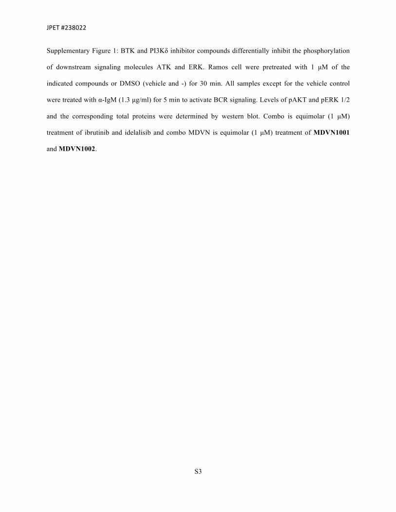
Supplementary Figure 1: BTK and PI3Kδ inhibitor compounds differentially inhibit the phosphorylation
of downstream signaling molecules ATK and ERK. Ramos cell were pretreated with 1 µM of the
indicated compounds or DMSO (vehicle and -) for 30 min. All samples except for the vehicle control
were treated with α-IgM (1.3 µg/ml) for 5 min to activate BCR signaling. Levels of pAKT and pERK 1/2
and the corresponding total proteins were determined by western blot. Combo is equimolar (1 µM)
treatment of ibrutinib and idelalisib and combo MDVN is equimolar (1 µM) treatment of MDVN1001
and MDVN1002.

JPET#238022
S4
% V
iabl
e C
ells
(nor
mal
ized
)Supplementary Figure 2
Compound IC50 (μM)
MDVN1001 1.47
MDVN1002 2.75
combo MDVN 0.87
-1 0 10
40
80
120
Log [Compound] (μM)
MDVN1001MDVN1002combo MDVN

JPET#238022
S5
Supplementary Figure 2: Combination treatment of MDVN1001 and MDVN1002 on cell viability of
DOHH-2 cells. DOHH-2 cells were treated with MDVN1001 or MDVN1002 or equimolar
concentrations of both compounds for 72 h. Cell viability was measured as described in the Materials and
Methods. The IC50 was calculated from the curve fitted to the data points by non-linear regression using
GraphPad Prism.

JPET#238022
S6
General Synthesis Information
1H NMR spectra and 13C NMR spectra were recorded on a Varian 400 MHz spectrometer.
Spectra are referenced to residual chloroform (δ 7.26, 1H), DMSO (δ 2.54, 1H) or methanol (δ 3.34, 1H)
unless otherwise noted. Chemical shifts are reported in ppm (δ); multiplicities are indicated by s (singlet),
d (doublet), t (triplet), q (quartet), quint (quintet), sext (sextet), m (multiplet) and br (broad). Coupling
constants, J, are reported in Hertz. Silica gel chromatography was performed using a Teledyne Isco
CombiFlash® Rf+ instrument using Hi-Purit Silica Flash Cartridges (National Chromatography Inco) or
RediSep Rf Gold C18 Cartridges (Teledyne Isco). Analytical HPLC was performed on a Waters
ACQUITY UPLC with a photodiode array detector using and a Waters ACQUITY BEH Shield RPC18
(2.1 × 50 mm, 1.7 µm) column. Analytical LCMS was performed on a Waters ACQUITY UPLC with a
Waters 3100 mass detector. Chiral HPLC was performed on a Waters Alliance e2695 with a photodiode
array detector using Daicel Chiralpak® AD-H, Chiralpak® IA, Chiralpak® IB, Chiralpak® IC, Chiralcel®
OD-H or Chiralcel® OJ-H columns. Optical rotations were obtained on a Jasco P-2000 digital polarimeter
and are reported as [α]DT temperature (T), concentration (c = g/100 mL) and solvent. Commercially
available reagents and solvents were used as received unless otherwise indicated.
Literature Preparations
The following compounds were prepared by literature methods: 5-bromo-2,3-dihydro-1H-inden-
2-ol (I)[Ref: Goeksu et al. Tetrahedron, 61(28), 6801-6807; 2005], 3-iodo-1H-pyrazolo[3,4-d]pyrimidin-
4-amine (V)[Ref: Rai et al. PCT Application WO2015/058084 A1, 2015], 1-(5-amino-2,3-dihydro-1H-
inden-2-yl)-3-(8-fluoro-3,4-dihydro-2H-benzo[b][1,4]oxazin-6-yl)-1H-pyrazolo[3,4-d]pyrimidin-4-amine
(MDVN1003).1
JPET#238022
S7
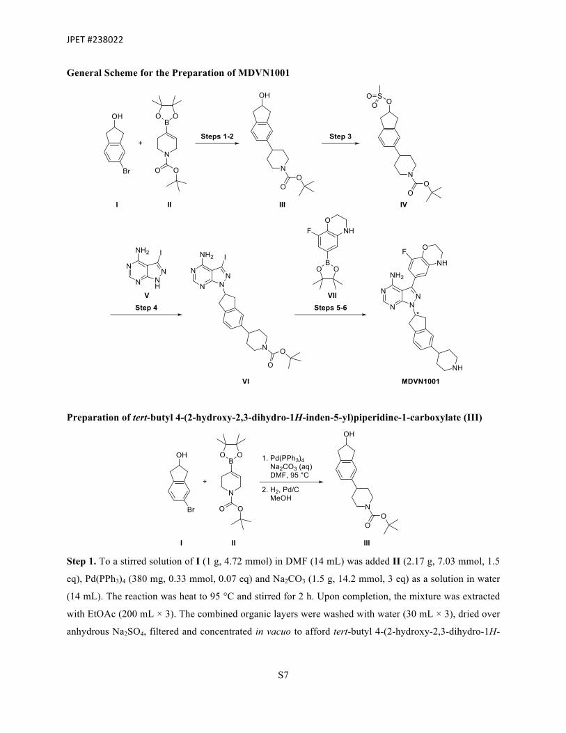
General Scheme for the Preparation of MDVN1001
Preparation of tert-butyl 4-(2-hydroxy-2,3-dihydro-1H-inden-5-yl)piperidine-1-carboxylate (III)
Step 1. To a stirred solution of I (1 g, 4.72 mmol) in DMF (14 mL) was added II (2.17 g, 7.03 mmol, 1.5
eq), Pd(PPh3)4 (380 mg, 0.33 mmol, 0.07 eq) and Na2CO3 (1.5 g, 14.2 mmol, 3 eq) as a solution in water
(14 mL). The reaction was heat to 95 °C and stirred for 2 h. Upon completion, the mixture was extracted
with EtOAc (200 mL × 3). The combined organic layers were washed with water (30 mL × 3), dried over
anhydrous Na2SO4, filtered and concentrated in vacuo to afford tert-butyl 4-(2-hydroxy-2,3-dihydro-1H-

JPET#238022
S8
inden-5-yl)-3,6-dihydropyridine-1(2H)-carboxylate (900 mg, 60%). The material was used without
further purification.
Analytical data
LC-MS 315 [M+H]+
Step 2. A round-bottom flask was charged with tert-butyl 4-(2-hydroxy-2,3-dihydro-1H-inden-5-yl)-3,6-
dihydropyridine-1(2H)-carboxylate (900 mg, 2.86 mmol), Pd/C (10 wt. %, 200 mg) and MeOH (50 mL).
The flask was then charged with H2 gas (1 atm). The reaction mixture was allowed to stir overnight at
room temperature. Upon completion, the contents were filtered through a bed of Celite® and washed with
MeOH (30 mL × 3). Residual Pd/C was carefully quenched with aqueous HCl. The supernatant was then
concentrated in vacuo to afford III (900 mg, 99%) which was used without further purification.
Analytical data 1H NMR (400 MHz, CD3OD) δ 7.71-7.58 (m, 2H), 7.58-7.51 (m, 1H), 4.60 (s, 1H), 4.19 (d, J =
13.2 Hz, 2H), 3.16-3.06 (m, 2H), 2.91-2.77 (m, 4H), 2.70-2.60 (m, 1H), 1.78 (d, J = 12.7
Hz, 2H), 1.58 (d, J = 3.9 Hz, 2H), 1.51-1.41 (m, 9H).

JPET#238022
S9
Preparation of tert-butyl 4-(2-((methylsulfonyl)oxy)-2,3-dihydro-1H-inden-5-yl)piperidine-1-
carboxylate (IV)
Step 3. To a solution of III (900 mg, 2.84 mmol) in CH2Cl2 (30 mL) was added Et3N (1.43 g, 14.2 mmol,
5 eq). The mixture was cooled to 0 °C and methanesulfonyl chloride (638 mg, 5.68 mmol, 2 eq) was
slowly added dropwise. The mixture was warmed to room temperature and allowed to stir for 2 h. Upon
completion the reaction mixture was extracted with EtOAc (200 mL × 3). The combined organic layers
were washed with water (100 mL × 3) followed by brine (30 mL × 2). The organic layer was dried over
anhydrous Na2SO4, filtered and concentrated in vacuo to afford IV (1.1 g, 98%) which was used without
further purification.
Analytical data 1H NMR (400 MHz, CD3OD) δ 7.66 (d, J = 7.0 Hz, 1H), 7.69 (d, J = 7.4 Hz, 1H), 7.48 (d, J = 7.8
Hz, 1H), 5.52 (br s, 1H), 3.33 (d, J = 4.7 Hz, 2H), 3.24 (br s, 2H), 3.15 (m, 2H), (3.03 (d,
J = 2.0 Hz, 3H), 2.79 (br s, 2H), 2.70-2.59 (m, 1H), 1.80 (d, J = 12.9 Hz, 2H), 1.67-1.59
(m, 2H), 1.50-1.44 (m, 9H).

JPET#238022
S10
Preparation of tert-butyl 4-(2-(4-amino-3-iodo-1H-pyrazolo[3,4-d]pyrimidin-1-yl)-2,3-dihydro-1H-
inden-5-yl)piperidine-1-carboxylate (VI)
Step 4. Cs2CO3 (1 g, 3.08 mmol, 2 eq) was added to a suspension of V (400 mg, 1.54 mmol) in DMF (10
mL). The reaction mixture was allowed to stir for 45 min at room temperature. Intermediate IV (1.1 g,
2.77 mmol, 1.8 eq) and tetrabutylammonium iodide (104 mg, 0.31 mmol, 0.2 eq) were then added. The
mixture was then heated to 95 °C and allowed to stir for 3 h. Upon completion, the mixture was allowed
to cool and water (50 mL) was added. The resulting precipitate was collected by vacuum filtration. The
filter cake was carefully washed with cold water (40 mL × 2) followed by cold n-pentane (40 mL × 3).
The solids were dried under reduced pressure to afford VI (350 mg, 41%) which was used without further
purification.
Analytical data
LC-MS 561[M+H]+
Preparation of 3-(8-fluoro-3,4-dihydro-2H-benzo[b][1,4]oxazin-6-yl)-1-(5-(piperidin-4-yl)-2,3-
dihydro-1H-inden-2-yl)-1H-pyrazolo[3,4-d]pyrimidin-4-amine (MDVN1001)

JPET#238022
S11
Step 5. A reaction vessel was charged with VI (350 mg, 0.625 mmol) and DMF (5 mL). To this was
added VII (261 mg, 0.938 mmol, 1.5 eq), Pd(PPh3)4 (58 mg, 0.05 mmol, 0.08 eq) and Na2CO3 (199 mg,
1.88 mmol, 3 eq) as a solution in water (5 mL). The reaction mixture was heated to 90 °C with stirring
and allowed to progress for 3 h. Upon completion, the reaction was quenched with ice cold water (30
mL). The resulting precipitate was collected by vacuum filtration. The filter cake was washed with cold
water (40 mL × 3) and cold n-pentane (30 mL × 3). The solids were dried under reduced pressure to
afford a crude product which was purified by reversed-phase SiO2 chromatography (55% CH3CN in 10
mM NH4OAc). The enantiomers were then separated by chiral preparative HPLC to afford tert-butyl 4-
(2-(4-amino-3-(8-fluoro-3,4-dihydro-2H-benzo [b][1,4] oxazin-6-yl)-1H-pyrazolo[3,4-d]pyrimidin-1-yl)-
2,3-dihydro-1H-inden-5-yl)piperidine-1-carboxylate (12 mg, 3%) as a single enantiomer.
Analytical data
LC-MS 586 [M+H]+
Step 6. To a reaction vessel containing tert-butyl 4-(2-(4-amino-3-(8-fluoro-3,4-dihydro-2H-
benzo[b][1,4]oxazin-6-yl)-1H-pyrazolo[3,4-d]pyrimidin-1-yl)-2,3-dihydro-1H-inden-5-yl)piperidine-1-
carboxylate (12 mg, 0.02 mmol) was added ethanolic HCl (4 mL) and the resulting mixture was allowed
to stir overnight at room temperature. Upon completion, the reaction mixture was concentrated and
lyophilized to afford MDVN1001 as a single enantiomer and hydrochloride salt (6 mg, 57%).
Analytical data
LC-MS 486 [M+H]+ 1H NMR (400 MHz, CD3OD) δ 8.33 (s, 1H), 7.29 (d, J = 7.8 Hz, 1H), 7.26-7.13 (m, 3H), 6.44 (d, J
= 6.1 Hz, 2H), 5.76 (d, J = 6.2 Hz, 1H), 4.25 (t, J = 4.3 Hz, 2H), 3.63-3.46 (m, 6H), 3.39
(dd, J = 25.4, 5.1 Hz, 6H), 3.14 (s, 2H), 2.92 (s, 1H), 2.09 (d, J = 14.2 Hz, 2H), 1.93 (t, J
= 12.9 Hz, 2H).

JPET#238022
S12
General Scheme for the Preparation of MDVN1002
Preparation of 1-(5-amino-2,3-dihydro-1H-inden-2-yl)-3-iodo-1H-pyrazolo[3,4-d]pyrimidin-4-
amine (IX)
Step 1. Cs2CO3 (3.49 g, 10.7 mmol, 2 eq) was added to a stirred suspension of V (1.4 g, 5.36 mmol) in
DMF (15 mL) at room temperature. The reaction mixture was allowed to stir for 45 min at room
temperature. Intermediate VIII (1.82 g, 8.04 mmol, 1.5 eq) and tetrabutylammonium iodide (396 mg,
0.107 mmol, 0.02 eq) were then added. The mixture was then heated to 75 °C and allowed to stir for 90
min. Upon completion, the mixture was allowed to cool and water (30 mL) was added. The resulting
precipitate was collected by vacuum filtration. The filter cake was triturated with hexane (10 mL × 2).
The solids were dried under reduced pressure to afford IX (1.7 g, 81%) which was used without further
purification.
Analytical data
LC-MS 393 [M+H]+

JPET#238022
S13
Preparation of 1-(5-amino-2,3-dihydro-1H-inden-2-yl)-3-(3-fluoro-4-isopropoxyphenyl)-1H-
pyrazolo[3,4-d]pyrimidin-4-amine (MDVN1002)
Step 2. A reaction vessel was charged with IX (200 mg, 0.51 mmol) and DMF (3 mL). To this was
added X (152 mg, 0.77 mmol, 1.5 eq), Pd(PPh3)4 (41 mg, 0.036 mmol, 0.07 eq) and Na2CO3 (162 mg,
1.53 mmol, 3 eq) as a solution in water (3 mL). The reaction mixture was heated to 85 °C with stirring
and allowed to progress overnight. Upon completion, the reaction was quenched with ice cold water (50
mL). The resulting precipitate was collected by vacuum filtration. The filter cake was washed with cold
water (30 mL × 2) and cold n-pentane (40 mL × 2). The solids were dried under reduced pressure to
afford a crude product which was purified by reversed-phase SiO2 chromatography (55-60% CH3CN in
10 mM NH4OAc) to afford MDVN1002 as a single enantiomer (90 mg, 42%).
Analytical data
LC-MS 419 [M+H]+ 1H NMR (400 MHz, CD3OD) δ 8.25 (s, 1H), 7.85 (s, 1H), 7.46–7.37 (m, 2H), 7.23 (t, J = 8.6 Hz,
1H), 6.99 (d, J = 8.0 Hz, 1H), 6.69–6.58 (m, 1H), 5.69 (q, J = 8.3 Hz, 1H), 4.79 (s, 1H),
4.69 (p, J = 6.1 Hz, 1H), 3.50 (ddd, J = 30.0, 15.5, 8.2 Hz, 2H), 3.35 (d, J = 5.7 Hz, 1H),
1.38 (d, J = 6.1 Hz, 6H).

JPET#238022
S14
General Scheme for the Preparation of Intermediate VIII
Preparation of 5-nitro-2,3-dihydro-1H-inden-2-ol (XII)
Step 1. To the stirred solution of XI (50 g, 0.282 mol) in MeOH (2 L) was added NaBH4 (21.5 g, 0.565
mol, 2 eq) at 0 °C portion wise. The reaction mixture was allowed to stir at 0 °C for 1 h. The reaction was
monitored by TLC. Upon completion, the reaction mixture was concentrated under reduced pressure. The
residue was treated with CH2Cl2, filtered and washed with additional CH2Cl2. The filtrate was
concentrated in vacuo to afford XII (43 g, 85%) as light brown solid. The material was used without
further purification.
Analytical data
LC-MS 180 [M+H]+

JPET#238022
S15
Preparation of 5-nitro-2,3-dihydro-1H-inden-2-yl methanesulfonate (XIII)
Step 2. To the stirred solution of XII (81 g, 0.45 mol) in CH2Cl2 (2.5 L) was added Et3N (206 g, 2.00
mol, 4.4 eq) at 0 °C followed by the slow addition of methanesulfonyl chloride (104 g, 0.91 mol, 2 eq).
The reaction was allowed to stir at 0 °C for 1.5 h. Upon completion, the reaction mixture was diluted with
water and extracted with CH2Cl2. The organic layer was washed with brine, dried over anhydrous
Na2SO4, filtered and concentrated in vacuo. The crude material was recrystallized with a mixture of
CH2Cl2 and MTBE to afford XIII (92 g, 79%) as an off-white solid.
Analytical data
LC-MS 258 [M+H]+
Preparation of (-)-5-amino-2,3-dihydro-1H-inden-2-yl methanesulfonate hydrochloride (VIII)
Step 3. To the stirred solution of XIII (46 g, 0.179 mol) in MeOH (2 L) was added Raney-Ni (46 g, wet).
The resulting reaction mixture was stirred for 3 h under H2 (30 psi). The reaction mixture was filtered and
the filter cake was washed with MeOH. The filtrate was concentrated in vacuo and treated with MTBE to
afford (±)-5-amino-2,3-dihydro-1H-inden-2-yl methanesulfonate (28 g, 68%) as an off-white solid. The
enantiomers were separated by chiral HPLC. The freebase was then diluted with CH2Cl2 and purged with
HCl (g) at 0 °C for 3 h. The precipitate was filtered and washed with CH2Cl2 and MTBE to give VIII as a
single enantiomer and hydrochloride salt. Peak 1HCl salt (batch 1, 10.8 g, 97.4% ee and batch 2 14.2 g,
98.9%ee) and peak 2 HCl salt (batch 1, 9.4 g, 97.7% ee and batch 2, 15.6 g, 98.4%ee) as an off-white
solid.
Analytical data
LC-MS 228 [M+H]+

JPET#238022
S16
1H NMR (400 MHz, DMSO-d6) δ 10.40 (br s, 2H), 7.39 (d, J = 8.0 Hz, 1H), 7.31 (s, 1H), 7.26–
7.19 (m, 1H), 5.50 (m, 1H), 3.36 (m, 2H), 3.22 (s, 3H), 3.14 (m, 2H).

JPET#238022
S17
Pharmacokinetic Analysis
Intravenous and Oral Pharmacokinetics in Mice. Female BALB/c mice (age, 10–12 weeks; 18–20 g
b.wt.) were used in the study. Food and water were given ad libitum. MDVN1003 was administered
intravenously, via the tail vein, at a dose of 2 mg/kg as a solution (1 mg/ml) in 20% DMSO, 40% PEG200
and 40% water at a dose volume of 2 ml/kg. The oral dose, 10 mg/kg, was administered by gavage at a
dose volume of 10 ml/kg in 0.5% methylcellulose at a concentration of 1 mg/ml. At defined time points
(5, 15 min, 30 min, and 1, 2, 4, 8, and 24 h after dose), groups of three mice per time point were sacrificed
by an overdose of CO2, and blood was collected by cardiac puncture and placed in tubes containing
K2EDTA as the anticoagulant. Plasma was obtained by centrifuging the blood samples at 800g for 10 min
and storing at -60 to -80°C until analysis.
Intravenous and Oral Pharmacokinetics in Rats. Male Sprague-Dawley rats (age, 7–9 weeks; 250–300
g b.wt.) were used in the study. Three rats were used for the intravenous and oral studies each, in a
parallel design. One day before the study, the jugular vein was cannulated in all the rats to be used. Rats
were fasted overnight before dosing and fed 4 h after dose. Water was given ad libitum. MDVN1003 (2
mg/kg) was administered intravenously as a bolus dose through the jugular vein, at a dose volume of 1
ml/kg (2 mg/ml) in saline. The oral dose (5 mg/kg) was administered by gavage at a dose volume of 5
mL/kg as a 1 mg/ml suspension in 0.5% methylcellulose in water. From each rat, serial blood samples
(≈0.2 ml per sample) were drawn at 5 (IV only), 15, 30, and at 1, 2, 4, 6, 8, 12 and 24 h after dose and
placed in tubes containing K2EDTA as the anticoagulant. Plasma was obtained by centrifuging the blood
samples at 1000g for 10 min and storing at -60 to 80°C until analysis.
Intravenous and Oral Pharmacokinetics in Dogs. Six male beagle dogs (age, 1–2 years; 9–11 kg b.wt.)
were used in the study. Three dogs were used in the intravenous and oral studies each, in a parallel design.
The dogs were fasted overnight before dose and fed 4 h after dose in the oral study. Water was given ad
libitum. The intravenous dose (1 mg/kg) was given as a bolus (1 ml/kg) in 20% DMSO, 40% PEG200

JPET#238022
S18
and 40% water at a concentration of 1 mg/ml. The oral dose (3 mg/kg) was administered by gavage at a
dose volume of 5 ml/kg as a suspension (0.6 mg/ml) in 0.5% methylcellulose in water. At defined time
points (3 (IV only), 5 (IV only), 15, and 30 min and 1, 2, 4, 8, 12, 24, 26 and 48 h after dose), serial blood
samples (≈1 ml) were collected from either the cephalic or jugular vein via a butterfly catheter and placed
into a chilled polypropylene tube with K2EDTA as the anticoagulant. Samples were centrifuged at 4°C at
a speed of 1000g for 10 min, and plasma was harvested and stored at -60 to -80°C until analysis.
Sample Analysis. Plasma samples from mouse, rat, and dog (10-30 µL) were precipitated with of
acetonitrile (200 µL) containing internal standard (warfarin) in a 96-well plate, vortexed for 5 min, and
then centrifuged at 1500g for 10 min in a refrigerated centrifuge (Sorvall Legend X1R centrifuge, Thermo
Scientific) at 4°C. Supernatants (100 µL) were transferred to a new 96-well plate containing 100 µL of
0.1% formic acid in each well. The plate was vortexed for approximately 10 minutes and then aliquots
were injected for LC/MS/MS analysis. Sample analysis was carried out AB/MDS Sciex API 4000
LC/MS/MS System (Framingham, MA) interfaced with Shimadzu SIL-20AC HT autosampler and
Prominence CBM-20A pumps (Kyoto, Japan). The mobile phase consisted of 0.1% formic acid in water
(A) and 0.1% formic acid in acetonitrile (B). A ballistic gradient (5% B for 0.5 min then increased to 95%
B in 1.5 min and hold for 1 min, then return to 5% B) was used to elute the analyte and internal standard
with a Waters Atlantis T3 (3 µ, 2.1 x 50 mm, Milford, MA) at a flow rate of 0.6 ml/min. The ion-optics
were optimize with Analyst software (v1.6) and MDVN1003 was analyzed with multiple reaction
monitoring experiment with the Q1 to Q3 transitions masses of m/z 418.1 – m/z 287.1, respectively. The
internal standard, warfarin, was monitored with m/z 309.0 – m/z 163.0. The assay was linear between 1
and 5000 nM (dog plasma: lower limit of quantitation, 1 nM) and between 2.0 and 1000 nM (mouse and
rat plasma: lower limit of quantitation, 2 and 3 nM, respectively). Criteria for accuracy and precision were
met.

JPET#238022
S19
Pharmacokinetic Analysis. Noncompartmental PK parameters such as the total volume of distribution
(Vd), systemic clearance (CL), elimination half-life (t1/2), area under the plasma concentration-time curve
up to the last nonzero concentration (AUC0-t), area under the plasma concentration-time curve from time 0
to infinity (AUC0-∞), the time of maximum concentration in plasma (tmax), and the maximum concentration
in plasma (Cmax) were estimated Phoenix WinNonlin version 6.4 (Pharsight Corp, St Louis, MO). The
terminal elimination rate constant was estimated from the terminal linear part of the log-linear plot (a
minimum of three points on the linear phase). The linear trapezoidal method was used to compute the
AUC. For rats and dogs, PK parameters were estimated in individual animals and averaged. For mice, the
PK parameters were estimated using the mean concentration data. The absolute oral bioavailability, F
(%), was calculated from, the dose normalized ratio of oral AUC to IV AUC.
1 Pujala B, Agarwal AK, Middya S, Banerjee M, Surya A, Nayak AK, Gupta A, Khare S, Guguloth R, Randive NA, Shinde BU, Thakur A, Patel DI, Raja M, Green MJ, Alfaro J, Avila P, Pérez de Arce F, Almirez RG, Kanno S, Bernales S, Hung DT, Chakravarty S, McCullagh E, Quinn KP, Rai R and Pham SM (2016) Discovery of Pyrazolopyrimidine Derivatives as Novel Dual Inhibitors of BTK and PI3Kδ. ACS Medicinal Chemistry Letters.