Drugs and Thyroid Function
7
1688 THE NEW ENGLAND JOURNAL OF MEDICINE Dec. 21, 1995 REVIEW ARTICLE DRUG THERAPY ALASTAIR J. J. WOOD, M.D., Editor DRUGS AND THYROID FUNCTION MARTIN I. SURKS, M.D., AND RUBENS SIEVERT, M.D. From the Division of Endocrinology, Department of Medicine, Montefiore Medical Center and Albert Einstein College of Medicine, Bronx, N.Y. Address reprint requests to Dr. Surks at the Montefiore Medical Center, 111 East 210th St., Bronx, NY 10467. T ESTING of thyroid function is common in clinical practice. Many patients who are tested, including those who have or are receiving treatment for thyroid disease, take medications that may affect thyroid func- tion. Therefore, the possible effect of these drugs both on the results of thyroid-function tests and on the effec- tiveness of treatment must always be considered in de- cisions regarding patient care. The pathways of thyroid hormone synthesis, secre- tion, transport in the circulation, and metabolism of- fer numerous targets for drug interaction (Fig. 1 and 2). Normal thyroid secretion depends on thyrotropin (TSH). Secretion of TSH is, in turn, inhibited by thy- roid hormones and stimulated by thyrotropin-releasing hormone (TRH). Iodide in serum is trapped by thyroid cells, after which it is oxidized and incorporated into some of the tyrosine residues of thyroglobulin, which then couple to form thyroxine (T 4 ) and triiodothy- ronine (T 3 ). The thyroid gland normally contains large stores of thyroglobulin, most of which is in the lumen of the thy- roid follicles. When thyroglobulin is resorbed into the follicular cells of the thyroid and hydrolyzed, T 4 and T 3 are secreted into the circulation. There they are bound to specific serum-binding proteins, so that very little circulates as free T 4 or T 3 . In extrathyroidal tissues, T 4 is converted to T 3 by the action of several T 4 5-deio- dinases; this process generates about 80 percent of the circulating T 3 . About 80 percent of T 4 and T 3 is metab- olized by deiodination and 20 percent by non-deiodina- tive pathways that include conjugation with glucuron- ides and sulfates, decarboxylation, and deamination. 1 In tissues, T 3 and — to a much smaller extent — T 4 are bound to specific nuclear receptor proteins that interact with regulatory regions of genes, influencing their ex- pression. In this paper we shall discuss the effects of groups of drugs on the production, secretion, transport, and me- tabolism of T 4 and T 3 and on the absorption of exoge- nously administered T 4 . DRUGS AFFECTING THE SECRETION OF TSH Measurement of serum TSH is the single best test of thyroid function, because of the sensitivity of TSH secre- tion to very small changes in serum T 4 and T 3 concentra- tions. Serum TSH concentrations are low in patients with hyperthyroidism and high in those with primary hy- pothyroidism. When hypothyroidism results from hypo- thalamic or pituitary disease, serum TSH values are usu- ally low or normal, but occasionally they are high because of the secretion of biologically inactive TSH. 2,3 Several drugs decrease TSH secretion and lower se- rum TSH concentrations, although not to values as low as those found in patients with hyperthyroidism (Table 1). These agents are dopamine (in doses of at least 1 mg per kilogram of body weight per minute), 4-8 glucocorti- coids (e.g., dexamethasone, in doses of 0.5 mg or more per day or hydrocortisone in doses of 100 mg or more per day), 9,10 and octreotide (in doses of more than 100 mg per day), which is a somatostatin analogue used for the treatment of acromegaly and certain other hor- mone-excess syndromes. 11,12 Patients who are receiving long-term glucocorticoid or octreotide therapy do not, however, have sustained reductions in TSH secretion, nor does hypothyroidism develop, probably because of the effect of decreased thyroid hormone secretion in in- creasing TSH secretion. Patients who require infusions of dopamine for more than a few days may have reduc- tions in secretion by the thyroid, which are difficult to distinguish from the changes in serum T 4 and T 3 con- centrations that result from the underlying illness. DRUGS AFFECTING THE SECRETION OF THYROID HORMONE In addition to methimazole and propylthiouracil, which are given deliberately to decrease thyroid hor- mone production in patients with hyperthyroidism, sev- eral other commonly used drugs may decrease thyroid hormone secretion. These include lithium carbonate and iodine-containing medications (Table 1). Drugs That Cause Hypothyroidism Lithium interferes with thyroid hormone synthesis and decreases thyroid hormone secretion. Long-term lithium treatment results in goiter in up to 50 percent of patients, subclinical hypothyroidism in up to 20 per- cent, and overt hypothyroidism in up to 20 percent. 13-15 Many lithium-treated patients have antithyroid anti- bodies in their serum; among them about 50 percent have subclinical hypothyroidism, as compared with 15 percent of patients with no antithyroid antibodies. 15 The antithyroid antibodies probably indicate the presence of preexisting chronic autoimmune thyroiditis, which would be expected to increase sensitivity to the antithy- roid actions of lithium, but which could, alternatively, be induced by lithium. Normal subjects given 1 to 2 mg of inorganic iodide per day (in addition to their usual diet) have a transient decrease in T 4 and T 3 secretion and a transient increase in TSH secretion. 16 The decrease in T 4 and T 3 secretion is much greater, but is usually also transient, in patients with hyperthyroidism. 17 However, in patients with chron- The New England Journal of Medicine Downloaded from nejm.org at REGIONALT CANCERCENTRUM VAST on June 5, 2014. For personal use only. No other uses without permission. Copyright © 1995 Massachusetts Medical Society. All rights reserved.
Transcript of Drugs and Thyroid Function

1688 THE NEW ENGLAND JOURNAL OF MEDICINE Dec. 21, 1995
REVIEW ARTICLE
DRUG THERAPY
A
LASTAIR
J. J. W
OOD
, M.D.,
Editor
DRUGS AND THYROID FUNCTION
M
ARTIN
I. S
URKS
, M.D.,
AND
R
UBENS
S
IEVERT
, M.D.
From the Division of Endocrinology, Department of Medicine, MontefioreMedical Center and Albert Einstein College of Medicine, Bronx, N.Y. Addressreprint requests to Dr. Surks at the Montefiore Medical Center, 111 East 210thSt., Bronx, NY 10467.
T
ESTING of thyroid function is common in clinicalpractice. Many patients who are tested, including
those who have or are receiving treatment for thyroiddisease, take medications that may affect thyroid func-tion. Therefore, the possible effect of these drugs bothon the results of thyroid-function tests and on the effec-tiveness of treatment must always be considered in de-cisions regarding patient care.
The pathways of thyroid hormone synthesis, secre-tion, transport in the circulation, and metabolism of-fer numerous targets for drug interaction (Fig. 1 and2). Normal thyroid secretion depends on thyrotropin(TSH). Secretion of TSH is, in turn, inhibited by thy-roid hormones and stimulated by thyrotropin-releasinghormone (TRH). Iodide in serum is trapped by thyroidcells, after which it is oxidized and incorporated intosome of the tyrosine residues of thyroglobulin, whichthen couple to form thyroxine (T
4
) and triiodothy-ronine (T
3
).The thyroid gland normally contains large stores of
thyroglobulin, most of which is in the lumen of the thy-roid follicles. When thyroglobulin is resorbed into thefollicular cells of the thyroid and hydrolyzed, T
4
and T
3
are secreted into the circulation. There they are boundto specific serum-binding proteins, so that very littlecirculates as free T
4
or T
3
. In extrathyroidal tissues, T
4
is converted to T
3
by the action of several T
4
5
�
-deio-dinases; this process generates about 80 percent of thecirculating T
3
. About 80 percent of T
4
and T
3
is metab-olized by deiodination and 20 percent by non-deiodina-tive pathways that include conjugation with glucuron-ides and sulfates, decarboxylation, and deamination.
1
Intissues, T
3
and — to a much smaller extent — T
4
arebound to specific nuclear receptor proteins that interactwith regulatory regions of genes, influencing their ex-pression.
In this paper we shall discuss the effects of groups ofdrugs on the production, secretion, transport, and me-tabolism of T
4
and T
3
and on the absorption of exoge-nously administered T
4
.
D
RUGS
A
FFECTING
THE
S
ECRETION
OF
TSH
Measurement of serum TSH is the single best test ofthyroid function, because of the sensitivity of TSH secre-
tion to very small changes in serum T
4
and T
3
concentra-tions. Serum TSH concentrations are low in patientswith hyperthyroidism and high in those with primary hy-pothyroidism. When hypothyroidism results from hypo-thalamic or pituitary disease, serum TSH values are usu-ally low or normal, but occasionally they are highbecause of the secretion of biologically inactive TSH.
2,3
Several drugs decrease TSH secretion and lower se-rum TSH concentrations, although not to values as lowas those found in patients with hyperthyroidism (Table1). These agents are dopamine (in doses of at least 1
m
gper kilogram of body weight per minute),
4-8
glucocorti-coids (e.g., dexamethasone, in doses of 0.5 mg or moreper day or hydrocortisone in doses of 100 mg or moreper day),
9,10
and octreotide (in doses of more than 100
m
g per day), which is a somatostatin analogue used forthe treatment of acromegaly and certain other hor-mone-excess syndromes.
11,12
Patients who are receivinglong-term glucocorticoid or octreotide therapy do not,however, have sustained reductions in TSH secretion,nor does hypothyroidism develop, probably because ofthe effect of decreased thyroid hormone secretion in in-creasing TSH secretion. Patients who require infusionsof dopamine for more than a few days may have reduc-tions in secretion by the thyroid, which are difficult todistinguish from the changes in serum T
4
and T
3
con-centrations that result from the underlying illness.
D
RUGS
A
FFECTING
THE
S
ECRETION
OF
T
HYROID
H
ORMONE
In addition to methimazole and propylthiouracil,which are given deliberately to decrease thyroid hor-mone production in patients with hyperthyroidism, sev-eral other commonly used drugs may decrease thyroidhormone secretion. These include lithium carbonateand iodine-containing medications (Table 1).
Drugs That Cause Hypothyroidism
Lithium interferes with thyroid hormone synthesisand decreases thyroid hormone secretion. Long-termlithium treatment results in goiter in up to 50 percentof patients, subclinical hypothyroidism in up to 20 per-cent, and overt hypothyroidism in up to 20 percent.
13-15
Many lithium-treated patients have antithyroid anti-bodies in their serum; among them about 50 percenthave subclinical hypothyroidism, as compared with 15percent of patients with no antithyroid antibodies.
15
Theantithyroid antibodies probably indicate the presenceof preexisting chronic autoimmune thyroiditis, whichwould be expected to increase sensitivity to the antithy-roid actions of lithium, but which could, alternatively,be induced by lithium.
Normal subjects given 1 to 2 mg of inorganic iodideper day (in addition to their usual diet) have a transientdecrease in T
4
and T
3
secretion and a transient increasein TSH secretion.
16
The decrease in T
4
and T
3
secretionis much greater, but is usually also transient, in patientswith hyperthyroidism.
17
However, in patients with chron-
The New England Journal of Medicine Downloaded from nejm.org at REGIONALT CANCERCENTRUM VAST on June 5, 2014. For personal use only. No other uses without permission.
Copyright © 1995 Massachusetts Medical Society. All rights reserved.
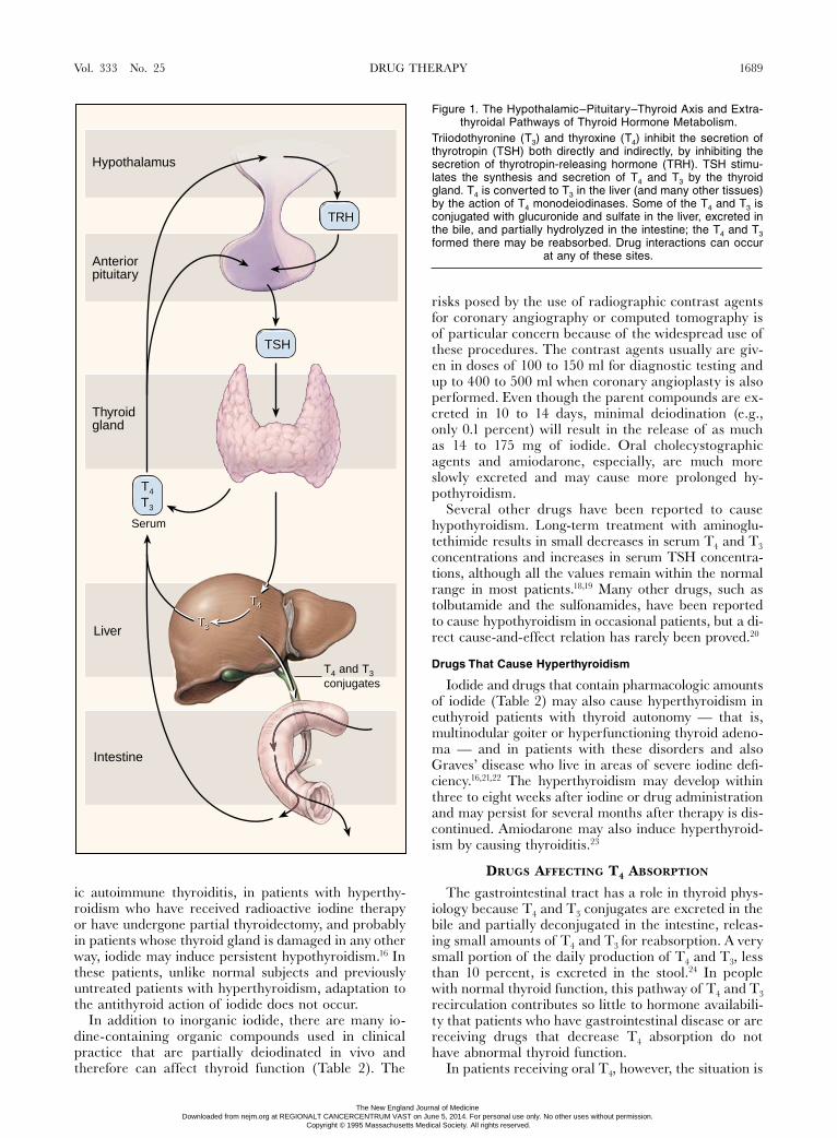
Vol. 333 No. 25 DRUG THERAPY 1689
Hypothalamus
Anteriorpituitary
Thyroidgland
Liver
T4 and T3conjugates
Intestine
Serum
T3
T4
T3
T4
T3
T4
T3
T4
TSHTSH
TRHTRH
Figure 1. The Hypothalamic–Pituitary–Thyroid Axis and Extra-thyroidal Pathways of Thyroid Hormone Metabolism.
Triiodothyronine (T
3
) and thyroxine (T
4
) inhibit the secretion ofthyrotropin (TSH) both directly and indirectly, by inhibiting thesecretion of thyrotropin-releasing hormone (TRH). TSH stimu-lates the synthesis and secretion of T
4
and T
3
by the thyroidgland. T
4
is converted to T
3
in the liver (and many other tissues)by the action of T
4
monodeiodinases. Some of the T
4
and T
3
isconjugated with glucuronide and sulfate in the liver, excreted inthe bile, and partially hydrolyzed in the intestine; the T
4
and T
3
formed there may be reabsorbed. Drug interactions can occurat any of these sites.
ic autoimmune thyroiditis, in patients with hyperthy-roidism who have received radioactive iodine therapyor have undergone partial thyroidectomy, and probablyin patients whose thyroid gland is damaged in any otherway, iodide may induce persistent hypothyroidism.
16
Inthese patients, unlike normal subjects and previouslyuntreated patients with hyperthyroidism, adaptation tothe antithyroid action of iodide does not occur.
In addition to inorganic iodide, there are many io-dine-containing organic compounds used in clinicalpractice that are partially deiodinated in vivo andtherefore can affect thyroid function (Table 2). The
risks posed by the use of radiographic contrast agentsfor coronary angiography or computed tomography isof particular concern because of the widespread use ofthese procedures. The contrast agents usually are giv-en in doses of 100 to 150 ml for diagnostic testing andup to 400 to 500 ml when coronary angioplasty is alsoperformed. Even though the parent compounds are ex-creted in 10 to 14 days, minimal deiodination (e.g.,only 0.1 percent) will result in the release of as muchas 14 to 175 mg of iodide. Oral cholecystographicagents and amiodarone, especially, are much moreslowly excreted and may cause more prolonged hy-pothyroidism.
Several other drugs have been reported to causehypothyroidism. Long-term treatment with aminoglu-tethimide results in small decreases in serum T
4
and T
3
concentrations and increases in serum TSH concentra-tions, although all the values remain within the normalrange in most patients.
18,19
Many other drugs, such astolbutamide and the sulfonamides, have been reportedto cause hypothyroidism in occasional patients, but a di-rect cause-and-effect relation has rarely been proved.
20
Drugs That Cause Hyperthyroidism
Iodide and drugs that contain pharmacologic amountsof iodide (Table 2) may also cause hyperthyroidism ineuthyroid patients with thyroid autonomy — that is,multinodular goiter or hyperfunctioning thyroid adeno-ma — and in patients with these disorders and alsoGraves’ disease who live in areas of severe iodine defi-ciency.
16,21,22
The hyperthyroidism may develop withinthree to eight weeks after iodine or drug administrationand may persist for several months after therapy is dis-continued. Amiodarone may also induce hyperthyroid-ism by causing thyroiditis.
23
D
RUGS
A
FFECTING
T
4
A
BSORPTION
The gastrointestinal tract has a role in thyroid phys-iology because T
4
and T
3
conjugates are excreted in thebile and partially deconjugated in the intestine, releas-ing small amounts of T
4
and T
3
for reabsorption. A verysmall portion of the daily production of T
4
and T
3
, lessthan 10 percent, is excreted in the stool.
24
In peoplewith normal thyroid function, this pathway of T
4
and T
3
recirculation contributes so little to hormone availabili-ty that patients who have gastrointestinal disease or arereceiving drugs that decrease T
4
absorption do nothave abnormal thyroid function.
In patients receiving oral T
4
, however, the situation is
The New England Journal of Medicine Downloaded from nejm.org at REGIONALT CANCERCENTRUM VAST on June 5, 2014. For personal use only. No other uses without permission.
Copyright © 1995 Massachusetts Medical Society. All rights reserved.

1690 THE NEW ENGLAND JOURNAL OF MEDICINE Dec. 21, 1995
different. Normally, about 80 percent of a usual dose(50 to 150
m
g per day) is absorbed, mostly in the je-junum and the upper part of the ileum. In patients whoare dependent on exogenous T
4
, drugs that decreaseT
4
absorption may induce hypothyroidism (Table 1).Among untreated patients, the severity of hypothyroid-ism might be expected to increase.
The bile acid sequestrants colestipol and cholestyra-mine bind T
4
and decrease its absorption, and theyhave proved useful in the treatment of patients withexogenous hyperthyroidism.
25
A decrease in serum T
4
concentrations and an increase in serum TSH concen-trations occurred when cholestyramine was adminis-tered to T
4
-treated patients with hypothyroidism.
26
Innormal subjects, thyroid function is not affected bythese drugs.
27
Decreased absorption of T
4
and increases in serumTSH concentrations have also been reported in T
4
-treated patients with hypothyroidism who are givenaluminum hydroxide,
28,29
ferrous sulfate,
30
or sucral-fate,
31
but absorption of T
4
was not decreased in themajority of the patients treated with ferrous sulfateand sucralfate who were studied.
30,32,33
The interactionscan be minimized by having the patient take T
4
andthe other drug several hours apart. Therefore, eventhough interference with T
4
absorption seems to occurin relatively few patients, it is prudent to advise all pa-tients to take their T
4
and other medications at differ-ent times.
D
RUGS
A
FFECTING
T
4
AND
T
3
T
RANSPORT
IN
S
ERUM
More than 99 percent of the T
4
and T
3
in serum isbound to one of the three major transport proteins: thy-roxine-binding globulin (TBG), transthyretin, and al-
bumin.
34,35
TBG binds approximately 70 percent of se-rum T
4
and a larger fraction of serum T
3
; it is thereforethe most important of the binding proteins. Althoughless than 0.1 percent of T
4
and T
3
circulates unbound toproteins, it is the concentration of free hormone thatdetermines the action of the hormones in tissues. Alter-ations in the serum concentrations of these binding pro-teins alter serum total T
4 and T3 concentrations, butnot serum concentrations of free T4 and T3; thereforethe patient remains euthyroid. Drugs may affect T4 andT3 transport either by raising or lowering the serumconcentration of a binding protein or by interfering withthe binding of T4 and T3 to a binding protein. Nearlyall the drugs that alter T4 and T3 transport do so by al-tering the serum concentration of TBG or its affinity forT4 and T3.
Increases in Serum TBG Concentrations
The most common causes of an increase in serumTBG concentrations are an increase in estrogen pro-duction and the administration of estrogen, either as acomponent of an oral contraceptive agent or as estro-gen-replacement therapy (Table 1).36-40 TBG is a gly-coprotein that is synthesized in the liver. Estrogens pro-duce increased sialylation of TBG, which decreases itsrate of clearance and raises its serum concentration.35
The increase in TBG in serum is dose-dependent. Theusual doses of ethinyl estradiol (20 to 35 mg per day)and conjugated estrogen (0.625 mg per day) raise se-rum TBG concentrations by approximately 30 to 50percent and serum T4 concentrations by 20 to 35 per-cent.36-40 The increases begin within two weeks, and anew steady state is attained in four to eight weeks. Inwomen with hypothyroidism who are receiving T4 andbecome pregnant, an increase of 45 percent in the doseis needed, on average, to maintain normal serum TSHconcentrations.41 Thus, the increase in serum total T4concentrations induced by estrogen occurs as a result ofat least a transient increase in T4 secretion.
Addition of a progestogen to estrogen therapy doesnot alter the estrogen-induced increase in the serumTBG concentrations, and progesterone alone has no ef-fect. Oral estrogen has a first-pass effect on the liver;transdermal administration of estrogen does not raiseserum TBG or T4 concentrations, even though serumestrogen concentrations are comparable to those meas-ured after oral administration.37 Tamoxifen has weakestrogen-agonist effects in the liver and raises serumTBG concentrations slightly.42
Serum TBG concentrations are increased in about 50percent of patients who use heroin for long periods orare treated with methadone.43-45 Many of these patientsalso have abnormal liver function, so that the increasein serum TBG may result from liver disease rather thanfrom specific effects of these drugs.43,44 Cocaine use hasnot been associated with changes in serum TBG, T4, orT3 concentrations.46
Mitotane and fluorouracil are also associated with in-creases in serum concentrations of total T4 and T3, butserum free T4 and TSH concentrations remain nor-
Figure 2. Thyroid Hormone Transport in Serum andHormone Action.
The binding proteins include thyroxine-binding globulin, trans-thyretin, and albumin. Drugs may alter the production or clear-ance of a binding protein or inhibit the binding of thyroxine (T4)
and triiodothyronine (T3) to the protein. TR denotesthyroid receptor.
Serum
Cell
Gene
Nucleus
B i n d i n g p r o t e i n s
Free T4 and free T3
T4 T3
T3
T3 T4
B i n d i n g p r o t e i n s
Free T4 and free T3
T4 T3
TRTR
The New England Journal of Medicine Downloaded from nejm.org at REGIONALT CANCERCENTRUM VAST on June 5, 2014. For personal use only. No other uses without permission.
Copyright © 1995 Massachusetts Medical Society. All rights reserved.

Vol. 333 No. 25 DRUG THERAPY 1691
mal.47,48 It is likely that these drugs also increase the se-rum concentration of TBG.
Decreases in Serum TBG Concentrations
In contrast to those treated with estrogens, patientstaking androgens or anabolic steroids have decreasedserum TBG and T4 concentrations (Table 1).49-51
These patients are clinically euthyroid, their serum freeT4 and TSH concentrations remain within the normalrange, and their production and turnover of T4 are nor-mal. The administration of androgen to women withbreast cancer who also had hypothyroidism and werebeing treated with T4 induced hyperthyroidism, with anincrease in serum free T4 and a decrease in serum TSHconcentrations.52 These results suggest that androgens
not only lower serum TBG concentrations but also slight-ly decrease T4 production. A decrease in the serumTBG concentration also occurs during long-term gluco-corticoid treatment.
Patients treated with nicotinic acid may have de-creased serum TBG and T4 concentrations.53-55 In onestudy, treatment of hypercholesterolemia with colesti-pol and niacin (3 to 6 g per day) resulted in a 25 per-cent decrease in the serum TBG concentration and asmall decrease in the serum T4 concentration (1.5 mgper deciliter [1.9 nmol per liter])53 but no change in theserum free T4 and TSH concentrations. The changeswere probably caused by the niacin, since colestipolalone has no effect on thyroid function.27
Inhibition of the Binding of T4 and T3 to TBG
At therapeutic concentrations, several drugs inhibitthe binding of T4 and T3 to TBG to varying degrees(Table 1). The initial effect of these drugs is to increaseserum free T4 concentrations, because the drug displac-es T4 from TBG; continued administration, however,results in a decrease in serum T4, normal serum free T4,and normal serum TSH concentrations.
Furosemide has no effect at the usual therapeuticconcentrations, but large intravenous doses (more than80 mg) result in a transient increase in serum free T4concentrations and a decrease in serum total T4 con-centrations.56-58 The changes in serum total and free T4vary depending on the length of time between the ad-ministration of the drug and the collection of the sam-
*TSH denotes thyrotropin, T4 thyroxine, T3 triiodothyronine, and TBGthyroxine-binding globulin.
Table 1. Drugs That Influence Thyroid Function.*
Drugs that decrease TSH secretionDopamineGlucocorticoidsOctreotide
Drugs that alter thyroid hormone secretionDecreased thyroid hormone secretion
LithiumIodideAmiodaroneAminoglutethimide
Increased thyroid hormone secretionIodideAmiodarone
Drugs that decrease T4 absorptionColestipolCholestyramineAluminum hydroxideFerrous sulfateSucralfate
Drugs that alter T4 and T3 transport in serumIncreased serum TBG concentration
EstrogensTamoxifenHeroinMethadoneMitotaneFluorouracil
Decreased serum TBG concentrationAndrogensAnabolic steroids (e.g., danazol)Slow-release nicotinic acidGlucocorticoids
Displacement from protein-binding sitesFurosemideFenclofenacMefenamic acidSalicylates
Drugs that alter T4 and T3 metabolismIncreased hepatic metabolism
PhenobarbitalRifampinPhenytoinCarbamazepine
Decreased T4 5�-deiodinase activityPropylthiouracilAmiodaroneBeta-adrenergic–antagonist drugsGlucocorticoids
CytokinesInterferon alfaInterleukin-2
Table 2. Iodine Content of Some Iodine-ContainingMedications and Radiographic Contrast Agents.
SUBSTANCE AMOUNT OF IODINE
ExpectorantsIophenOrganidin (iodinated glycerol)Par GlycerolR-Gen
25 mg/ml15 mg/tablet5 mg/ml6 mg/ml
IodidesPotassium iodide (saturated solution)Pima syrup (potassium iodide)Lugol’s solution (potassium iodide
� iodine)Iodo-Niacin
~25 mg/drop255 mg/ml~7 mg/drop
115 mg/tablet
Antiasthmatic drugsMudraneElixophyllin-KI (theophylline) elixirIophylline
195 mg/tablet6.6 mg/ml2 mg/ml
Antiarrhythmic drugsAmiodarone 75 mg/tablet
Antiamebic drugsIodoquinol 134 mg/tablet
Topical antiseptic agentsPovidone–iodineClioquinol cream
10 mg/ml12 mg/g
DouchesPovidone–iodine 10 mg/ml
Radiographic contrast agentsIopanoic acidIpodate sodiumIntravenous preparations
333 mg/tablet308 mg/tablet140–380 mg/ml
The New England Journal of Medicine Downloaded from nejm.org at REGIONALT CANCERCENTRUM VAST on June 5, 2014. For personal use only. No other uses without permission.
Copyright © 1995 Massachusetts Medical Society. All rights reserved.

1692 THE NEW ENGLAND JOURNAL OF MEDICINE Dec. 21, 1995
ple, on the rate of renal clearance of the drug, and onthe serum concentrations of albumin (which also bindsfurosemide) and TBG. Several nonsteroidal antiinflam-matory drugs have similar effects.57
Salicylates (in doses of �2.0 g per day) and sal-salate (in doses of 1.5 to 3.0 g per day) also inhibit thebinding of T4 and T3 to TBG; salicylates inhibit bind-ing to transthyretin as well.59 As with furosemide, theinitial effect is an increase in serum free T4 concentra-tions.60 When therapeutic serum concentrations aresustained, salicylates result in a 20 to 30 percent de-crease in serum total T4 concentrations and normalserum free T4 concentrations. Salsalate may result ina greater decrease in the serum T4 concentration (by30 to 40 percent) and a decrease in the serum free T4index,61-63 but the latter change is probably an in vitroartifact.60
Serum free T4 concentrations increase transientlyafter the administration of heparin.64 This increase iscaused in vitro by the inhibition of protein binding ofT4 by the free fatty acids generated as a result of theability of heparin to activate lipoprotein lipase.65-67
METABOLISM OF T4 AND T3 T4 and T3 are metabolized mostly by deiodination
but also by glucuronidation and sulfation.1,68 The ac-tivity of the enzymes that facilitate these reactionsis affected by a variety of drugs (Table 1). Their ac-tions, in general, vary according to whether the pa-tient has normal pituitary–thyroid function and there-fore can compensate for any alteration in T4 and T3metabolism or has hypothyroidism and therefore littleability to increase whatever thyroid secretion persists.Among these drugs are phenobarbital and rifam-pin,68-71 which increase T4 and T3 metabolism by stim-ulating hepatic microsomal drug-metabolizing enzymeactivity. Hypothyroid patients treated with T4 maybecome hypothyroid again when rifampin is adminis-tered.71
Phenytoin and carbamazepine have more complexeffects. Like phenobarbital and rifampin, the two anti-convulsant drugs increase the rate of T4 and T3 metab-olism and can cause hypothyroidism in patients withhypothyroidism who are treated with T4.72 Phenytoinand carbamazepine also cause a decrease of 20 to 40percent in serum total and free T4 concentrations anda smaller decrease in serum total and free T3 concen-trations in patients who have no thyroid disease73-76;most have normal serum TSH concentrations, are clin-ically euthyroid, and have a normal resting metabolicrate.73,77 These paradoxical findings, notably the de-crease in serum free T4 and T3 concentrations in the ab-sence of any other evidence of hypothyroidism, may beexplained by recent measurements of free T4 in undilut-ed human serum by ultrafiltration (Surks MI, DeFesiCR: unpublished data). In these assays, in contrast toprevious measurements in which serum was diluted, se-rum free T4 concentrations were normal in patientstreated with phenytoin and carbamazepine. Transient
hypothyroidism has been reported in a few patientswith hypersensitivity reactions to phenytoin.20
T4 5�-Deiodinase
Most of the T3 produced outside the thyroid resultsfrom the action of the T4 5�-deiodinase (type I) that isfound mainly in liver, kidney, and muscle.1,68 Drugs thatinhibit this enzyme result in a decrease in T3 produc-tion and lower serum T3 concentrations (Table 1). Oc-casionally, serum T4 concentrations increase as well.
Although amiodarone may cause either hypothyroid-ism or hyperthyroidism, most patients treated withamiodarone remain euthyroid but have altered serumT4 and T3 concentrations.21,78 Their serum total and freeT4 concentrations increase to the high-normal range orjust above normal, and their serum T3 concentrationsdecrease to low-normal. Serum TSH concentrations re-main normal, although occasionally they are slightlyhigh during the first several months of treatment.
Small decreases in serum T3 concentrations occur inpatients treated with large doses (�160 mg per day) ofpropranolol, and a few have small increases in serumT4 concentrations.79,80 The patients are clinically eu-thyroid and have normal serum TSH concentrations.Among patients with hyperthyroidism, atenolol, alpren-olol, and metoprolol decrease serum T3 concentrationsslightly, but serum T4 concentrations do not change.81,82
Large doses of glucocorticoids — for example, 4 mgof dexamethasone per day — also cause a 30 percentdecrease in serum T3 concentrations within severaldays.83-87 There is minimal short-term change in serumT4 concentrations, but, as noted above, they may de-cline slightly during long-term glucocorticoid therapybecause of decreased production of TBG.
THYROID DYSFUNCTION CAUSED BY CYTOKINES
Thyroid dysfunction may develop in patients withchronic inflammatory disorders or tumors who receivelong-term treatment with cytokines. Therapy with in-terferon alfa is associated with the development of an-tithyroid microsomal (antithyroperoxidase) antibodiesin 20 percent of patients, and some have transient hy-perthyroidism, hypothyroidism, or both.88-90 Patientswho have antithyroid antibodies before treatment are athigher risk for thyroid dysfunction during treatment.Thyroid dysfunction has not been reported during treat-ment with interferon beta or gamma.91,92 Therapy withinterleukin-2 was associated with transient painless thy-roiditis in about 20 percent of patients.93,94
CONCLUSIONS
Drugs can affect thyroid economy in numerous ways.They may cause hyperthyroidism or hypothyroidism,subclinical or overt hypothyroidism in patients treatedwith T4, or abnormalities on any of the tests used toevaluate patients in whom thyroid dysfunction is sus-pected. Knowledge of the site of drug interaction andthe physiologic features of the thyroid hormone systemshould enable the clinician to anticipate these changes.
The New England Journal of Medicine Downloaded from nejm.org at REGIONALT CANCERCENTRUM VAST on June 5, 2014. For personal use only. No other uses without permission.
Copyright © 1995 Massachusetts Medical Society. All rights reserved.

Vol. 333 No. 25 DRUG THERAPY 1693
REFERENCES
1. Curran PG, DeGroot LJ. The effect of hepatic enzyme-inducing drugs onthyroid hormones and the thyroid gland. Endocr Rev 1991;12:135-50.
2. Beck-Peccoz P, Amr S, Menezes-Ferreira M, Faglia G, Weintraub BD. De-creased receptor binding of biologically inactive thyrotropin in central hy-pothyroidism: effect of treatment with thyrotropin-releasing hormone.N Engl J Med 1985;312:1085-90.
3. Horimoto M, Nishikawa M, Ishihara T, Yoshikawa N, Yoshimura M, InadaM. Bioactivity of thyrotropin (TSH) in patients with central hypothyroid-ism: comparison between in vivo 3,5,3�-triiodothyronine response to TSHand in vitro bioactivity of TSH. J Clin Endocrinol Metab 1995;80:1124-8.
4. Cooper DS, Klibanski A, Ridgway EC. Dopaminergic modulation of TSHand its subunits: in vivo and in vitro studies. Clin Endocrinol (Oxf ) 1983;18:265-75.
5. Agner T, Hagen C, Andersen AN, Djursing H. Increased dopaminergic ac-tivity inhibits basal and metoclopramide-stimulated prolactin and thyrotro-pin secretion. J Clin Endocrinol Metab 1986;62:778-82.
6. Boesgaard S, Hagen C, Hangaard J, Andersen AN, Eldrup E. Effect of do-pamine and a dopamine D-1 receptor agonist on pulsatile thyrotrophin se-cretion in normal women. Clin Endocrinol (Oxf ) 1990;32:423-31.
7. Kerr DJ, Singh VK, McConway MG, et al. Circadian variation of thyrotro-phin, determined by ultrasensitive immunoradiometric assay, and the effectof low dose nocturnal dopamine infusion. Clin Sci 1987;72:737-41.
8. Brabant G, Prank K, Hoang-Vu C, Hesch RD, von zur Muhlen A. Hypotha-lamic regulation of pulsatile thyrotropin secretion. J Clin Endocrinol Metab1991;72:145-50.
9. Brabant A, Brabant G, Schuermeyer T, et al. The role of glucocorticoids inthe regulation of thyrotropin. Acta Endocrinol 1989;121:95-100.
10. Samuels MH, Luther M, Henry P, Ridgway EC. Effects of hydrocortisoneon pulsatile pituitary glycoprotein secretion. J Clin Endocrinol Metab 1994;78:211-5.
11. Bertherat J, Brue T, Enjalbert A, et al. Somatostatin receptors on thyrotro-pin-secreting pituitary adenomas: comparison with the inhibitory effects ofoctreotide upon in vivo and in vitro hormonal secretions. J Clin EndocrinolMetab 1992;75:540-6.
12. Christensen SE, Weeke J, Orskov H, et al. Long-term efficacy and tolerabil-ity of octreotide treatment in acromegaly. Metabolism 1992;41:Suppl 2:44-50.
13. Spaulding SW, Burrow GN, Bermudez F, Himmelhoch JM. The inhibitoryeffect of lithium on thyroid hormone release in both euthyroid and thyro-toxic patients. J Clin Endocrinol Metab 1972;35:905-11.
14. Perrild H, Hegedus L, Baastrup PC, Kayser L, Kastberg S. Thyroid functionand ultrasonically determined thyroid size in patients receiving long-termlithium treatment. Am J Psychiatry 1990;147:1518-21.
15. Bocchetta A, Bernardi F, Pedditzi M, et al. Thyroid abnormalities duringlithium treatment. Acta Psychiatr Scand 1991;83:193-8.
16. Braverman LE. Iodine induced thyroid disease. Acta Med Austriaca 1990;17:Suppl 1:29-33.
17. Philippou G, Koutras DA, Piperingos G, Souvatzoglou A, Moulopoulos SD.The effect of iodide on serum thyroid hormone levels in normal persons, inhyperthyroid patients, and in hypothyroid patients on thyroxine replace-ment. Clin Endocrinol (Oxf ) 1992;36:573-8.
18. Dowsett M, Mehta A, Cantwell BMJ, Harris AL. Low-dose aminoglutethi-mide in postmenopausal breast cancer: effects on adrenal and thyroid hor-mone secretion. Eur J Cancer 1991;27:846-9.
19. Figg WD, Thibault A, Sartor AO, et al. Hypothyroidism associated withaminoglutethimide in patients with prostate cancer. Arch Intern Med 1994;154:1023-5.
20. Gupta A, Eggo MC, Uetrecht JP, et al. Drug-induced hypothyroidism: thethyroid as a target organ in hypersensitivity reactions to anticonvulsants andsulfonamides. Clin Pharmacol Ther 1992;51:56-67.
21. Figge HL, Figge J. The effects of amiodarone on thyroid hormone function:a review of the physiology and clinical manifestations. J Clin Pharmacol1990;30:588-95.
22. Martin FIR, Tress BW, Colman PG, Deam DR. Iodine-induced hyperthy-roidism due to nonionic contrast radiography in the elderly. Am J Med1993;95:78-82.
23. Bartalena L, Grasso L, Brogioni S, Aghini-Lombardi F, Braverman LE,Martino E. Serum interleukin-6 in amiodarone-induced thyrotoxicosis.J Clin Endocrinol Metab 1994;78:423-7.
24. Hays MT. Thyroid hormone and the gut. Endocr Res 1988;14:203-24.25. Shakir KMM, Michaels RD, Hays JH, Potter BB. The use of bile acid se-
questrants to lower serum thyroid hormones in iatrogenic hyperthyroidism.Ann Intern Med 1993;118:112-3.
26. Harmon SM, Seifert CF. Levothyroxine-cholestyramine interaction reem-phasized. Ann Intern Med 1991;115:658-9.
27. Witztum JL, Jacobs LS, Schonfeld G. Thyroid hormone and thyrotropinlevels in patients placed on colestipol hydrochloride. J Clin EndocrinolMetab 1978;46:838-40.
28. Sperber AD, Liel Y. Evidence for interference with the intestinal absorptionof levothyroxine sodium by aluminum hydroxide. Arch Intern Med 1992;152:183-4.
29. Liel Y, Sperber AD, Shany S. Nonspecific intestinal adsorption of levothy-roxine by aluminum hydroxide. Am J Med 1994;97:363-5.
30. Campbell NRC, Hasinoff BB, Stalts H, Rao B, Wong NC. Ferrous sulfatereduces thyroxine efficacy in patients with hypothyroidism. Ann Intern Med1992;117:1010-3.
31. Havrankova J, Lahaie R. Levothyroxine binding by sucralfate. Ann InternMed 1992;117:445-6. [Erratum, Ann Intern Med 1993;118:398.]
32. Campbell JA, Schmidt BA, Bantle JP. Sucralfate and the absorption ofL-thyroxine. Ann Intern Med 1994;121:152.
33. Khan F, Jeanniton E, Renedo M. Does sucralfate impede levothyroxinetherapy? Ann Intern Med 1993;118:317.
34. Bartalena L. Recent achievements in studies on thyroid hormone-bindingproteins. Endocr Rev 1990;11:47-64.
35. Bartalena L, Robbins J. Variations in thyroid hormone transport proteinsand their clinical implications. Thyroid 1992;2:237-45.
36. Knopp RH, Bergelin RO, Wahl PW, Walden CE, Chapman MB. Clinicalchemistry alterations in pregnancy and oral contraceptive use. ObstetGynecol 1985;66:682-90.
37. Steingold KA, Matt DW, DeZiegler D, Sealey JE, Fratkin M, ReznikovS. Comparison of transdermal to oral estradiol administration on hormonaland hepatic parameters in women with premature ovarian failure. J Clin En-docrinol Metab 1991;73:275-80.
38. Kuhl H, Jung-Hoffman C, Weber J, Boehm BO. The effect of a biphasicdesogestrel-containing oral contraceptive on carbohydrate metabolism andvarious hormonal parameters. Contraception 1993;47:55-68.
39. Geola FL, Frumar AM, Tataryn IV, et al. Biological effects of various dosesof conjugated equine estrogens in postmenopausal women. J Clin Endo-crinol Metab 1980;51:620-5.
40. Ben-Rafael Z, Mastroianni L Jr, Struass JF III, Flickinger GL, Arendash-Durand B. Changes in thyroid function tests and sex hormone binding glob-ulin associated with treatment by gonadotropin. Fertil Steril 1987;48:318-20.
41. Mandel SJ, Larsen PR, Seely EW, Brent GA. Increased need for thyroxineduring pregnancy in women with primary hypothyroidism. N Engl J Med1990;323:91-6.
42. Mamby CC, Love RR, Lee KE. Thyroid function test changes with adjuvanttamoxifen therapy in postmenopausal women with breast cancer. J Clin On-col 1995;13:854-7.
43. Azizi F, Vagenakis AG, Portnay GI, Braverman LE, Ingbar SH. Thyroxinetransport and metabolism in methadone and heroin addicts. Ann Intern Med1974;80:194-9.
44. English TN, Ruxton D, Eastman CJ. Abnormalities in thyroid function asso-ciated with chronic therapy with methadone. Clin Chem 1988;34:2202-4.
45. Novick DM, Poretsky L, Kalin MF. Methadone and thyroid-function tests.Clin Chem 1989;35:1807-8.
46. Dhopesh VP, Burke WM, Maany I, Ravi NV. Effect of cocaine on thyroidfunctions. Am J Drug Alcohol Abuse 1991;17:423-7.
47. van Seters AP, Moolenaar AJ. Mitotane increases the blood levels of hor-mone-binding proteins. Acta Endocrinol 1991;124:526-33. [Erratum, ActaEndocrinol 1991;125:336.]
48. Beex L, Ross A, Smals A, Klopenborg P. 5-Fluorouracil-induced increaseof total serum thyroxine and triiodothyronine. Cancer Treat Rep 1977;61:1291-5.
49. Deyssig R, Weissel M. Ingestion of androgenic-anabolic steroids inducesmild thyroidal impairment in male body builders. J Clin Endocrinol Metab1993;76:1069-71.
50. Malarkey WB, Strauss RH, Leizman DJ, Liggett M, Demers LM. Endocrineeffects in female weight lifters who self-administer testosterone and anabol-ic steroids. Am J Obstet Gynecol 1991;165:1385-90.
51. Graham RL, Gambrell RD Jr. Changes in thyroid function tests during dan-azol therapy. Obstet Gynecol 1980;55:395-7.
52. Arafah BM. Decreased levothyroxine requirement in women with hypothy-roidism during androgen therapy for breast cancer. Ann Intern Med 1994;121:247-51.
53. Cashin-Hemphill L, Spencer CA, Nicoloff JT, et al. Alterations in serumthyroid hormonal indices with colestipol-niacin therapy. Ann Intern Med1987;107:324-9.
54. O’Brien T, Silverberg JD, Nguyen TT. Nicotinic acid-induced toxicity asso-ciated with cytopenia and decreased levels of thyroxine-binding globulin.Mayo Clin Proc 1992;67:465-8.
55. Shakir KMM, Kroll S, Aprill BS, Drake AJ III, Eisold JF. Nicotinic acid de-creases serum thyroid hormone levels while maintaining a euthyroid state.Mayo Clin Proc 1995;70:556-8.
56. Newnham HH, Hamblin PS, Long F, Lim CF, Topliss DJ, Stockigt JR. Effectof oral frusemide on diagnostic indices of thyroid function. Clin Endocrinol(Oxf ) 1987;26:423-31.
57. Stockigt JR, Lim CF, Barlow JW, et al. Interaction of furosemide with se-rum thyroxine binding sites: in vivo and in vitro studies and comparisonwith other inhibitors. J Clin Endocrinol Metab 1985;60:1025-31.
The New England Journal of Medicine Downloaded from nejm.org at REGIONALT CANCERCENTRUM VAST on June 5, 2014. For personal use only. No other uses without permission.
Copyright © 1995 Massachusetts Medical Society. All rights reserved.

1694 THE NEW ENGLAND JOURNAL OF MEDICINE Dec. 21, 1995
58. Stockigt JR, Topliss DJ. Assessment of thyroid function during high-dosefurosemide therapy. Arch Intern Med 1989;149:973.
59. Larsen PR. Salicylate-induced increases in free triiodothyronine in humanserum: evidence of inhibition of triiodothyronine binding to thyroxine-bind-ing globulin and thyroxine-binding prealbumin. J Clin Invest 1972;51:1125-34.
60. Faber J, Waetjen I, Siersbaek-Nielson K. Free thyroxine measured in undi-luted serum by dialysis and ultrafiltration: effects of non-thyroidal illness,and an acute load of salicylate or heparin. Clin Chim Acta 1993;223:159-67.
61. McConnell RJ. Abnormal thyroid function test results in patients taking sal-salate. JAMA 1992;267:1242-3.
62. Kabadi UM, Danielson S. Misleading thyroid function tests and several ho-meostatic abnormalities induced by “disalcid” therapy. J Am Geriatr Soc1987;35:255-7.
63. Bishnoi A, Carlson HE, Gruber BL, Kaufman LD, Bock JL, Lidonnici K.Effects of commonly prescribed nonsteroidal anti-inflammatory drugs onthyroid hormone measurements. Am J Med 1994;96:235-8.
64. Hershman JM, Jones CM, Bailey AL. Reciprocal changes in serum thyrot-ropin and free thyroxine produced by heparin. J Clin Endocrinol Metab1972;34:574-9.
65. Mendel CM, Frost PH, Kunitake ST, Cavalieri RR. Mechanism of the hep-arin-induced increase in the concentration of free thyroxine in plasma.J Clin Endocrinol Metab 1987;65:1259-64.
66. Mendel CM, Frost PH, Cavalieri RR. Effect of free fatty acids on the con-centration of free thyroxine in human serum: the role of albumin. J Clin En-docrinol Metab 1986;63:1394-9.
67. Hollander CS, Scott RL, Burgess JA, Rabinowitz D, Merimee TJ, Oppen-heimer JH. Free fatty acids: a possible regulator of free thyroid hormonelevels in man. J Clin Endocrinol Metab 1967;27:1219-23.
68. Engler D, Burger AG. The deiodination of the iodothyronines and of theirderivatives in man. Endocr Rev 1984;5:151-84.
69. Oppenheimer JH, Bernstein G, Surks MI. Increased thyroxine turnover andthyroidal function after stimulation of hepatocellular binding of thyroxineby phenobarbital. J Clin Invest 1968;47:1399-406.
70. Cavalieri RR, Sung LC, Becker CE. Effects of phenobarbital on thyroxineand triiodothyronine kinetics in Graves’ disease. J Clin Endocrinol Metab1973;37:308-16.
71. Isley WL. Effect of rifampin therapy on thyroid function tests in a hypothy-roid patient on replacement L-thyroxine. Ann Intern Med 1987;107:517-8.
72. Blackshear JL, Schultz AL, Napier JS, Stuart DD. Thyroxine replacementrequirements in hypothyroid patients receiving phenytoin. Ann Intern Med1983;99:341-2.
73. Smith PJ, Surks MI. Multiple effects of 5,5�-diphenylhydantoin on the thy-roid hormone system. Endocr Rev 1984;5:514-24.
74. Liewendahl K, Tikanoja S, Helenius T, Majuri H. Free thyroxin and free tri-iodothyronine as measured by equilibrium dialysis and analog radioimmu-noassay in serum of patients taking phenytoin and carbamazepine. ClinChem 1985;31:1993-6.
75. Isojarvi JIT, Pakarinen AJ, Myllyla VV. Thyroid function in epileptic pa-tients treated with carbamazepine. Arch Neurol 1989;46:1175-8.
76. Bentsen KD, Gram L, Veje A. Serum thyroid hormones and blood folic acidduring monotherapy with carbamazepine or valproate: a controlled study.Acta Neurol Scand 1983;67:235-41.
77. Herman R, Obarzanek E, Mikalauskas KM, Post RM, Jimerson DC. Theeffects of carbamazepine on resting metabolic rate and thyroid function indepressed patients. Biol Psychiatry 1991;29:779-88.
78. Trip MD, Wiersinga W, Plomp TA. Incidence, predictability, and pathogen-esis of amiodarone-induced thyrotoxicosis and hypothyroidism. Am J Med1991;91:507-11.
79. Kristensen BO, Weeke J. Propranolol-induced increments in total and freeserum thyroxine in patients with essential hypertension. Clin PharmacolTher 1977;22:864-7.
80. Cooper DS, Daniels GH, Ladenson PW, Ridgway EC. Hyperthyroxinemiain patients treated with high-dose propranolol. Am J Med 1982;73:867-71.
81. Perrild H, Hansen JM, Skovsted L, Christensen LK. Different effects of pro-pranolol, alprenolol, sotalol, atenolol and metoprolol on serum T3 and se-rum rT3 in hyperthyroidism. Clin Endocrinol (Oxf ) 1983;18:139-42.
82. Reeves RA, From GLA, Paul W, Leenen FHH. Nadolol, propranolol, andthyroid hormones: evidence for a membrane-stabilizing action of propran-olol. Clin Pharmacol Ther 1985;37:157-61.
83. LoPresti JS, Eigen A, Kaptein E, Anderson DP, Spencer CA, Nicoloff JT.Alterations in 3,3�5�-triiodothyronine metabolism in response to propylthi-ouracil, dexamethasone, and thyroxine administration in man. J Clin Invest1989;84:1650-6.
84. Degroot LJ, Hoye K. Dexamethasone suppression of serum T3 and T4.J Clin Endocrinol Metab 1976;42:976-8.
85. Gamstedt A, Jarnerot G, Kagedal B. Dose related effects of betamethasoneon iodothyronines and thyroid hormone-binding proteins in serum. ActaEndocrinol 1981;96:484-90.
86. Duick DS, Warren DW, Nicoloff JT, Otis CL, Croxson MS. Effect of singledose dexamethasone on the concentration of serum triiodothyronine inman. J Clin Endocrinol Metab 1974;39:1151-4.
87. Chopra IJ, Williams DE, Orgiazzi J, Solomon DH. Opposite effects of dex-amethasone on serum concentrations of 3,3�5�-triiodothyronine (reverseT3) and 3,3�5-triiodothyronine (T3). J Clin Endocrinol Metab 1975;41:911-20.
88. Schultz M, Muller R, von zur Muhlen A, Brabant G. Induction of hyperthy-roidism by interferon-a-2b. Lancet 1989;1:1452.
89. Primo J, Hinojosu J, Moles JR, et al. Development of thyroid dysfunctionafter a-interferon treatment of chronic hepatitis C. Am J Gastroenterol1993;88:1976-7.
90. Baudin E, Marcellin P, Pouteau M, et al. Reversibility of thyroid dysfunctioninduced by recombinant alpha interferon in chronic hepatitis C. Clin Endo-crinol (Oxf ) 1993;39:657-61.
91. Pagliacci MC, Pelicci G, Schippa M, Liberati AM, Nicoletti I. Does inter-feron-b therapy induce thyroid autoimmune phenomena? Horm Metab Res1991;23:196-7.
92. Kung AWC, Jones BM, Lai CL. Effects of interferon-gamma therapy on thy-roid function, T-lymphocyte subpopulations and induction of autoantibod-ies. J Clin Endocrinol Metab 1990;71:1230-4.
93. Vassilopoulou-Sellin R, Sella A, Dexeus FH, Theriault RL, Pololoff DA.Acute thyroid dysfunction (thyroiditis) after therapy with interleukin-2.Horm Metab Res 1992;24:434-8.
94. Kruit WHJ, Bolhuis RLH, Goey SH, et al. Interleukin-2-induced thyroiddysfunction is correlated with treatment duration but not with tumor re-sponse. J Clin Oncol 1993;11:921-4.
The Journal’s E-Mail Addresses:
For letters to the Editor:[email protected]
For information about submitting material for Images in Clinical Medicine:[email protected]
For information about the status of a submitted manuscript:[email protected]
The New England Journal of Medicine Downloaded from nejm.org at REGIONALT CANCERCENTRUM VAST on June 5, 2014. For personal use only. No other uses without permission.
Copyright © 1995 Massachusetts Medical Society. All rights reserved.