DRUG DISCOVERY APPROACH ON Tectonagrandis Linn LEAVES ...
174
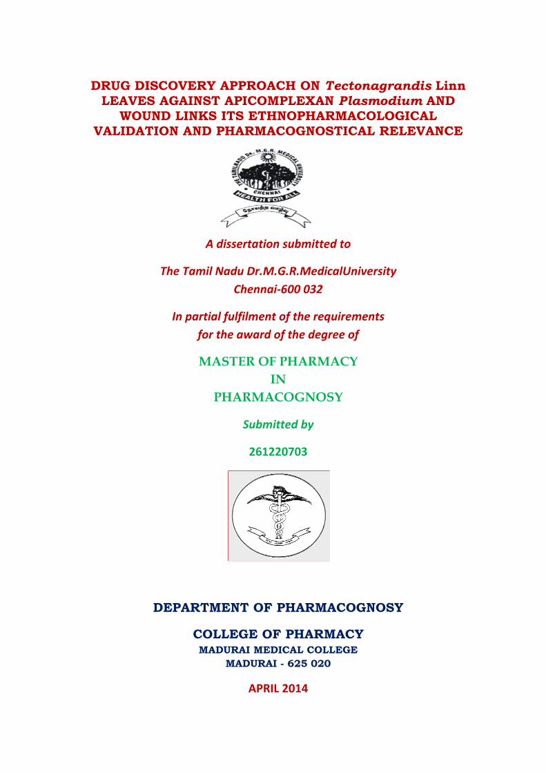
DRUG DISCOVERY APPROACH ON Tectonagrandis Linn LEAVES AGAINST APICOMPLEXAN Plasmodium AND WOUND LINKS ITS ETHNOPHARMACOLOGICAL VALIDATION AND PHARMACOGNOSTICAL RELEVANCE A dissertation submitted to The Tamil Nadu Dr.M.G.R.MedicalUniversity Chennai-600 032 In partial fulfilment of the requirements for the award of the degree of MASTER OF PHARMACY IN PHARMACOGNOSY Submitted by 261220703 DEPARTMENT OF PHARMACOGNOSY COLLEGE OF PHARMACY MADURAI MEDICAL COLLEGE MADURAI - 625 020 APRIL 2014
Transcript of DRUG DISCOVERY APPROACH ON Tectonagrandis Linn LEAVES ...
DRUG DISCOVERY APPROACH ON Tectonagrandis Linn
LEAVES AGAINST APICOMPLEXAN Plasmodium AND WOUND LINKS ITS ETHNOPHARMACOLOGICAL
VALIDATION AND PHARMACOGNOSTICAL RELEVANCE
A dissertation submitted to
The Tamil Nadu Dr.M.G.R.MedicalUniversity Chennai-600 032
In partial fulfilment of the requirements for the award of the degree of
MASTER OF PHARMACY IN
PHARMACOGNOSY
Submitted by
261220703
DEPARTMENT OF PHARMACOGNOSY
COLLEGE OF PHARMACY MADURAI MEDICAL COLLEGE
MADURAI - 625 020
APRIL 2014

Dr.A.ABDUL HASAN SATHALI, M.Pharm., Ph. D., I/c Principal& Head, Department of Pharmaceutics, College of Pharmacy, MaduraiMedicalCollege, Madurai-625020
CERTIFICATE
This is to certify that the dissertation entitled “DRUG DISCOVERY APPROACH ON
Tectona grandis Linn LEAVES AGAINST APICOMPLEXANPlasmodium
AND WOUND LINKS ITS ETHNOPHARMACOLOGICAL VALIDATION
AND PHARMACOGNOSTICAL RELEVANCE” submittedbyMiss.R.Jancy
Gracelet (261220703)in partial fulfilment of the requirement for the award of thedegree of
MASTER OF PHARMACY in PHARMACOGNOSYby The Tamil Nadu
Dr.M.G.R.Medical University is a bonafied work done by herduring the academic year
2013-2014 at the Department of Pharmacognosy, College of Pharmacy,Madurai Medical
College,
Madurai-625020.
(Dr.A.ABDUL HASAN SATHALI)

Dr.K.PERIYANAYAGAM, M.Pharm., Ph.D., Assistant Reader, Department of Pharmacognosy, College of Pharmacy, MaduraiMedicalCollege, Madurai-625020
CERTIFICATE
This is to certify that the dissertation entitled “DRUG DISCOVERY
APPROACH ON T.grandis Linn. LEAVES AGAINST APICOMPLEXAN
PLASMODIUM AND WOUND LINKS ITS ETHNOPHARMACOLOGICAL
VALIDATION AND PHARMACOGNOSTICAL RELEVANCE’’submitted
byMiss.R.JancyGracelet (261220703)in partial fulfilment of the requirement for the
award of thedegree of MASTER OF PHARMACY in PHARMACOGNOSYby The
Tamil Nadu Dr.M.G.R.Medical University is a bonafied work done by her under my
guidance during the academic year 2013-2014 at the Department of Pharmacognosy,
College of Pharmacy, Madurai Medical College,Madurai-625020.
(DR. K. PERIYANAYAGAM)

Dr. D. STEPHEN., M.Sc., Ph.D.,
ASSISTANT PROFESSOR,
DEPARTMENT OF BOTANY,
THE AMERICAN COLLEGE,
MADURAI-625002.
CERTIFICATE
This is to certify that the specimen brought by Miss.R.JANCY GRACELET,
II.M.Pharmacy, Department of Pharmacognosy, College of Pharmacy, Madurai Medical
College, Madurai is identified asTectonagrandisLinn.belonging to the
familyVerbenaceae.
Station : Madurai. (Dr. D. STEPHEN)
Date :08-07-2013

ACKNOWLEDGEMENTS Ifirst and foremost express my revered regard and obeisance to the ALMIGHTY
GODwith whose blessings I was able to complete my project work.
I am grateful to express my sincere thanks to Dr.B.SANTHA KUMAR,M.Sc(F.Sc),
MD (F.M)Dean, Madurai Medical College, Madurai, for giving an opportunity to carry
outmy project work.
I heartfelt sense of gratitude to Dr.ABDUL HASAN SATHALI,
M.Pharm.,Ph.D.,Principal I/C, College of Pharmacy, Madurai Medical College, Madurai.
I owe a great debt of gratitude and heartfelt thanks to
Dr. K. PERIYANAYAGAM, M. Pharm., Ph.D., P.G. Diploma in Clinical Pharmacy
(Australia), Assistant Reader in Pharmacognosy, College of Pharmacy, Madurai Medical
College, Madurai, for his enthusiastic co-operation as my project guide and all the constant
encouragement, suggestions, contribution and support rendered during the project work. As
a gesture of respect I would like to extend my thanks for his patience in listening and
answering to my questions.
I express my thanks and honourable regards to Ms. R. GOWRI, M. Pharm., Mr.
T. VENKATARATHINAKUMAR, M.Pharm., (Ph.D)., Assistant readers inDept of
Pharmacognosy College of Pharmacy, Madurai Medical College, Madurai.
I thank Mrs.A.KRISHNAVENI,M.Pharm.,
andMrs.A.SETHURAMANI,M.Pharm., Tutors in Pharmacy,Department of
Pharmacognosy, College of Pharmacy,Madurai Medical College, Madurai for their help.
I am thankful to Mr. P. SIVAKUMAR, M.Sc., DMLT, Lab Supervisor,
Mr.MAGUDESWARAN, DMLT, Lab Technician and Mrs. P. ELLAYEEfor their
support during my study of this work.

I thank Mrs. P.USHA RAVI KUMAR and other staffmembers, Institute of
pathology,Madurai Medical Collegefor their help to complete my project.
Dr. STEPHEN, M.Sc.,Ph.D.,Department of Botany, American College, Madurai
for plant authentication. I place on record my gratitude toDr.Sasikala Director of Siddha
Central Research Institute, Arunbakkam, Chennai who helped me in the microscopic
studies
I express heartfelt sense of gratitude to Mr.P.UDAYA KUMAR, District Malaria
Officer,Ramnad supply Plasmodium falciparun to carryout my bioassay.
I also thank my ever loving classmatesMs.P.ANITHA,Mr.S. JEGADEESHMs.D.
SUGANYA, Ms.KALAIYARASI, Mrs.S.R. NANDHINI BOSE, Mrs. S. NATHIYA
PRAKASH, Ms. K.VIJAYALAKSHMI, Ms.E.AJILA, Ms.R.ELAVARASI,
Ms.S.KARPAGAM, Mr.P.KANNIAPPAN and my juniorsMr.SEVA KUMAR,
Mr.BALA SUBRAMANIYAM, Mrs.PREMALATHA, Mrs.VIJAYALAKSHMI,
Ms.GOKILA for their constant motivation and help.
My heartfelt thanks to my lovable friend Miss P.BALA, Department of
Pharmacognosy, Madurai Medical College, Madurai for the help and encouragement,
endless patience to complete my work successfully.
I thank to Mr.PRABHAKAR RAO,M.pharm.,Pavan college of pharmacy,kolar for
his motivation to complete my studies.
I also thanks to my friends SUJI, SARASU, JEMI, DEEPA, SHAMILI, SHYNIfor their
encouragement and co-operation to my studies.
Above all, I am foreverindebted to myparentsfor their understanding, endless
patience,help and encouragement which made me to complete this work in a successful
manner.

CONTENTS
S. No. TITLE Page No.
1. INTRODUCTION 1
2. REVIEW OF LITERATURE 15
3. AIM AND OBJECTIVE 56
4.
MATERIALS AND METHODS
4.1 PLANT COLLECTION AND AUTHENTICATION
4.2 PHARMACOGNOSTICAL STUDIES
4.2.1 Morphological studies
4.2.2 Microscopical studies
4.2.3 Powder microscopy
4.2.4 Microscopic schedules
4.2.5 Scanning Electron Microscopy study
4.2.6 Physicochemical Parameters
59
60
61
61
63
64
65
66
4.3 PHYTOCHEMICAL STUDIES
4.3.1 Preliminary phytochemical screening
4.3.2 Fluorescence analysis
4.3.3 Estimation of flavonoid content
4.3.4 Estimation of Total phenolic content
4.3.5Energy Dispersive X-ray spectrometer (EDS)
4.3.6 HPTLC analysis
69
69
76
76
77
78
80
4.4 PHARMACOLOGICAL STUDIES 4.4.1 Acute Toxicological study using Brine shrimp Lethality
Assay (BSLA) 4.4.2In-vitro Antimalarial activity of TGEAE of the leaves 4.4.3 Effect of TGEAE leaves on ex-vivoporcine skin wound
healing model (PSWHM)
81
82
86

5.
RESULTS
5.1 PHARMACOGNOSTICAL STUDIES
5.1.1 Morphological studies
5.1.2 Microscopical studies
5.1.3Scanning Electron Microscopy study
5.1.4 Powder microscopy
5.1.4Microscopic schedules
5.1.5 Physicochemical Parameters
87
87
88
91
91
92
95
5.2 PHYTOCHEMICAL STUDIES
5.2.1 Preliminary phytochemical screening
5.2.2 Fluorescence analysis
5.2.3 Estimation of flavonoid content
5.2.4 Estimation of Total phenolic content
5.2.5Energy Dispersive X-ray spectrometer (EDS)
5.2.6 HPTLC analysis of TGEAE and TGEAE
96
100
101
101
102
102
103
5.3 PHARMACOLOGICAL STUDIES 5.3.1 Acute Toxicological Study Using Brine Shrimp Lethality Assay (BSLA) 5.3.2 In vitro Anti-malarial activity of TGEAEof the leaves 5.3.3 Effect of TGEAEleaves on ex-vivo porcine skin wound healing model (PSWHM)
106
107
108
6. DISCUSSION 109
7. CONCLUSION AND RECOMMENDATION 120
8. REFERENCES 125

Figure Contents
S.No Figures Page No
1. Diagram showing the Aerial Parts of T.grandis 87
1.A T.grandis Aerial Parts 87
2. T.S. of Leaf of T.grandis through Midrib Ground Plan 88
3. T.S. of Petiole 90
4. T.grandis Leaf Powder Microscopy 91
5. Calibration curve of quercetin for estimation of flavonoid content 101
6. Calibration curve of gallic acid for estimation of totalphenolic content 102
7. Energy Dispersive X-ray Spectrogram for T.grandis Leaves 102
8. HPTLC Profile of the TGEAE and TGEE of the leaves 105
9. HPTLC Graph of TGEAE and TGEE of the leaves 105
10. Percentage of Infected RBC’s 107

Table Contents
S.No Tables Page No
1. Vein Islet and vein termination number of T.grandis leaves 92
2. Stomatal number of T.grandis leaves 93
3. Stomatal index of T.grandis leaves 93
4. Palisade ratio of T.grandis leaves 94
5. Ash value of the leaves of T.grandis 95
6. Loss on Drying (LOD) for T.grandis leaves 95
7. Extractive values for T.grandis leaves (Individual solvents) 96
8. Extractive values for T.grandis leaves (Successive solvents) 96
9. Preliminary Phytochemical screening of leaves of T.grandis 100
10. Fluorescence analysis 101
11. T.grandisleaves elements weight & atomic Percentage 103
12. HPTLC profile of the TGEAE and TGEE leaves 104
13. Rf value for TGEAE and TGEE leaves 105
14. Various concentrations of TGEAE leaves on Artemia nauplii 106
15. Percentage of Infected RBC’s 108

Plate Contents
S.No Plates Page No
1. Habit and Habitat of T.grandis 87
2. Leaf arrangement ofT.grandis 87
3. Dorsal and Ventral View of the Leaves of T.grandis 87
4. Inflorescence of T.grandis 87
5. Flowers of T.grandis 87
6. Fruits of T.grandis 87
7. Seeds of T.grandis 87
8. T.S. of Leaf of T.grandisthrough the midrib 88
9. Adaxial Epidermis – A Portion Enlarged 88
9. A. Adaxial Epidermis Surface View (Apostomatic) 88
10. Abaxial Epidermis – A Portion Enlarged 88
10.A. Abaxial Epidermis (Showing Stomata) 88
11. Vascular Bundle – A Portion Enlarged 88
12. T.S. of Lamina – A Portion Enlarged 89
13. Venation Pattern 89
14. Trichomes of T.grandis 89
15. T.S. of Petiole 89
16. T.S. of Petiole – A Portion Enlarged 89
17. Trichomes – Various Magnificatios 90
18. Vessels – Various Magnificatios 90
19. Lamina 90 20. Upper Epidermis surface view 90
21. HPTLC Plates of TGEAE and TGEE of the leaves under UV 254nm 104
22. Antimalarialactivity 108
23. Histology showing effect of TGEAE on ex-vivo PSWHM 108

CHAPTER – I
INTRODUCTION
The role of Medicinal plants in the Human history:
Human relied on plants for basic needs such as food,clothing and shelter, all
products of plant matrices such as leaves, woods, fibres etc and storage parts like fruits,
tubers for many centuries. Plants was also used for additional purposes like arrow and dart
poisons for hunting, poisons for murder, hallucinogens used for ritualistic purposes,
stimulants for endurance hunger suppression as well as inebriants and medicines. The plant
constituents used for these latter purposes are largely secondary metabolites (like
alkaloids,flavonoids,glycosides etc.) which are derived biosynthetically from plant primary
metabolites (e.g carbohydrates, aminoacids and lipids) and are not directly involved in the
growth, development or reproduction of plants.
Arrow and dart poisons have been used in certain parts of the world with the
principal ingredients derived from the genera Aconitum, Antiaris, Strophanthus, Strychnos
etc. In some cultures some plants well documented for murder were henbane, mandrake,
deadly nightshade, calabar bean etc. Certain plants formerly used for arrow poisons have
also been used as medicines at lower dosages due to their desirable pharmacological
actions.[Bisset NG., 1991]
The hallucinogens used in the past was usually associated with magic and ritual
egCannabis sativa, Erythroxylum coca, Papaversomniferum etc. Tea, coffee, cocoa, cola
were used in the production of stimulant beverages and inebriants or intoxicants (Wine,
beer, kava) in many cultures since ancient times.[SneaderW .,1996]

Plants were the basis of sophisticated traditional medicine (TM) practices and used
for thousands of years in India, China and many other countries. Some of the earliest
records of the usage of plants as medicines are found in the Atharvaveda (which is the basis
of Ayurveda) in India, the clay tablets in Mesopotamia and the Eber Papyrus in Egypt, De
MateriaMedica of Dioscorides, Pen Ts’aoChing Classic of MateriaMedica.[Nerukar PV et
al., 2004,Sneader W 2005].
Before the realization that pharmacologically active phytoconstituents present in the
plants are responsible for their effect, the “Doctrine of Signatures” was used to identify
plants for treating diseases, for e.g. golden rod with yellow hue was used to treat jaundice,
red coloured herbs for blood diseases, liverworts for liver diseases, pile worts for
hemorrhoids, toothworts for tooth ache. [Sneader.,2005]
Morphine, atropine, caffeine, cocaine, ephedrine, pilocarpine, physostigmine,
quinine, salicin, theobromine, theophylline, tubocurarine were isolated in 19th
century.[Sneader W1996, Samuelsson G 2004].
The correlation between the ethnomedical usage of herbs and modern medicines
discovered from them has been studied . In this analysis 88 single chemical entities isolated
from 72 medicinal plants was introduced in to modern therapy many of which have the
same or similar therapeutic purpose as their original ethnomedical use. [Fabricant., DS and
Farnsworth NR., 2001].Some of these plant derived constituents such as atropine, codeine,
morphine, pilocarpine etc. are still being used widely as single agent or combination
formulations in prescription drugs [Snedear W.,1996]. Now a days , plants are still
important sources of medicines, this era especially in developing countries that still use
plant derived TM for their healthcare.
Plant based compounds and their role in drug development:
In spite of the recent resurgence of interest in drug discovery by molecular
modeling, combinatorial chemistry and other synthetic chemistry methods, natural product
derived compounds are still proving to be an invaluable source of medicine for humans.
Other than the direct usage of plant based metabolites in their original forms as drugs, these
compounds can also be used as drug precursors, templates for synthetic modification and
pharmacophores all of which will be discussed briefly here.
Secondary metabolites from plants as drug precursors:
Natural products derived from plants can be used as small molecule drug precursors
which can be converted into the compound of interest by chemical modifications or
fermentation methods. The semisynthetic approach is a valuable to resolve the shortage of
supply due to the low yield of compounds from plants and or the high cost of total
synthesis. The following are some secondary metabolites from plants which are useful
drug precursors, though they are not necessarily pharmacologically active form naturally.
To meet the market demand of the anti-tumour drug paclitaxel cropping of the bark of slow
growing Pacific yew tree ,Taxusbrevifolia Nutt. and the synthesis are not feasible method
to provide sufficient amounts. But 10-deacetylbaccain III can be isolated comparatively
larger amounts from the needles of the other related yew species, such as Taxusbaccata L.,
a renewable resource, can be converted chemically in several steps into paclitaxel.[Denis
JN,Greene AE1988, Holton RAet al., 1995].
Diosgenin, a steroidal sapogenin obtained from the tubers of various
Dioscoreaspecies that grow in Mexico and Central America is chemically converted into

progesterone which is the key intermediate for the production of cortisone an important
anti-inflammatory.
[Mancera Oet al., 1952, 1953].
Tamiflu (Oseltamivir phosphate) an orally active neuraminidase inhibitor developed
for the prophylaxis and treatment of influenza viruses A& B is synthesized from (-)
shikimic acid which is an important biochemical intermediate in plants and
microorganisms. Though initially it was isolated from star anise, shikimi tree
(Illiciumverum), later on it was obtained from the fermentation of genetically engineered
E.coli strains.Currently, Roche, the drug manufacturer still relies on both extraction and
fermentation methods to obtain ton quantities of shikimic acid. [AbrechtS et al.,
2004,Yarnell A., 2005].
Secondary metabolites from plants as drug precursors:
Prototype defined as “ the first compound discovered in a series of chemically
related therapeutic agents” [Sneader W., 1998]. Podophyllotoxin, camptothecin and
guanidine are drug prototypes with analogs having the same pharmacological action as the
parent compound while atropine is a drug prototype that has furnished many analogs that
have additional pharmacological properties. Guanidine is a natural product with good
hypoglycemic activity isolated from Galegaofficinalis but is too toxic for clinical use.
Though many derivatives of guanidine have been synthesized metformin
(dimethylbiguanide) was later found to be clinically suitable for treatment of type II
diabetes.[Krentz AJ and Bailey CJ., 2005].

Taxol 10-deacetyl baccatin
Tamiflu Shikimic acid Podophyllotoxin
Camptothecin Vepesid Hycamtin
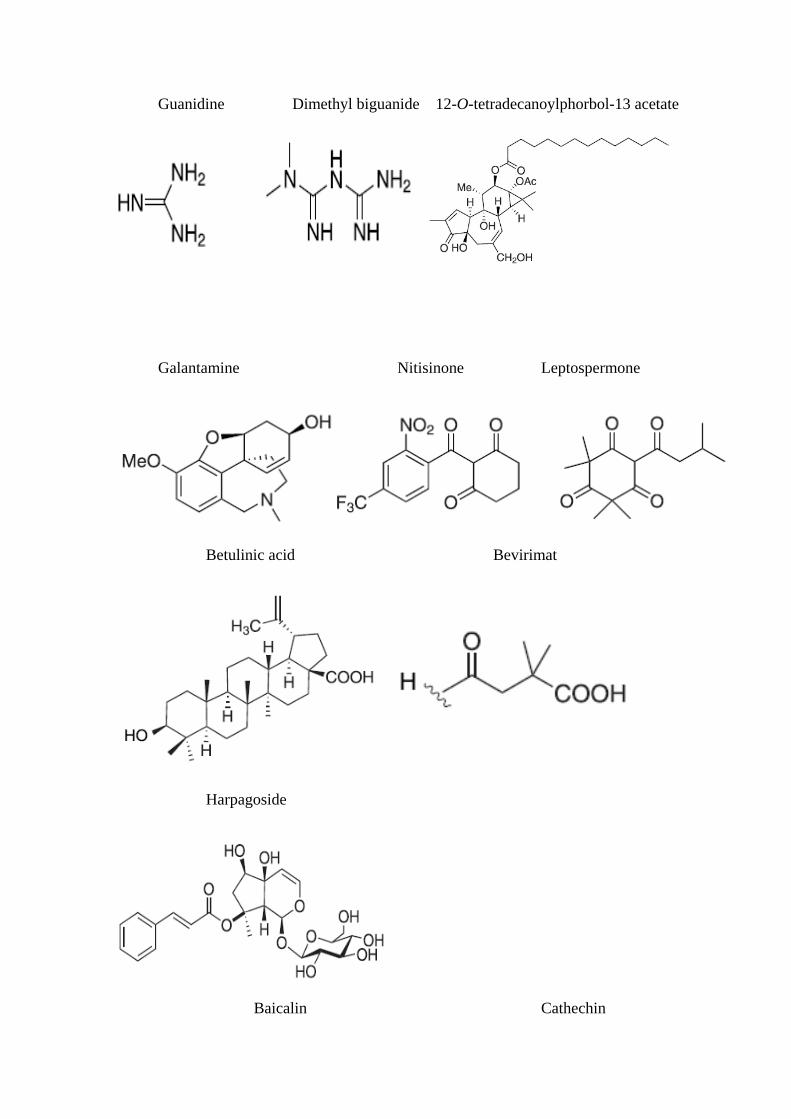
Guanidine Dimethyl biguanide 12-O-tetradecanoylphorbol-13 acetate
Galantamine Nitisinone Leptospermone
Betulinic acid Bevirimat
Harpagoside
Baicalin Cathechin

Ginkgolides A Ginkgolides B
Ginkgolides C Bilobalide
Secondary metabolites from plants as pharmacological probes:
Secondary metabolites of plant origin like phorbol esters and genistein can be uses
as “pharmacological probes” which help researchers to understand the mechanism of action
of intracellular signal transductions and biological mechanisms related to human diseases
which can aid the design of better drugs. An isoflavone found naturally in soyabean,

Genistein, is an inhibitor of various protein tyrosine kinase (PTK) which are essential
enzymes involved in intracellular signal transduction. Genistein has been used to probe the
interaction between PTK and cyclic nucleotide gated channels which are important in
mammalian olfactory and visual systems. Phorbol, a tetracyclic diterpenoid plant secondary
metabolite isolated as hydrolysis product of croton oil from the seeds of Croton tiglium has
been used in biomedical research as carcinogen promotions [Molokanova E and Kramer
RH., 2000].
Drug discovery from plants- recent developments:
Plant derived natural products still contribute to the overall total number of new
chemical entities(NCE) that continue to be launched to the market.
Plant derived drugs launched from 2001:
In the past 6yrs five new drugs derived from plants namely
apomorphinehydrochloride( for Parkinson’s disease), galantaminehydrobromide( for early
onset of Alzheimer’s disease), nitisinone (for hereditary tyrosinemia type1), tiotropium
bromide(bronchospasm with COPD and varenicline( aid to smoking cessation) have been
approved by the US FDA.
Plant derived compounds involved currently in clinical trials:
Many medicinal plant-derived compounds are currently undergoing clinical trial for
the potential treatment of various diseases in which majority are in the oncological area
including new analogs of known anticancer drugs based on the camptothecin, taxane-
podophyllotoxin or vinblastin type skeletons. Compounds with carbon skeletons different
from the existing plant derived drugs used in cancer are betulinc acid, ceflatonine,

combretastatin A4 phosphate, ingenol-3 angelate, phenoxodiol and
protopanaxadiol.Bevrimat and celgosivir are currently undergoing clinical trials for the
treatment of HIV and hepatitis C viral (HCV) infections respectively. Capsaicin is in
clinical trial for the treatment of severe postoperative pain and huperzine is developed for
Alzheimer’s disease. Betulinic acid is a lupine type triterpene that is mostly distributed in
the plant kingdom and this constituent along with various derivatives shown to have anti-
inflammatory, anticancer, antimalarial, anti-HIV, anthelminthic, and antioxidant properties.
(Cichewicz RH and Kouzi SA., 2004). In 1995, University of Illinois at Chicago research
group reported that betulinc acid inhibited human melanoma both in vitro and in-vivo
systems and induced apoptosis in Mel-2 human melanoma cells. It was further developed
under the Rapid Access to Intervention Development program of the United States
National Cancer Institute and currently undergoing I/II clinical trials for the treatment of
dysplatic melanocytic nevi, a preliminary symptom that may lead to melanomas of the
skin. Bevirimat a semisynthetic compound derived from betulinc acid is being developed
by Panacos Pharmaceuticals, Watertown, MA, USA as a new class of antiretroviral drug.
Bevirimat blocks HIV-1 maturation by distrupting a late step in the Gag processing
pathway, causing the virons released to be noninfectious, thus terminating the viral
replication. It is currently undergoing Phase II clinical trials and Phase III trials are
expected to start soon. (www .clinicaltrials .gov/ct /show/ NCT00346502,
www.panacos.com)
Plant extracts currently involved in clinical trials:
New forms of registered phytomedicines are not single chemical entities. These
complex drugs are subjected to quality control via extract standardization procedures
involving either or both constituents with known biological activity or inactive marker

compounds present in high concentration. Harpagophytumprocumbens, Devil’s claw
extract from Africa is currently undergoing phase II clinical trials in the USA for the
treatment of hip and knee osteoarthritis. Flavocoxid (Limbrel) a proprietary blend of
natural flavonoids from Scutellariabaicalensis and Acacia catechu is being marketed in the
USA by Primus Pharmaceutical under prescription as a medical food therapy for
osteoarthritis Ginkgo extracts from the dried leaves of Ginkgo biloba used for the treatment
of early stage of Alzhemer’s disease, vascular dementia, peripheral claudication, tinniturs
of vascular origin. Mistletoe (Viscum album) extract used in the treatment of cancer in
European countries. Sativex developed by GW pharmaceuticals is Cannabis sativa extract
for the treatment of neuropathic pain in patients with multiple
sclerosis.(www.Clinicaltrials.gov/ct/gui/show/NCT00391079).
Hence it is clear that natural products are an integral part of human health care
system nowadays because there is now popular concern over toxicity and side effects
modern drugs. The untapped wealth of the plant kingdom has become a target for the
research by multinational drug companies and research institutes for new drugs and lead
compounds.
There is recent increased participation of pharmaceutical companies in the
antimalarial drug discovery and development process offers hope for the development of
new, affordable drugs. Indeed, an unprecedented number of malaria discovery and
development projects are now executed, involving many organizations including the
Medicines for Malaria Venture (MMV) as malaria remains one of the most important
diseases of the developing countries killing 1-3 million people and causing disease in 300-
500 million people annually.

Recent malaria discovery and development projects and associated
organizations[Fidock, DA et al., 2004].
Discovery projects Development projects
Improved quinoline GSK, Liverpool Rectal artesunates
Farnesyltransferase inhibitors BMS,Wasington Chlorproguanil, dapsone,artesunate
Manzamine derivatives Pyronaridineartesuanate
Cysteine protease inhibitors GSK Amodiaquineartesunate
Fatty acid biosynthesis inhibitionMMV,Texas A&M U., AECOM Jacobus
Mefloquineartesunate
Chalcones (National U.Singapore, LicaPharmaceuticals)Short chainchloroquine
third generationantifolates.
The failure to eradicate malaria is due to a number of factors including resistance of
the causative vector female Anopheles mosquitoes to insecticides such as DDT and
avoidance of insecticide use because of toxicological and ecological considerations.
Malaria caused by Plasmodium faciparum is the most serious type because it often proves
fatal unless prompt treatment is given. P.falciparum resistance to clinically used drugs such
as chloroquin, mefloquineetc is a serious problem and in parts of South East Asia and
Africa, the only effective antimalarial drug is artemisinin or one of its derivatives. In many
tropical countries extensive list of plants useful for the treatment of malaria is available.
The ethnomedical information of Tectonagrandis reveals that it is one among them.
Reason for selection of this plant:
It was reported that Verbenaceaefamily members have phytoconstituents with
various pharmacological properties useful in the treatment of bacterial, fungal, malaria and

pain arising from various causes including rheumatoid arthritis. It was also claimed that
these plants merit detailed study which can prove useful in the discovery of lead
compounds leading to novel and more efficacious drugs. [Rahmatullah Md.,2011]
Tectonagrandis belonging to the family Verbenaceaereallydo not have any match as a
cheap natural and easily available plant. Its leaves traditionally known to be useful for the
treatment of wide panel of diseases like tuberculosis, various kinds of wounds especially
burn wound, malaria, anaemia, leprosy, skin diseases like pruritis, stomatitis, indolent
ulcers, haemorrhages, menstrual disorder, bone joint disease. Young leaves and fruits
prescribed for women for fertility control and fumes of dry leaves to kill guinea worms. It
also possesses anti- ulcer and anti- cancer activity. Various scientific investigations of the
leaves showed anti-diabetic activity, anti-microbial activity, anti-oxidant, analgesic, anti-
inflammatory, anti-amoebic, anti-hypertensive and wound healing activity.
Bark is used as astringent, expectorant, anti-inflammatory, anthelmintic and for
constipation, leucoderma, headache, piles, bronchitis, hyperacidity, and dysentery, burning
sensation, diabetes, difficult in labour, leprosy, skin diseases, and indigestion. Researches
proved it is anti-fungal, anti-bacterial, uterine relaxant, anti-histaminic, anti-asthmatic, anti-
oxidant, analgesic and anti-inflammatory, anti-diabetic, hepatoprotective and cardiac
activities.
Wood traditionally used as diuretic, stimulant, hepatic astringent, toothaches, sedative to
gravid uterus, leucoderma. It was reported that wood possesses anti-oxidant, anti-termite,
anti-hyperglycaemic, nephroprotective activities.
Flowers used for bronchitis, biliousness, urinary discharge, diuretic, depurative,
anti-inflammatory, leprosy, skin diseases, diabetes, congestion of liver.
Seed is used in poisoning. Seed oil in skin disease. Oil obtained from seeds and
flowers promote the growth of hair, eczema, ring worm, scabies. Scientific investigation of
seed extract proved hair growth activity, diuretic, anti-inflammatory activity.
Roots are useful in anuria and retention of urine, anaemia. Anti-ulcer activity, anti-
inflammatory, anti-tussive, anti-oxidant, hypoglycemic, anti-pyretic activities have been
evaluated and proved.
It has been reported that bioflavones and flavonol glycosides were found to possess
anti- malarial activity.[Khaomek P.,2008].
Betulinic acid is a lupane type triterpene that is widely distributed in the plant
kingdom and this compound along with various derivatives has been shown to have anti-
cancer, anti-bacterial, anti-malarial, anti-HIV, anthelmintic, anti-inflammatory and anti-
oxidant properties.[Cichewicz RH and Kouzi SA .,2004, Yogeeswari P and Sriram D.,
2005]
The survey of literature on T.grandisalso reveals that leaves contain various
phytoconstituents like flavonoids especially apigenin and steroidal compounds like
betulinic acid, glycosides like anthraquinones etc.

Review of literature showed lacunae exist in the pharmacognostic, phytochemical
and pharmacological studies. The present study assesses the potential of T.grandisleaves in
relation to its traditional uses (in malaria) and in terms of findings on modern bio scientific
research.
CHAPTER-2
LITERATURE SURVEY
VERBENACEAE

Verbenaceaefamily plants used in Bangladesh was described. It was reported that
they have constituents with various pharmacological property useful in the
treatment of bacterial and fungal infection, malaria and pain arising from various
causes including rheumatoid arthritis. The importance of plants used in Bangladesh
lies more so in the potential for treatment of malaria and rheumatoid arthritis. It was
claimed that the plants merit detailed studies which can prove useful in the
discovery of lead compounds leading to novel and more efficacious drugs.
[Rahmatullah Md. , 2011]
Taxonomical classification of T. grandis plant[Aradhana R et al. , 2010]
Kingdom : Plantae
Subkingdom : Tracheobionta
Super division :Spermatophyta
Division : Magnoliophyta
Class : Magnoliopsida
Subclass : Asteridae
Order : Lamiales
Family : Verbenaceae
Genus : Tectona
Species : grandis
Botanical Name :Tectonagrandis. Linn
Vernacular names for T. grandis:
Eng : Indian Teak
Hindi : Sagun, Sagwan
Mar : Sag, Saga, Sagwan

Guj : Saga, Sagach
Oriya :Singuru
Tel : Adaviteeku, peddateeku
Kan : Jadi, sagwani, tega, tyagadamara
Mal : Thekku, tekka
Tam : Tekkumaram, tekku [Anonymous. , 2005]
Cultivation History of T. grandis:
T. grandisnatural distribution is in Southeast Asia, from the Indian subcontinent
through Myanmar and Thailand to Laos. T. grandis early cultivation by Hindu settlers,
possibly in the 7th century, is not clearly established. T. grandis is believed T. grandis was
introduced to Java 400-600 years from India. At the beginning of this century, T. grandis
was introduced to both East and West Africa and ‘Trinidad T. grandis’has become
particularly well known in the Caribbean region. T. grandis is planted for timber or
ornament and in botanical gardens. T. grandis was the most important export timber in
Thailand. [Aradhana R et al. ,2010]
Methods for cultivation:
The new plants can also be propagated from cuttings apart from the seed sowing. T.
grandis usually planted when it is 4 to 6 weeks old. Plough the land thoroughly and levelit.
The best season to plant the T. grandis is monsoon, most probably after the first shower.
Carry out weeding operations regularly. T. grandis requires loamy soil rich in humus and
having the right content of moisture with good drainage. T. grandis grows well in hilly and
dry areas. T. grandis requires a dry tropical climate for its growth. T. grandis flowers in
February and March. [Aradhana R et al. ,2010]
Natural Regeneration:
T. grandis naturally reproduces from seed but the extent of natural regeneration
depends on climatic and edaphic factors. Fertile seeds begin to be produced on trees from
near about the 20th year. Coppice shoots however, flower and fruit abnormally early, taking
not more than 10 years to produce fertile seeds. The T. grandis produces seeds every year,
poor seed years being occasional.
Factors influencing natural regeneration:
• Spread of the seed
• Germination
• Survival and development of the seedling
These factors almost possibilities for natural regeneration. The amount of
regeneration increases with the increase in moisture and depth of the soil. The soil areas
classified as, semi-moist T. grandis type, moist T. grandis type, very moist evergreen type.
Semi moist T. grandis type:
This type show less incidence of fire, but grazing continues to be heavy. Generally
in this type, the ground cover is fair and natural regeneration of T. grandis is fairly
adequate. The regeneration process, though slow, is steady and this tendency may be
attributed to:
• Beneficial effect of mixed vegetation on soil
• Survival ofT. grandis seedlings as a result of fire protection
• Moderate growth of bamboos, eliminating grass and other
herbaceous growth of which T. grandis is intolerant
• Establishment of T. grandis seedlings under moderate shade of
bamboos

• Protection of seedlings from desiccation by the under storey
• Relatively greater resistance of T. grandis seedlings to damage by
cattle and fires as compared to the seedlings of its companions
• The capacity of seedlings to regenerate after cutting back, if
sufficient overhead light is available.
Moist T. grandis type:
This type is better protected from fire and grazing is low. It appears that, before
bamboos and other shrubby growth become dense and the upper canopy starts closing up,
the conditions are most favourable for regeneration ofT. grandis towards the drier end of
this type. In the more humid parts, where the overwood is comparatively denser, canopy
manipulation does bring in regeneration, but the progress is slow, and such areas cannot be
relied upon for natural regeneration. The ground cover is dense and consequently natural
regeneration occurs only in patches.
Very moist ever green type:
In this type prolonged fire protection results in the formation of dense understorey
of semi ever green shrubs which inhibit regeneration of T. grandis. Consequently this type
is particularly devoid of natural regeneration and the percentage of T. grandis is generally
quite slow. [Anonymous. ,2005]
Artificial Regeneration:
T. grandis has been made to propagate by vegetative methods. Cleft grafting,
budding and branch cuttings have been successfully employed for establishing clones of
desired quality. Almost 80-100% success has been obtained in case of cleft grafting and

bud grafting but in branch cuttings only 60% success has been recorded. Other methods
like air-layering etc. .were not successful in this plant. [Anonymous. ,2004]
Propagation:
T. grandis is propagated mainly from seeds. Germination of the seeds is often poor
and sporadic. It involves pre-treatment to remove dormancy arising from the thick pericarp.
Pre-treatment involves alternate wetting and drying of the seed. T. grandis seeds are soaked
in water for 12 hours and then spread to dry in the sun for 12 hours. This method is
repeated for 10-14 days. Dry heat method is better for drying. Seeds are heated for 1-5
weeks at 50˚C or at 48 hour at 80˚C. Then the seeds are sown in shallow germination beds
of coarse peat covered by sand. T. grandis seeds are germinate after 15 to 30 days.
[Aradhana R et al. ,2010]
Collection of T. grandis:
In dry situations and seasons, T. grandis leaves fall from November to January,
while in moist localities the tree may remain in leaf until March or even later. As a rule, T.
grandis trees are leafless throughout the greater part of the hot season, the new leaves
appear from April to June but in wet seasons they sprout early. T. grandis flowers come up
from June to August or September but, like the leaves, they may begin to appear in April
under abnormally wet conditions. T. grandis fruits ripen from November to January and
fall gradually and may be collected from under T. grandis trees. For easy storage T.
grandisfruits, The calyx is removed by filling the bag with fruits and vigorously rubbing
and shaking it; the remains of the calyces can be separated from the nuts by winnowing.
The T. grandis nuts vary in weight( Madhya Pradesh), the number of fruits varied from 2,
000 to 3, 000/kg. [Anonymous. ,2005]

Natural Habitat:
T. grandis will grow and survive under a wide range of climatic and edaphic
conditions(warm, moist, tropical climate)with significant difference between dry and wet
seasons. T. grandis is deciduous, while its grown in non-seasonal climates are semi-
deciduous. T. grandis is often a dominant member of a mixed deciduous forest, where it is
main associates are Xyliaspp, Afzeliaxylocarpa, Terminaliaspp and Lagerstroemia spp. The
forest floor is often covered with Bamboo. It is generally occurs scattered but can form
almost pure stands under suitable conditions. Young plants show a remarkable capability to
recover after fire. [Aradhana R et al. ,2010]
Geographical Distribution of T. grandis:
T. grandis is native in India, Western peninsula, Konkani, Burma, Myanmur,
Thailand and Laos and exotic to Antigua and Barbuda, Bangladesh, Barbados, Dominica,
Dominican Republic, Ghana, Grenada, Jamaica, Kenya, Brazil, Cambodia, China,
Brunei, Cote d′lvoire, Cuba, Guadeloupe, Malaysia, Mauritius, Nepal, Nigeria, Pakistan,
Panama, Philippines, Puerto Rico, South Africa, Sri Lanka, St Lucia, St Vincent and the
Grenadines, Tanzania, Togo, Trinidad and Tobago, Uganda, Vietnam, USA, Virgin
Islands(US). [Anonymous. ,2005, Aradhana R et al. , 2010].
Provenances:
Several provenances of T. grandis differing in growth characteristics,
morphological characters of stem, leaf and timber have been recognized. Five distinct
ecotypes of T. grandisforests(very dry, dry, semi dry, moist and very moist) are found in
India. Dry and very dry area timbers has good figure and is valued for panelling and
furniture. T. grandis from western regions ( Malabar) is superior in strength to that from
drier areas and is practically equivalent to the T. grandis from Burma. Hudsa plantation of
Virnoli Range in North Kanara was observed to have smaller leaves with smooth, shiny,

dorsal surface. This seems to be a mutant type and known as Teli type which is stronger,
durable and much cleaner than the common T. grandis, more cylindrical with very little
fluting with less lower branches, better germinating seeds, smooth bark and also lefing
and flowering are in advance. It appears to be resistant to defoliation by
HyblaeapueraCramer. Another provenances from Ramanathapuram and Tirunelveli
districts is reported to have different phenology. It have leaves of smaller, elliptic-ovate
with acuminate tip with silver-glaucous under surface. Based upon a study of
morphological differences, especially of leaf, a key, prepared by Bor, distinguishes
several Burmese and Indian provenances, including those of Nilambur and other parts of
Kerala, Shimoga, Dhulia, Kamara and Jhansi. It has been recommended that for planting
a particular site, seed from a local or nearby and similar climatic regions should be
considered the safest, unless by provenances test, some other provenance has shown better
performance than the local source of seed. (Anonymous. ,2005)
Climate:
T. grandis thrives best reaches its largest dimensions in a fairly moist, warm,
tropical climate, although it occurs even in dry localities, subjected to great heat and
drought during the hot season. In highly moist, tropical regions T. grandis tends to be
replaced by evergreen species. T. grandis tree may extend into regions of slight frost, but
throughout T. grandis distribution the frost is generally unknown. T. grandis thrives best in
areas having a normal rainfall, varying from 125-250 cm, with a marked dry season of 3-5
months, but T. grandis is known to grow, though not adequately well, in places with a
rainfall as low as 75 cm or even less(Dhulia, Jalgaon, Nimar, Buldana, Ahmadnagar and
West Karnool)and as high as 500 cm(West Coast). In the Indian Peninsula, it experiences
in places absolute maximum shade temperature of 48˚ and absolute minimum shade

temperature of 2˚ but these extremes denote a drier climate than is favourable to T. grandis
tree development. In the moist parts of the West Coast, however, where T. grandis
reaches larger dimensions as compared to the trees in the drier parts of the peninsula, the
climate is much more equable, the absolute maximum shade-temperature varying from 39
to 44˚C and the absolute minimum from 13-17˚C. (Anonymous. ,2005)
Soil:
T. grandis prefers rich soils. T. grandis tree is quite hardy, but the extent to which
T. grandis flourishes and the quality of timber not only depend on the physical nature of
the soils, but also on their depth, drainage, moisture content and fertility. T. grandis shows
very good development on the fertile lower slopes of the hills, where the soil is deep , but
in shallow soils and along dry ridges T. grandis becomes stunted. T. grandis comes up on
soils produced by a variety of geological formations like traps,basalt, granitic gneisses,
calcareous crystalline rocks, phyllites and schists. T. grandis also grows well on easily
disintegrable sandstones like Vindhyan sandstones, but on quartzite and hard, metamorphic
sandstones growth is poor. The great trap areas of the peninsula are extensively covered
with T. grandis, but the trees are small in size as the soil is of no great depth;in the valleys
and along the lower slopes, where the soil is deep, the trees attain fair dimensions. Good
growth is noticed on soil produced by rocks rich in calcium oxide and other bases. T.
grandis also thrives well on freshly deposited river- alluvia, having sufficient contents of
exchangeable calcium. T. grandis tree avoids laterite;only where the laterite is highly
disintegrated and mixed with other rocks does T. grandis attain any size. T. Grandisis not
found on water logged ground or sites liable to prolonged inundation nor does T.
grandisgrow on stiff clay, black cotton soils and deep dry sands. Good growth of T.
grandis is also not generally found on Gondwana rocks of plateau region. Rocks yielding

coarse-textured, nutrient-deficient, shallow soils are not good for T. grandis growth. T.
grandis was found that soils having a lower silica sesquioxide ratio, lower dispersion
coefficient and a very low or very high watertable are unfavourable for good growth of T.
grandis. The root being very sensitive to the deficiency of oxygen, T. grandis does not
withstand water-logging and requires well-aerated soils. The pH of soils in the T. grandis
forests is between 5. 0 and 7.0, reaching 7.5 in drier tracts.In Madhya Pradesh the soils
having more than 0.39% exchangeable calcium, support the growth of T. grandis and also
good natural regeneration of soils containing 5-7mg phosphorus/100gm. [Anonymous.
,2005]
ELEMENTAL DEFICIENCY
Nitrogen:
It causes stunted growth, strong chlorosis, premature defoliation and absence of
branching in seedlings.
Phosphorus:
It causes the seedlings exhibit scorched leaf margins and chlorosis, followed by
necrosis, wrinkled leaf surface with restricted shoot growth and absence of branching.
Potassium:
In seedlings, it causes intervenalchlorosis and scorching of margins. In younger
leaves the surface wrinkles and margins curl inwards.
Calcium:
In seedlings, is exhibited by severe intervenalchlorosis, distortion of leaves, and
premature defoliation with restricted branching. [Anonymous. ,2005]
Diseases:

Oliveatectonae(rust stem and leaf); Phyllactiniaguttataand
Uncinulatectonae(mildew); Cercosporatectonae(leaf spot); Nectriahaematococca(stem
canker); Corticiumsalmonicolor(pink disease); Phomopsistectonaein combination with
Colletotrichumgloeosporioides(leaf spots); Pseudomonas tectonaeand P. tectonae(wilt);
Fusariumoxyporum(damp. off); Armillariellamellea(root rot); Helicobasidiumcompactum,
Phellinusnoxius, Rigidoporuslignous, R. zonalisandPeniophorarhizomorpho-
sulphurea(butt rot); Pellinusnoxius, P. lamaoensis, Ustulinadeusta, Polyporusrubidus,
Ganodermaapplanatumand R. zonalis(heart rots); Cossuscadambae(trunk borer);
Phialophorarichardsiae(die-back of trees); Dendrophthoefalcata(the mistletoe)
insectsHolotrichia spp. (white grubs); Sahyadrassusmalabaricus(sapling borer);
Cossuscadambae(trunk borer); Hyblaeapuera(defoliators); Eutectonamachaeralis,
Pyraustamachaeralis, Hapaliamachaeralisand Eutectonamachaeralis(skeletonizer);
Xyleutesceramica(bee hole borer and shoot borer); Duomitusceramicus(bee hole borer);
Zouzeracoffeae(shoot borer);Pagidasalvaris; Machaerotaelegans;
Mylabrisphalerata;Dichocrosispunctiferalis; Eublemma spp. ;
Xyleborusdestructuens;Calotermestectonae;X. padestruens and Coptotermeselisae
(termites)Pelliculariasalmonicolor(bark flaking)Peniophorarhizomorpho-sulphurea causes
serious mortality due to rootrot in plantations in North India. The fungus produces
subterranean rhizomorphs through which the disease spreads. Polyporuszonalis causes root
and buttrot in plantations, In dry coppice T. grandis forests, 50%trees exhibit hollowness
due to decay of heartwood by Polyporuszonalis and Fomeslividus at the end of rotation.
Heartrot is present in the callus shoots arising from cambial activity at the cut end of stools;
as these shoots develop intimate organic connection with the stools during their growth, the
fungi migrate from the stools to the heartwood of shoots. However, coppice shoots arising
from dormant buds at or below the ground level, develop their own root system and in due

course become independent of stools and thus remain free from heartrot. [Anonymous.
,2005]
LEAVES:
ETHNOMEDICAL INFORMATION:
The young leaves and fruits are prescribed to women for fertility control by the
herbal vendors. In Rajasthan, the fumes of dry leaves are used to kill guinea worms
in infested patients. [Jayakumaret al. , 1987]
T. grandis have revealed that the plant has anti-ulcer [Pandey et al. , 1982], anti-
microbial [Sumthonget. al. , 2006], anti-cancer[Khan RM and Miungwana SM. ,
1999]
Leaves are used for stomachic, astringent and vermifuge.
[Kamat SV. ,2001]
Leaves have also been used in indigenous medicine. Extracts of the leaves showed
complete inhibition of Mycobacterium tuberculosis [Anonymous. , 2005]
Tumours were treated with leaves of F. glomerata, T. grandis and E. scaber
repeatedly and then with a honey mixed fine paste of A. roxburghiana, C. sappa, S.
racemosa, T. arjuna, X. strumarium was applied. [Premalatha and Rajgopal. , 2005]
The leaves of T. grandis are widely used in India folklore for the treatment of
various kinds of wounds especially burn wound [Manoharan KP et al. , 2007,
Majumdar M et. al. , 2007] Leaves of T. grandis is used as anti-inflammatory and
topical treatment for burn injuries like burn inflicted wounds and skin ulcers.
[Yogesh Sharma et al. , 2013]
The leaves of T. grandis with or without J. curcas and F. flavenscens are
traditionally used in Togo to treat malaria and other anaemia diseases. [Diallo A et
al. , 2008]
T. grandis leaves used for cooling, haemostatic, depurative, anti-inflammatory
and vulnerary, leprosy, skin diseases, pruritis, stomatitis, indolent ulcers,
haemorrhages, haemoptysis, vitiated conditions of pitta. [Aradhana R et al. ,
2010, Khera N and Bhargava S. , 2013]
T. grandis leaf, root, flower usedfor constipation, blood dysentery, skin diseases,
powdered roots and boiled leaves are orally administered for constipation and blood
dysentery, macerated leaves and flowers are applied to affected areas in skin
diseases. [SajjadulkarimMdet al. , 2011]
Decoction of T. grandis fresh or dried leaves are used for menstrual disorder and
haemorrhages[Kothale KV et al. , 2012]
T. grandis leaves used as remedy for bone joint disease by valaiyar community in
Madurai district. [Palaniappan P et al. , 2012]
T. grandis young leaves, bark, wood traditionally used for fever, malaria,
stimulant, anaemia, diabetes II, anti-ulcer, clothes industry of bits of matches.
[Bangou MJ et al. , 2012]
PHARMACOGNOSTICAL STUDIES
T. grandis leaves are elliptic or obovate, acute or acuminate, the upper surface
rough but usually glabrous, the lower clothed with dense stellate grey or tawny
tomentum, entire, base usually cuneate, main nerves 8-10 pairs with 2 or 3 large
branches near the edge of the leaf, joined by numerous parallel transverse veins.
[Kiritikar Basu. , 1987]
T. grandis leaves was studied for the drug weight loss and chemical changes
(decomposition process) in a small fresh water lake using plastic net bags. (1mm
pore size). The results revealed that dry weight loss and changes in the levels
ofcellulose, hemicellulose, lignin, sugar, amino acid and nitrogen of T. grandis

leaves at different depths in a shallow freshwater lake. The rate of decomposition
was faster when compared with similar studies in Canada and Europe. [Tiwari BK
and Mishra RR. , 1983]
The leaf extract of fallen leaves showed inhibitory effect on the growth of Arachis
hypogea and Zea mays. Chlorogenic, coumaric and ferulic acids present in the
extract are probably responsible for the allelopathic effects. [Anonymous. ,2004] .
Naphthotectone showed high level of activity(aqueous solution concentration
ranging from 10-3 to 10-5 M) in etiolated wheat coleoptile bio assay as well as
standard target species lettuce, cress, tomato and onion suggested that it may be
involved in the allelopathic activity and in other defence mechanism their by this
makes naphthotectone and important future target in the development of drugs and
eco herbicides. [Lacret R et al. , 2011]
Structures of isolated compounds from T. grandis leaves were determined and
studied (using etiolated wheat coleoptiles). The activities showed that the isolated
lignans and nolignans should be part of the defence mechanisms of this plant.
[Lacret R et al. , 2012]
PHYTOCHEMICAL STUDIES
A novel compound tectograndone derived from the interaction of two
prenylatednaphthoquinones has been isolated from the leaves of T. grandis.
Thestructure was elucidated as 10-(2, 5, 8-trihydroxy naphthaquinone-3yl)-5, 12-
dihydroxy-2, 2, 8 trimethyl-2(H)-pyrano(2, 3, -b)anthracene-6, 11-dione, primarily
by 1D and 2D NMR techniques. [Aguinaldo et al. , 1993]
Leaves are containing a yellow or red dye which has been used and strongly
recommended for dyeing silk yellow olive or related shades. Crushed leaves are
produce a red colour when rubbed with saliva [Anonymous. 2005]

T. grandis leaf reported to contain carbohydrates, alkaloids, tannins, sterols, saponins,
proteins, calcium, phosphorus, crude fibre and dyes(yellowish-brown or reddish).
[Anonymous . , 2006, Krishnan KS. , 2006, Majumdar M et al. , 2007, Goswami DV et
al. , 2009, Nayeem N Karvekar . , 2010]
A new Anthraquinonetectone(1), 14 known compounds (2-5)[5 terpenoids2-5, 15],
4flavanoids(6-9), 3 flavone glycosides(10-12), 2 phenolic glycosides(13-14)were
isolated and identified synthesized from the chloroform and n-butanol fractions of
ethanolic extract of T. grandis leaves. [Shukla N et al. , 2010]
T. grandis leaves contain reported chemical constituents are
• Quinones(tectoquinone, lapachol, deoxylapachol and its isomer, tectoleafoquinone,
anthraquinone-napthaquinone pigment)
• Steroidal compounds(squalene, poly isoprene-α-tolyl methyl ether, betulinic
acid,tectograndone, monoterpene, Apocarotenoids: Tectoionols A, Tectoionols B.
• Glycosides (Anthraquinone glycosides)
• Phenolic acids(tannic acid, gallic acid, ferulic acid, caffeic acid and ellagic acid).
• Flavonoids (rutin and quercetin). [Aradhana R et al. , 2010]


A comparative phytochemical analysis of methanolic extract of frontal and mature
leaves of T. grandis were showed that the total phenolic and flavonoid content was
more in the frontal leaf extract than that of matured leave[Nayeem N and Karvekar
MD. , 2010]a
The presence of gallicacid, ellagicacid, rutin, quercitin, identified and isolated from
methanolic extract of the frontal leaves of T. grandis.
[ Nayeem N and Karvekar MD. , 2010]b
An isoprenoidquinone and dimericanthraquinone, two new quinones named as
naphthotectone and anthratectone respectively from bio-active extract of T. grandis. [
Lacret Ret al. , 2011]
Chloroform extract of T. grandis leaves isolated active ingredients of anthraquinones,
naphthoquinones, juglone, lapachol and deoxylapachol, emodin, parietinic acid
possessed potent anti-bacterial activity[Mahesh S Krishna, Jayakumaran Nair A. ,
2011]
The compounds gallicacid, rutin, quercitin, ellagicacidandsitosterol were isolated from
the methanolic leaf extract of T. grandis. [Nayeem N and Karvekar MD. , 2011]a
Preliminary phytochemical screening showed the presence of saponins, carbohydrates,
cardiacglycosides, steroids, triterpenes. Alkaloids, tannins, phenols, flavonoids,
proteins, aminoacids, fats and oil found to be absent. Extractive value found to be as
follows by soxhlet petroleum ether:6. 5, chloroform:6. 9, ethanol:14. 6, aqueous:27.
14(maceration). [Pradeep G et al. , 2012]

Cardiac glycosides are found to be present in T .grandis. Alkaloids shown positive results with Dragendroff’s reagent in T. grandisleaves. Anthraquinones, flavonoid, leucoanthocyanin present only in T. grandis leaves. [Kothale KV et al. , 2012]
It was reported that the methanolic extract of T. grandis leaves found to contain more
flavonoids and also contain alkaloids, tannins, anthraquinones, naphthaquinones.
[JangameCM and Burande MD. , 2013]
Ethyl acetate and n-butanol fractions of T. grandis leaves were isolated(2 phenolic
acids and 8 flavanoids) and purified by different column chromatography. The isolated
compounds were elucidated(UV, IR, NMR, Co-PC, Co-TLC and Co-mp) and
identified (acid hydrolysis) as quercetin, apigenin, gallic acid, quercetrin,
chlorogenic acid, diosmin, kaempferol, toxifolin and hesperidin. [Ghareeb MA et al.
, 2013]
Quercetin Apigenin gallic acid
Quercetrin Chlorogenic acid diosmin

Taxifolin hesperidin
Defatted 90% methanol leaf extract of T. grandis showed the presence of alkaloids,
flavonoids, saponins, terpenoids, steroid, tannins, anthraquinones, coumarins and
sterols. [Ghareeb MA et al. , 2014]
PHARMACOLOGICAL STUDIES
ANTI DIABETIC ACTIVITY
Chloroform and n-butanol fractions of the ethanolicextractofT. grandis leaves
compounds(tectone, 2 terpenoids, phenolic glycoside)showed significantanti-
hyperglycemic activity(100/kg/bw)in STZ induced diabeticrats, when compared to
metformin. [Shukla N et al. , 2010]
Significant anti-diabetic activity on glucose tolerance in alloxan induced diabetic rats
was reported at 30thmts for ethanolic extract(250mg/kg P. O). [Pradeep G et al. , 2012]
CYTOTOXIC ACTIVITY
Against chick embryo fibroblast cells hexane extract of T. grandis leaves showed 54%
inhibition(but chloroform bark extract showed 87%)[Mahesh S Krishna and
Jeyakumaran Nair A. , 2010]
T. grandis leaf extracts(defatted 90%methanol, n-BuOH, EtOAc)was evaluated using
preliminary brine shrimp and towards liver cancer cell line(Hep G2 using
Sulphorhodamine B assay). T. grandis leaf extracts of n-butanol(IC50=11. 6µg/mL),
90% defatted methanol(IC50=19. 7 µg/mL), ethyl acetate(IC 50=22%µg/mL)showed
high cytotoxic activity toward the Hep G2cell line comparing with the standard
doxorubicin(IC50=4µg/Ml). [Ghareeb MA et al. , 2014]
ANTI MICROBIAL ACTIVITY
Anti-bacterial activity of T. grandis leaf extracts(hexane, methanol, ethylacetate,
chloroform)were evaluated against S. aureus, K. pneumoniae, S. paratyphi and Proteus
mirabilis(disc diffusion assay). Chloroform leaf extract showed inhibition to growth of
S. aureus(14mm)and K. pneumoniae(8mm). [Mahesh S Krishna andJeyakumaran Nair
A. , 2010]
The methanolic extract of T. grandis leaves were examined (MIC&MBC)anti-bacterial
activity(disc diffusion method). Leaf extract showed anti-bacterial activityagainst
Streptococcusspecies(Grampositive), K. pneumoniae, P. aeruginosa, P. mirabilis, E.
coli, S. typhimurium(Gram negative).
Methanolic extract of T. grandis leaves were alsoevaluated(MFC) foranti-fungal
assay(disc diffusion method). Leaf extract showed significant activity against fungal
strains of C. albicans, C. neoformans, A. niger, A. flavus, A. fumigatus, Rhizopussp(T.
rubrum, T. gypseum, T. mentagrophytes)[Purushotham KG et al. , 2010]a
Methanolic extract of T. grandis leaves showed maximum synergitic activity against
different gram positive and gram negative bacteria along with tetracycline. The
highest synergitic rate was against S. typhimurium(MTCC

98)Klebsiellapneumoniae(MTCC 432)and lowest action againstPichiapastoris(MTCC
34)E. coli(MTCC 729)and no synergitic activity against Citrobacterfreondii(MTCC
1658). [Purushotham KG et al. , 2010]b
Anti-microbial activity of aqueous leaf extract T. grandis was tested (MIC & MBC)
against S. typhimurium, S. typhi, P. aeruginosa (macrobroth dilution method). Aqueous
leaf extract showed inhibition against S. typhi ranging from 2. 5to 5mg/ml(MBC). The
results support for the use of T. grandis in traditional medicine for treatment of
typhoid fever.
[Bolou GEK et al. ,2011]
Rutin isolated from the methanolic leaf extract showed significant anti-microbial
activity against S. aureus, B. substilis, E. coli, K. pneumoniae(Gram positive and Gram
negative)and fungi C. albicans. It was suggested that the activity was attributed to that
number and position of phenolic-OH groups present. [Nayeem N and Karvekar MD.
,2011]b.
Leaf extracts ofT. grandis(ethanol, petroleumether, chloroform, aqueous)showed in-
vitro anti-microbial activity against E. coli but maximal activity in methanolic extract.
[Pradeep G et al . , 2012]
T. grandismethanolic leaf extracts 0. 5%, 1%, 2% and 4%(w/v) were evaluated anti-
fungal activity against A. phaeospermum causing wood decay(diffusion method on
potato dextrose agar). The leaf extract at a concentration as low as 0.
5%(w/v)suppressed significantly the growth of A. phaeospermum (81. 4%w/v) with
MIC (0. 4%w/v. The leaf extract inhibited significantly the fungal radial growth, the
total bio mass and sporulation. [Ni Putu and Dewa Ngurah. , 2012]
ANTI OXIDANT ACTIVITY
Hexane, methanol, ethylacetate, chloroform extract of T. grandis leaf showed anti-
oxidant activity. [Mahesh S Krishna, Jeyakumaran Nair A. , 2010]
Quercetin isolated from the methanolic leaf extract showed significant anti-oxidant
activity byDPPH method (IC50 60. 38). The decreasing order of activity was as follows
quercetin>rutin>gallic acid>ellagic acid>sitosterol. [Nayeem N and Karvekar MD. ,
2011]
Different leaf extracts of T. grandis(petroleum ether , CHCl3, Ethyl acetate and n-
butanol)was evaluated by three assays(DPPH, Phosphomolybdenum, RPAA). T.
grandis leaf extract showed strong antioxidant activity when compared with
standard(L-ascorbic acid). [Ghareeb MA et al. , 2014]
ANALGESIC AND ANTI-INFLAMMATORY ACTIVITY
The methanolic extract of the frontal leaves of T. grandis showed dose
dependent(250mg/kg bw)analgesic activity at 15, 30, 60, 120mts(Eddy’s hot plate
method)and significant anti-inflammatory activity after 15mts better than that of the
standard(carrageenan induced paw edema method). The researches claimed the action
is due to the presence of poly phenolic compounds like phenolic acids, flavonoids and
tannins. [NayeemN, Karvekar MD 2010]c
The methanolic extract of T. grandis frontal leaves(250 mg/kg bw) and mature leaves
(500 mg/kg bw)showed analgesic activity at 15, 30, 60, 120 mts(Eddy’s hot plate
method)and significant anti-inflammatory activity after 15mts better than that of the
standard (carrageenan induced paw edema method). The results of the frontal leaf

extract has shown better activity when compared to the mature leaves by virtue of the
difference in the amount of phytoconstituents. [ NayeemNandKarvekar MD. , 2012]
ANTI-ANAEMIC ACTIVITY
Oral administration of T. grandis extract at 1gm/kg/day and 2gm/kg/day to the rats
previously treated with phenylhydrazine, increased the concentration of haemoglobin,
red blood cells number, haemoctocrit and reticulocytes rate. Moreover, the extract of T.
grandis enhanced the osmotic resistance of the red blood cells that confirm the
important presence of young red blood cells. These results support partially the
traditional use of T. grandis in the treatment anaemia. [Diallo Aet al. , 2008]
ANTI-HYPERTENSIVE ACTIVITY
Ethanolic extract of T. grandis leaves has significantly decreased (p<0. 05)blood
pressure of renal artery occluded hypertensive rats at the dose levels of 20, 40, 80
mg/kg/I. V and captopril showed (p<0. 05)elevated blood pressure at the dose of
1mg/kg. This result would justify the traditional use of the herb for the management
hypertension and thus may be due to the action on renin angiotension system. [Ajay
GO et al. , 2011]
WOUND HEALING ACTTVITY
Oral administration of T. grandis leaf extract showed wound healing activity (incision
wounds, &excision wounds)in wistar rats when compared with control. T. grandis leaf
extract increased the tensile strength, decreased the epithelisation period and showed
faster rate of wound contraction as compared to control. [Upadhya VK et al. , 2000]
The hydrochloric extract of T. grandis leaves on excision, incision, burn, dead space
wound showed; significant reduction in period of epithelisation and wound

contraction(50%)[excision], a significant increase in the breaking
strength[incision]topical(5%&10%), significant increase in the breaking strength, dry
weight hydroxyl proline content of the granulation tissue by oral route(250&500 mg/kg
bw)[dead space wound]. It was concluded that it possesses wound healing activity and
support folklore claim. [Majumdar M et al. , 2007]
Comparative study of wound healing activity in excision wound model using
methanolic extract of frontal and mature leaves T. grandis showed significant activity
which was 100% on the 12th day and 16th day in the frontal leaves (p<0. 001)and the
mature leaves (p<0. 05)respectively. This support the ethnopharmacological claim that
only the frontal leaves of T. grandis have been used for treatment of wounds. It was
further proposed that more total phenolic and flavonoid content of frontal leaf may be
the reason for this difference. [Nayeem N and Karvekar MD . , 2010]a
0. 2% ointment formulated using the isolated constituents are gallicacid, rutin,
quercetin, ellagicacid, sitosterol in emulsifying ointment base showed complete wound
healing from the 10th day in the following order rutin>gallic acid >ellagic acid
>sitosterol>quercetin in excision , incision rat wound model. Stability study of the
ointment containing rutin was reported. [Nayeem N and Karvekar MD . , 2011]
T. grandis leaf extract applied topically or given orally promoted the breaking strength,
woundcontraction, collegenation. [Fatima S et al. , 2001, Yogesh Sharma et al. , 2013]
BARK
ETHNOMEDICAL INFORMATION
Bark is used as astringent, constipation, anthelmintic, leucoderma, headache, piles,
laxative, expectorant, anti-inflammatory. [Qudhia P. , ][Sharma PV et al. ,1986]and
depurative. It is used in bronchitis, hyperacidity, dysentery, verminosis,
burningsensation, diabetes, difficult in labour[Shiddamallayya N et al. , 2008], leprosy
and skin diseases[Ghasias M et al. , 2009], indigestion, expels worms from the body
and in vitiated conditions of pitta[Khera N and Bhargava S. , 2013].
The stem bark of T. grandis is made in to a paste with water ½ tumbler(above 50
ml)and given orally for relieving constipation daily one time by Irulas and Paniyas of
Nilgiri District. [Rajan S et al. , 2001]
PHYTOCHEMICAL STUDIES
T. grandis bark was found to containlapachonone& a naphthoquinone (lapachol).
[Varma SB and Jaybhaye DL. , 2010]
Phytoscreening of the T. grandis bark revealed the presence of flavonoids, tannins,
glycosides and carbohydrates and also reported that extractive value of petroleum
ether, ethylacetate, aqueousextract were found to be 2. 81, 6. 48, 5. 19% respectively.
[Goswami DV et al. , 2010]
Preliminary screening of stem bark of T. grandis revealed the presence of alkaloids,
carbohydrates, cardiacglycosides, saponinglycosides, steroids, tannins, proteins and
amino acids, flavonoids in ethanol and aqueous extract. [Asif Md. , 2011]
Quinones and sterols have been reported in the ethanolic extract of T. grandis
bark[Bagalli. , 2011]
70% alcohol extract of stem bark of T. grandis has shown the presence of
carbohydrates, proteins, steroids, anthraquinoneglycosides, phenoliccompounds and
saponins while glycosides, steroids and proteins are absent in aqueous extract.
[Gaikwadet al. , 2011]
A phytochemical study on bio-active extract from T. grandis was isolated two new nor-
lignans(tectonoelinA, tectonoelin B)with ten known compounds[six lignans(5-10), 4

phenolic compounds(1-4)]. Structures of isolated compounds were determined and
studied (using etiolated wheat coleoptiles). The activities showed that the isolated
lignans and norlignans should be part of the defence mechanisms of this plant. [Macias
et al. , 2008], [ Lacret Ret al. , 2012]
PHARMACOLOGICAL ACTIVITY
ANTI-FUNGAL AND ANTI-BACTERIAL
The stem of reforestation specimen and fractionation of its hexane extract by classical
procedures led to isolation of naphthaquinones, lapacholanddehydro-α lapachone and
anthraquinones, tectoquinona and obtusifolina. It was also reported the presence of a
substance belonging to the class of anthraquinones. Thetectoquinona (1)which are
assayed anti-fungal, anti-bacterial and repellant attacks and some insects and is
therefore liable the durability of wood and export to the rigors. [Rafael YOM et al. ,
2006]
UTERINE RELAXANT ACTIVITY
T. grandis bark (70%w/v methanol extract)has showed uterine relaxant activity in dose
dependent manner and complete inhibition of contraction noted 32mg (p<0. 001)
comparable to standard uterine relaxant i. e Injection of Magnesium sulphate (75 mg),
Nifidipine(0. 18 mg), Isoxsuprine (0. 18 mg). It was suggested that mechanism due to
the inhibition of prostaglandin bio-synthesis. [Jaybhaye D et al. , 2010]
ACUTE TOXICITY
No toxicity found in ethanolic extract of T. grandisbark(72 hrs)[Varma SBandJaybhaye
DL. , 2010]
The acute toxicity study for petroleum ether, ethanol, ethyl acetate using swiss albino
mice as per OECD guidelines were found safe upto the dose of 2000 mg/kg for all
extracts. [Goswami DV et al . , 2010]
Acute toxicity study did not reveal any toxic symptom or mortality upto the dose of
2gm/kg bw both ethanol and aqueous extract of T. grandis(TGEE&TGAE). [Asif Md. ,
2011]
There was no lethality toxicity reaction found upto the dose of 6. 4gm/kg bw. [Bera S
et al. , 2011]
ANTI HISTAMINIC ACTIVITY
Ethyl acetate extract of T. grandis bark possesses anti-histaminic activity, inhibited
clonidine induced catalepsy on not inhibited haloperidol induced catalepsy (100
mg/kg)in swiss albino mice model than that of petroleum etherðanol extract.
[Goswami DV et al. , 2010]
ANTI-ASTHMATIC ACTIVITY
Ethyl acetate extract of T. grandis bark possesses significant (p<0. 001)anti-asthmatic
activity compare to petroleum ether and ethanolic extract at a dose of 100mg/kg(P. O)
and it was suggested that it may be attributed to mast cell stabilizing adaptogenic
activity suggestive of its potential use in the treatment and prophylaxis of asthma.
[Goswami DV et al. , 2010]
CYTOTOXIC ACTIVITY
T. grandis bark extracts (hexane, methanol, ethyl acetate, chloroform)were checked for
cytotoxic potential (MTT assay). Chloroform bark extract showed very high inhibition

activity against chick embryo fibroblast (87%)and human embryonic kidney cells(95.
3%). [Mahesh S Krishna and Jeyakumaran Nair A. 2010]
ANTI-BACTERIAL ACTIVITY
Different extracts (hexane, methanol, ethylacetate, chloroform)ofT. grandis bark
showed anti-bacterial activity. [Mahesh S Krishna andJeyakumaran Nair A. 2010]
ANTI-OXIDANT ACTIVITY
Ethyl acetate extract of T. grandis bark showed anti-oxidant activity against DPPH and
ABTS+free radicals. [Mahesh S Krishna andJeyakumaran Nair A. 2010]
ANALGESIC AND ANTI-INFLAMMATORY
Both ethanol(TGEE) and aqueous(TGAE)extract of T. grandis showed significant(p>0.
001)dose dependent(200, 300, 500mg/kgp. o)analgesic and anti-inflammatory effect in
wistar rats with hot plate test and carrageenan induced paw edema model. [Asif Md. ,
2011]
ANTI-DIABETIC ANTI-OXIDANT ACTIVITY
Oral administration of ethanol extract of T. grandisbark(2. 5 &5g/kg bw)showed anti-
hyperglycemic effect in alloxan induced diabetic rats. Significant activity showed
within 30days(250±6. 5 to50±2. 5mg/dL). These results support the folk medicine of T.
grandis used as anti-hyperglycemic agent[Varma SB&Jaybhaye DL. , 2010]
The methanolic extract of bark of T. grandis showed significant reduction in elevated
plasma level along with decrease in serum creatinine, urinealbumin, urine total protein
and significant increase in serum albumin, total protein and %body weight and showed
absence of sclerotic lesion showed by diabeticconditioninalloxan induced diabeticrats,

henceindicate its potential to treat diabetes mellitus prevent associate the renal damage.
[Ghaisas M et al. , 2010]
The hydro methanolic extract of T. grandis bark recovered FBG activities of
carbohydrate metabolic enzymes like G6P, G6P dehydrogenase, hexokinase in liver
along with quantity of glycogen in the liver and skeletal muscle significantly(p>0. 05).
A significant recovery also reported in the activities of anti-oxidant enzymes in
catalysedperoxidase, TBR, substances in liver and kidney and hence it was indicated
that the hydromethanolic extract of bark possess anti-hyperglycemic and oxidative
stressin STZ diabetic rats(400mg/kg bw)Glybenclamide used as standard. [Bera S et al.
, 2011]
Ethanolic extract of the T. grandis bark showed potent anti-oxidant activity. Acute oral
toxicity study showed 2gm/kg bwp. o. It showed significant anti-diabetic activity in
alloxan induced diabetic rats in single and multiple dose study. Regeneration of islets
of Langerhans and β cells were observed in histopathological examination of the
pancreas(200mg/kgp. o). [BagalliRS . , 2011]
The ethanolic extract of the bark of T. grandis showed significant anti-diabetic effect in
alloxan induced diabetic rats (50, 100, 200mg/kg bwp. o)with significant decrease in
bio-chemical parameters, atherogenic index and haemodynamic parameters were
reported prevention of cardiac dysfunction alloxan induced rats. [Ghaisas M et al. ,
2011]
Methanolic extract ofT. grandis bark altered parameters (blood glucose,
glycosylatedhaemoglobin, protein, totalcholesterol, urea, serumcreatinine,
Aspartatetransaminase, Alaninetransaminase, Lactate dehydrogenase, thiobarbituric

acid reactive substance, superoxidedismutase, catalase and glucose 6 phosphatase)
were significantly controlled in the dose level of 300mg/kg bwin alloxan induced
albino diabetic rats and hence claim to be potent anti-oxidant and anti-diabetic.
[Rajaram Ke. , 2013]
ANTI- MITOTIC ACTIVITY
While 70% alcohol extract of stem bark of T. grandis was subjected to column
chromatography. It was reported that chloroform fraction was found to be the most
anti-mitotic activity Allium cepamethod andbrine shrimp lethality test usingArtemia
Salina eggs than that of the aqueous extract. [Gaikwadet al. , 2011]
HEPATOPROTECTIVE ACTIVITY
Ethanolic extract of bark of T. grandis showed significant hepatoprotective activity in
CCl4treated rat(200mg/kg PO). [Ghaisas M et al. , 2011]
CARDIAC ACTIVITY
Oral administration of ethanol extract of T. grandis (50. 100, 200 mg/kg)stem bark
showed significant effect on cardiac dysfunction in alloxan induced (single i. p
administration)diabetic rats. Within 42 days T. grandis showed significant dose
dependent decrease (p<0. 01) in Bio-chemical, hemodynamic parameters(SAP, DAP,
MAP)atherogenic index compared with diabetic control. The bark of T. grandis is
traditionally used in the treatment of diabetes and associated with cardiovascular
disease. [Ghaisas Met al. , 2011]
WOOD
Teak wood is most important wood in this world. It is used for ship-building, heavy
construction in general and bridge-building in particular, piles in harbours, railways and other
coach-building, furniture and cabinet-making, flooring, joinery and general carpentry , poles
and posts. Teak wood waste is used as fuel. [Anonymous. ,2005]

DISEASES:
Anthraquinone substituted compounds(lapachol, deoxylapachol and o-cresyl methyl
ether) are resistance to termites. Toxicity of 4, 5-dihydroxy methyl anthraquinone resistance to
Cryptotermesbrevis and Coptotermeslacteus.Wood extractives are against
Microcerotermesbeesonii, Heterotermesindicola and Polyporusversicolor. Tectol,
dehydrotectol and some naphthaquinones inhibit drying of polyester lacquers. Some
constituents like lapachol have sensitizing effect and may cause allergic eczema or severe
itching to some persons. Desoxylapachol is also dangerous. β-methyl anthraquinone act as
synergist for DDT.
ETHNOMEDICAL INFORMATION
Wood oil is for Ring Worm in Rajasthan &MP[Jain SK . , 2004]
Paste made from the wood is used as adiuretic, stimulant, hepaticastringent, relief
from toothache. Wood ash is applied to the swollen eyelids to strengthen the eye sight.
Oily product obtained from the wood chips applied to eczema[Aradhana R et al. ,
2010]
Acrid, cooling, laxative, sedative to gravid uterus useful in treatment of piles,
leucoderma and dysentery. Oil extracted from the wood is best for headache,
bioliousness, buningpains, particularly over a region of liver[Khera N and Bhargava
S. , 2013].
PHARMACOGNOSTIC STUDIES:
Timber of T. grandis have remarkable physical and mechanical properties, particularly
shape-retention and durability. Teak is distinguished into two types on the basis of origin:
• Central Indian teak

It is darker(Madhya Pradesh)in shade and ornate in appearance, broad,
wavy streaks.
• South Indian Teak
It is light coloured and more straight-grained.
Sap wood of teak:
It is white to pale-yellowish brown, the heart wood is golden yellow, darker streaks
,turning dark brown on exposure, moderately hard but rather coarse, uneven-textured and
strongly odorous. It is medium heavy(sp. gr:0. 55-0. 70, wt:608-688 kg/cu. m), strong, and
average hardness. Thewood is ringporous with the growth rings delimited distinctly by a
lighter zone of large and clearly visible pores of spring wood and the darker zone of summer
wood consisting of denser, fibrous tissue and smaller pores. Stronger wood have 2-5 growth
rings/cm. The vessels are partly occluded with tyloses and occasionally filled with white and
yellowish deposits. The rays are widely spaced, broad and distinct, conspicuous silvery flock
on the radial surface. Teak is model wood for air-seasoning. It is stacked in open piles under
cover with a free air circulation through the piles. Teak dries quickly and with little or no
depreciation. [Anonymous. ,2005]
DURABILITY
Heartwoodteak:
The heart wood of teak is one of the most durability of woods. Graveyard test indicate
the average life of teak 160 months(Madhya Pradesh). Heart wood has a moderate resistance
to marine borers. Teak is almost immune to the attack of white ants and other insects, the
immunity being due to the presence of toxic tectoquinones. Sapwood is perishable and quickly
destroyed by rot, whiteants and borers. Sapwood is erratic to preservative treatment. Teak has

better resistance to decay against the whiterot fungi than the brownrot fungi. In older trees of
inner heartwood is less resistant to decaying fungi than the outer heartwood, but in younger
trees of entire heartwood has same resistance and comparable to the inner heartwood. Alcohol
or benzene and methyl alcohol soluble heartwood is more durable. No marked difference
between natural and plantation grown timber of teak but taken from different parts of a tree
may differ in strength properties. Strength and specific gravity are higher at the bottom and top
regions and lower in the intermediate segments. This variation is comparatively greater in
plantation grown teak wood thanin a natural crop. Strength also increases with distance from
the pith, but decreases near the periphery. Strength of young poles is low. [Anonymous.
,2005]
South Indian teak:
It has same properties of Madhya Pradesh teak(weight:95, beam strength:95, beam
stiffness:85, suitability as a post:85, shock resisting ability:95, retention of shape:105,
shear:115 and hardness:95). The acoustic content is very high but low equilibrium constant
and penetration of water. South Indian teak tolerates changes in atmospheric humidity. It
has good resistance to acids. It does not corrode iron, copper and aluminium.
[Anonymous. ,2005]
PHYTOCHEMICAL STUDIES:
Teak wood showed the value for cold water extraction(4. 00), hot water
extraction(5. 68), petroleum ether extraction(3. 06), alcohol benzene(6. 59),
pentosans(19. 43), lignin(30. 05), holocellulose(61. 11), α-cellulose(36.
31), hemi-cellulose A(11. 37), hemi cellulose B(2. 97), alcohol soluble
portion of holo-cellulose(10. 46), acetyl content(1. 66), ash(1. 33) and

silica(0. 25%). The sugars present in the hydrolysate of cellulose fractions
are xylose, glucose, mannose, galactose and arabinose. The wood waste
contain carbohydrates and lignin. Starch, tannin or alkaloid has not been
reported from the wood. Percentage of silica which is found in vitreous
form in parenchyma. The wood also contains 4. 8% of caoutchouc in
parenchyma of xylem. This imparts good abrasion resistance, resistance to
mineral acids. CaHPO4. 2H2o has been reported in Japanese teak.
[Anonymous. , 2005]
In teak wood extractives, reported organic compounds are anthraquinones,
naphthalene compounds and triterpenic and hemi-terpenic compounds.
[Anonymous. , 2005]
Teak wood contain reported chemical constituents are,
Squalene
Lapachol
Lapachonone
Deoxylapachol
Tectol
Dehydrotectol
2-methyl anthraquinone(tectoquinone)
2-hydroxy methyl anthraquinone
3-methyl anthraquinone
3-hydroxy 2-methyl anthraquinone
4, 5 dihydroxy methyl anthraquinone

1-hydroxy 2-methyl anthraquinone
1, 4-dihydroxy 2- methyl anthraquinone
Anthraquinone 2-carboxylic acid
Anthraquinone 2- aldehyde
2-(3, 3-dimethyl allyl)1, 4-naphthaquuinone
Ο-tolyl methyl ether
Cis and trans 1, 4-polyisoprene
2-iodobenzoic acid
Betulinic acid
Teak wood also contains a number of free volatile fatty acids like formic ,acetic,
propionic, butyric, crotonic and acrylic acids. [Anonymous. , 2005]
PHARMACOLOGICAL ACTIVITY
CYTOTOXIC ACTIVITY
Methanolic extract of T. grandiswood showed 61%inhibition against chick embryo
fibroblast cells. [Mahesh S Krishna and Jeyakumaran Nair A. 2010]
ANTI-OXIDANT ACTIVITY
Anti-oxidant activity of T. grandis wood extracts(hexane, methanol, ethyl acetate,
chloroform)were checked with DPPH and ABTS+ free radical. Ethyl acetate wood
extract showed very high inhibition activity(98. 6%)than the standard compounds.
[Mahesh S Krishna and Jeyakumaran Nair A. , 2010]
ANTI- TERMITE ACTIVITY
T. grandis wood of different ages and origins against the termite C. Curvignathus by
anti-fedency test exhibited different degrees of termite mortality acetone, water extract
showed highest mortality[Rudi et al. , 2012]

Ethanolic extract of T. grandis heart wood (TGHV)showed significant anti-oxidant
activity in DPPH(4-250µg/ml), nitric oxide and hydroxyl radical(250-400µm) in vitro
assay methods. [Agnihotri A et al. , 2013]
ACUTE TOXICITY
Acute toxicity study (as per the OECD guidelines) showed no signs of toxicity upto a
dose level of 2000mg/P. O ofethanolic extract. [Agnihorti A, Singh V. , 2013]
ANTI-HYPERGLYCEMIC ACTIVITY
Ethanolic extract of (TGHW)heart wood of T. grandis showed significant anti-
hyperglycemic effect(p<0. 01) in alloxan induced diabetic rats at the dose of 500
mg/kg bwpo. [AgnihortriA, Singh V. , 2013]
NEPHROPROTECTIVE ACTIVITY
Ethanolic extract of (TGHW) heart wood of T. grandis showed significant
nephroprotective effect in alloxan induced diabetic rats at the dose of 500 mg/kg bwp.
o. [AgnihortriA, Singh V. , 2013]
ANTIMICROBIAL ACTIVITY
Methanolic extract of T. grandis(root and aerial parts)showed inhibitory action against
Alternariacalaniand Helminthosporium(phytopathogenic fungi)[Srivastava SR. , 2013]
FLOWERS AND SEEDS
ETHNOMEDICAL INFORMATION
Flowers are used for bronchitis, biliousness, urinary discharge[Sharma PV et al. , 1986,
KheraNet al. , 2013]diuretic, depurative, anti-inflammatory, burningsensation, dipsia,
leprosy, skindiseases, strangury, diabetes and vitiated conditions of pitta and kapha.
Seed in poisoning, seed oil in skin disease. [Shiddamallayya N et al. , 2008]

Oil obtained from seeds and flowers promotes the growth of hair and is useful in
eczema, ring worm and to check scabies. [Aradhana R et al. , 2010]
Infusion of flowers is taken in congestion of liver. [Aradhana R et al. , 2010]
PHARMACOGNOSTICAL STUDIES
T. grandis flowers shortly pedicellate, in large erect terminal branched
tomentosecymose panicles 0. 3-0. 9m long with lanceolate bracts at the forks, bracts
beneath the calyx 2. 5mm long, linear lanceolate. Calyx in flower 3mm long,
stellatelytomentose, semiglobose-campanulate, the lobes 5 or 6 spreading, subequal 1.
2mm long.
PHYTOCHEMICAL STUDIES
The preliminary phytochemical screening of the aqueous extract of flowers and seeds
of T. grandis revealed the presence of phenolic compounds, carbohydrates, saponins,
tanninsandflavonoids[Phalphale SG et al. , 2012]
Methanolic extract of T. grandis seed showed the presence of flavonoids, tannins,
glycosides, steroids. Petroleum ether extract of T. grandis seed contain carbohydrates,
saponins, glycosides, flavonoids, tannins, steroids. This report is irrelavent
components are denoted as present. [JangameCM, Burande MD. , 2013]
PHARMACOLOGICAL ACTIVITY
HAIR GROWTH ACTIVITY
Hair growth initiation time was significantly reduced to half and greater number of hair
follicles (64 and 51%) in anagenic phase by topical application of 5%, 10% petroleum
seed extract of T. grandis than the standard 2%minoxidil (49% only)in albino mice was

observed. Hence this study provide support to the traditional claim of use of seeds as
hair tonic. [Jaybhaye D et al. , 2010]
DIURECTIC ACTIVITY
Oral administration of the aqueous extract of flowers and seeds of T. grandis in three
doses (100, 200&400 mg/kg)showed diuretic activity in rats at 2, 4, 6, 8 and 24 hrs.
Significant activity showed at different time interval and there was increase in urinary
Na+ and Cl- excretion, when compared to hydrochlorthiazide. It was also reported that
the LD50 value of aqueous extract. [Phalphale SG et al. , 2012]
ANTI-INFLAMMATORY ACTIVITY
Methanolic and petroleum ether extract of T. grandis showed significant Þ>0. 04 and
dose dependent (100, 200, 400mg/kgbwpo)anti-inflammatory activity in carrageenan
induced foot pad oedema model. [JangameCM, Burande MD. , 2013]
ROOT
ETHNOMEDICAL INFORMATION
Roots are useful in anuria and retention of urine, anaemia. [KheraN, Bhargava S. ,
2013, Qudhia P][ Jung M et al. , 2006]
Traditionally roots are used in the treatment of anurea and urine retention. In the tribal
community of Dhule District of Maharashtra is being using the root extract for their
common diseases.
[Poojaet al., 2010]
PHYTOCHEMICAL STUDIES
A phytochemical tests showed that aqueous and water extracts of T. grandis
rootspositive for carbohydrates, reducingsugars, alkaloids, glycosides, flavonoids,
sterols and saponins. [Kavshik A et al. , 2009]
The preliminary phytochemical study indicated the presence of flavonoids, and
tannins. [Poojaet al. , 2010]

T. grandis root contains lapachol, tectol, tectoquinone, β-sitosterol and a diterpene,
tectogradinol. [Poojaet al. , 2011]
HPLC finger print of methanolic extract of T. grandis roots and aerial parts showed
gallic acid 17. 48µg/ml, ferulic acid 7. 27µg/ml. [Srinivastava SR. , 2013]
PHARMACOLOGICAL ACTIVITY
ANTI- ULCER ACTIVITY
Lapachol, a naphthaquinone isolated from the roots of T. grandis possesses anti-
ulcerogenic effect when given at a dose of 5mg/kg/p. o twice daily for 3 days on
subsequently induced experimental gastric and duodenal ulcers in rats and guinea pigs
and this action appears to be associated with and effect on the protein content of the
gastric juice and reversed aspirin induced changes in peptic activity protein and sialic
acid. [Goel RK et al. , 1987]
ANTI- INFLAMMATORY
It was reported that both methanol and aqueous extract of T. grandis root significantly
suppressed (P>0. 05) the development of paw oedemainduced by carrageenan in rats at
the dose level of 500 mg /kg. [Kavshik A et al. , 2009]
ANTI-OXIDANT ACTIVITY
Maximum scavenging of nitric oxide, superoxide radicals the methanolic extract of T.
grandis root was found to be 26. 61 and 46. 64% respectively at the concentration of
250µg/ml and compare with rutin as a standard. It indicates as its effective in
scavenging free radicals and to be a powerful anti-oxidant. [Poojaet al. , 2010]
ANTI-TUSSIVE ACTIVITY
Aqueous and methanol extract of T. grandis roots significantly suppressed (P<0. 05)
the asthmas in sulfur dioxide gas induced cough model in rats at the dose level of 500
mg/kg. [Kaushik A etal. , 2011]

LD50
No toxic effect lethality of methanolic extract of T. grandis root was observed upto
3000 mg/kg p. o in rat only the consumption of food was increased by 20% the dose of
2000&3000 mg/kg during 4h but remaining normal afterwards. [Poojaet al. , 2011]
HYPOGLYCEMIC ACTIVIY
Methanolic extract of T. grandis root exhibited significant (p<0. 05)
hypoglycaemicactivity inalloxan induced albino rats at 500mg/kg p. o dose level and
compare with Glibenclamide. [Poojaet al. , 2011]
ANTI-PYRETIC ACTIVITY
The methanolic root extract of T. grandis 250, 500mg showed significant (p>0. 5)
reduction in body temperature on yeast induced pyrexia in wistar albino rats and
compared with paracetamol. It was claimed that the constituent lapachol may be
responsible for the anti-pyretic activity. [Priyanka Set al. , 2011]
FRUITS
ETHNOMEDICAL INFORMATION
Fruits are diuretic, demulcent, strangury, vesiclecalculi, pruritis, stomatitis[Aradhana R
et al. , 2010]
PHARMACOGNOSTICAL STUDIES
T. grandis fruits are hard, bony, irregularly globose, somewhat pointed at the apex, the
pericarp soft with dense felted stellate hairs, 10-15 mm in diam, 4-celled, enveloped by
light brown, bladder like calyx. [Kiritikar Basu. , 1987, Anonymous. , 2005]

PLANT MEDICINAL VALUE AND MALARIA CONTROL
Malaria, the disease, is a clinical diagnosis and traditional medicines have been used to
treat symptomatic malaria for hundreds of years . These medicinal herbs are still used today by
the majority of the rural populations in developing countries. A need for traditional medicine
as a source of malaria treatment has been recognized in view of the difficulties faced in areas
where populations are either unable to afford or access effective antimalarials or are unwilling
to use allopathic medicines.
Natural products have played a key role in the discovery of leads for the development
of drugs for malaria . For example, the bark of the Peruvian Cinchona tree was one of the anti-
fever herbs that led to the discovery of natural quinine (an alkaloid), the first antimalarial drug
which is still used today as well as several synthetic quinolones, particularly chloroquine
This plant was used to treat malaria since the year 1632 . Another example includes
artemisinin, which was isolated from a sweet or annual wormwood. Tree, Artemisia annuaby
Chinese researchers in 1972. This plant has been used as a traditional remedy for chills and
fevers for more than 2000 years by the Chinese. Artemether, a derivative from artemisinin, is
found to be more active than its precursor and is the most frequently used artemisinin
derivative as a first line treatment for uncomplicated malaria (World Health Organization,
2008). New antimalarial leads are, therefore directed towards plant sources. There is a
consensus that traditional medicines as antimalarials are effective but safety and efficacy
concerns of these medicinal plants prevents them from being an integral part of the current
health care systems. Plants, in general, constitute of a wide array of phytochemicals, and
when used in high dosages the herbal remedies can elicit harmful effects on the body instead
To assure the development of efficient and safe malaria phytomedicines, the uses of these

medicinal plants needs to be scientifically studied (i. e. critiqued and standardized). The
pharmacological efficacy, phytochemical composition, as well as the toxicity of these plants
needs to be investigated as potential antimalarial medicines.
DRUG DISCOVERY FROM ANTIMALARIAL NATURAL PRODUCTS
Drug discovery from natural products remain a focal point. Plants have been used for
centuries for various purposes, including the treatment of diseases. The experimental use of
plants has led to the discovery and isolation of drugs (i. e. the pharmaceutical active
components within the plant) such as cocaine (Erythroxylum coca), opium
(Papaversomniferum), and important antimalarial drugs such as artemisinin (Artemisia
annua), quinine (Cinchona species) and their respective analogues. As a result, more
emphasis is put on medicinal plants in the quest to develop novel drugs for malaria.
STATEMENT OF THE PROBLEM
Historically plants have been used as medicine to treat malaria. Today still,
indigenous flora is used as curative means to treat disease and numerous ailments. This
knowledge has been passed on orally from generation to generation among the locals. Even
though these plants may be used as herbal remedies, in very few instances have their uses
been recorded, and more specifically validated. As a result, this knowledge can become lost
Although it is premature to conclude that herbal combinations can be used as effective
antimalarials, the screening of herbal medicines provides a foundation for further exploration
of their use as antimalarial medicines. There is a need to record and validate the medicinal
uses of these plants to expand their use to include integration into modern medical healthcare
systems. In this study an attempt was made to validate the ethno-medicinal uses of medicinal
plants TectonagrandisLinn. through botanical identification and biological assessment of its
value as complementary medicine for malaria

CHAPTER-3
AIM AND OBJECTIVE
The resurgent idea of multicomponent drug therapy preferred to be better than
monotherapy illustrated by the recent resolution of World Health Organization to support
artemisinin based combination therapies(ACT) instead of mono therapy with artemisinin.
Literature reveals that some flavonoid show anti-malarial activity(Dehydrosilibin, Di methyl
allylcampferide)but rutin and silibin reported to be inactive chloroquine sensitive and resistant
to P.falciparum were affected luteolin>quercetin>apigenin. When flavonoids were combined
there was additive effect of the flavonoids. This indicates that flavonoid can play an important
role highly endemic malaria region. Recent evidences showed common dietary flavonoids
such as quercetin, apigenin, luteolin and kaempferol had both individual and synergetic effect
against P.falciparum in-vitro. [Lehane A, Salibak .,2008,Ferreira JFS et al.,2010]
The urgent need for new anti-malarials due to massive worldwide use of
chloroquine(CQ).The gold standard treatment of malaria was followed the reports of CQ
resistant strains of P.falciparum. CQ resistant today has spread to the vast majority of malaria
endemic areas rendering CQ increasingly ineffective. However inspite of prevalence of CQ
resistance P.falciparum it continues to be widely used and this almost certainly contribute to
the recent finding that malaria associated mortality is on the increase in Africa. Sulfodoxine-
pyrimethamine (sp) a combination antifolate drug is the only other widely used inexpensive
antimalarial but resistance is also leading to unacceptable levels of therapeutic failure in many
areas in Asia, South America and Africa. The mid-term replacements are artemisinin
derivative which have very short half- lives and necessitates the use in combination with long
acting drugs clearly additional new drugs are needed.
If we are to avoid an ever- increasing toll of malaria on tropical areas, it is imperative to
rapidly put in to action strategic plans for the discovery and development of novel anti-
malarial compound that are not encumbered by pre-existing mechanisms of drug
resistance.[Fidock DA et al.,2004]
AIM:
To study the pharmacognostic , preliminary phytochemical, invitro antimalarial activity
along with enhancement of ex-vivo Porcine skin wound healing model of the leaves
ofTectonagrandisLinn. (Family:Verbenaceae).
OBJECTIVE:
The objective of the study was divided into three parts.
Part 1: Phramacognostical study:-
Collection and authentication of plant
Morphological study of the plant
Microscopy of the leaf
1. Anatomical study using light microscope
2. SEM analysis
3. Powder microscopy
4. Microscopic schedules
Physio-chemical parameters

1. Ash values
2. Loss on drying
3. Extractive values
Part 2: Preliminary Phytochemical Screening:-
Preparation of ethyl acetate extract of T.grandisleaves (TGEAE)
Qualitative analysis of the leaves for the presence of various phyto constituents
Determination of flavonoid content, total phenolic content,
determination trace elements by SEM
Identification and quantitative determination of apigenin by HPTLC
Part 3: Pharmacological study:-.
Acute toxicological study using brine shrimp lethality bioassay (BSLA)
Production of Artemianauplii
Cytotoxicity bioassay
Lethality concentration determination
In Vitro anti malarial activity of TGEAE of the leaves
In-vitro cultivation of Plasmodium falciparum
Collection and identification of Plasmodium falciparum infected blood sample:
Preparation of medium for cultivation of parasite
In-vitro cultivation of Plasmodium falciparum
Testing of Anti-malarial Sensitivity of TGEAE of the leaves
To evaluate the effect of TGEAE of the leaves on the ex-vivo porcine skin
wound healing model to provide scientific confirmation of the ethno medical
claim.

CHAPTER – 4
MATERIALS AND METHODS
RESEARCH DESIGN
Identification & collection of healthy leaves of T.grandis
Preparation of voucher specimen
Scientific identification & authentication
Microscopica studies & SEMl
Quantitative microscopy &
Physico chemical
Drying in Ambient Temp
Prepatation of Extract & Storage
Phytochemical screening
Preliminary phyto chemical screening
Determination of flavonoid & total phenolic content
HPTLC Profile of flavonoid
Culturing and maintenance of P.falciparum
Anti malarial bioassay
Determination of inhibitory
concentration
Histopathological studies
Collection of young porcine ear & maintenance
Determination of wound Healing
Activity
PESWM assay
Quantitative microscopy & Physico chemical
Analysis

4.1. PLANT COLLECTION AND AUTHENTIFICATION
Leaves of the plant TectonagrandisLinn. selected for our study was collected from
Karungal,Kanyakumari District, Tamil Nadu, India during the month of July 2013 and was
authenticated byDr.Stephen, Department of Botany, American college, Madurai and
Dr.Sasikala Director of Siddha Central Research Institute, Arunbakkam, Chennai.
Leaf drying and pulverizing
The leaves were collected and shade dried. It was powdered in a mixer. The powder
was sieved in a No.60 sieve and kept in a well closed container in a dry place.
4.2. PHARMACOGNOSTIC STUDIES
Morphological and micro morphological examination and characterization of medicinal
plants have always been accorded due credentials in the pharmacognostical studies. Botanical
identity of the plants is an essential prerequisite for undertaking the analysis of medicinal
properties of any plant. A researcher may succeed in getting a new compound or may find
many useful pharmacological active properties in the plant. If the botanical identity of the
plant happens to be dubious or erratic, the entire work on the plant becomes invalid. Thus it is
needless to stress the botanical identity of the crude drug is the threshold in the processes of
pharmacological investigations. The researchers should be equipped with all possible
diagnostic parameters of the plant on which the researchers plan to work.

4.2.1. Morphological studies of TectonagrandisLinn.
Aerial part, leaf and petiole, flower and fruits were studied individually for its
morphological characters by organoleptic test.
4.2.2. Microscopical studies on the leaf of T.grandis
Collection of specimen
Care was taken to select healthy plants and for normal organs. Leaf, Petiole specimens
were collected from a healthy plant by making a cut with petioles.The materials were cut into
pieces and immediately immersed in fixative fluid FAA (Formalin – 5ml + Acetic acid – 5ml
+70% Ethyl alcohol – 90ml).
Dehydration
After 24 hours of fixing , the specimens were dehydrated with graded series of ethyl
alcohol and tertiary-butyl alcohol as per the schedule given by Sass, 1940. The specimen is
kept is in each grade of the fluid for about 6 hrs.Every time the fluid is decanted and
immediately the specimen were flooded with next grade of fluid.
Infiltration with paraffin wax
After dehydration, the shavings of paraffin wax were added to the vial containing the
plant material with pure TBA. The paraffin shavings are added every 30mts at about 40-45ºC
four or five times. Then the vials were filled with wax without damaging the tissues. The vial
filled with wax is kept open in warm condition to evaporate all TBA, leaving the specimen in
pure molten wax. The specimen filled with pure molten wax for 2 or 3 times by decanting the
old wax every time.

Casting to mold
A boat made out of chart board, by folding the margin, is used to prepare a mold of
wax containing specimens. The paraffin along with the leaf and petiole specimen was poured
into the boat. With the help of heated needles, the specimen were arranged in parallel rows
with enough space in between the specimens. The block was then immersed in chilled water
and allowed to cool for few hours.
Sectioning:
The paraffin embedded specimens were sectioned with the help of microtome. The
thickness of the sections was 10-12μm. Dewaxing of the sections was by customary procedure.
The sections were stained with Toluidine blue as per the method published by O’Brien et al
(1964). Since toluidine blue is a poly chromatic strain, the straining results were remarkably
good and some cytochemical reactions were also obtained. The dye rendered pink colour to
the cellulose walls, blue to the lignified cells, dark green to suberin, violet to the mucilage,
blue to the protein bodies etc. Where ever necessary sections were also stained with safranin
and fast-green and potassium iodide (for starch ).
For studying the stomatal morphology, venation patttern and trichome distribution,
paradermal sections (sections taken parallel to the surface of leaf as well as clearing of leaf
with 5% sodium hydroxide or epidermal peeling by partial maceration employing Jeffrey’s
maceration fluid (Sass, 1940) were prepared. Glycerin mounted temporary preparations were
made for macerated/cleared materials. Powdered materials of different parts were cleared with
sodium hydroxide and mounted in glycerin medium after staining. Different cell components
were studied and measured.
Photomicrographs
Microscopic descriptions of tissues are supplemented with micrographs wherever
necessary. Photographs of different magnifications were taken.with Nikon labphot 2
Microscopic unit. For normal observations bright field was used. For the study of crystals,
starch grains and lignified cells, polarized light were employed. Since these structures have
birefringent property, under polarized light they appear bright against dark background.
Magnifigations of the figures are indicated by the scalebars. (Johansen DA, 1940, Purvis MJ
et.al., 1966).
4.2.3. POWDER MICROSCOPY:
Maceration technique
Maceration is the process of separation of individual cells by selectively dissolving the
pectic middle lamella between the cells. The middle lamella binds the cells with each other
forming different tissues. The middle lamella is dissolved by employing a chemical that
dissolves the lamella to free the cells to obtain their three dimensional view.
Maceration fluid
Jaffrey’s maceration fluid is one that is commonly used for maceration (Johnsen DA,
1940). The fluid consists of equal volumes of 5% chromic acid and 5% nitric acid. The plant
material is cut into small pieces and immersed in the maceration fluid. The fluid with the
materials is kept at 55ºC for 3-5 hrs. Then the material is washed thoroughly with water and
placed on a glass slide in a drop of safranin (0.5%) for 15-20 min. The stain is drained
carefully and mounted with a drop of dilute glycerin. The cells are spread well with a needle
and the material is covered with cover slip. The slide so prepared is examined under the
microscope to study different components of the macerate.

4.2.4. MICROSCOPIC SCHEDULES (Wallis TE, 1953, Wallis TE, 1965, Iyengar MA,
1994, Anonymous, 2001)
The vein islet number, vein terminal number, stomatal number and stomatal index were
determined on fresh leaves using standard procedures.
A. Vein islet number and Vein termination number
The term vein islet in used to denote the minute area of photosynthetic tissue encircled
by the ultimate divisions of the conducting strands. The number of vein islets per sq.mm. Area
is called vein islet number.
Vein terminal number may be defined as the number of vein terminals present in one
sq., mm. area of the photosynthetic tissue.
B. Determination of Vein Islet Number and Vein Termination Number
Small square portion from the lamina region of the leaf was cleared in chloral hydrate,
stained and mounted on a slide. A camera Lucida is set up and by means of a stage micrometer
the paper is divided into squares of 1mm2 using a 16mm objective. The stage micrometer is
then replaced by the cleared preparation and the veins are raced in four continuous squares,
either in a square 2mm x 2mm (or) rectangle 1mm x 4 mm.
When counting, it is convenient to number each vein-islet on the tracing. Each
numbered area must be completely enclosed by veins, and those which are incomplete are
excluded from the count if cut by the top and left-hand sides of the square (or) rectangle but
included if cut by the other two sides. Ten readings for vein islet and vein termination number
were recorded.
C. Stomatal Index
It is the percentage, which the numbers of stomata from the total number of epidermal
cells, each stoma being counted as one cell.

I, Stomatal index = S/S+E x 100
Where S = Number of stomata per unit area
E = Number of epidermal cells in the same unit area
D. Determination of Stomatal Index
The procedure adopted in the determinations of stomatal number was observed under
high power (45 X).The epidermal cells and the stomata were counted. From these values the
stomatal index was calculated using the above formula.
4.2.5. MICROSCOPICAL STUDY OF LEAF USING SCANNING ELECTRON
MICROSCOPE
Scanning Electron Microscope (SEM)
Movement of beam of foccussed electrons across an object forms a 3D image on a
cathode - ray tube in a Scanning Eectron Microscope and it reads both the electrons scattered
by the object and the secondary electrons produced by it. The electromagnetic lenses are used
in SEM and focussing is done by the current. On photographic plate of screen the image is
projected which gives comprehensive, quasi 3-D representation of the objects gives the ultra
structure of plant cells. In addition , shows the unsuspected details and any undescribed
characters. In other words the micrograph from SEM, shows the best possible structural details
of the specimens. (Robards, 1970)
Usage
SEM info was handled as conventional character (or) character complexes as “pure”
information without being broken down (or) interpreted as individual character using
computer processing. The SEM information can be used some what at the superficial level just
described to assist in solving taxonomic problem by confirming, changing (or) other grounds.

It is also used often as diagnostic feature to avoid misleading by over simplified descriptions
and one may find new kinds of microstructures not previously recognised and apprently simple
structures may be extremely complex. Remarkably, poor conventional descriptions enabling
taxonomic process of reducing a complex pattern to a few simple characters (Heywood VH,
1971). SEM plays a vital role when a specimen need to be satisfactorily defined in terms of
characters. For most biological materials, maximum information is obtained by employing
light and electron microscopy jointly and an attempt was made by applying SEM to the leaf of
T.grandis, to pinpoint the positions of specific characters with in the cell, which can be easily
seen in final image.
SEM sample preparation
Sample for SEM analysis were mounted on the specimen stub using carbon adhesive
sheet. Small sample were mounted with I sq. cm glass slide And kept in carbon adhesive
sheet.. Samples were coated with gold to a thickness of 100 AO using hitachi vacuum
evaporator. Coated sample were analysed in a Hitachi Scanning electron Microscope 3000 H
model.
4.2.6. PHYSICOCHEMICAL PARAMETERS: (Anonymous, 1996, 1998, 2001)
Determination of Ash Values
Ash Value
The ash values were determined by using air dried powder of the leaf as per the official
method.
Total ash
Two grams of the air dried leaf powder was accurately weighed in a silica crucible
separately. The powder was scattered into a fine even layer on the bottom of the crucible and
incinerated by gradually increasing the temperature not exceeding 450oc, until free from

carbon. Then it was cooled and weighed for constant weight. The percentage of ash with
reference to the air dried powder was calculated.
Water soluble Ash
The ash obtained from the total ash procedure was boiled with 25ml of water for 5
minutes and the insoluble matter was collected on an ash less filter paper and washed with hot
water. Then it was ignited for 15 minutes at a temperature not exceeding 450oc. The weight
of the insoluble matter was subtracted from the weight of the total ash. The difference in
weight represents the water soluble ash. The percentage of water soluble ash was calculated
with reference to the air dried powder.
Acid insoluble ash
The ash obtained from the total ash was boiled for five minutes with 25ml of dilute
hydrochloric acid. The insoluble matter was collected in a tarred sintered glass crucible. The
residue was washed with hot water, dried and weighed. The percentage of acid insoluble ash
with reference to the air dried drug was calculated.
Determination of Loss on Drying
For the determination of loss on drying, the method described by Wallis was followed.
One gram of dried powdered leaf was accurately weighed in a tarred Petri dish, previously
dried under the conditions specified in IP’96. The powder was distributed as evenly as
practicable, by gentle sidewise shaking. The dish was dried in an oven at 100 – 105°C for 1
hour. It was cooled in desiccators and again weighed. The loss on drying was calculated with
reference to the amount of the dried powder taken.

Extractive Values (Individual Solvent)
Petroleum Ether Soluble Extractive Value
Five gram of the coarsely powder was macerated separately with 100ml of petroleum
ether in a closed flask for 24 hrs. It was frequently shaken for the first 6 hours and allowed to
stand for 18 hours. Thereafter it was filtered rapidly taking precaution against loss of
petroleum ether. 25ml of the filtrate was evaporated to dryness in a tarred flat bottomed
shallow dish, dried at 105oC and weighed. The percentage of the petroleum ether soluble
extractive value was calculated with reference to the air dried powder.
Ethyl Acetate Soluble Extractive Value
Five gram of the coarsely powder was macerated separately with 100ml of ethyl
acetate in a closed flask for 24 hrs. It was frequently shaken for the first 6 hours and allowed
to stand for 18 hours. Thereafter it was filtered rapidly taking precaution against loss of ethyl
acetate. 25 ml of the filtrate was evaporated to dryness in a tarred flat bottomed shallow dish,
dried at 105oC and weighed. The percentage of the ethyl acetate soluble extractive value was
calculated with reference to the air dried powder.
Ethanol Soluble Extractive Value
Five gram of the coarsely powder was macerated separately with 100ml of ethanol in a
closed flask for 24 hrs. It was frequently shaken for the first 6 hours and allowed to stand for
18 hours. Thereafter it was filtered rapidly taking precaution against loss of ethanol. 25ml of
the filtrate was evaporated to dryness in a tarred flat bottomed shallow dish, dried at 105oC and
weighed. The percentage of the ethanol soluble extractive value was calculated with reference
to the air dried powder.

Water Soluble Extractive Value
Five gram of the coarsely powder was macerated separately with 100ml of chloroform
water in a closed flask for 24 hrs. It was frequently shaken for the first 6 hours and allowed to
stand for 18 hours. Thereafter it was filtered rapidly taking precaution against loss of
chloroform water. 25ml of the filtrate was evaporated to dryness in a tarred flat bottomed
shallow dish, dried at 105oC and weighed. The percentage of the chloroform water soluble
extractive value was calculated with reference to the air dried powder.
4.3. PHYTOCHEMICAL STUDIES
[Anoymous, 1998, Chaudhri RD, 1999, Kokate CK, 2005, Agarwal, 2007, Horbone
JB, 1973]
4.3.1 PRELIMINARY PHYTOCHEMICAL SCREENING
PREPARATION OF EXTRACT
The leaf powder was sieved in a No.60 sieve and refluxed with ethyl acetate for 4hrs
and filtered. The filtrate evaporated under vacuum (Rotavapor RII, Buchi). The pale green
residue obtained (TGEAE) was stored in the refrigerator until further use.
TEST FOR ALKALOIDS
Various procedures to liberate alkaloids
Powdered drug was mixed thoroughly with 1ml of 10% ammonia solution and then
extracted for 10 minutes with 5 ml methanol, under reflux. The filtrate was then
concentrated.
Powdered drug was mixed thoroughly with 1ml of 10% sodium carbonate solution and
then extracted for 10 minutes with 5ml methanol, under reflux. The filtrate was then
concentrated.
Powdered drug was ground in a mortar for about 1 minute with 2ml of 10% ammonia
solution and then thoroughly mixed with 7 gram basic Aluminium oxide. The mixture
was then loosely packed into a glass column and 10ml chloroform was added, eluted,
dried and methanol was added.
Powdered drug was shaken for 15 minutes with 15ml of 0.1 N sulphuric acid and then
filtered. The filter was washed with 0.1 N sulphuric acid to a volume of 20ml filtrate;
1ml concentrated ammonia was then added. The mixture was then shaken with two
portions of 10ml diethyl ether. The ether was dried over anhydrous sodium sulphate,
filtered and evaporated to dryness and the resulting residue was dissolved in methanol.
Powdered drug was mixed with one gram of calcium hydroxide and 5ml of water,
made into a smooth paste and set aside for 5 minutes. It was then evaporated to
dryness in a porcelain dish on a water bath. 20ml of 90% alcohol was added, mixed
well and then refluxed for half an hour on a water bath. It was then filtered and the
alcohol was evaporated. To that dilute sulphuric acid was added.
The above made extracts were tested with various alkaloid reagents as follows.
1. Mayer’s reagent
2. Dragendorff’s reagent
3. Hager’s reagent
4. Wagner’s reagent
Test for purine group (Murexide test)
The residue obtained after the evaporation of chloroform was treated with 1ml of
hydrochloric acid in a porcelain dish and 0.1g of potassium chlorate was added and evaporated

to dryness on water bath. Then the residue was exposed to the vapors of dilute ammonia
solution.
TEST FOR CARBOHYDRATES
Molisch’s test
The aqueous extract of the powdered material was treated with alcoholic solution of α
- naphthol in the presence of sulphuric acid.
Fehling’s test
The aqueous extract of the powdered material was treated with Fehling’s I and II
solution and heated on a boiling water bath.
Benedict’s test
The aqueous extract of the powdered drug was treated with Benedict’s reagent and
heated over a water bath.
TEST FOR GLYCOSIDES
General test
Test A
200 mg of the powdered drug was extracted with 5ml of dilute sulphuric acid by
warming on a water bath, filtered and neutralized with 5% sodium hydroxide solution. Then
0.1ml of Fehling’s solution I and II were added, until it becomes alkaline and heated on a
water bath for 2 minutes.
Test B
200 mg of the powdered drug was extracted with 5ml of water instead of sulphuric
acid. Boiled and equal amount of water was added instead of sodium hydroxide solution.
Then 0.1ml of Fehling’s solution I and II were added, until it becomes alkaline and heated on a
water bath for 2 minutes.
Anthraquinones
Borntrager’s test
The powdered leaf was boiled with dilute sulphuric acid, filtered and to the
filtrate benzene was added and shaken well. The inorganic layer was separated and ammonia
solution was added slowly.
Modified Borntrager’s test
About 0.1gram of the powdered leaf was boiled for two minutes with dilute
hydrochloric acid and few drops of ferric chloride solution was added, filtered while hot and
cooled. The filtrate was then extracted with benzene and the benzene layer was separated.
Equal volume of dilute ammonia solution was added to the benzene extract and shaken well.
Test for cyanogenetic glycosides
Small quantity of the powdered leaf was placed in a stoppered conical flask with just
sufficient water to cover it. A sodium picrate paper strip was inserted through the stopper so
that it was suspended in the flask and it was set aside for 2 hours in a warm place.
Test for cardiac glycosides
Keller Killiani test
About 1gram of the powdered leaf was boiled with 10ml of 70% alcohol for two
minutes, cooled and filtered. To the filtrate 10ml of water and 5 drops of solution of lead sub
acetate were added and filtered. The filtrate was then extracted with chloroform and the
chloroform layer was separated and evaporated to dryness. The residue was dissolved in 3ml
of glacial acetic acid containing a trace of ferric chloride. To this 3ml of concentrated
sulphuric acid was added along the sides of the test tube carefully.
Raymond Test
To the alcoholic extract of the leaf, hot methanolic alkali was added.
Legal’s Test
To the alcoholic extract of the powdered drug, pyridine and alkaline sodium nitro
pruside solution were added.
Coumarin glycosides
A small amount of powdered leaf was placed in test tube and covered with a filter
paper moistened with dilute sodium hydroxide solution. The covered test tube was placed on
water bath for several minutes. Then the paper was removed and exposed to UV light.
TEST FOR PHYTOSTEROLS
The powdered leaf was first extracted with petroleum ether and evaporated. The
residue obtained was dissolved in chloroform and tested for sterols.
Salkowski Test
Few drops of concentrated sulphuric acid were added to the above solution, shaken
well and set aside.
Libermann – Burchard’s Test
To the chloroform solution few drops of acetic anhydride was added and mixed well.
1ml of concentrated sulphuric acid was added through the sides of the test tube and set aside
for a while.

Test for Saponins
About 0.5gram of the powdered leaf was boiled gently for 2 minute with 20 ml of
water and filtered while hot and allowed to cool. 5ml of the filtrate was then diluted with
water and shaken vigorously.
Determination of Foaming Index
One gram of the coarsely powdered leaf was weighed and transferred to 500 ml conical
flask containing 100 ml of boiling water. The flask was maintained at moderate boiling, at 80-
90oC for about 30 minutes. Then it was cooled and filtered into a volumetric flask and
sufficient water was added through the filter to make up the volume to 100 ml (V1).
Ten Stoppard test tubes were cleaned (height 16 cm, diameter 1.6 cm) and marked
from 1 to 10. Measured and transferred the successive portions of 1, 2, 3ml up to 10ml and
adjusted the volume of the liquid in each tube with water to 10ml. Then the tubes were
Stoppard and shaken lengthwise for 15 seconds, uniformly and allowed to stand for 15 minutes
and measured the length of the foam in every tube.
TEST FOR TANNINS
To the aqueous extract of the powdered leaf, few drops of ferric chloride solution were
added.
Gold beater’s skin test
2% hydrochloric acid was added to a small piece of gold beater skin and rinsed with
distilled water and placed in the solution to be tested for five minutes. Then washed with
distilled water and transferred to a 1% ferrous sulphate solution.
TEST FOR PROTEINS AND FREE AMINOACIDS
Millon’s test
The acidulous alcoholic extract of the powdered leaf was heated with Millon’s reagent.
Biuret test
To the alcoholic extract of the powdered leaf 1ml of dilute sodium hydroxide was
added. Followed by this one drop of very dilute copper sulphate solution was added.
Ninhydrin Test
To the extract of the powdered drug, ninhydrin solution was added, and boiled.
TEST FOR MUCILAGE
To the aqueous extract of the powdered leaf, ruthenium red solution was added.
TEST FOR FLAVONOIDS
Shinoda Test
A little amount of the powdered leaf was heated with alcohol and filtered. To the
alcoholic solution a few magnesium turnings and few drops of concentrated hydrochloric acid
were added, and boiled for 5 minutes.
Alkaline reagent test
To the alcoholic extract of the powdered leaf, few drop of sodium hydroxide solution
was added.
Zinc Hydrochloride Test
To the alcoholic extract, mixture of zinc dust and concentrated hydrochloric
acid was added.

TEST FOR TERPENOIDS
The powdered leaf was shaken with petroleum ether and filtered. The filtrate was
evaporated and the residue was dissolved in small amount of chloroform. To the chloroform
solution tin and thionyl chloride were added.
TEST FOR VOLATILE OIL
About 100gram of fresh leaves, were taken in a volatile oil Clevenger apparatus and
subjected to hydro distillation for four hours.
TEST FOR FIXED OIL
A small amount of the powdered leaf was pressed in between in the filter paper and the
paper was heated in an oven at 105oC for 10 minutes.
4.3.2. FLUORESCENCE ANALYSIS
Powdered leaf material ofT.grandis was subjected to analysis under UV light after
treatment with various chemical and organic reagents like Ethanol, Ethyl acetate, Chloroform,
Water, 50% sulphuric acid, 10% sodium hydroxide, 50% nitric acid and dried leaf powder
(Horbone JB, 1973).
4.3.3. ESTIMATION OF FLAVONOID CONTENT
[Chang CC et al.,2002, Mabry TG et al., 1970 and Siddiquie MA et al., 2010].
The flavonoid content of plant extract was estimated by aluminium chloride method. In
this method, aluminium chloride complexes with flavonoids of C3-C5 hydroxyl group and to
produce intense colour in acidic medium. The intensity of the colour is proportional to the
amount of flavonoids and can be estimated as quercetin equivalent at wavelength of 415nm.

Materials required
1. Ethyl acetate extract of leaves of Tectonagrandis(TGEAE)
2. 10%w/v aluminium chloride
3. 1M Potassium acetate
4. 95%v/v ethanol
Procedure
0.5ml of TGEAE (1mg/ml) was transferred to a test tube. To this solution, 0.1ml of
aluminium chloride, 0.1ml of potassium acetate, 1.5ml ethanol and made to 5ml with distilled
water. The mixture was allowed to stand for 30 min with intermittent shaking. The absorbance
was measured at 415nm. The calibration curve was generated using quercetin as a standard at
different concentrations (5-50µg/ml). The reaction mixture without aluminium chloride was
used as a blank. The flavonoids content in TGEAE was expressed as mg of quercetin
equivalent per gram of extract.
4.3.4. ESTIMATION OF TOTAL PHENOLIC CONTENT (Singleton VL et al.,
1979,Gouthamchandra K et al., 2010)
Principle
The total phenolic content of the TGEAE was determined by FolinCio-calteau reagent.
This reagent consists of phosphotungstate and phosphomolybdate mixture which is reduced to
mixture of blue molybdenum and tungsten oxides while phenolic content of the extract was
oxidized. The intensity of colour is proportional to the amount of phenolic content of the
extract and which was measured at 765nm. The total phenolic content in TGEAE was
expressed as milligrams of gallic acid equivalent (GAE) per gm of extract.

Materials
1. Ethyl acetate extract of leaves of Tectonagrandis(TGEAE)
2. 10%w/v sodium carbonate solution
3. Gallic acid
4. Folin-Ciocalcateau reagent
Procedure
0.5ml and 1ml of TGEAE was transferred into separate test tube. To this solution, FCR
0.5ml and 1ml of sodium carbonate were added and final volume made upto 10ml with
distilled water. The mixture was allowed to stand for 1hr with intermittent shaking. The
absorbance was measured at 765nm. A calibration curve was generated using gallic acid as a
standard at different concentrations (2, 4, 6, 8, 10µg/ml). The reaction mixture without sample
was used as a blank. The total phenolic content in TGEAE was expressed as milligrams of
gallic acid equivalent (GAE) per g of extract.
4.3.5 DETERMINATION OF TRACE ELEMENTS IN LEAF OF T.grandis BY
ENERGY DISPERSIVE X-RAY SPECTROMETER (EDS)
The SEM allows the observation of materials in macro and submicron ranges. SEM is
capable of generating 3-D images for analysis of topographic features . When SEM is used
along with EDS the analyst can perform an elemental analysis on specimens of microscopic
sections or contaminants that may be present.
EDS analytical capabilities
Backscattered electron images in the SEM display compositional contrast that results
from different atomic number elements and their distribution. EDS is used to find particular
elements are and their Atomic % . The Y-axis shows the counts (number of X-rays received

and processed by the detector) and the X-axis shows the energy level of those counts[Bob
Hofner].
By Viewing 3-D images of specimens solves some of the problem in an analysis and it is
also necessary to detect different elements associated with the specimen. This is accomplished
by using the “built-in” spectrometer called an Energy Dispersive X-ray Spectrometer.
EDS system comprises of 3 basic components
1. An X-ray Detector - detects and converts X-ray into electronic signals.
2. A Pulse Processor - measures the electronic signals to find out energy of each X-ray
detected; and
3. A Multiple Channel Analyser - interprets and displays analytical data.
EDS is an analytical technique in which the specimen emits X-rays due to the
bombardment of electron beam on it which is used to identify the elemental composition of the
specimen due to the ejection of electrons from the atoms on the specimen surface. To explain
further, when the sample is bombarded by the electron beam of the SEM, electrons are ejected
from the atoms on the specimens surface. A resulting electron vacancy is filled by an electron
from a higher shell, and an X-ray is emitted to balance the energy difference between the two

electrons. The EDS X-ray detector measures the number of emitted X-rays emitted versus their
energy.
The energy of the X-ray is characteristic of the element from which the X-ray was
emitted. A spectrum of the energy versus relative counts of the detected x-rays is obtained and
evaluated for the determinations of the elements.
4.3.6HIGH PERFORMANCE THIN LAYER CHROMATOGRAPHY OF TGEAE:
High performance thin layer chromatography (HPTLC) is a modern adaptation of TLC
with improved versatility, separation efficiency and detection limits. HPTLC is a useful tool
for identification of plant extract because each plant species produces a distinct chromatogram,
with unique marker compounds used for the plant identification. It is used as a quality control
tool since comparison of chromatograms of different lots can demonstrate the similarities and
differences between the test samples and their standard chemical markers. HPTLC is a reliable
method for quantification of nanogram level even when present in complex formation. HPTLC
fingerprint analysis is used for rapid identity check, for monitoring purity of drugs, for
detection of adulterants, for determining whether a material is derived from a defined botanical
species and also to know whether the constituents are clearly characterized
(Wagner H et al., 1996)

4.4 PHARMACOLOGICAL STUDIES:
4.4.1 ACUTE TOXICOLOGICAL STUDY USING BRINE SHRIMP LETHALITY
ASSAY (BSLA)
(Michael A.S et.al., 1956, Vanhaecke P et.al.,1981, Sleet R.B and Brendel K.,1983)
The importance of medicinal plants and traditional health systems solving the health
care problems of the world is gaining increasing attention. Most of the developing countries
have adopted traditional medical practice as an integral part of their culture.
In order to study the toxicity of these medicinal plants we performed Brine Shrimp
Lethality Bioassay which based on the ability to kill laboratory cultured brine shrimp
(Artemianauplii). The brine shrimp assay is a useful tool for preliminary assessment of
toxicity and it has been used for the detection of fungal toxins, plant extract toxicity, heavy
metals, pesticides and cytotoxicity testing of dental materials.
The method is attractive because it is very simple, inexpensive and low toxin amount
are sufficient to perform the test in the micro well scale.
Production of Artemianauplii
Artemiais a non-selective filter feeder of organic detritus, micro algae and bacteria.
Artemia are naturally found in saltpans, hyper saline lakes and coastal lagoons as well as in the
man made saltpans. When the cysts are inoculated in seawater for 24 hrs, the free-swimming
nauplii are hatched out.
Cytotoxicity Bioassay
Ten nauplli were drawn through a glass capillary and placed in each vial containing
4.5ml of brine solution. In each experiment, 0.5ml of the extract was added to 4.5 ml of brine
solution and maintained at room temperature for 24 hrs under the light and surviving larvae

were counted. Experiment was conducted along with control (vehicle treated), different
concentrations of the aqueous extract (100-7500 ppm) & volatile oil (5-50 ppm) in a set of
three tubes per dose.
Lethality Concentration Determination
The percentage lethality was determined by comparing the mean surviving larval of the
test and control tubes. LC50 value was obtained from the best – fit line, plotted concentration
verses percentage lethality. Podophyllotoxin was used as a positive control in the bio assay.
Abbot’s formula :
Corrected mortality (%) = Test mortality (%) - Control mortality (%)
x 100 100 - Control mortality (%)
4.4.2 In Vitro ANTI MALARIAL ACTIVITY OF TGEAE THE OF LEAVES
INTRODUCTION[WHO Geneva.,1998]
Disease caused by protozoa are responsible for considerable mortality and
morbidity,especially in the developing world.Malaria is by far the world’s most important
tropical parasitic disease,than any other communicable disease except tuberculosis.
It is a public health problem,inhabited by a total of some 2400 million people 40% of the
world’s population.Around 3 million people were affected by malaria in Indian subcontinent.
The causative agents in humans are four species of plasmodium
protozoa,P.falciparum,P.vivax,P.ovale and P.malariae of these P.falciparum accounts for the
majority of infections and is the most lethal.
In 1950s,itt was confidently expected that the disease would be eradicated because of
effective drugs which would eliminate P.falciparum infection in man and potent insecticides
which would destroy the Anopheles vector mosquitoes.Despite the efforts,today,malaria has

again reacted epidemic proportions on a world wide scale due to resistance of P.falciparum to
clinically useful drugs such as chloroquine and pyrimethamine.Resistance of the vector
mosquitoes to DDT & their insecticides has also contributed to the resurgence of malaria.
Many plant species are used in the preparation of traditional medicines for the treatment
of protozoal diseases and plants are the sources of the clinically used antiprotozoal drugs
quinine,artemisinin(from Artemisia annua).Now inexpensive and affordable new drug is
needed.
The ethno medical information of T.grandis reveals that it has been used for the
treatment of malaria .The aqueous,hydro methanol and chloroform extract of leaves,roots and
stem barks of P.inermis induced a significant decrease of parasite proliferation when evaluated
for antimalarial activity[Traore Keita F et al .,2000] .The present study focused on evaluation
of the antimalarial activity of the ethyl acetate extract of T.grandisagainstP.falciparum.
In-vitro cultivation of Plasmodium falciparum[Usha Devi C and Pillai CR]
Collection and identification of Plasmodium falciparum infected blood sample:
Plasmodium falciparum infected blood was collected and identified by
P.Udayakumar, District Malarial Officer, Ramnad, Tamilnadu from Primary Health Centre,
Pamban after getting the necessary consent from the patient.
Preparation of medium for cultivation of parasite
10.4 gms of RPMI-1640 was dissolved in 900ml of double distilled water and 5.94 gms
of HEPES was added to this solution for buffering.The volume was madeup to 960 ml and this
solution was sterilized by filtration through a Millipore filter of 0.22µm porosity.This filtered
solution was divided into aliquots and canbe stored at 4˚C up to a period of one month.The pH
was adjusted to7.4 by the addition of 4.2 ml of sterile 5%Sodium bicarbonate.This incomplete

medium was used for washing the erythrocytes.Complete media was prepared by the addition
of 10 ml of normal human serum to this incomplete medium.
Serum
Blood was collected without anticoagulant and kept overnight at 4˚C.The serum was
separated by centrifugation.The type of serum depends on the cells to be used.The best suited
is AB+serum because this is compatible with any type of human cells.The serum can be stored
frozen at -20˚C.
Erythrocytes
The cells were obtained from the blood collected in ACD( Acid Citrate Dextrose).The
cells can be stored at 4˚C for one month.Any type of cells (erythrocyte)can be used with AB+
serum,butA+cell are preferable.
Red cells are prepared as follows
Appropriate volume of blood was taken in a centrifuge tube and centrifuged for 10
minutes at 650g(2000rpm).The supernatant portion along with the buffy coat was
removed.The cells were mixed and resuspended in the incomplete medium and
centrifuged.This washing procedure was repeated thrice.Finally the supernatant was removed
and the cells resuspended in equal volume of complete medium to make a 50%suspension.
Method
The infected blood was collected from a malaria patient.The blood was
processed(washed)exactly like the washing procedure followed for the normal erythrocytes
and finally a 50%suspension was prepared.An appropriate volumeof the infected cells,was
added to the uninfected erythrocytes to get an initial parasitaemia of 0.1%.This suspension was
diluted with complete medium to make an 8% cell suspension and it was dispensed in the

culture vials.The vial should contain a thin layer of erythrocytes and the height of the medium
should not exceed 4mm.
The vials were placed in a desiccator with a candle.The candle was lit and the cover put
on with the stopcock open.When the candle flame goes out,the stopcock was closed and the
desiccator was set in an incubator at 37˚C.
The medium of the culture should be changed after every 24 hours and fresh medium
added.Slides were prepared to see the parasitaemia.Then the subculture of the cells were done.
The percentage of infected cells was calculated by examining under the oil immersion
lens(1000 x).The number of infected RBC’Sand number of non infected RBCS were counted to
estimate the percentage of the parasite for 100 RBCs.
Testing of Anti-malarial Sensitivity of TGEAE of the leaves
Micro in-vitro test against the ethanol extract of the plant was carried out in microtitre
plates.(Pl250µl of infected blood from in-vitrocultivation of P.falciparum was taken in five
wells each.The first well was kept as control.In the second well 50µl of Chloroquine at the
concentration of 0.01mg/µl was added and kept as a standard.
In the 3rd,4th and 5th wells 50µl of ethanol extract of T.grandis in the concentration
0.01mg/50µl was added and incubated at37˚C in an incubator under an atmosphere with 7%
CO2 and low oxygen .Slides were prepared during ‘0’hrs and incubation of 48 hrs,72 hrs and
96 hrs from all the wells(control,standard,test)and stained with JSB I and IIexamined under oil
immersion lens and the percentage of infected cells was calculated as explained.

4.4.3 EFFECT OF TGEAE LEAVES ON ex-vivo PORCINE SKIN WOUND HEALING
MODEL
Wound healing is the process of repair that follows injury to the skin and other soft
tissues. Following injury an inflammatory response occurs and the cells below thedermis
begins to increased collagen production. Later the epithelial tissue is regenerated.
The objective of screening of drugs for healing activity based on various measures on
healing (Nayak S 2006).
Wound healing evaluated by ex-vivo porcine skin wound healing model (PSWHM).
Architecture of pig skin is closer to human (Bollen Peter JA et al, 1999, Laberet al, 2002).
Porcine model is an excellent tool for the evaluation of therapeutic agent meant for wound
healing (Sullivan TP et al, 2001).
Porcine (6 months old) ears were obtained from a local slaughter house were washed
and disinfected with PBS and 70% ethanol respectively. Circular porcine skin (6mm diameter)
taken by using circular biopsy punch. Subsequently on the excised skin small circular wound
(3mm diameter) made by using circular biopsy punch. Removing epidermis and upper dermis
from the centre for making the wound under sterile conditions. The PSWHMs were divided
into five groups (n=6). Control, 1%, 2%, 3% test drug (TGEAE) Ointment, Standard drug
(Mupirocin) 2% Ointment applied on the wound and immersed in PBS. Incubate the PSWHM
with 5% CO2 at 37°C for 2 days. Histopathological evaluation done after staining with
hematoxylin / Eosin. The migration was normalized with the PBS group and expressed as
mean % ± SE. statistical analysis was performed using one way analysis of variance
(ANOVA). P value 0.01 was considered to be statistically significant (Khamule R et al, 2012).

CHAPTER - 5
RESULTS
5.1 PHARMACOGNOSY: (Kiritikar basu 1987, Anonymous 2002,2005)
5.1.1 Morphological features of T.grandis Linn.
It is a large deciduous tree, 10-20m tall(Plate-1, Fig -1,1A ).
Leaves : (Plate -2,3, Fig 1, 1A)
Shape : Elliptic or obovate,acute or acuminate,the upper surfacerough
But usually glabrous, the lower clothed with dense stellategrey
or tawny tomentum, petiolate, opposite or whorled, large, entire
Size : Length 30-60cm × 20-30 cm.
Colour : Green
Margin : Entire or crenulate
Base : Usuallycuneate: main nerves 8-10 pairs with 2 or 3
Large branches near the edge of the leaf, joined by numerous
Parallel transverse veins
Flowers :(Plate-4,5Fig 1,1A )
Shape :Shortly pedicellate,in large erect terminal branched tomentose Cymose
panicles 0.3-0.9 m long,with lanceolate bracts at the
forks. : Bracts beneath the calyx2.5mm.long,linear lanceolate
Size :Large erect
Colour :White
Taste :Sweet-scented
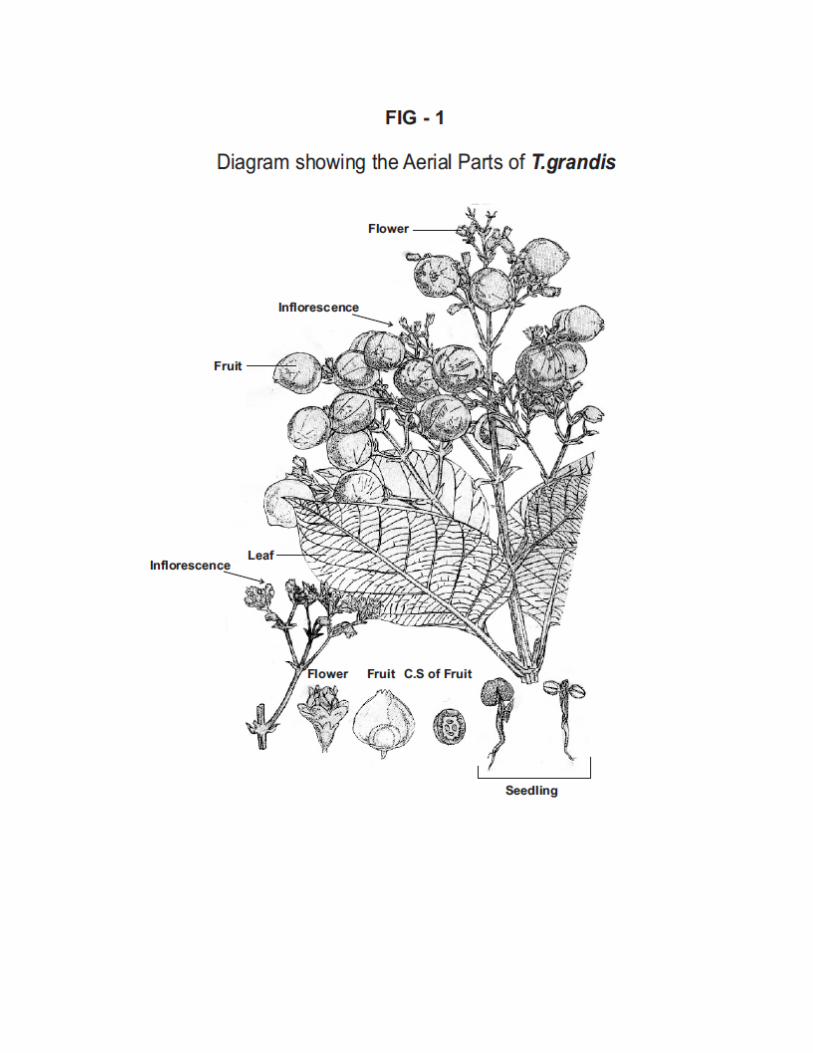


Calyx :stellately tomentose semi globose campanulate,3mm long,5-6
lobed,enlarged and becomingbladder like infruit,ovoid or
urceolate
Corolla :White,tube short,limb with 5-6short subequal spreading
Imbricate lobes
Stamens :Inserted near the base of the corolla,equal,exserted(5-6)
Anthers :ovate or oblong with distinct parallel cells.
Ovary :Fleshy,4-celled,ovule solitary in each cell,style linear,stigma
shortly 2-fid.
Fruits:(Plate-6, Fig 1, 1A)
Shape : DrupeHard,bony,irregularly globose,somewhat
pointed at the apex enclosed in the enlarged
calyx
Size :10-15mm in diameter
Colour :Stellate hairy,enveloped by light brown
Endocarp :Thick, bony,4-celled
Seeds:(Plate-7)
Seeds 1-3,rarely 4 in a fruit,marble-white,ovate,4-8mm long.
5.1.2 MICROSCOPY OF THE LEAF:
The leaf is dorsiventral with prominent midrib. Lamina is thick dorsiventral
differentiated.
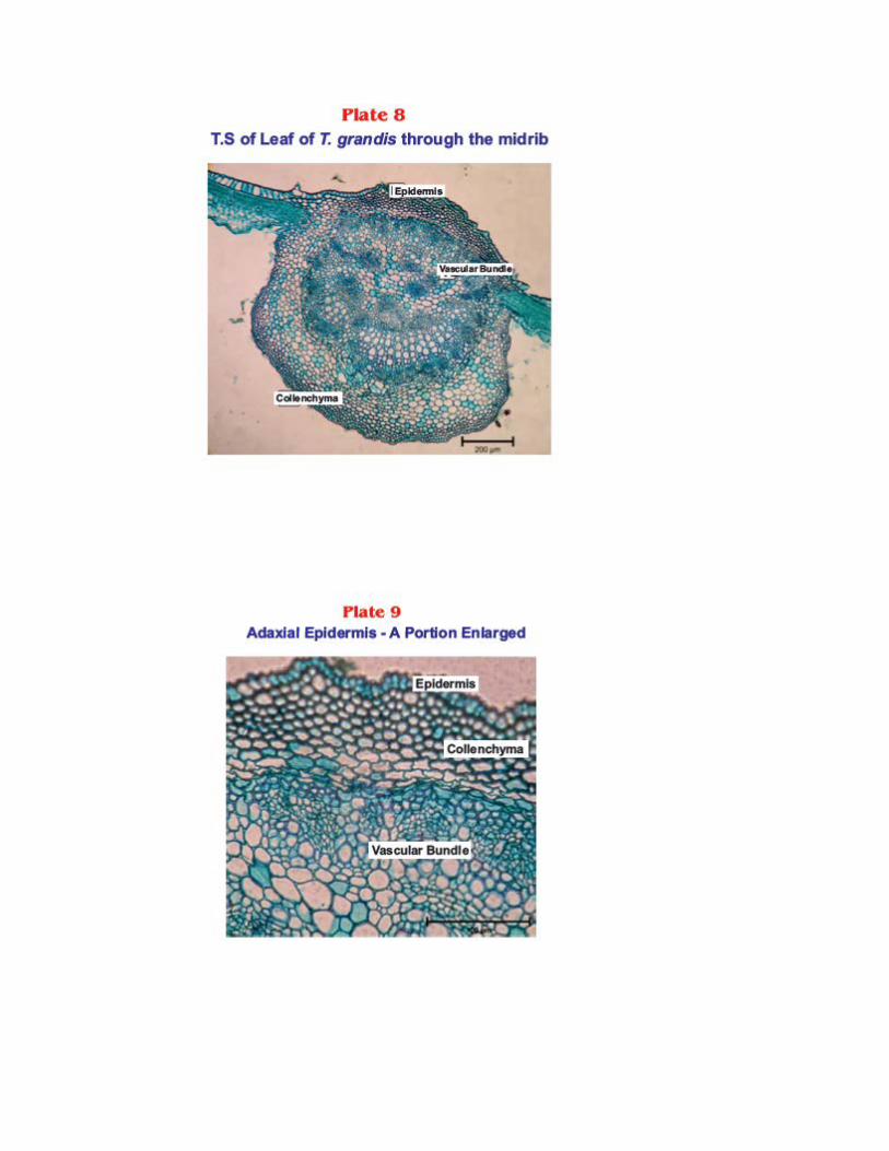

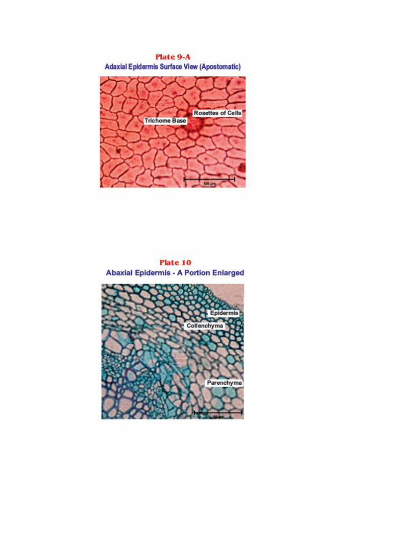
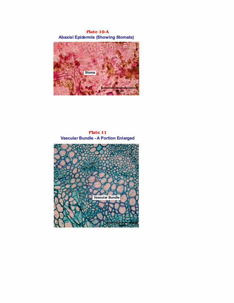


LEAF MIDRIB (Plate 8, Fig 3)
Shape : Plano convex
Adaxial side : Slightly raised round in shape,thick
Abaxial side : Semi circular broad and thick
Epidermis :
Adaxial epidermis:(Plate :9,9 A)
It is composed of larger cells and pentagonal in surface view with very slightly wavy
contour.Apostomatic.It is covered by thick cuticle.
Abaxial epidermis: (Plate :10)
It is made up of smaller cells with wavy outline. Silicified rosettes of cellscontain
cystolith like bodies are seen.
Stomata: (Plate : 10 A)
Deep sheatedranunculaceous stomata are present.
Hypodermis : (Plate : 9 )
Hypodermal region composed of 3-6 rows of collenchyma cells.
Vascular Bundles (Plate : 8,11)
Large,situated in the centre and occupies the major portion of the midrib.It contains closed
cylinder of xylem and surrounded by completely closed cylinder of phloem and fibres.

Ground Tissue : (Plate : 8)
It is made up of closely compactly packed parenchyma cells.Some cells contain cluster crystals
of calcium oxalate.
Lamina: (Plate : 12)
Dorsiventral. The adaxial epidermis composed of larger and transversely elongated
cells. Abaxial epidermal cells are rectangular and smaller in size. Mesophyll differentiated into
palisade and spongy parenchyma. Palisade is composed of single layer of closely packed
columnar parenchyma. Spongy mesophyll is composed of 2 or 3 layers of loosely arranged
parenchyma cells.
Venation Pattern : (Plate : 13)
Distinct vein islets are formed by the secondary and tertiary veins.Vein islets are small and
clearly seen;some of the vein terminations are forked.
Trichomes: (Plate :14)
It is numerous non-glandular hairs with cystolith in the cells surrounding the base.
T.S OF PETIOLE : (Plate : 15,16, Fig -4)
Shape:
More or less triangular with flat adaxial surface.
Epidermis:
Single layer made up of small rectangular cells covered by a striated cuticle.
Cortex:
It is divided into 2 zones with outer 6-8 rows of collenchyma cells and inner 10-15 rows of
closely arranged polyhydral parenchyma cells.
Pericyclic Fibres:
2-3 layers of fibres seen as ring.
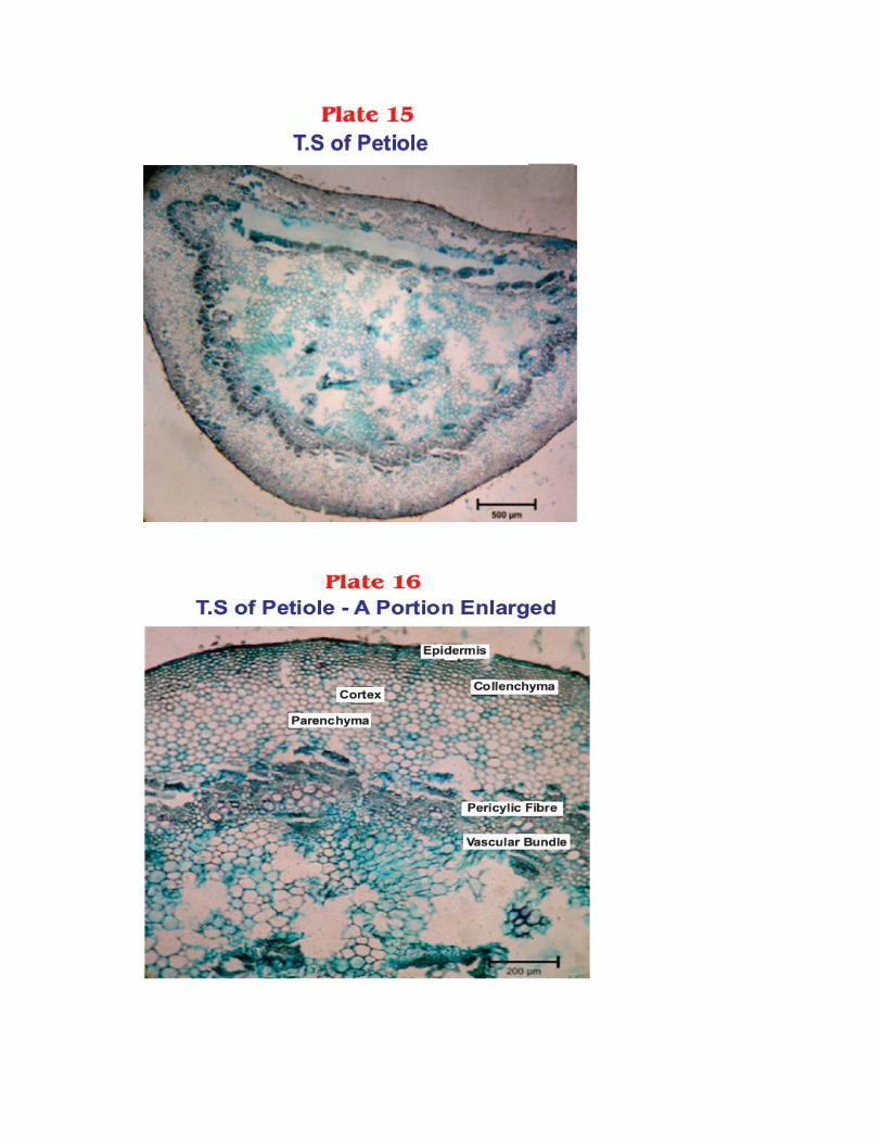


Vascular Bundle:
It contain closed cylinder of xylem and phloem.
Pith:
Central large pith region composed of compactly arranged parenchyma cells.
5.1.3 SEM study of leaf (Plate 17,18,19,20)
Scanning Electron Microscopy study of the leaves showed no diagnostic feature and
new kind of micro constituents not previously recognised and apparently simple struture
which maybe extremely complex were observed.Trichomes,vessels,epidermis and other area
studied in various magnifications.
5.1.4 POWDER MICROSCOPY (Fig -2) Organoleptic characters
1. Nature : Coarse
2. Colour : Green
3. Odour : Characteristic
4. Taste : Characteristic,mild bitter
5. Shaken with water : Froathing occurs
6. Pressed in between two filter paper : No oil mark on the paper
We have observed the following microscopical cell structures,
♦ Ranunculaceous stomata(Anomocytic)
♦ Parenchyma
♦ Xylem vessels
♦ Calcium oxalate crystals
♦ Phloem
♦ Fibres
♦ Collenchyma
♦ Uniseriate multi cellular trichomes

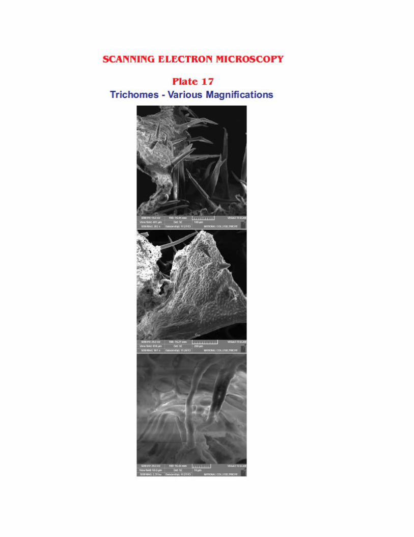


5.1.5 MICROSCOPIC SCHEDULES
As per the methods described in materials and methhods, microscopic schedules were
carried out and the results tabulated from the Tables 1- 4.
Table – 1
VEIN ISLET AND VEIN TERMINATION NUMBER OF T.grandisLEAVES
Observation number
Vein Islet number
Vein termination
number
1 26 9
2 27 7
3 30 6
4 32 5
5 31 8
6 28 9
7 34 8
8 29 6
9 33 7
10 28 6
Table – 2
STOMATAL NUMBER OF T.grandisLEAVES
Range Minimum Average Maximum
Vein islet number 26 29.8 34
Vein termination 5 7.1 9

Observation
number
Lower
epidermis
1 65
2 60
3 62
4 66
5 61
6 67
7 64
8 59
9 63
10 55
Range Minimum Average Maximum
Lower epidermis 55 62.2 67

Table – 3
STOMATAL INDEX OF T.grandisLEAVES
Observation
number
Lower epidermis
1 31
2 34
3 30
4 32
5 36
6 33
7 34
8 32
9 31
10 35
Range Minimum Average Maximum
Lower epidermis 30 32.8 36

Table – 4
PALISADE RATIO OF T.grandisLEAVES
5.1.6 PHYSICO CHEMICAL PARAMETERS
As per the methods described in materials and methods, physicochemical parameters
were carried and the results were as follows
Observation number Palisade
ratio
1 3
2 4
3 4
4 2
5 3
6 3
7 2
8 4
9 3
10 3
Minimum Average Maximum
2 3.125 4

Table – 5
ASH VALUE OF THE LEAVES OF T.grandis
Observation
Number Total Ash (%) Acid Insoluble Ash (%) Water soluble Ash (%)
1 6..39 0.86 -
2 6.73 1.44 -
3 5.64 0.84 -
4 6.28 1.41 -
5 5.91 0.99 -
6 6.58 - 4.23
7 6.62 - 4.46
8 5.77 - 3.84
9 6.58 - 4.17
10 6.32 - 3.99
Minimum 5.64 0.84 3.84
Average 6.28 1.10 4.13
Maximum 6.73 1.44 4.46
Table – 6
LOSS ON DRYING (LOD) FOR T.grandisLEAVES
Observation number LOD %w/w
1 0.63
2 1.02
3 1.13

Table – 7
EXTRACTIVE VALUES FOR T. GrandisLEAVES(INDIVIDUAL SOLVENTS)
Solvents Extractive value (%)
Petroleum ether 1.2
Ethanol 4.5
Water 5.1
Table – 8
EXTRACTIVE VALUESFOR T. GrandisLEAVES(SUCCESSIVE SOLVENTS)
Solvents Extractive value (%)
Petroleum ether 1.21
Ethyl acetate 4.3
Ethanol 4.4
Water 5.12
4 0.68
5 1.07
Material Minimum Average Maximum
Leaf
powder 0.63 0.90 1.13

5.2 PRELIMINARY PHYTOCHEMICAL SCREENING
5.2.1 Qualitative Phytochemical Test
Preliminary phytochemical screening of the powdered mature leaves were carried out
and the results are as follows (Table 9)
TEST FOR ALKALOIDS
Mayer’s test : No cream precipitate shows the absence of
alkaloids
Dragendorff’s test : Reddish brown precipitate shows the absence of
alkaloids
Hager’s test : No yellow precipitate shows the absence of
alkaloids
Wagner’s test : No reddish brown precipitate shows the absence of
alkaloids
Muroxide test : No appearance of purple colour shows the absence
of purine alkaloids
TEST FOR CARBOHYDRATES
Molish’s test : Appearance of purple colour shows the presence of
carbohydrates.
Fehling’s test : Formation of reddish brown precipitate shows the
presence of free reducing sugars.
Benedict’s test : Formation of reddish brown precipitate shows the
presence of free reducing sugars.

TEST FOR GLYCOSIDES
TES
T
FOR
PHY
TO
STE
ROL
S
Salkowski’s test : Appearance of red colour in lower layer shows the
Presence of sterol
Liebermann –
Burchard’s test
: Brown ring at the junction of two layers and green
colour in the upper layer shows the Presence of
sterols
TEST FOR SAPONINS Frothing occurs indicates the presence of Saponins
TEST FOR TANNINS
Ferric chloride test : Appearance of bluish black colour shows the
presence of tannins
Gold beater’s skin
: Appearance of brown colour shows the presence
of tannins
Keller killiani’s test
Borntrager’s test
Modified Borntrager’s test
:
:
:
No reddish brown colour ring at the junction shows
the absence of cardiac glycosides.
Appearance of pink colour in ammonical layer
shows the presence of anthraquinone glycosides
Appearance of pink colour in ammonical layer
shows the presence of anthraquinone glycosides
TEST FOR PROTEINS AND FREE AMINOACIDS
Millon’s test : Appearance of red colour on heating shows the

TE
ST
FO
R
FL
AV
ON
OIDS
Shinoda test : Appearance of purple colour shows the presence
of flavonoids
Alkaline reagent test : Appearance of yellow - orange colour shows the
presence of flavonoids
Acid test : Appearance of yellow – orange colour shows the
presence of flavonoids
Zinc hydrochloride
test
: Appearance of red colour shows the presence of
flavonoids
TEST FOR
VOLATILE OIL
: Volatile oil not obtained shows the absence of
volatile oil
TEST FOR FIXED OIL : No translucent greasy spot shows the absence of
fixed oil
presence of proteins
Biuret test : Appearance of violet colour shows the presence of
proteins
Ninhydrin test : Formation of violet colour shows the presence of
amino acids
TEST FOR
MUCILAGE
:
No appearance of reddish pink colour shows the
absence of mucilage
TEST FOR
TERPENOIDS
: Appearance of pink colour shows the presence of
terpenoids

Table – 9
PRELIMINARY PHYTOCHEMICAL SCREENING OF LEAVES OF T.grandis
S.NO TEST OBSERVATION
I. ALKALOIDS
Mayer’s reagent -
Dragondorff’s reagent -
Hager’s reagent -
Wagner’s reagent -
Test for purine Group(Muroxide test) -
II. CARBOHYDRATES
Molisch’s test +
Fehling’s test +
Benedict’s test +
III. GLYCOSIDES
Anthroquinone glycosides +
Borntrager’s test +
Modified Borndrager’s test +
Cardiac glycosides
Keller Killiani test -
Raymond test -
Legal test -
Cyanogenetic glycosides -
Coumarin glycosides -

IV. STEROLS
Salkowski test +
LibermanBurchard’s test +
V. SAPONINS +
VI. TANNINS
Ferric chloride +
Gold Beater’s skin test +
VII. PROTEINS AND FREE AMINO ACIDS
Millon’s test +
Biuret test +
Ninhydrin test +
VIII. MUCILAGE -
IX. TERPENOIDS +
X. FLAVONOIDS
Shinoda test +
Alkali test +
Acid test +
Zn/Hcl test +
XI. VOLATILE OIL -
XII. FIXED OIL -
5.2.2 FLUORESCENCE ANALYSIS OF POWDERED LEAF
The fluorescence analysis of the leaf powder ofT.grandis was studied. The results were
as follows (Table -10)

Table -10
FLUORESCENCE ANALYSIS
Reagent Observation
Powder as such Faint green
Powder + 50% Hydrochloric acid Yellowish Green
Powder + 50% Nitric acid Light brown
Powder + Petroleum ether Light orange
Powder + 50% Sulphuric acid Dark brown
Powder + 1N NaOH in water Dark green
Powder + 1N NaOH in methanol Dark Green
Powder +5% Ferric chloride solution Greenish brown
Powder + Picric acid Flourescence green
Powder + Chloroform Green
Powder + 5% Iodine solution Green
Powder + (HNO3 + NH3) Green
5.2.3 Estimation of flavonoid content of TGEAE Figure: 5 Calibration curve of quercetin for estimation of flavonoid content

Report:
Flavonoid content of TGEAE in terms of quercetin by aluminium chloride was found
to be 13.5µg/g.
5.2.4 Estimation of Total phenolic content of TGEAE
Figure :6
Calibration curve of gallic acid for estimation of total phenolic content
Report:
Total phenolic content of TGEAE in terms of gallicacid was found to be 22.2µg/g.
5.2.5 IDENTIFICATION OF INORGANIC MINERALS OF THE LEAVES OF T.grandis by Energy Dispersive X-ray Spectrometer (EDS)
Figure-7
Energy Dispersive X-Ray Spectrum For T.grandisLeaves
Calibration curve of gallic acid
y = 0.116x - 0.004R2 = 0.9998
-0.2
0
0.2
0.4
0.6
0.8
1
1.2
1.4
0 5 10 15
Conc. in mcg/mL
Abso
rban
ce Absorbance
Linear(Absorbance)

Estimation of the elements like O, Mg, P, S, Cl, K, Ca, Zn showed the following mg
weight percentage and atomic percentage.
Table-11 T. grandisLeaves Elements weight & Atomic Percentage
5.2.6. HPTLC PROFILE OF THE TGEAE &TGEE OF THE LEAVES Development of HPTLC fingerprint
About 2,6,8,10 µl of standard Apigenin ,4,8 µl of TGEAE,3 µl Quercetin,6µl of TGEE was applied as a band using CAMAG Linomat sample applicator on aluminium sheets pre-coated with silica gel 60 GF 254 HPTLC plates used as a stationary phase. The plates were developed in the mobile phase Toluene : Ethyl acetate: Formic acid : Methanol (3:6:1.6:0.4) to a distance of 8cm in CAMAG-twin trough glass chamber. The tracks were scanned using WIN CATS 1.43 software at 254nm. The fingerprint profiles were recorded and presented in Table-13 Plate size : HPTLC plates silica gel 60 F 254
(E.MERCK KGaA)
Sample solvent type : Methanol
Dosage speed : 150 nl/s
Number of tracks : 8
SI.NO ELEMENTS WEIGHT(%) ATOMIC(%)
1. OK 85.25 93.05
2. MgK 0.62 0.45
3. PK 0.97 0.55
4. SK 0.48 0.26
5. ClK 1.24 0.61
6. KK 9.24 4.13
7. CaK 2.19 0.96
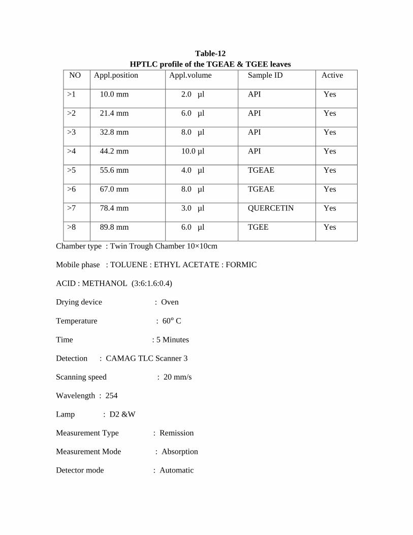
Table-12 HPTLC profile of the TGEAE & TGEE leaves
NO Appl.position Appl.volume Sample ID Active
>1 10.0 mm 2.0 µl API Yes
>2 21.4 mm 6.0 µl API Yes
>3 32.8 mm 8.0 µl API Yes
>4 44.2 mm 10.0 µl API Yes
>5 55.6 mm 4.0 µl TGEAE Yes
>6 67.0 mm 8.0 µl TGEAE Yes
>7 78.4 mm 3.0 µl QUERCETIN Yes
>8 89.8 mm 6.0 µl TGEE Yes
Chamber type : Twin Trough Chamber 10×10cm
Mobile phase : TOLUENE : ETHYL ACETATE : FORMIC
ACID : METHANOL (3:6:1.6:0.4)
Drying device : Oven
Temperature : 60ᵒ C
Time : 5 Minutes
Detection : CAMAG TLC Scanner 3
Scanning speed : 20 mm/s
Wavelength : 254
Lamp : D2 &W
Measurement Type : Remission
Measurement Mode : Absorption
Detector mode : Automatic

Figure-9 HPTLC graph of TGEAE & TGEE of the leaves
STANDARD : APIGENIN STANDARD : QUERCETIN
SAMPLE: TGEAE SAMPLE : TGEE
Table – 13 Rf values for TGEAE & TGEE leaves
SI NO
SAMPLES
Start Rf
Max Rf
End Rf
Area
Area %
1
Standard Apigenin
0.73
0.78
0.81
15860.2
100.00
2
Sample TGEAE
0.73
0.77
0.81
5696.2
100.00
3
Standard Quercetin
0.73
0.76
0.79
6814.8
100.00

4
Sample TGEE
0.75
0.79
0.81
662.3
100.00


5.3 .PHARMACOLOGICAL STUDIES
5.3.1.Acute toxicological study
EFFECT OF VARIOUS CONCENTRATIONS OF ETHYL ACETATE EXTRACT
FROM THE LEAVES OF T.grandisL ON Artemianauplii
Table-14
Various Concentrations Of TGEAE leaves on Artemianauplii
Concentration
(ppm)
Number of
Larvae
released
Number of
Larvae
dead after
24hrs
Mortality
(%)
Corrected (%)
Mortality using
Abbot’s
Formula
100
10 2 20
20 10 2 20
10 2 20
200
10 2 20
20 10 2 20
10 3 20
500
10 4 40
36.6 10 4 40
10 3 30
1000
10 4 40
43.3 10 5 50
10 4 40

2500
10 3 30
43.3 10 5 50
10 5 50
5000
10 5 50
50.0 10 5 50
10 5 50
6000
10 6 60
63.3 10 7 70
10 6 60
7500
10 7 70
66.6 10 7 70
10 6 60
Control
10 - -
- 10 - -
10 - -
5.3.2 In-vitro Anti-Malarial Activity of TGEAE of The Leaves
Anti-malarial activity of the ethyl acetate extract T.grandis against P.falciparum. (Plate-22)
After the incubation the percentage of parasitemia in the control well has
increased drastically after 96 hrs as compared to the parasite at 0 hrs.This increase shows the
normal growth of the parasite in the RBC’s.The percentage of increase of the parasite at 48
hrs,72 hrs and 96 hrs have been tabulated(Table-16).
In the standard wells, a 100% reduction was noticed during 48 hrs,72hrs and 96hrs
which indicates the action of the known anti-malarial drug chloroquine against the parasite.

In the test the percentage of reduction of the parasite at 48 hrs is 33%,72 hrs is 25%
and 96 hrs is 18%.This showed that ethanol extract of leaves ofT.grandisinduced a significant
decrease of P.falciparum proliferation.
This shows it is an inexpensive and affordable crude extract and can be evaluated
further for formulations.
Fig-10
Anti–malarial activity of TGEAE of the leaves


Table-15 PERCENTAGE OF INFECTED RBC’s
5.3.3.EFFECT OF TGEAE OF THE LEAVES ON ex-vivo PORCINE SKIN WOUND HEALING MODEL:
Wound healing effect was evaluated by ex-vivo porcine skin wound healing model
(PSWHM). Architecture of pig skin is closer to human (Bollen Peter JA et al, 1999, Laberet
al, 2002). Porcine model is an excellent tool for the evaluation of therapeutic agent meant for
wound healing (Sullivan TP et al, 2001).
Histopathological evaluation has showed all treated wounds were sound with no signs of
apoptosis, necrosis or bacterial contamination and no toxicity of the tested concentrations of
TGEAE of the leaves.(Plate ). Morphology of the wound margins, epidermis and dermis layer
were found to be normal. Epidermal cell migration in each side of the wound was measured
from the edge of the wound to the migration tip.
Measurement of epidermal migration distances from the wound showed statistically
significant dose dependent wound healing effect (Anova p<0.05).The efficacy of wound
healing promoting effect was expressed as normalised epidermal migration % mean SE against
PBS control groups.( Fig 10)
Sl No
Samples
0hrs
48 hrs
72 hrs
96 hrs
1 Control 32±1.3 47±1.5 55±1.3 65±0.9
2
Test
10 µg/ml
33±1.12
28±0.85
12±1.12
5±0.79
3 20µg/ml 33±0.96 20±1.12 2±1.3 NIL
4 30µg/ml 35±1.1 NIL NIL NIL


CHAPTER-6
DISCUSSION
The dissertation covers a study on widely available member of the family Verbenaceae
known botanically as TectonagrandisLinn.commonly called as ‘Teak’. The leaves of
T.grandis really do not have any match as a cheap natural and easily available plant.It is
traditionally known to be useful for the treatment of wide panel of diseases like tuberculosis,
various kinds of wounds especially burn wound, malaria, anaemia, leprosy, skin diseases
like pruritis, stomatitis, indolent ulcers, haemorrhages, menstrual disorder, bone joint disease.
Young leaves and fruits prescribed for women for fertility control and fumes of dry leaves to
kill guinea worms.It also possesses anti- ulcer and anti- cancer activity. Various scientific
investigation of the leaves showed anti-diabetic activity, anti-microbial activity, anti-oxidant,
analgesic, anti-inflammatory,anti-amoebic, anti-hypertensive and wound healing activity.
Bark is used as astringent, expectorant, anti-inflammatory, anthelmintic and for
constipation, leucoderma,headache, piles, bronchitis, hyperacidity, and dysentery,burning
sensation, diabetes, difficult in labour, leprosy, skin diseases, and indigestion.Researches
proved it is anti-fungal, anti-bacterial, uterine relaxant, anti-histaminic, anti-asthmatic, anti-
oxidant, analgesic and anti-inflammatory, anti-diabetic, hepatoprotective and cardiac activities.
Wood traditionally used as diuretic, stimulant, hepatic astringent, toothaches, sedative
to gravid uterus,leucoderma.It was reported that wood possesses anti-oxidant,anti-termite, anti-
hyperglycaemic, nephroprotective activities.
Flowers used for bronchitis, biliousness, urinary discharge, diuretic, depurative, anti-
inflammatory, leprosy, skin diseases, diabetes, congestion of liver.
Seed is used in poisoning.Seed oil in skin disease.Oil obtained from seeds and flowers
promote the growth of hair,eczema,ring worm, scabies. Scientific investigation of seed extract
proved hair growth activity, diuretic, anti-inflammatory activity.
Root is useful in anuria and retention of urine, anaemia. Anti-ulcer activity, anti-
inflammatory, anti-tussive, anti-oxidant, hypoglycemic, anti-pyretic activities have been
evaluated and proved.
The economic aspect of this crop evidently proved that as commercial crop. In fact the
revenue generated by this crop can be further magnified by many folds, if its medicinal
applications are scientifically explored well. By a well-coordinated effort, we can properly
exploit this plant. Therefore research on development of herbal products from this plant is
required to be initiated immediately for exploring the unique potential of this crop which
would also minimize the menacing wastage especially the leaves. It may be further envisaged
that the revenue generated by this plant would easily exceed that generated by any major crop
of the country even with a present level of traditional agro economic practices. Therefore a
well-coordinated effort by the farmers, traders, scientist, technologists, extension workers,
physician, administrators, and policy makers is required to be initiated to boost up the national
economy as well as the proper exploitation of this for proper therapeutic purpose. The review
of literature showed some lacuna exists in the pharmacological, phytochemical, and
pharmacological studies in the leaves of T. grandis.
PHARMACOGNOSTICAL STUDIES:
Morphological and micromorphological examination and characterization of medicinal
plants have always been accorded due credentials in the pharmacological studies. There was
no detailed pharmacognostical work has been carried out including botanical identity based on
micro morphology in this leaves of this plant.

The application of morphological studies in drug analysis is pertinent in the field of
crude drug authentication. It was studied for the leaf. Interpretation of the morphological
characteristics based on different parameters, for the plant organs give a guideline for the
diagnosis of the original plant and its adulterants.
Colour, size, shape, margin, texture, arrangement were observed and compared with
previous data.
Microscopic techniques help to magnify the fine structure of minute objects and there by
confirm the structural details of the plant drug. Though the microscopical evaluation cannot
provide complete profile, still it can offer supporting evidences which when combined with
other analytical parameters can be used to obtain full evidence for standardization and
evaluation of herbal drugs. Consideration must therefore be given to the types of cells and cell
inclusions and the manner in which they are distributed in different organ of the plants. The
habit and habitat and the various morphological characters of the various parts have been
studied after proper identification and authentication.
PHARMACOGNOSTICAL STUDIES (Plate : 8-14)
The leaves are dorsiventral elliptic or obovate or acuminate.The upper surface is rough,
glabrous, the lower clothed with dense stellate grey or tawny tomentum petiolate, opposite or
whorled, large entire,Length 30-60 cm ×20-30 cm green entire margin with cuneate base.
In microscopicaltransectional view plano convex with slightly raised round thick
adaxial side and semi-circular broad thick abaxialside.The upper epidermis apostomatic with
larger cells and pentagonal surface view with very slightly wavy walls but the abaxial
epidermis is made up of smaller cells with wavy outline with frequent silicified rosettes of
cells containing cystolith like body. It contains ranunculaceous stomata, collenchyma cells is

present in the hypodermal region of hump and lower epidermis. The lamina contain single
layer of palisade and spongy parenchyma. The vascular bundle situated in the centre of midrib
contains closed cylinder of xylem surrounded by phloem and fibres. The ground tissue is made
up of compactly packed parenchyma cells with frequent calcium oxalate crystals. Numerous
non-glandular hairs with cystolith were observed. Some distinct vein islets and vein
terminals(some are forked) were clearly seen.
The petiole more or less triangular with flat adaxial surface. The epidermis is covered with
striated cuticle. Cortex contain outer collenchyma cells and inner polyhydral parenchyma cells.
Vascular bundle contain xylem and phloem fibres. Central large pith region composed of
parenchyma is present.(Plate : 15,16)
Scanning electron microscopy (SEM) study showed no diagnostic features and new kind of
micro-constituents not previously recognized and apparently simple structure which may be
extremely complex were observed.(Plate : 17-20)
The plant drugs are generally used in the powdered form where the macro morphology
is generally destroyed, so the diagnosis of the plant through the microscopicalcharacter is
essential. The powdered crude drugs can be identified based on the presence or absence of
different cell types.
In powdered microscopy observed ranunculaceous stomata, wavy walled epidermal
cells, spiral annular vessels, phloem cells, sieve tube and companion cells, fibres, collenchyma
cells, parenchyma cells, calcium oxalate crystals.(Fig-2)
Quantitative microscopy includes certain measurements to distinguish some closely related
species which are not easily differentiated by general microscopy. The stomatal number is

the oldest technique but a simple method of diagnosis of fragmentary leaf parts. The stomatal
index is the percentage of stomata in relation to the epidermal cells. Both are very specific
criteria for the identification and characterization of leafy drugs. Vein islet and vein
terminationnumber are another simple technique for distinguishing fragmentary specimens at
specific levels. It is used as the distinguishing character for the leaf of the same species or
different one.
Palisade Ratio is another criterion for identification and evaluation of herbal drugs.
This value remains constant within a range for a given plant species and is of diagnostic value
in differentiating the species. This value does not alter based on geographical variation and
differs from species to species and that is why it is a very useful diagnostic feature for
characterization and identification of different plant species. (Table 1-4)
The ash content of the crude drug is generally taken to be the residue remaining after
incineration. If usually represents the inorganic salts naturally occurring in the drug and
adhering to it, but it may also involve the inorganic matter added for the purpose of
adulteration. There is a considerable difference within narrow limits in the case of individual
drug. Hence ash determination furnishes a basis for judging the identify and cleanliness of a
drug and gives information related to its adulteration with inorganic matter. The ash or residue
yielded by an organic chemical compound is a rule to measure the amount of inorganic matter,
which is present as impurity. In most cases the inorganic matter is present in small amounts
which are difficult to remove in the purification process and which are not objectionable if
only traces are present. Ash values are helpful in determining the quality and purity of the
crude drug in especially in powdered from. The acid insoluble ash is of more value to detect
the earthy matter adhering to the drug. In this way one can obtain evidence of the presence of

foreign matter, which likely to occur with root, rhizomes and also in pubescent leaves. The
water soluble ash is used to detect the presence of matter exhausted by water. Insufficient
drying favours spoilage by molds and bacteria and makes possible the enzymatic destruction
of active principles (Table -5).
Extractive values of crude drugs determine the amount of active constituents in a
given amount of medicinal plant material when exhausted with solvents. It is employed for
that material for which no chemical or biological assay method exist. As mentioned in
different official books [Anonymous., 1996, Anonymous.,2006,Horborne JB.,1973] the
determination of water-soluble and alcohol soluble extractive, is used as means of evaluating
crude drugs which are not readily estimated by other means. The extraction of any crude with
a particular solvent yields a solution containing different phytoconstituents. The composition
of these phytoconstituents in that particular solvent depends upon the nature of the drug and
solvent used. The use of single solvent can be the means of providing preliminary information
on the quality of a particular drug sample. The water soluble extractive values play an
important role for the evaluation of crude drugs. It can be used to indicate poor quality,
adulteration with any unwanted material or incorrect processing of the crude drug during the
drying, storage etc. The alcohol soluble extractive is also indicative for the same purpose as
water soluble extractive values (Table -7, 8).
Loss on drying at 105oC is determined as the presence of excess moisture is
conductive to the promotion of mold and bacterial growth, and subsequently to deterioration
and spoilage of the drug (Table -6)
THE PRELIMINARY PHYTOCHEMICAL STUDIES:

The preliminary phytochemical screening reveals the presence of carbohydrates,
proteins and amino acids,anthraquinoneglycosides, flavonoids, terpenoids, tannins, saponins,
sterols. Alkaloids,Volatile oil, fixed oil were found to be absent. (Table -9)
The reaction of drugs in powdered form in ordinary light and with filtered UV light is
of importance in several cases by the luminosity in UV light by fluorescent analysis. Many
flavonoids showed distinctive colours under UV light: Bright yellow (6-hydroxy flavanoids
and flavones and some chalcones), dark brown (most flavanol glycosides, dark may be
(isoflavones and flavonols). Hence this parameter can also be used as a diagnostic tool for the
standardization of herbal drugs for the detection of adulterants in crude drugs (Table-10)
(Horborne JB., 1973).
Determination of total flavonoid content was found to be 13.5µg/g in
TGEAE.Determination of total phenolic content was found to be 22.2µg/g in TGEAE.
Identification of inorganic minerals of the leaves of T.grandisbyenergy dispersive X-
ray spectrometer(EDS)showed the presence of
mineralsoxygen(85.25%),magnesium(0.62%),phosphorous(0.97%),sulphur(0.48%),chlorine(1.
24%),potassium(9.24%),calcium(2.19%)(Table-13). No special characters were observed in
SEM analysis.
It was reported that essential oils, phenolic compounds, iridoids, triterpenes were
isolated from the different member of the Verbenaceaefamily. In an investigation presence of
quercetin, apigenin, gallic acid, quercetrin, chlorogenic acid, diosmin, kaempferol, hesperidin
etc. in the ethylacetate extract of the leaves have been reported[Ghareeb MA et al.,2013]. In
the therapeutically important constituents apigenin, and other flavonoids may be responsible
for the various pharmacological activity of the leaf. In an attempt identify the presence of
apigenin and quercetin by HPTLC. The apigenin(5,7,4′ tri hydroxy flavone) and quercetin

were identified and quantified in our study. Apigenin was found in ethyl acetate extract of
T.grandis(TGEAE) (0.7%) and Quercetin was found in ethanol extract of T.grandis(TGEE)
(0.01%).(Plate-21, Fig-8,9,Table-12,13)
PHARMACOLOGICAL STUDIES: Acute Toxicological study:
In continuation of our efforts to verify the safety of TGEAE we performed Brine
shrimp lethality assay (BSLA) using free swimming hatched out Artemianauplii which based
on the ability to kill laboratory cultured brine shrimp . It was observed that 100% of mortality
above 7500ppm for TGEAE . LC50 for TGEAE were about 5000ppm and 10ppm respectively
in 24hrs.(Table-14)
As a part of larger effect to develop anti-malarial agent we performed P.falciparum growth
assay, a whole organism method allows all relevant blood stage target to be screened
simultaneously using 96 well microtitre plates.[Fidock DA et al.,2004,Diallo A et al.,2008]
After the incubation the percentage of parasitemia in the control well has increased
drastically after 96 hrs as compared to the parasite at 0 hrs. This increase shows the normal
growth of the parasite in the RBCs . The percentage of increase of the parasite at 48 hrs, 72 hrs
and 96 hrs have been tabulated.(Table-15)
The results were encouraging that is comparable to that of the standard drug
chloroquine. The results was statistically significant(p<0.05).
Therefore the above studies proved that the ethyl acetate extract of the leaves of
T.grandis is therapeutically effective agent in malaria without toxicity and its effectiveness
comparable to currently available therapeutic agent chloroquine.
In the standard well a 100% reduction was noticed during 48 hrs,72hrs and 96 hrs
which indicates the action of the known anti-malarial drug chloroquine against the parasite. In
the test percentage of reduction of the parasite at 48 hrs is, 72 hrs is,96hrs is (Plate-22). This
showed that ethyl acetate extract of T.grandis leaves induced a significant decrease
P.falciparum proliferation. This shows it is an in expensive and affordable crude extract and
can be evaluated further for formulations.From this investigation it is confirmed that T
.grandisleaves is a cheap, easily accessible, safe and additive to conventional treatment of
malaria.The activity may be due to the presence of polyphenols, betulinic acid,
apigenin.[Aradhana R et al.,2010,Ghareeb MA et al.,2013]
The result provide scientific background for the leaves used as anti-malarial in folk
medicine.
Limitations of study:
Some of the limitations include the use of organic extracts instead of aqueous extracts
as prepared in the traditional setting. This means some of the compounds which may have a
therapeutic effect in an aqueous extract may be absent in the test samples. This may have
excluded antimalarial compounds that are polar resulting in lower antimalarial activity.
Another limitation is the use of parasites at different growth stages. Plasmodium parasites
were unsynchronized at the time of the experiments.This means that if an extract had an effect
on a particular growth stage and not others, the antiplasmodial effect would be reduced.
Additionally, a limited number of replicates of antiplasmodial activity assays were done. This
may have introduced bias, as parasite growth patterns are variable depending on culture
conditions; hence using more replicates would have resulted in more representative outcomes
in the antiplasmodial assays. Finally, metabolic assays have to be carried out on only. Further
information on the mechanism of action of the plant extracts could have been obtained using
this Plasmodium strain.

It was reported that leaves of T.grandisused for wound healing in folk medicine. So we
have planned to provide scientific background of this ethnomedical claims. So the restoration
of damaged tissue as closely as possible to its normal state mainly studied. Our study confirms
the T.grandistraditional claim for wound healing activity. Epidermal migration was measured
(Nayak S, 2006).
Epidermal migration or keratinocyte migration distances from the edges of each wound
were measured, normalized with the PBS control group and expressed as mean%. The result
clearly showed TGEAE( 3%) promoted statistically significant wound healing effect is
comparable to the standard drug mupirocin.
Wound healing activity of ethyl acetate extract of T.grandis leaves may be due to its
flavonoids, triterpenoids constituents. Both of them known to have astringent property which
is responsible for wound contraction and increased rate of epithelialisation along with the
supportive anti-microbial activity (Nayak S., 2006).
Here we want to emphasise the traditional use of the leaves for the treatment of
diabetes and the several supportive scientific research of this claim as anti-diabetic and for
wound healing activity[Manoharan KP et al., 2007,Majumdar M et.al.,2007,Shukla N et
al.,2010, Aradhana R et al.,2010, SajjadulkarimMdet al.,2011,Pradeep G et al.,2012, Bangou
MJ et al.,2012, Khera N and Bhargava S.,2013, Yogesh Sharma et al.,2013].
The common complication of diabetic patient is wound as a adverse effect which are an
enormous burden on the health care system, both in terms of cost and intensity of care
required. Conclusively our study showed significant enhancement of wound repair and
therefore can be beneficially, safely used as auxiallary therapy in diabetic patient with foot
ulcers in addition to the other available treatment.

CHAPTER - 7
CONCLUSION AND RECOMMENDATION
The present investigation highlights the pharmacognostical, phytochemical and
potential,(a new hope in the fight against apicomplexanPlasmodiumfalciparum) more
effective antimalarial therapeutically comparable to the synthetic counterpart, without toxicity
of the ethyl acetate extract of the leaves of TectonagranidisLinn family Verbenaceae, a
widely, easily available plant commonly called as Teak plant. Ethno medical information
revealed that it was used in various ailments for long time all over the world. It is traditionally
known to be useful for the treatment of wide panel of diseases like tuberculosis, various kinds
of wounds especially burn wound, malaria, anaemia, leprosy, skin diseases like pruritis,
stomatitis, indolent ulcers, haemorrhages, menstrual disorder, bone joint disease. Young
leaves and fruits prescribed to women for fertility control and fumes of dry leaves to kill
guinea worms. It also possesses anti- ulcer and anti- cancer activity. Various scientific
investigations of the leaves showed anti-diabetic activity, anti-microbial activity, anti-oxidant,
analgesic, anti-inflammatory, anti-amoebic, anti-hypertensive and wound healing activity.
So we have also investigatedex vivo wound healing activity to provide scientific
grounds of its ethno medical claims and to support the previous reports. The tremendous
economic potentiality of this cash crop remains neglected by the scientists, technologists,
physician, traders, administrators, policy makers, farmers etc.
The morphological evaluation showed the adherence of general character to the family.
Detailed microscopical characters of the leaves showed the presence of usual leaf
anatomical characters.T.S through midrib showed planoconvexdorsivenrtal leaf with slight
raised in the centre hump, palisadecells, spongyparenchyma,few layesrs of collenchymas

below both the epidermis in thr midrib, vascular bundle, ground tissue with frequent calcium
oxalate crystals,non glandular multicellular uniseriatetrichomes, apostomatic upper epidermis
and lower epidermis with ranunculaceous stomata. Powder microscopy, microscopic
schedules (Vein islet and termination numbers, stomatal number and index, palisade ratio),
physicochemicalparameters(Ash values, extractive values, LOD) were evaluated and
presented.
T.S of petiole showed more or less triangular with flat adaxial surface. The epidermis
is covered with striated cuticle. Cortex contain outer collenchyma cells and inner polyhydral
parenchyma cells. Vascular bundle contain xylem and phloem fibres. Central large pith region
composed of parenchyma is present.
Scanning Electron Microscopy of midrib showed many folded appearance. No
diagnostic feature and new kind of microcostituents not previously recognised and apparently
simple structure which may be extremely complex was observed.
Preliminary phytochemical screening showed the presence of carbohydrates, proteins
and amino acids, anthroquinone glycosides, flavonoids, saponin, terpenoids, tannins and
phytosterols. Fixed oil, alkaloids, volatile oil were found to be absent.
Flavonoid content of TGEAE in terms of quercetin by aluminium chloride was found
to be 13.5µg/g. Total phenolic content of TGEAE in terms of gallic acid was found to be
22.2µg/g. Trace elements analysis by energy dispersive X ray spectrometer (EDS) showed Mg
(0.62), P (0.97), S (0.48),Cl (1.24), K(9.24),Ca (2.19% weight).The presence of apigeninwas
identified and quantified (0.7%) by HPTLC which is reported to possesses antimalarial
activity(quercetin 0.01% in TGEE) The 3R’s ethical principle (Reduction, Refinement,

and Replacement) was implemented that help to minimize harms to vertebrate animals used in
science.
Acute toxicity assessment using Artemianauplii bybrineshrimp lethality bioassay
(BSLA)showedLC50 of TGEAE of the leaves 5000ppm& found non- toxic to the animal.
This study has been a contribution to the assessment of possible therapeutic properties of
TGEAE of the leaves in an attempt to validate theirethnomedicinal uses in malaria. The
presence of apigenin and polyphenols which have anti-malaria properties. Together with their
antiplasmodial activities, it strongly supports theposition that the plant traditional usage is
rational. Presence of lupine type betulinic acid may also be responsible for the activity. The
antimalarial activity shown by this leaf also confirms the importance of using an
ethnopharmacological approach in screening plants as a source for potential antimalarials. Its
dual benefit is of scientifically proved antianaemic activity (Diallo A et al., 2008) of another
traditional use for anaemia which is a serious symptom in the malaria patient
The results were encouraging that it is comparable to that of the standard drug
chloroquine. This research provide a pathway to a newer drug lead or herbal formulation
Traditionally claimed wound healing activity was evaluated by using ex-vivo porcine
ear wound healing model (PEWHM). TGEAE of the leaves (3%) showed statistically
significant wound healing effect which is comparable to the standard drug mupirocin which is
the currently recommended drug for wounds and diabetic foot ulcer.
A previous reports confirm the antidiabetic activity of the leaves of T.grandis this drug
may be useful in diabetic foot ulcer as an additional benefit[Shukle N et al., 2010, Pradeep G
et al., 2012].
Further studies are needed for purification compounds in extract and to understand the
complete mechanism of wound healing activity of T.grandisleavesIt may be suggested for
treating various types of wounds in human beings. Development of wound healing agent from
a natural drug is cheaper, safer and effective.
We recommend further investigations in animal model and clinical trials to confirm this
potential therapeutic effect. These aspects remain to be studied.
Moreover the vast economic potentiality of this crop can be adequately exploited
and can create employment opportunity to an agricultural worker throughout the year.
Recommendation:
In conclusion this study shows the value of traditional knowledge in the fight against
apicomplexan parasite Plasmodium.It is possible to balance people’s cultural believes and
practices with the WHO objective of malaria elimination. The validation of plants used by the
indigenous populations as described will facilitate in the ongoing efforts to eliminate malaria
by providing treatment to the isolated rural populations that do not have access or to do not
believe in conventional medicine and rely mostly on ethnomedicine.
Further in vitro investigations should include antimalarial screening and mode of action
studies. Combination treatments based on use of plants with different mechanisms of action
should be evaluated as well as repeat treatments with the extracts as these may be
plasmodicidal and not just suppressive. In vivo studies in a small animal model of malaria
should also be conducted to investigate how liver metabolism affects the efficacy of the plant
treatments. These studies will also provide toxicological and pharmacological data to advise
on dosage regimens. It is foreseen that the sum of these studies on scientific validation of the

medicinal uses of such plants may eventually contribute to the integration of ethno-medicine
into mainstream malaria case management.
In addition our study showed significant enhancement of wound repair and therefore
can be beneficially, safely used as auxiallary therapy in diabetic patient with foot ulcers in
addition to the other available treatment along with its proved antidiabetic activity.

REFERENCES
1. Abrecht S, Harrington P, Iding H, Karpf M, Trussadi R, Wirz B, Zutter U. 2004, Chi-
mia, 58,621.
2. Agarwal SS, Paridhavi. 2007, ‘Herbal Drug Technology’, 1stedn Universities Press
(India) Private Ltd, Hyderabad.’
3. Agnihotri A, Singh V.2013,‘Effect of alcoholic extract of T.grandis heart wood
against oxidative stress and diabectic conditions’,Wor J PharmaPharma Sci,2(1)367-
378.
4. Aguinaldo, Alcia M, Ocampo, Osler Paul M, Bowden, Bruce F, Gray, Alexander I,
Waterman, Peter G.1993,‘Ananthraquinone –naphthoquinone pigment from the leaves
of T.grandis’, Phytochem (oxford)) 33(4) 933-935.
5. Ajay GO, Olowe JA , Ajuluchukwu JN.2011,‘T. grandis leaf ethanol extract in
renal artery occluded hypertensive rats’, Delicious planta, DOI:10.1055/s-0031-
12282980.
6. Anonymous.1996, Indian Pharmacopoeia’, Vol II, Ministry of Health and Family
Welfare, New Delhi, A-53: 54, 89.
7. Anonymous.1998,‘Quality control Methods for Medicinal Plant Materials’, WHO, 28-
35.
8. Anonymous.2001,‘ The Ayurvedic Pharmacopoeia of India’, Part I, Istedn, Vol 1,
Government of India, Ministry of Health and Family Welfare, Indian Systems Medicine
and Homeopathy, 140-145.
9. Anonymous.2002,‘Database on medicinal plants used in Ayurveda’,vol:5,295-303.
10. Anonymous.2004,‘The Wealth of India’,vol 5(R-Z)195-197.

11. Anonymous.2005,‘The Wealth of India.’ vol X( sp-w)136-150.
12. Anonymous.2006,‘TheAyurvedic Pharmacopoeia of India’, Part I, vol I, Ist edn,137-
143.
13. Aradhana R, Rao KNV, Davidbanji and R.K.Chaithanya.2010,‘Review on
T.grandisLinn : Chemistry and medicinal uses’. Herbal Tech Industry,6-9.
14. AsifMd .2011,‘In-vivo Analgesic and Anti-inflammatory effects of T.grandis stem
bark extracts’, Malaysian J Pharm Sci,9(1)1-11.
15. Bagalli RS .2011,‘Screening of anti-oxidant, anti-diabectic and hepatoprotective
activities of selected medicinal plants’,Ph .D thesis submitted, The Kle deemed
university Belgaum, Karnataka, India.
16. Bangou MJ, Zeba B, MillogoRasolodimby J, Coulidiaty Y.D and YougbareZiebrou
M.2012, ‘Anti-oxidant and Anti-bacterial activities of five verbenaceae species from
Burkina Faso’ Cur Res J Bio Sci, 4(6)665-672
17. Bera S, Chatterjee K, De D, Monjur K, Ghosh D.2011,‘Effect of hydro
methanolic(2.3) extract of the bark of T.grandis on the management of hyperglycemia
and oxidative stress in STZ induced diabetes in rats’,J Nat.Pharma,2(4)196-202.
18. Bisset NG.1989, J Ethno pharmacol, 25,1.
19. Bob Hofner.‘Energy Dispersive Spectroscopy on the SEM’A Primer:1,15.
20. Bollen peter JA, Hansen, Konerup A, Rasmussen, Helle J, Bollen PJA. 1999, ‘The
Laboratory Swine, The Laboratory animal pocket reference series’, CRC press, Boca
Roton.

21. Bolu GEK, Bagre I, Ouattara K and Djaman AJ.2011,‘Evaluation of the anti-
bacterial activity of 14 medicinal plants in Cote d lvoire’, Trop J pharm Res,10(3)335-
340.
22. Chang CC, Yang MH, Wen HM, Chern JC, 2002, ‘Estimation of total flavonoid
content in Propolisby two complementary colorimetric methods’, J Food Drug
Analysis, 10(3)178-182.
23. Chaudhri R.D,1999,‘Herbal Drug Industry ’Eastern publishers’, New Delhi,489-501.
24. Denis J-N, Greene AE .1988, J Am ChemSoc ,110,5917.
25. Diallo A, Gbeasssor M, Vovor, Eklu A, Gadegbeku K, Agbonan A, Abena A,
Adesouza C, Akpagana K.2008a,‘Effect of T.grandis on phenyl hydrazine induced
anaemia in rats’, Fitoterpia,79(5),332-336.
26. Estensen RD.1984, J ExpPathol, 1,71.
27. Fatima S, Farooqi AHA, Kumar R, Kumar TRS, Khanuja SPS.2001,‘Anti-bacterial
activity possessed by medicinal plants used in tooth powders’, J Med Aromatic plants
Sci, 22:187-189.
28. Ferreira JFS, Luthra DL ,Sasaki T, Heyerick A.2010,‘Flavonoids from Artemisia
annua L as anti-oxidants and their potential synergism with Artemisinin against
malaria and cancer’,Molecules,15,3135-3170.
29. Fidock DA, Rosenthal PJ, Croft SL, RetoBrun, Solomon Nwaker.2004,‘Anti-malarial
drug discovery efficacy models for compound screening’,Nat Rev Drug
Discovery,3,509-519.
30. Gaikwad SB, Krishna MG, Anerthe SJ.2011,‘ Anti-mitotic activity and Brine Shrimp
lethality test of T.grandisbark’, Res J Pharm Bio and ChemSci, 2(4)1014-1022.

31. Ghaisas M, Navghare V, Takawale A, Zope V, Tanwar M, Deshpande A. 2009, ‘Effect
of T.grandis on dexamethasone induced insulin resistance in mice’, J Ethno Pharma,
122:304-307.
32. Ghaisas M, Navghare V, Takawale AR, Zope VS, Tanwar MB, Phanse MA.2011, ‘T.
grandis prevent the cardiac dysfunction in alloxan induced diabetic rats’ J Nat
Remedies,11(2)132-142.
33. Ghaisas M, Navghare V, Takawale AR, Zope VS, Phanse MA, 2010,‘Anti-diabectic
and nephro protective effect of T.grandis in alloxan induced diabetes’, Ars
Pharm,51(4)195-206.
34. Ghareeb MA, Shoeb HA, FauzyMadkour HM, Abdel-Ghany L, Abdel-Motagaly M,
Mohamed Saad, 2014,‘Anti oxidant and cytotoxic activities of Tectonagrandis leaves’,
Int J PhytoPharmacol, 5(2),143-157.
35. Ghareeb MA, Shoeb HA, Madkour MF, Rafahy LA, Mohamed MA, Saad AM.2013,
‘Radical scavenging potential and cytotoxic activity of phenolic compound from
T.grandis Linn’, Global J Pharmacol, 7(4) 486-497.
36. Goel RK, Pathak NK, Biowas M, Pandey VB, Sanyal AK.1987,‘Effect of lapachol , a
naphthaquinone isolated from T.grandis on experimental peptic ulcer and gastric
secretion’, J Pharm Pharmacol, 39(2)138-140.
37. Goswami DV, Nirmal SA, Patil MJ, Dighe NS, Laware RB.2009,‘An over view of
T.grandis chemistry and pharmacological profile’,Pharmacog Rev,3(5)170-174.
38. Goswami DV, Sharma S, Modi A, Umesh B, Patil MJ.2010 b,‘Efffect of various
extracts of T.grandis bark on bronchitis’, Pharmacol online:1:816-820.

39. Goswami DV, Sonawane LL, Nirmal SA, Patil MJ.2010a,‘Evaluation of anti- asthmatic
activity of T.grandis bark’,Int J Pharm Sci and Res,1(1)10-16.
40. Gouthamchandra K, Mahmood R, Manjunatha H. 2010, ‘Free radical scavenging, anti-
oxidant enzymes and Wound healing activities of leaves extracts from
Clerodendruminfortunatum L.’, Environ Toxicol and Pharmacol, 30, 11-18.
41. Heywood VH.1971‘The Systemics Associations, “Scanning Electron Microscopy,
Systemics and evolutionary appplications”,Proceedings of an International
Symposium held at the Department of Botany’, University of Reading, Academic
Press, London, (4)1-16.
42. Holton RA, Biediger RJ, Boatman PD .1995,‘ Semisynthesis of taxol and taxotere’
43. Horborne JB.1973, ‘Phytochemical Analysis- A guide to modern techniques of plant
analysis’, Chapman & Hall, London.
44. Iyengar MA, Nayak SGK.1994,‘Pharmacognosy Lab Manual’, Manipal press,
Manipal, 78-87.
45. Jain Sk .2004,‘Credibility of traditional knowledge-the criterion of multi locational and
multi ethnic use’, Ind J Trad Knowledge,3(2)137-153.
46. Jangame CM, Burande MD.2013,‘Anti-inflammatory activity of methanolic and
petroleum ether extracts of T.grandis’, Res J pharma, Bio and ChemSci, 4(1)642-647.
47. Javbhave D, Varma S, Gagne N, Bonde V, Gite A, Bhosle D. 2010.‘Effect of
T.grandis seeds on hair growth activity of albino mice’,IntAyur Res,1(4)211-215.
48. Jayakumar M, Eyini M, Pannirselvam A.1987,‘Allelopathic effect of teak leaf extract
on the seeding of groundnut and corn’ Geobios ,(14)66-69.

49. Jaybhaye D, Varma S, Bonde V, Gite A.2010,‘Effect of T.grandis stem bark on
estradiol benzoate injected uterus of female albino wistar rats’, Asian J Pharma and
Clin Res,3(2)123-124.
50. Johansen DA, 1940, Plant Micro technique, McGraw Hill Book Company 27-93, 126-
154.
51. Jung M, Park M, Lee HC, Kang YH, Kang ES, Kim SK. 2006,‘Anti-diabetic agents
from medicinal plants’, Current Med Chem,13:1203-1218.
52. Kamat SV.2001,‘Folk medicines of Satter Dem Village’, Zoo’s print J.,16(3)450.
53. Kavshik A, Kumari M, Ghebre M.2011,‘A studies on anti-tussive effect of T.grandis
roots using a cough model induced by sulfur dioxide gas in guinea pigs’Int J Phyto
med3(2)
54. Kavshik A, Kumari M, Jyoti J, Mital M, Lal N, Agarwal AC. 2009, ‘Anti
inflammatory effect of T.grandis in experimental animal models’, Pharma Rev,
55. Khamule R, Naksupan N, Ounaroon A, Saelim N. 2012,‘ Skin wound healing
promoting effect of polysaccharide extracts from Tremellafuciformis and
Auriculariaauricula on the ex-vivo porcine skin wound healing model’, IPCBEE,
43(20), 93-98.
56. Khan RM, Miungwana SM.1999,‘ 5- hydroxylapachol:a cytotoxic agent from
T.grandis ’,Phyto chem,50,439-442.
57. Khaomek P, Ichino C, Ishyama A, Sekiguchi H, Namatama M, Ruangrungsi, Saifa E,
2008 ‘Invitro antimalarial activity of prenylated flavonoids from Erythrinafusca’ J Nat
Med,62, 217-220.

58. Khera N and Bhargava S.2013,‘Phytochemical and pharmacological evaluation of
T.grandisLinn’,’Inter J Pharm and Pharm Sci, 5(3)923-927.
59. Kiritikar Basu.1987,Indian Medicinal Plants,vol:3,1924-1926.
60. Kokate C.K, Purohit AP, Gokhale S.B.2005, ‘Pharmacognosy’ 32thedition,
NiraliPraksahan, 99, 100, 111, 112.
61. Kothale KV, Thoke BT, and Rothe SP.2012,‘Pharmacognostic investigation on some
members of family Verbenaceae’,Bionano frontier.,5(a) (2-II)
62. Krentz AJ, Bailey CJ .2005, Drugs, 65,385.
63. Krishnan KS.2006,‘The useful Plants of India’623.
64. Laber, Kathy.2002, Laboratory Animal Medicine, 2ndedn, Academic press, Newyork.
65. Lacret R, Rosa M Varela, Jose MG, Molinills, Nogueiras C, Francisco A Macias.
2011, ‘Anthratectone and naphthotectone, two quinines from bioactive extracts of
T.grandis’, J Chem Eco, DOI 10.1007/S 10886-011-0048-8.
66. Lacret R, Rosa M, Jose MG, Clara N, Franciscs A.2012, ‘Tectonoelins, new norlignans
from a bio-active extract of T.grandis’, J Phytol, DOI:10.1016/2012.03.008.
67. Lehane A and Saliba K.2008,‘Common dietary flavonoids inhibit the growth of the
intra erythrocyte malaria parasite’,BMC Res Notes,1,26.
68. Mabry TJ , Markham KR, Thomas MB. 1970,‘The systematic identification of
flavonoids’, Springer verlay, Newyork.
69. Mahesh S Krishna , Jeyakumaran Nair A.2010,‘Anti- bacterial cytotoxic and anti-
oxidant potential of different extracts from leaf, bark, wood of T.grandis’,Int J
PharmaSci Drug Res,2(2)155-158.

70. Mahesh S Krishna, Jayakumaran Nair A.2011,‘Anthraquinone from the leaves of
T.grandis :A detailed study on its antibacterial activity and other biological properties’,
Int J Phy Med,3,50-58.
71. Majumdar M, Nayeem N, Kamath JV, Asad M. 2007, ‘Evaluation of T.grandisleaves
for wound healing activity’, Pak J Pharm Sci, 20(2)120-124.
72. Mancera O, Ringold HJ, Djerassi C, Rosenkranz G, Sondheimer F .1953,J Am
ChemSoc75,1286.
73. Mancera O, Zaffaroni A, Rubin BA, Sondheimer F, Rosenkranz G, Djerassi C. 1952, J
Am ChemSoc ,74,3711.
74. Manoharan KP, Song FS, Benny, TKH, Yang D.2007, ‘Spectra assignment and
reference data tri terpenoids from Eugenia grandis structure elucidation by NMR
spectroscopy’, Mag Res Chem,45,279-281.
75. Michael AS, Thompson CG, Abramovitz M. 1956,‘Artemia Salina as a test Organism
for a bioassay’, Science, 123-464.
76. Molokanova E, Savchenko A, Kramer RH .2000, J Gen Physiol, 115,685.
77. Nayak S, Nalabothu P, Standiford S, Bhogadi V, Adogwa A. 2006, ‘Evaluation of
wound healing activity of Allamandacatharatica L. and Laurusnobilis L. extracts on
rats’, BMC Complementary and Alternative medicine, 6(12), 1-6.
78. Nayeem N and Karvekar MD.2011a,‘Anti-microbial and anti- oxidant properties of the
isolated compounds from the methonolic extract from the leaves of T.grandis’,J Basic
and Clinpharma, 2(4)163-165.

79. Nayeem N ,Karvekar MD.2011b,‘Stability studies and evaluation of the semi solid
dosage form of the rutin, quercetin, ellagicacid, gallic acid and sitosterol isolated from
the leaves of T.grandis for wound healing activity’, Arch App Sci Res,3(1)43-51.
80. Nayeem N and Karvekar MD.2012, ‘Effect of plant stages on analgesic and anti-
inflammatory activity of the leaves of T.grandis’,Euro J Exp Bio,2(2) 396-399.
81. Nayeem N, Karvekar MD. 2010 c ,‘Analgesic and anti-inflammatory activity of the
methanolic extract of the frontal leaves of T.grandis’,Int J
Pharmacol,8(1):1055580/941.
82. Nayeem N, Karvekar MD. 2010a,‘Comparative phytochemical and pharmacological
screening of the methonolic extracts of the frontal and mature leaves T.grandis’, Int J
Pharm Bio Sci,1(3).
83. Nayeem N, Karvekar MD. 2010b‘ Isolation of phenolic compound from the methonolic
extract of T.grandis’, Res J PharmaBiolChemSci, 1(2)221-225.
84. Ni Putu A, DrianiAstiti and DewaNgurah Suprapta.2012,‘Anti-fungal activity of
T.grandis leaf extract against Arthriniumphaeospermum(corda)M.E.Ellis,the cause of
wood decay on Albiziafalcataria (L)fosberg’,J IntSoci South Asian Agri Sci,18(1)62-
69.
85. O’Brien TP, Feder N, MC Cull M.E.1964,‘Polychromatic staining of plant cell walls
by Toluidine Blue-O’,Protoplasma,59,364-373.
86. Palaniappan P, Pandian M, Natarajan, Pitchairaman. 2012, ‘Ethnomedical wisdom of
Alagar hills in Esternghats, Tamil Nadu, India’ ,Int J Bio Res., 6,28-34.
87. Pandey BL, Goel RK, Pathak NKR, Biswas M, Das PK.1982‘Effect of T.grandison
experimental ulcers and gastric secretion’,Ind J Med Res,76,89-94.

88. Phalphale SG, Gawari A, Biyani KR, Shete RV, KoreKJ,ChawdhariSR,Magar. S
.2012,‘Evaluation of diurectic activity of T.grandis in rats’Wor J PharmaPharma
Sci,2(1)245-252.
89. Pooja, Chandra Samantha K, Kokra SL, Sharma P, Sharma V.2010,‘Free radical
scavenging activity of T.grandisroots’,Int J Pharm Sci Res,1(12)159-163.
90. Pooja, Sharma V, Samantha KC.2011,‘Hypoglycemic activity of methanolic extract of
T.grandis root in alloxan induced diabetic rats’, J App Pharma Sci,1(4)106-109.
91. Pradeep G, Naresh A, Jeyarami Reddy A, Nagarjuna Reddy G, Narayana TV,
Ramanarayana Reddy V.2012,‘Anti- microbial and anti-diabectic activity of T.grandis
and prosopischilensis extract against alloxan induced diabectic rats’,Int J Adv Pharm
Res,3(6)959-967.
92. Premalatha and Rajgopal. 2005, ‘Cancer an ayurvedic perspective’, Pharmacol Res.,
51,19-30.
93. Priyanka S, Pooja, Samantha KC, Rathore KS.2011,‘Antipyretic activity of methanolic
extract of root of T.grandis on albino rats’, J Pharmacol and Toxicol,28-33.
94. Purushotham KG, Arun P, JohnsyJeyarani J, VasanthakumariR,Sankar L, Raviprakash
Reddy B.2010a,‘Synergistic invitro anti-bacterial activity of T.grandis leaves with
tetracycline’,Int J Pharm Tech Res,2(1)519-523.
95. Purushotham KG, Arun P, JohnsyJeyarani J, Vasnthakumari R and Chamundeeswari
D.2010b,‘In vitro anti-bacterial and anti-fungal assay of T.grandis,’Res J
PharmacogPhytochem, 2(1)1-9.
96. Purvis MJ, Collier DC, Wallis D.1966, ‘Laboratory Techniques in Botany, 2ndedn,
Butter worth & Co Ltd, London,82 -169.

97. Qudhia P. Medicinal herbs of Chattisgarh, ‘India having less known traditional uses ’,
http://www.botanical.com.
98. Rafael YOM, Arruda MSP, Arruda AC, Santos SL, Muller AH, Guilton GMSP,
Santos AS, Terezo E.2006,‘Naphthoquinones and anthraquinones from the stem of a
specimen reforestation of T.grandis’, Brazil J pharmacog, 16(3)392-396.
99. RahmatullahMd, Jahan R, SafulAzam FM, Hossan S, Molik MAH, Rahman T.2011,
‘Folk medicinal uses of verbinaceae family plants in Bangladesh’, Afr J Trad,Comp
and Alter Med,8(5)53-65.
100. Rajan S, Baburaj DS, Sethuraman M, Parimala S.2001,‘Stem and stem bark used
medicinally by the tribalsIrulas and Paniyas of Nilgiri District, Tamil Nadu,1(1)49-54.
101. Rajaram K.2013,‘Anti-oxidant and anti-diabetic activity of T.grandis in alloxan
induced albino rats’, Asian J Pharm Clin Res 6(3)174-177.
102. Robards,1970, ‘Electron micrcoscopy and plant ultra structure,’ McGraw Hill, London,
14-15, 36-59.
103. Rudi, Irshad, Abdul H.P.S, Naif A and Hermawan.2012,‘Evaluation of anti-termitic
activity of different extracts obtained from Indonesian teak wood’,Bio Reso,7(2)1452-
1461.
104. SajjadulKarimMd, MahfluzurRahmanMd, SadiaBintashahid, IshitaMalek,
AtiqurRahmanMd, SharminJahan, FarhanaIsratJahan, Mohammed Rahmatullah.2011,
‘Medicinal plants used by the folk medicinal practioners of Bangladesh: a randomized
survey in a village of Narayangan district’, AmeEur J Sustain Agric.,5(4)405-414.
105. Samuelsson G.2004,‘ Drugs of Natural Origin’, 5th edn,Apotekarsocieteten, Stock-
holm
106. Sass JE.1940,‘Elements of botanical microtechnique’,McGraw Hill Book Co,
NewYork, 222.
107. Sharma PV, Shaka RC.1986,’Text book of dravyaguna’,791-793.
108. Shiddamallya N, Azra Y, Gopakumar K. 2008, ‘Hundred common forest medicinal
plants of Karnataka in primary health care, Ind J Trad Knowledge,9(1)90-95.
109. Shukla N Kumar M, Akansha Ahmad G, Rahuja N, Singh AB, Srivastava AK,
Rajendran SM, Mourya R.2010 ‘Tectone ,a new anti-hyperglycemic anthrax quinone
from T.grandisleaves’, Nat prod commun,5(3)427-430.
110. Siddique MA, Mujeeb M, Najim AK, Akram M, 2010, ‘Evaluation of antioxidant
activity, quantitative estimation of phenols and flavonoids in different parts of
Aeglemarmelos’, Afr J Plant Sci, 4(1), 1-5.
111. Singleton VL, Orthofer R, Raventos RML. 1999, ‘Analysis of total phenols and other
oxidation substrates and anti-oxidants by means of FolinCiocalteu reagent’,
MethoEnzymol, 299, 152-178.
112. Sleet RB and Brendel K,1983, ‘Improved Methods for harvesting and counting
synchronous populations of Artemianauplil for use in developmental toxicology’,
Ecotoxicol Environ safety, 7, 435-446.
113. Sneader W .1996,‘ Drug Prototypes and their Exploitation’, Wiley, Chichester, UK
114. Sneader W. 2005,‘ Drug Discovery: a History’, Wiley, Chichester, UK
115. Srivastava SR.2013,‘Anti-fungal a activity screening and HPLC analysis of crude
extract from T.grandis ’, Int J Ayur Med,5(2).
116. Sullivan TP, Eaglestein WH, Davis SC, Martz P, 2001, ‘The pig as a model for wound
healing’, Wound Repair Regen, 9(2), 66-76.

117. Sumthong P, Damveld RA, Choi YH, Arentshorst M, Ram,VandenHondel C.A.M.J.J,
Verpoorte R.2006,‘Activity of quinones from teak on fungal cell wall
stress’,PlantaMedica, 72,943-944.
118. Tiwari B.K, Mishra R.R.1983, ‘Dry weight loss and changes in chemical composition
of Pine (Pinuskesiyaroyle) Teak (Tectonagrandis L) leaves during processing in a
fresh water lake’.Dr.W.Junk Publishers,98,249-256.
119. Traore Keita F, Gasquet M, Di-Giorgia C, Ollivier E, Dalmas F, KeitaA, Doumbo O,
Balansard G, Timon David P,2000,‘Anti-malarial activity of four plants used in
traditional medicine in Mali’,PhytoTher Res,14(1)45-47.
120. Upadhaya VK, Udupa AL, Udupa SL. 2000,‘Abstracts of research papers presented at
the international congress on frontiers in pharmacology and therapeutics in
21stcentury,New Delhi’, Ind J Pharmacol,32:132-175.
121. Usha Devi C and Pillai CR. ‘National Institute of Communicable Disease and Malaria
Research Centre’, Department of Biochemistry, New Delhi.
122. Vanhaecke P, Persoone G, Claus C, Sorgeloos P, 1981,‘ Proposal for a short-term
toxicity test with Artemianauplil, Ecotoxicol Environ safety’, 5, 382-387.
123. Varma SB and Jaybhaye DL.2010, ‘Antihyper glycemic activity of T.grandis bark
extract on alloxan induced diabetes in rats, Int J Ayur Res,1(3),163-166.
124. Wagner H, Bladt XS, Gain Z, Suie EM. 1996,‘Plant Analysis, Springer Verlag,
Berlin, Germany, 360.
125. Wallis TE.1953, ‘Practical Pharmacognosy’,6thedn, J& A Churchill Ltd, London,170-
175.

126. Wallis TE.1965, Analytical Microscopy, 3rdedn, J&A Churchill Ltd, London, 180, 183,
184.
127. WHO Geneva, 1998,‘ Health communication and Public relations, WHO press
Releases,Fact sheetNo.94, (WHO Home page http://www.who.ch/)
128. Yarnell, A .2005, ChemEng News, 83,22.
129. Yogesh Sharma, Jeyabalan G, Raman Deepsingh, Alok semwal.2013,‘Current aspects
of wound healing agents from medicinal plants’,J Med Plant Stud.,
1(3)1-11.