DRUG DELIVERY VIA THE BUCCAL PATCH A NOVEL … · · 2014-03-11 e 7 Vol 3, Issue 5, 2013. Tarun...
18
www.iajpr.com Page3466 Indo American Journal of Pharmaceutical Research, 2013 ISSN NO: 2231-6876 Journal home page: http://www.iajpr.com/index.php/en/ INDO AMERICAN JOURNAL OF PHARMACEUTICAL RESEARCH DRUG DELIVERY VIA THE BUCCAL PATCH – A NOVEL APROACH Pokhariyal Tarun* 1 , Singh Vikas Kumar 1 , Tiwari Ajay Kumar 1 1 Department of Pharmaceutics Jaipur National University, Jaipur Rajasthan Corresponding author Tarun Pokhariyal M. Pharm. (Pharmaceutics), School of Pharmaceutical Sciences, Jaipur National University, Jaipur Copy right © 2013 This is an Open Access article distributed under the terms of the Indo American journal of Pharmaceutical Research, which permits unrestricted use, distribution, and reproduction in any medium, provided the original work is properly cited. ARTICLE INFO ABSTRACT Article history Received 27/04/2013 Available online 15/05/2013 Keywords: Buccal Patch, Mucoadhesion, Polymers, Evaluation of Patch. The successful delivery of drugs across the oral mucosa represents a continuing challenge, as well as a great opportunity. Buccal delivery has progressed far beyond the use of traditional dosage forms with novel approaches emerging continuously. Buccal drug delivery system in which drug enters directly in systemic circulation thereby by passing the first pass effect. Contact with digestive food of gastrointestinal tract is avoided which might be unsuitable for stability of many drugs.. Moreover it shows better stability, patient compliance; uniform and sustained drug release and above all easy and cheap methods of preparation which can be done with various commonly available biocompatible polymers. The buccal route has been researched for a wide variety of drugs like miconazole nitrate, Aceclofenac, Cyproheptadine Hydrochloride and various different categories and has gained significant attention and momentum since it offers remarkable advantages. Over past few decades, buccal route for systemic drug delivery using mucoadhesive polymers to significantly improve the performance of many drugs has been of profound interest. Many ways of research work may be carried out to develop more this formulation for delivery of drug. Please cite this article in press as Tarun Pokhariyal et.al. Drug Delivery via the Buccal Patch – A Novel Approach. Indo American Journal of Pharm Research.2013:3(5).
Transcript of DRUG DELIVERY VIA THE BUCCAL PATCH A NOVEL … · · 2014-03-11 e 7 Vol 3, Issue 5, 2013. Tarun...
www.iajpr.com
Pag
e34
66
Indo American Journal of Pharmaceutical Research, 2013 ISSN NO: 2231-6876
Journal home page:
http://www.iajpr.com/index.php/en/
INDO AMERICAN
JOURNAL OF
PHARMACEUTICAL
RESEARCH
DRUG DELIVERY VIA THE BUCCAL PATCH – A NOVEL APROACH
Pokhariyal Tarun*1, Singh Vikas Kumar
1, Tiwari Ajay Kumar
1 1Department of Pharmaceutics Jaipur National University, Jaipur Rajasthan
Corresponding author
Tarun Pokhariyal
M. Pharm. (Pharmaceutics),
School of Pharmaceutical Sciences, Jaipur National University, Jaipur
Copy right © 2013 This is an Open Access article distributed under the terms of the Indo American journal of Pharmaceutical
Research, which permits unrestricted use, distribution, and reproduction in any medium, provided the original work is properly
cited.
ARTICLE INFO ABSTRACT
Article history Received 27/04/2013
Available online
15/05/2013
Keywords: Buccal Patch,
Mucoadhesion, Polymers,
Evaluation of Patch.
The successful delivery of drugs across the oral mucosa represents a continuing challenge, as
well as a great opportunity. Buccal delivery has progressed far beyond the use of traditional
dosage forms with novel approaches emerging continuously. Buccal drug delivery system in
which drug enters directly in systemic circulation thereby by passing the first pass effect.
Contact with digestive food of gastrointestinal tract is avoided which might be unsuitable for
stability of many drugs.. Moreover it shows better stability, patient compliance; uniform and
sustained drug release and above all easy and cheap methods of preparation which can be
done with various commonly available biocompatible polymers. The buccal route has been
researched for a wide variety of drugs like miconazole nitrate, Aceclofenac, Cyproheptadine
Hydrochloride and various different categories and has gained significant attention and
momentum since it offers remarkable advantages. Over past few decades, buccal route for
systemic drug delivery using mucoadhesive polymers to significantly improve the
performance of many drugs has been of profound interest. Many ways of research work may
be carried out to develop more this formulation for delivery of drug.
Please cite this article in press as Tarun Pokhariyal et.al. Drug Delivery via the Buccal Patch – A Novel Approach. Indo American
Journal of Pharm Research.2013:3(5).
www.iajpr.com
Pag
e34
67
Vol 3, Issue 5, 2013. Tarun Pokhariyal et al. ISSN NO: 2231-6876
INTRODUCTION
Amongst the various routes of administration tried so far in the novel drug delivery systems, localized
drug delivery to tissues of the oral cavity has been investigated for the treatment of periodontal disease,
bacterial and fungal infection. Over the decades mucoadhesion has become popular for its potential to optimize
localized drug delivery, by retaining a dosage form at the site of action (e.g. within the gastrointestinal tract) or
systemic delivery by retaining the formulation in intimate contact with the absorption site (e.g. buccal cavity).
Well defined bioadhesion is the ability of a material (synthetic or biological) to adhere to a biological tissue for
an extended period of time. The biological surface can be epithelial tissue or it can be the mucus coat on the
surface of a tissue. If adhesion is to a mucous coat, the phenomenon is referred to as a mucoadhesion. The use
of mucoadhesive polymers in buccal drug delivery has a greater application. Various mucoadhesive devices,
including tablets, films, patches, disks, strips, ointments and gels, have recently been developed. However,
buccal patch offer greater flexibility and comfort than the other devices. In addition, a patch can circumvent the
problem of the relatively short residence time of oral gels on mucosa, since the gels are easily washed away by
saliva. Buccal route of drug delivery provides the direct access to the systemic circulation through the jugular
vein bypassing the first pass hepatic metabolism leading to high bioavailability. Other advantages such as
excellent accessibility, low enzymatic activity, suitability for drugs or that mildly and reversibly damage or
irritate the mucosa, painless administration, easy withdrawal, facility to include permeation enhancer/ enzyme
inhibitor or pH modifier in the formulation, versatility in designing as multidirectional or unidirectional release
system for local or systemic action.1,2
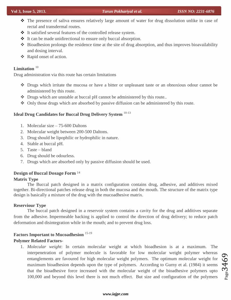
Components or structural features of oral cavity 2,3
Oral cavity is that area of mouth delineated by the lips, cheeks, hard palate, soft palate and floor of
mouth. The oral cavity consists of two regions. Outer oral vestibule, which is bounded by cheeks, lips, teeth and
gingival (gums).Oral cavity proper, which extends from teeth and gums back to the fauces (which lead to
pharynx) with the roof comprising the hard and soft palate.
The tongue projects from the floor of the cavity.
Buccal Drug Delivery
Difficulties associated with parenteral delivery and poor oral availability provided the impetus for
exploring alternative routes for the delivery of such drugs. These include routes such as pulmonary, ocular,
nasal, rectal, buccal, sublingual, vaginal, and transdermal. Substantial efforts have recently been focused on
placing a drug or drug delivery system in a particular region of the body for extended periods of time. The
mucosal layer lines a number of regions of the body including the oral cavity, gastro intestinal tract, the
urogenital tract, the airways, the ear, nose and eye. Hence the mucoadhesive drug delivery system can be

www.iajpr.com
Pag
e34
68
Vol 3, Issue 5, 2013. Tarun Pokhariyal et al. ISSN NO: 2231-6876
classified according to its potential site of applications. The buccal region of oral cavity is an attractive site for
the delivery of drugs owing to the ease of the administration. Buccal drug delivery involves the administration
of desired drug through the buccal mucosal membrane lining of the oral cavity. This route is useful for mucosal
(local effect) and transmucosal (systemic effect) drug administration. In the first case, the aim is to achieve a
site-specific release of the drug on the mucosa, whereas the second case involves drug absorption through the
mucosal barrier to reach the systemic circulation. Based on current understanding of biochemical and
physiological aspects of absorption and metabolism of many biotechnologically produced drugs, they cannot be
delivered effectively through the conventional oral route. Because after oral administration many drugs are
subjected to pre-systemic clearance extensive in liver, which often leads to a lack of significant correlation
between membrane permeability, absorption, and bioavailability. Direct access to the systemic circulation
through the external jugular vein by pass the drugs from the hepatic first pass metabolism which may lead to
higher bioavailability.4,5
Advantages of Buccal Drug Delivery System ,5-9
Excellent accessibility
Presence of smooth muscle and relatively immobile mucosa, hence suitable for administration of
retentive dosage forms
Direct access to the systemic circulation through the internal jugular vein bypasses drugs from the
hepatic first pass metabolism leading to high bioavailability
Low enzymatic activity
Suitability for drugs or excipients that mildly and reversibly damages or irritates the mucosa
Painless administration
Facility to include permeation enhancer/enzyme inhibitor or pH modifier in the formulation
Versatility in designing as multidirectional or unidirectional release systems for local or systemic actions
etc.
Oral mucosal drug delivery systems are easy and painless to administer and well accepted by the patient.
Precise dosage form localization is possible and there is ability to terminate delivery when required.
Flexibility in physical state, shape, size and surface.
For patient suffering with nausea or vomiting or in the state of unconsciousness, with an upper
gastrointestinal tract disease or surgery which affects oral drug absorption, the oral cavity a useful site
for drug delivery for upper symptoms.
Maximized absorption rate due to intimate contact with the absorbing membrane and decreased
diffusion barriers.
Excellent route for the systemic delivery of drug with high first pass metabolism, thereby offering a
greater bioavailability.
A significant reduction in dose can be achieved, thereby reducing dose dependent side effects.
Drugs which are unstable in the acidic environment of the stomach or are destroyed by the enzymatic or
alkaline environment of the intestines can be administered by this route.
It offers a passive system for drug absorption and does not require any activation.
It allows for the local modification of tissue permeability, inhibition of protease activity or reduction in
immunogenic response. Thus, selective use of therapeutic agents like peptides, proteins and ionized
species can be achieved.
The oral mucosa lacks prominent mucus secreting goblets cells and therefore there is no problem of
diffusion limited mucus buildup beneath the applied dosage form.
www.iajpr.com
Pag
e34
69
Vol 3, Issue 5, 2013. Tarun Pokhariyal et al. ISSN NO: 2231-6876
The presence of saliva ensures relatively large amount of water for drug dissolution unlike in case of
rectal and transdermal routes.
It satisfied several features of the controlled release system.
It can be made unidirectional to ensure only buccal absorption.
Bioadhesion prolongs the residence time at the site of drug absorption, and thus improves bioavailability
and dosing interval.
Rapid onset of action.
Limitation 10
Drug administration via this route has certain limitations
Drugs which irritate the mucosa or have a bitter or unpleasant taste or an obnoxious odour cannot be
administered by this route.
Drugs which are unstable at buccal pH cannot be administered by this route..
Only those drugs which are absorbed by passive diffusion can be administered by this route.
Ideal Drug Candidates for Buccal Drug Delivery System 10-13
1. Molecular size – 75-600 Daltons
2. Molecular weight between 200-500 Daltons.
3. Drug should be lipophilic or hydrophilic in nature.
4. Stable at buccal pH.
5. Taste – bland
6. Drug should be odourless.
7. Drugs which are absorbed only by passive diffusion should be used.
Design of Buccal Dosage Form 14
Matrix Type
The Buccal patch designed in a matrix configuration contains drug, adhesive, and additives mixed
together. Bi-directional patches release drug in both the mucosa and the mouth. The structure of the matrix type
design is basically a mixture of the drug with the mucoadhesive matrix.
Reserviour Type
The buccal patch designed in a reservoir system contains a cavity for the drug and additives separate
from the adhesive. Impermeable backing is applied to control the direction of drug delivery; to reduce patch
deformation and disintegration while in the mouth; and to prevent drug loss.
Factors Important to Mucoadhesion 15-19
Polymer Related Factors-
1. Molecular weight: In certain molecular weight at which bioadhesion is at a maximum. The
interpenetration of polymer molecule is favorable for low molecular weight polymer whereas
entanglements are favoured for high molecular weight polymers. The optimum molecular weight for
maximum bioadhesion depends upon the type of polymers. According to Gurny et al. (1984) it seems
that the bioadhesive force increased with the molecular weight of the bioadhesive polymers upto
100,000 and beyond this level there is not much effect. But size and configuration of the polymers
www.iajpr.com
Pag
e34
70
Vol 3, Issue 5, 2013. Tarun Pokhariyal et al. ISSN NO: 2231-6876
molecule are also important factors e.g. polyethylene oxide adhesive strength increases even upto
molecular weight of 4,000,000. These molecules are of highly linear configuration.
2. Concentration of active polymer: Bremecker (1983) maintains that there is an optimum concentration of
polymer corresponding to the best bioadhesion. In highly concentrated system, the adhesive strength
drops significantly.
3. Flexibility of Polymer Chains: It is important for interpenetration and enlargement. As water-soluble
polymers become cross-linked, the mobility of the individual polymer chain decreases. As the cross-
linking density increases, the effective length of the chain which can penetrate into the mucus layer
decreases even further and mucoadhesive strength is reduced.
4. Spatial conformation: Spatial conformation of molecule is also important. Despite a high molecular
weight of 19,500,000 for dextrans, they have similar adhesive strength to that of polyethylene glycol
with a molecular weight of 200,000. The helical conformation of dextran may shield many adhesive
active groups. Primarily responsible for adhesion unlike PEG polymers which have a linear
conformation.
Environment Related Factors-
1. Applied Strength: To place a solid bioadhesive system, it is necessary to apply a defined strength the
adhesion strength increased with the applied strength or with the duration of its application upto an
optimum. If high pressure is applied for longer period of time, polymers become mucoadhesive even
though they do not have attractive interaction with mucin.
2. Initial contact time: The mucoadhesive strength increased as the initial contact time increases.
3. Selection of the model substrate surface: It should be necessary for examining the properties like
permeability, electrophysiology or histology.
4. Swelling: Swelling depends both on polymer concentration and on presence of water. When swelling is
too great, a decrease in bioadhesion occurs.
Physiological Variables
1. Mucin turnover: It is important for two reasons.
a. The mucin turnover is expected to limit the residence time of the mucoadhesive on the mucus
layer. Mucoadhesives are detached from the surface due to mucin turnover.
b. b) Mucin turnover result in substantial amounts of soluble mucin molecules. These molecules
interact with mucoadhesive before they have a chance to interact with mucus layer.
2. Disease states: The physiochemical properties of the mucus are known to charge during disease
condition such as common cold, gastric ulcers, ulcerative colitis etc.
Methods to Increase Drug Delivery via Buccal Route
Penetration enhancers 20
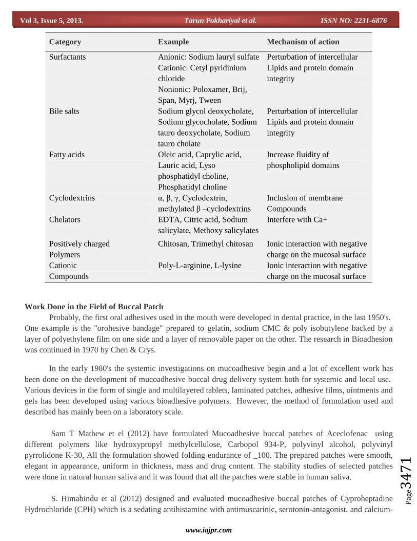
Absorption enhancers have demonstrated their effectiveness in delivering high molecular weight
compounds, such as peptides, that generally exhibit low buccal absorption rates. These may act by a number of
mechanisms, such as increasing the fluidity of the cell membrane, extracting inters/intracellular lipids, altering
cellular proteins or altering surface mucin. The most common absorption enhancers are azone, fatty acids, bile
salts and surfactants such as sodium dodecyl sulfate.
Penetration Enhancers and their Mechanism of Action21
www.iajpr.com
Pag
e34
71
Vol 3, Issue 5, 2013. Tarun Pokhariyal et al. ISSN NO: 2231-6876
Category Example Mechanism of action
Surfactants
Anionic: Sodium lauryl sulfate
Cationic: Cetyl pyridinium
chloride
Nonionic: Poloxamer, Brij,
Span, Myrj, Tween
Perturbation of intercellular
Lipids and protein domain
integrity
Bile salts
Sodium glycol deoxycholate,
Sodium glycocholate, Sodium
tauro deoxycholate, Sodium
tauro cholate
Perturbation of intercellular
Lipids and protein domain
integrity
Fatty acids
Oleic acid, Caprylic acid,
Lauric acid, Lyso
phosphatidyl choline,
Phosphatidyl choline
Increase fluidity of
phospholipid domains
Cyclodextrins
α, β, γ, Cyclodextrin,
methylated β –cyclodextrins
Inclusion of membrane
Compounds
Chelators EDTA, Citric acid, Sodium
salicylate, Methoxy salicylates
Interfere with Ca+
Positively charged
Polymers
Chitosan, Trimethyl chitosan
Ionic interaction with negative
charge on the mucosal surface
Cationic
Compounds
Poly-L-arginine, L-lysine
Ionic interaction with negative
charge on the mucosal surface
Work Done in the Field of Buccal Patch
Probably, the first oral adhesives used in the mouth were developed in dental practice, in the last 1950's.
One example is the "orohesive bandage" prepared to gelatin, sodium CMC & poly isobutylene backed by a
layer of polyethylene film on one side and a layer of removable paper on the other. The research in Bioadhesion
was continued in 1970 by Chen & Crys.
In the early 1980's the systemic investigations on mucoadhesive begin and a lot of excellent work has
been done on the development of mucoadhesive buccal drug delivery system both for systemic and local use.
Various devices in the form of single and multilayered tablets, laminated patches, adhesive films, ointments and
gels has been developed using various bioadhesive polymers. However, the method of formulation used and
described has mainly been on a laboratory scale.
Sam T Mathew et el (2012) have formulated Mucoadhesive buccal patches of Aceclofenac using
different polymers like hydroxypropyl methylcellulose, Carbopol 934-P, polyvinyl alcohol, polyvinyl
pyrrolidone K-30, All the formulation showed folding endurance of _100. The prepared patches were smooth,
elegant in appearance, uniform in thickness, mass and drug content. The stability studies of selected patches
were done in natural human saliva and it was found that all the patches were stable in human saliva.
S. Himabindu et al (2012) designed and evaluated mucoadhesive buccal patches of Cyproheptadine
Hydrochloride (CPH) which is a sedating antihistamine with antimuscarinic, serotonin-antagonist, and calcium-
www.iajpr.com
Pag
e34
72
Vol 3, Issue 5, 2013. Tarun Pokhariyal et al. ISSN NO: 2231-6876
channel blocking action. Buccal films were made with Hydroxy propylcellulose (HPC EF) and Hydroxy Propyl
Methyl Cellulose (HPMC E15) as mucoadhesive polymers. The formulation F8 of HPMC E15 was found to
give the better results and release of drug from the film followed Higuchi and Korsmeyer and Peppas models.
G. Shaji et al (2011) designed and optimized an oral controlled release Nebivolol mucoadhesive tablet
by using HPMC K4M, HPMC K15M and Carbomer-940 as mucoadhesive polymers, which significantly
influence characteristics like swelling index, ex-vivo mucoadhesive strength and in-vitro drug release. The
results indicate that suitable mucoadhesive buccal tablet with desired property can be prepared.
Bazigha K Abdul Rasool et al (2010) have prepared Five different film formulations containing 20 mg
of miconazole nitrate, drug solubilizers (propylene glycol 10% w/w, polyethylene glycol 3% w/w, tween20 6%
w/w, and oleic acid 5% w/w) and chitosan as film forming polymer, had been prepared. These preliminary
results indicate that the selected film formulation (MC 0.524 mg/cm2, PG 10% w/w and chitosan 2% w/w) can
represent a valid mean for the management of oral candidiasis.
Dheeraj Baviskar et al (2009) have prepared Mucoadhesive buccal patch of Aceclofenac using polymer
like Gelatin, Poly Sodium CMC and Poly Vinyl Alcohol. Eight formulations were prepared with varying the
concentration of Poly Sodium CMC and evaluated for various parameters like weight variation, patch thickness,
volume entrapment efficiency %, and measurement of % elongation at break, folding endurance, in vitro
mucoadhesive time, in vitro release and stability study.
R. Manivannan et al (2008) Mucoadhesive buccal tablets of Diltiazem hydrochloride were prepared
using carbopol-934, Sodium carboxy methyl cellulose (SCMC), Hydroxy propyl methyl cellulose (HPMC),
sodium alginate and guar-gum as mucoadhesive polymers. Eight formulations were developed with varying
concentrations of polymers. FTIR studies show no evidence on interaction between drug and polymers.
Formulation FA2 showed maximum release of 76.98% in 8hours
Bhupendra G. Prajapati et al (2007) prepared the buccal adhesive tablets musing sodium
carboxymethylcellulose (SCMC) and Carbopol-934 (CP) as bioadhesive polymers to impart mucoadhesion and
ethyl cellulose (EC) to act as an impermeable backing layer. Buccal devices were evaluated by different
parameters such as weight uniformity, content uniformity, thickness, hardness, surface pH, swelling index, ex
vivo mucoadhesive strength, ex vivo mucoadhesion time, in vitro drug release, and in vitro drug permeation. As
compared with bilayered tablets, multilayered tablets showed slow release rate of drug with improved ex vivo
bioadhesive strength and enhanced ex vivo mucoadhesion time.
A.P. Munasur et al (2006) designed a Box–Behnken experimental to optimise a polymeric blend for the
preparation of propranolol HCl matrices with maximum mucoadhesivity and were thereafter modified for
achieving controlled drug release. The quantitative effects of the polymers used i.e. poly (acrylic acid) (PAA)
and poly(vinyl pyrrolidone) (PVP) on mucoadhesion could be predicted using polynomial equations. A
formulation of 20% PAA, 20% CMC and 20% PVP was identified for maximizing mucoadhesivity and
obtaining a controlled drug release profile.
Lobna Mohamed Mortada et al (2003) Mucoadhesive patches for delivery of cetylpyridinium chloride
(CPC) were prepared using polyvinyl alcohol (PVA), hydroxyethyl cellulose (HEC) and chitosan. Swelling and
www.iajpr.com
Pag
e34
73
Vol 3, Issue 5, 2013. Tarun Pokhariyal et al. ISSN NO: 2231-6876
bioadhesive characteristics were determined for both plain and medicated patches. The results showed a
remarkable increase in radial swelling (SD) after addition of the water-soluble drug (CPC) to the plain
formulae. A decrease in the residence time was observed for PVA and chitosan-containing formulae. Higher
drug release was obtained from PVA patches compared to HEC ones, while both are non-ionic polymers.
K. Balamlfugam, J.K. Pandit 2001, was developed a systemic absorption of Propranolol hydrochloride
delivered through rabbit mucosa was studied from buccoadhesive films by using polymers combination sodium
CMC and carbopol with different ratio. S1 & S6 formulation, means alone SCMC (3) and SCMC (2.5) : CP
(0.5) having maximum inhibition of the heart rate for longer period of time.
Khurana R. et al. 2000, developed and evaluated mucoadhesive films of Miconazole nitrate for the
treatment of oral candidiasis. A film was prepared by casting procedure using various polymer combinations
and was evaluated for their in-vitro bioadhesive performance and release characteristics. The formulations
containing carbopol-934P and HPMC-M combination was found to give best result.
Shakoor O. et al. 1999 formulated bioadhesive buccal tablets for Nicotine replacement therapy and
demonstrated the ability to produce zero-order release from buccal adhesive tablets.
C. Li, P. Bhatt, 1998, has done evaluation of mucoadhesive buccal patch for delivery of peptides; in-
vitro screening of Bioadhesion
Khanna et al., 1997 designed muco-adhesive buccal films of clotrimazole using different polymers and
propylene glycol. The film was evaluated on the basis of their physical characteristics. A combination of
Carbopol-934P and HPMC in the ratio of 1: 5 and using ethanol as the solvent was found to give satisfactory
results
`Taylan B. et al. 1996, designed and evaluated sustained release buccoadhesive Propranol hydrochloride
tablets using HPMC, carbopol as polymers and 1% magnesium stearate was used as a lubricant. The result of
this study demonstrate that buccal adhesive propranol hydrochloride tablets containing PAA: HPMC of 2 : 8
showed suitable release and adhesive properties to the buccal mucus membrane.
Miyazaki S. et al. 1995, prepared bioadhesive tablets of Diltiazem by directly compressing the drug with
a mixture of chitosan and sodium alginate. They found that the maximum force of adhesion of these tablet to
the membrane increased with increasing alginate content of tablets. They also found that the release from
tablets composition of 1 : 4 and 1 : 1 chitosan/ Alginate was rapid with almost 100% release within 3 hrs.
Guo J.H. et al. 1994, prepared bioadhesive buccal patches for Buprenorphine controlled delivery using
carbopol 934, HPMC and chitosan and evaluated the in-vitro adhesion and release properties. They found
bioadhesive strength of buccal patches increases with increasing thickness up to a maximum value and swelling
is the major mechanism of buprenorphine release from buccal patches.
Cassidy et al. 1993, has done the in-vitro study of Buprenorphine to determine its buccal flux. Smart
J.D. 1992, evaluated the rate of release of a model water soluble drug from various polyacrylic acid containing
www.iajpr.com
Pag
e34
74
Vol 3, Issue 5, 2013. Tarun Pokhariyal et al. ISSN NO: 2231-6876
matrices. It was shown that a formulation containing carbopol-934P cross linked with calcium chloride was
found to give the slowest rate of drug release (t50% of 7.74) with release kinetics nearest to the ideal zero order.
Bottenberg P. et al. 1991, investigated bioadhesive characteristics of tablet made from modified starch,
polyacrylic acid (PAA), PEG and sodium CMC. They observed that despite its long adhesion time in-vivo,
clinical application of PAA in the oral cavity is not recommended but when PAA used in small amount together
in a non-irritating polymer, PAA improves the bioadhesive qualities of the formulation.
Collins A.E. and Deasy P.B. 1990, studied the release of cetyl pyridinium chloride from two-three
layered bioadhesive flavoured device in six healthy human volunteers. It was observed that in comparison with
a proprietary lozenge, the device produced more uniform and effective level of drug (20mg/ml), with adequate
comfort, taste and non-irritancy over a period of 3 hrs.
Anders R. and Merkle H.P. 1989, developed and evaluated laminated mucoadhesive patches of
Protirelin for buccal drug delivery. The patches consisted of the two-ply laminates of an impermeable backing
layer and a hydrocolloid polymer layer containing the drug. The duration of mucosal adhesion in-vivo was
found to be dependent on the type of polymer used, its viscosity grade, the polymer load per patch and drying
procedure for the preparation.
Khar R.K. et al. 1988, developed buccoadhesive erodible films of Triamcinoline acetonide for the
treatment of oral lesions using carbopol 934P, hydroxy propyl cellulose-M, HPMC-EUM, HPMC alone and in
different ratio. Propylene glycol was used as plasticizer and films were prepared by solvent casting method.
They found results obtained from erodible patches of different formulations were comparable.
Yotsuyangi et al. 1985, designed a mucoadhesive, moderately water-soluble polymeric film containing
Analgesics and Antibiotics for the treatment of lesions and also to ease the accompanying pain. The film
consisted of HPC-M and contained tetracaine, thiamphenicol and triacetin.
Bremecker K.D. et al. 1984, formulated a novel mucosal adhesive ointment based partly on neutralized
polymethacrylic acid methyl ester. The rhelogical behavior as well as the adhesion on the mucosal membrane
could be varied by the type and concentration of the polymer used and the base used for neutralization. Both
vehicle and preparation were found to be pleasant for patients use.
www.iajpr.com
Pag
e34
75
Vol 3, Issue 5, 2013. Tarun Pokhariyal et al. ISSN NO: 2231-6876
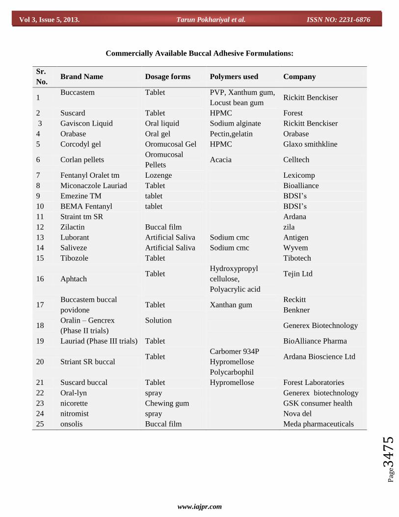
Commercially Available Buccal Adhesive Formulations:
Sr.
No. Brand Name Dosage forms Polymers used Company
1 Buccastem
Tablet
PVP, Xanthum gum,
Locust bean gum Rickitt Benckiser
2 Suscard Tablet HPMC Forest
3 Gaviscon Liquid Oral liquid Sodium alginate Rickitt Benckiser
4 Orabase Oral gel Pectin,gelatin Orabase
5 Corcodyl gel Oromucosal Gel HPMC Glaxo smithkline
6 Corlan pellets Oromucosal
Pellets Acacia Celltech
7 Fentanyl Oralet tm Lozenge Lexicomp
8 Miconaczole Lauriad Tablet Bioalliance
9 Emezine TM tablet BDSI’s
10 BEMA Fentanyl tablet BDSI’s
11 Straint tm SR Ardana
12 Zilactin Buccal film zila
13 Luborant Artificial Saliva Sodium cmc Antigen
14 Saliveze Artificial Saliva Sodium cmc Wyvem
15 Tibozole Tablet Tibotech
16 Aphtach Tablet
Hydroxypropyl
cellulose,
Polyacrylic acid
Tejin Ltd
17 Buccastem buccal
povidone Tablet Xanthan gum
Reckitt
Benkner
18 Oralin – Gencrex
(Phase II trials)
Solution
Generex Biotechnology
19 Lauriad (Phase III trials) Tablet BioAlliance Pharma
20 Striant SR buccal Tablet
Carbomer 934P
Hypromellose
Polycarbophil
Ardana Bioscience Ltd
21 Suscard buccal Tablet Hypromellose Forest Laboratories
22 Oral-lyn spray Generex biotechnology
23 nicorette Chewing gum GSK consumer health
24 nitromist spray Nova del
25 onsolis Buccal film Meda pharmaceuticals
www.iajpr.com
Pag
e34
76
Vol 3, Issue 5, 2013. Tarun Pokhariyal et al. ISSN NO: 2231-6876
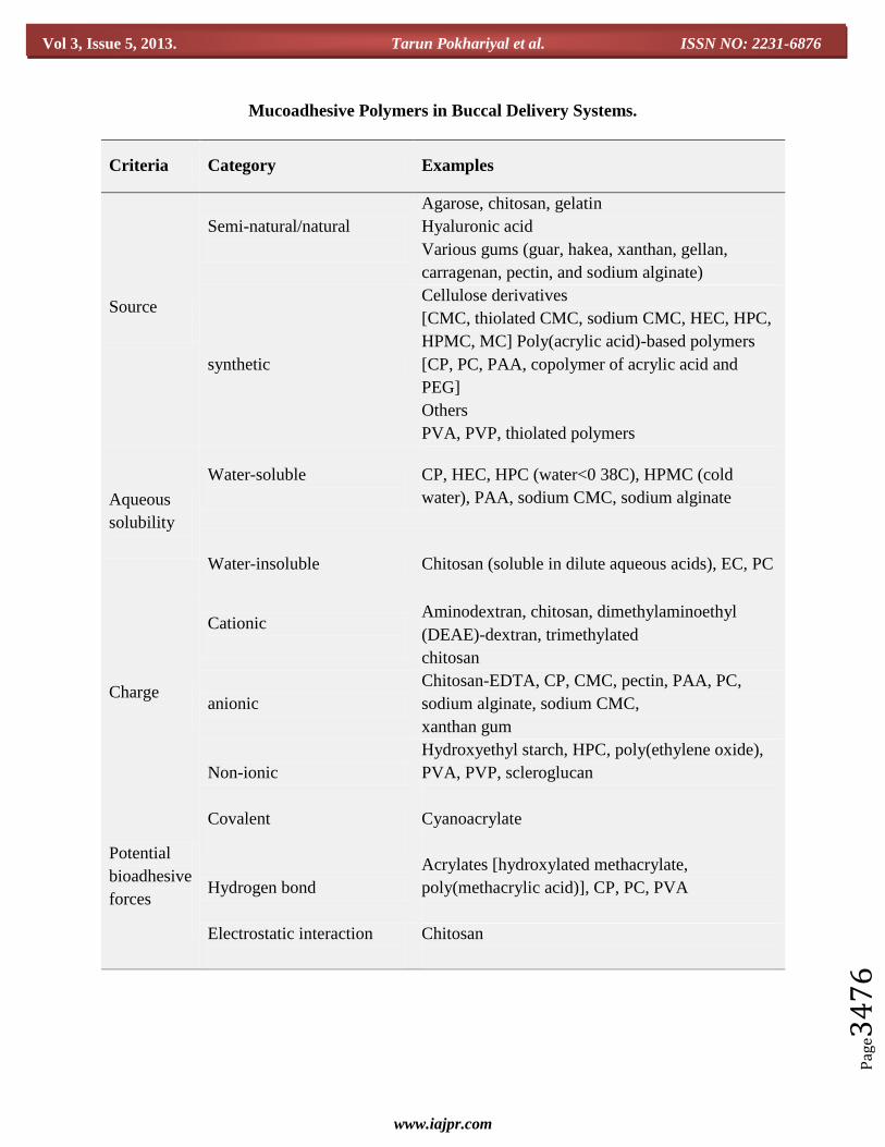
Mucoadhesive Polymers in Buccal Delivery Systems.
Criteria Category Examples
Source
Semi-natural/natural
Agarose, chitosan, gelatin
Hyaluronic acid
Various gums (guar, hakea, xanthan, gellan,
carragenan, pectin, and sodium alginate)
synthetic
Cellulose derivatives
[CMC, thiolated CMC, sodium CMC, HEC, HPC,
HPMC, MC] Poly(acrylic acid)-based polymers
[CP, PC, PAA, copolymer of acrylic acid and
PEG]
Others
PVA, PVP, thiolated polymers
Aqueous
solubility
Water-soluble
CP, HEC, HPC (water<0 38C), HPMC (cold
water), PAA, sodium CMC, sodium alginate
Water-insoluble Chitosan (soluble in dilute aqueous acids), EC, PC
Charge
Cationic
Aminodextran, chitosan, dimethylaminoethyl
(DEAE)-dextran, trimethylated
chitosan
anionic
Chitosan-EDTA, CP, CMC, pectin, PAA, PC,
sodium alginate, sodium CMC,
xanthan gum
Non-ionic
Hydroxyethyl starch, HPC, poly(ethylene oxide),
PVA, PVP, scleroglucan
Potential
bioadhesive
forces
Covalent
Cyanoacrylate
Hydrogen bond
Acrylates [hydroxylated methacrylate,
poly(methacrylic acid)], CP, PC, PVA
Electrostatic interaction
Chitosan

www.iajpr.com
Pag
e34
77
Vol 3, Issue 5, 2013. Tarun Pokhariyal et al. ISSN NO: 2231-6876
Composition of Buccal Patches:-24-26
A. Active ingredient.
B. Polymers (adhesive layer):- Hydroxy ethylcellulose, hydroxypropyl cellulose, polyvinyl pyrrolidone,
polyvinyl alcohol, carbopol and other mucoadhesive polymers.
C. Diluents:- Lactose DC is selected as diluent for its high aqueous solubility, its flavouring characteristics,
and its physico-mechanical properties, which make it suitable for direct compression. Other example:
microcrystalline starch and starch.
D. Sweetening agents:- Sucralose, aspartame, mannitol, etc.
E. Flavouring agents:- Menthol, vanillin, clove oil, etc.
F. Backing layer:- Ethyl cellulose, etc.
G. Penetration enhancer:- Cyano acrylate, etc.
H. Plasticizers:- PEG-100, 400, propylene glycol, etc
Methods of Preparation of Buccal Patch
26-30
(a) Solvent casting technique
In this technique the required quantity of mucoadhesive polymer is treated with required volume of
solvent system and vortexed to allow polymer to swell. After swelling, mixture is treated with, measured
quantity of plasticizer (propylene glycol or glycerin or dibutyl phthalate) and vortexed. Finally the required
quantity of drug is dissolved in small volume of solvent system and added to the polymer solution and mixed
well. Then set aside for some time to remove any entrapped air and transferred into a previously cleaned petri
plate. The formed patches were stored in a desiccator till the evaluation tests were performed.
(b) Hot melt extrusion technique
The Hot-melt extrusion (HME) technique is an attractive alternative to traditional processing methods
and offers many advantages over the other pharmaceutical processing techniques. Molten polymers during the
extrusion process can function as thermal binders and act as drug depots and/or drug release retardants upon
cooling and solidification. Since solvents and water are not necessary, the numbers of processing and time-
consuming drying steps are reduced. A matrix can be massed into a larger unit independent of compression
properties. The intense mixing and agitation imposed by the rotating screw cause de-aggregation of suspended
particles in the molten polymer resulting in a more uniform dispersion and the process is continuous and
efficient. Bioavailability of the drug substance may be improved when it is solubilized or dispersed at the
molecular level in HME dosage forms. Pharmaceutical Hot-Melt Extrusion processes can be categorized as
either ram extrusion or screw extrusion
(c) Solvent evaporation method
In this technique the required quantity of mucoadhesive polymer is treated with required volume of
solvent system and heat on water bath to dissolve polymer properly than dissolved the drug in that solution by
heating and add plasticer in required quantity. . Than set aside for some time to remove any entrapped air and
transferred into a previously cleaned petri plate. The formed patches were stored in a desiccator till the
evaluation tests were performed.

www.iajpr.com
Pag
e34
78
Vol 3, Issue 5, 2013. Tarun Pokhariyal et al. ISSN NO: 2231-6876
Evaluations of Buccal Patch: 30-34
1. Surface pH:-
a. Buccal patches are left to swell for 2 hr on the surface of an agar plate. The surface pH is
measured by means of a pH paper placed on the surface of the swollen patch.
2. Thickness measurements:-
a. The thickness of each film is measured at five different locations (centre and four corners) using
an electronic digital micrometer.
3. Swelling study:-
a. Buccal patches are weighed individually (designated as W1), and placed separately in 2% agar
gel plates, incubated at 37°C ± 1°C, and examined for any physical changes. At regular 1- hour
time intervals until 3 hours, patches are removed from the gel plates and excess surface water is
removed carefully using the filter paper The swollen patches are then reweighed (W2) and the
swelling index (SI) is calculated
4. Folding endurance:-
The folding endurance of patches is determined by repeatedly folding 1 patch at the same place until it
breaks or is folded up to 200 times without breaking.
5. Thermal analysis study:-
Thermal analysis study is performed using differential scanning calorimeter (DSC).
6. Morphological characterization:-
Morphological characters are studied by using scanning electron microscope (SEM).
7. Water absorption capacity test:-
Circular Patches, with a surface area of 2.3 cm2 are allowed to swell on the surface of agar plates
prepared in simulated saliva (2.38 g Na2HPO4, 0.19 gKH2PO4, and 8 g NaCl per liter of distilled water
adjusted with phosphoric acid to pH 6.7), and kept in an incubator maintained at 37°C ± 0.5°C. At
various time intervals (0.25, 0.5, 1, 2, 3, and 4 hours), samples are weighed (wet weight) and then left to
dry for 7 days in a desiccators over anhydrous calcium chloride at room temperature then the final
constant weights are recorded. Water uptake (%) is calculated using the following equation.
Water uptake (%) = (Ww – Wi )/Wf x 100
Where,
Ww is the wet weight and Wf is the final weight. The swelling of each film is measured.
8. Ex-vivo bioadhesion test:-
The fresh sheep mouth separated and washed with phosphate buffer (pH 6.8). A piece of gingival
mucosa is tied in the open mouth of a glass vial, filled with phosphate buffer (pH 6.8). This glass vial is

www.iajpr.com
Pag
e34
79
Vol 3, Issue 5, 2013. Tarun Pokhariyal et al. ISSN NO: 2231-6876
tightly fitted into a glass beaker filled with phosphate buffer (pH 6.8, 37°C ± 1°C) so it just touched the
mucosal surface. The patch is stuck to the lower side of a rubber stopper with cyano acrylate adhesive.
Two pans of the balance are balanced with a 5-g weight. The 5-g weight is removed from the left hand
side pan, which loaded the pan attached with the patch over the mucosa. The balance is kept in this
position for 5 minutes of contact time. The water is added slowly at 100 drops/min to the right-hand side
pan until the patch detached from the mucosal surface. The weight, in grams, required to detach the
patch from the mucosal surface provided the measure of mucoadhesive strength.
a. Measurement of mucoadhesive strength
9. In Vitro Drug Release:-
The United States Pharmacopeia (USP) XXIII-B rotating paddle method is used to study the
drug release from the bilayered and multilayered patches. The dissolution medium consisted of
phosphate buffer pH 6.8. The release is performed at 37°C ± 0.5°C, with a rotation speed of 50 rpm. The
backing layer of buccal patch is attached to the glass disk with instant adhesive material. The disk is
allocated to the bottom of the dissolution vessel. Samples (5 ml) are withdrawn at predetermined time
intervals and replaced with fresh medium. The samples filtered through whatman filter paper and
analyzed for drug content after appropriate dilution. The invitro buccal permeation through the buccal
mucosa (sheep and rabbit) is performed using Keshary-Chien/Franz type glass diffusion cell at 37°C±
0.2°C. Fresh buccal mucosa is mounted between the donor and receptor compartments. The buccal patch
is placed with the core facing the mucosa and the compartments clamped together. The donor
compartment is filled with buffer.
www.iajpr.com
Pag
e34
80
Vol 3, Issue 5, 2013. Tarun Pokhariyal et al. ISSN NO: 2231-6876
1. Schematic diagram of Franz diffusion cell for buccal patch
10. Permeation study of buccal patch:-
The receptor compartment is filled with phosphate buffer pH 6.8, and the hydrodynamics in the
receptor compartment is maintained by stirring with a magnetic bead at 50 rpm. Samples are withdrawn
at predetermined time intervals and analyzed for drug content.
11. Ex-vivo mucoadhesion time:-
The ex-vivo mucoadhesion time performed after application of the buccal patch on freshly cut
buccal mucosa (sheep and rabbit). The fresh buccal mucosa is tied on the glass slide, and a
mucoadhesive patch is wetted with 1 drop of phosphate buffer pH 6.8 and pasted to the buccal mucosa
by applying a light force with a fingertip for 30 seconds. The glass slide is then put in the beaker, which
is filled with 200 ml of the phosphate buffer pH 6.8, is kept at 37°C ± 1°C. After 2 minutes, a 50-rpm
stirring rate is applied to simulate the buccal cavity environment, and patch adhesion is monitored for 12
hours. The time for changes in colour, shape, collapsing of the patch, and drug content is noted.
12. Measurement of mechanical properties:-
Mechanical properties of the films (patches) include tensile strength and elongation at break is
evaluated using a tensile tester. Film strip with the dimensions of 60 x 10 mm and without any visual
defects cut and positioned between two clamps separated by a distance of 3 cm. Clamps designed to
secure the patch without crushing it during the test, the lower clamp held stationary and the strips are
pulled apart by the upper clamp moving at a rate of 2 mm/sec until the strip break. The force and
elongation of the film at the point when the strip break is recorded. The tensile strength and elongation
at break values are calculated using the formula.
T = m x g/ b x t Kg/mm2
Where,
M - is the mass in gm, g - is the acceleration due to gravity 980 cm/sec 2
B - is the breadth of the specimen in cm
T - is the thickness of specimen in cm.
Tensile strength (kg/mm2) is the force at break (kg) per initial cross- sectional area of the specimen
(mm2)
13. Stability study in human saliva:-
The stability study of optimized bilayered and multilayered patches is performed in human
saliva. The human saliva is collected from humans (age 18-50years). Buccal patches are placed in
separate Petri dishes containing 5ml of human saliva and placed in a temperature controlled oven at
37°C ± 0.2°C for 6 hours.
FUTURE CHALLENGES AND OPPORTUNITIES
The main impediment to the use of many hydrophilic macromolecular drugs as potential therapeutic
agents is their inadequate and erratic oral absorption. The relatively recent evolution of recombinant DNA
research and modern synthetic and biotechnological methodologies allow the biochemist and chemist to
produce vast quantities of variety of peptides and proteins possessing better pharmacological efficacy.
However, therapeutic potential of these compounds lies in our ability to design and achieve effective and stable
delivery systems. The future challenge of pharmaceutical scientists will not only be polypeptide cloning and

www.iajpr.com
Pag
e34
81
Vol 3, Issue 5, 2013. Tarun Pokhariyal et al. ISSN NO: 2231-6876
synthesis, but also to develop effective non-parenteral delivery of intact proteins and peptides to the systemic
circulation. Buccal permeation can be improved by using various classes of transmucosal and transdermal
penetration enhancers such as bile salts, surfactants, fatty acids and derivatives, chelators and cyclodextrins.
Researchers are now looking beyond traditional polymer networks to find other innovative drug. systems are
particularly interesting as they offer protection to therapeutic entities as well as the enhanced absorption that
result from increased contact time provided by the bioadhesive component. Exciting challenges remain to
influence the bioavailability of drugs across the buccal mucosa. Many issues are yet to be resolved before the
safe and effective delivery through buccal mucosa. Successfully developing these novel formulations requires
assimilation of a great deal of emerging information about the chemical nature and physical structure of these
new materials. The future direction of buccal adhesive drug delivery lies in vaccine formulations and delivery
of small proteins/peptides. Another important aspect concerns the in vitro and ex vivo techniques which are
employed for evaluation of the performance of the materials and dosage forms. Efforts should be made to
develop standard in vitro and ex vivo biological models that allow one to characterize and compare different
material and formulation in terms of their capability to promote drug absorption via the buccal route.
CONCLUSION
The need for research into drug delivery systems extends beyond ways to administer new
pharmaceutical therapies. Buccal adhesive systems offer innumerable advantages in terms of accessibility,
administration and withdrawal, retentivity, low enzymatic activity, economy and high patient compliance.
Adhesions of these drug delivery devices to mucosal membranes lead to an increased drug concentration
gradient at the absorption site and therefore improve bioavailability of systemically delivered drugs. In addition,
buccal adhesive dosage forms have been used to target local disorders at the mucosal surface (e.g., mouth
ulcers), to reduce the overall required dosage and minimize side effects that may be caused by systemic
administration of drugs. Investigations are continuing beyond traditional polymer networks to find other
innovative drug transport systems. At the current global scenario, scientists are finding ways to develop buccal
adhesive systems through various approaches to improve the bioavailability of drugs used orally by
manipulation of the formulation strategies like inclusion of pH modifiers, enzyme inhibitors, permeation
enhances etc.
REFERENCES
1. R. Venkatalakshmi, Yajaman Sudhakar , Madhuchudana Chetty C., Sasikala C. and Mohan Varma M “Buccal
Drug Delivery using Adhesive Polymeric Patches”. International Journal of Pharmaceutical science and
research 2012; Vol. 3(1): 35-41.
2. Ajeet, Brajpal Singh, Rashmi Juneja “Recent Findings in Concern To Buccal Patches: A Review” International
Journal of Current Pharmaceutical Research 2012 Vol 4, Issue 1, 12-17.
3. Vivekanand Prajapati, Mayank Bansal, Pramod Kumar Sharma “Mucoadhesive Buccal Patches and Use of
Natural Polymer in Its Preparation – A Review” International Journal of PharmTech Research Vol.4, No.2, pp
582-589.
4. Kumar V, Aggarwal G, Zakir F, Choudhary A “ Buccal Bioadhesive Drug Delivery- A Novel Technique”
International Journal of Pharmacy and Biological Sciences |Volume 1| Issue 3 |july-sept |2011|89-102.
5. A. Puratchikody, Prasanth V.V, Sam T. Mathew, Ashok Kumar B. “Buccal Drug Delivery: Past, Present and
Future – A Review” International Journal of Drug Delivery 3 (2011) 171-184.
www.iajpr.com
Pag
e34
82
Vol 3, Issue 5, 2013. Tarun Pokhariyal et al. ISSN NO: 2231-6876
6. Kumar V, Aggarwal G, Zakir F, Choudhary A “Buccal Bioadhesive Drug Delivery- A Novel Technique”
International Journal Of Pharmacy And Biological Sciences (Eissn: 2230-7605) Ijpbs |Volume 1| Issue 3 |July-
Sept |2011|89-102.
7. Khar K, Ahuja A, Javed A. Mucoadhesive drug delivery. Controlled and novel drug delivery by Jain NK. 1st
Ed. New Delhi; 1997.
8. Vyas SP, Khar RK. Controlled drug delivery concepts and advances.Vallabh Prakashan, 1st Ed. Delhi; 2002.
9. Tripathi K.D. “essential of medical Pharmacology” 6th edition New Delhi 2008.
10. Rowe C. Reymond “Handbook of Pharmaceutical Excipients” 5th edition London 2006
11. P.Ilavarasan, K.Ezhumalai, A.N.Rajalakshmi “Buccal Patches As Emerging Trend” International Journal of
Pharmacy and Technology | June-2011 | Vol. 3 | Issue No.2 | 973-986.
12. Chinna Reddy P, Chaitanya K.S.C,Madhusudan Rao Y. “ A review on bioadhesive buccal drug delivery
systems: current status of formulation and evaluation methods” DARU Vol. 19, No. 6 2011
13. .Patel K.V., Patel N.D, Dodiya H.D, Shelat P.K. “ Buccal Bioadhesive Drug Delivery System: An Overview”
International Journal of Pharmaceutical & Biological Archives 2011; 2(2): 600-609.
14. Biswajit Basu, Kevin Garala, Thimmasetty j. “formulation and evaluation of pimozide buccal mucoadhesive
patches” International journal of pharmaceutical sciences and nanotechnology volume 2 • issue 4 • january –
march 2010
15. Prasanth V.V, Mamatha. Y, Selvi Arunkumar, Sam T Mathew, Abin Abraham “Formulation and Evaluation Of
Mucoadhesive Buccal Patches Of Aceclofenac” Scholars Research Library Der Pharmacia Lettre, 2012, 4
(1):297-306.
16. Yashwant T. Dangat, Nilesh A. Bari, Swati C. Jagdale Aniruddha R. Chabukswar and Bhanudas S. Kuchekar
“Controlled release from bisoprolol fumarate buccal patches” Pelagia Research Library Der Pharmacia Sinica,
2012, 3 (3):317-320.
17. S. Himabindu, D. Sathish and Shayeda “Formulation and In-vitro Evaluation of Mucoadhesive Buccal Patches
of Cyproheptadine Hydrochloride” Journal of Applied Pharmaceutical Science 02 (07); 2012: 196-201.
18. K.S.C. Chopparapu, P. Chinna Reddy, Narender Doodipala and Y.M. Rao “Development of Promethazine
Hydrochloride Mucoadhesive Patches for Buccal Delivery: In vitro, Ex vivo and In vivo Characterization”
International Journal of Pharmaceutical Sciences and Nanotechnology Volume 5 • Issue 2• July – September
2012.
19. Varinder Kumar, Foziyah Zakir, Geeta Agarwal, Ankush Choudhary “ Formulation And Evaluation Of Buccal
Patches Of Venlafaxine” International Journal Of Pharmacy And Biological Sciences |Volume 1| Issue 3 |July-
Sept |2011|170-182.
20. Anuj Kumar, Vikas Phatarpekar, Naveen Pathak, Kumud Padhee, Minakshi Garg And Neeta Sharma
“Formulation Development & Evaluation Of Carvedilol Bioerodable Buccal Mucoadhesive Patches” Pharmacie
Globale International Journal Of Comprehensive Pharmacy Vol. 02, Issue 03.
21. P.Suresh Kumar, B.Srikanth, T.Satyanarayana, G.Shaji, S.Navaneetha Krishnan, P.Saranya “Formulation and
Evaluation Of Nebivolol Mucoadhesive Buccal Tablet” Pharmacologyonline 3: 869-885 (2011).
22. Santosh Kumar Mishra, Navneet Garud and Ranjit Singh “Development And Evaluation Of Mucoadhesive
Buccal Patches Of Flurbiprofen” Acta Poloniae Pharmaceutica N Drug Research, Vol. 68 No. 6 Pp. 955n964,
2011.
23. Bazigha K Abdul Rasool, Saeed A. Khan “In Vitro Evaluation Of Miconazole Mucoadhesive Buccal Films”
International journel of applied pharmaceutics Vol 2, Issue 4, 2010.
24. Parikh Bhavik Anjankumar “Design And Evaluation Of Buccal Patches Of Valsartan” IJPI’s Journal of
Pharmaceutics And Cosmetology Vol 1: 2 (2011) 51-55
www.iajpr.com
Pag
e34
83
Vol 3, Issue 5, 2013. Tarun Pokhariyal et al. ISSN NO: 2231-6876
25. Doshi Abha, Koliyote Sheeja, Joshi Bhagyashri “Design And Evaluation Of Buccal Film Of Diclofenac
Sodium” International Journal of Pharmacy and Biological Sciences (ISSN:2230-7605) Volume 1, Issue 1,
JAN-MARCH 2011.
26. Bhanja Satyabrata, P Ellaiah, Rohit Choudhury, KVR Murthy, Panigrahi Bibhutibhusan, Martha Sujit Kumar
“Design and evaluation of methotrexate buccal mucoadhesive patches” Pharma Inter Science Publishers
International Journel of Pharmacy Biomededical Science 2010, 1(2), 31-36.
27. M. Alagusundaram, B. Chengaiah, S. Ramkanth, S. Angala Parameswari, C. Madhu Sudhana Chetty and D.
Dhachinamoorthi “Formulation and Evaluation of Mucoadhesive Buccal Films of Ranitidine” International
Journal of PharmTech Research Vol.1, No.3, pp 557-563.
28. Amit Khairnar, Parridhi Jain, Dheeraj Baviskar and Dinesh Jain “Developmement of Mucoadhesive Buccal
Patch Containing Aceclofenac: In Vitro Evaluations” International Journal of PharmTech Research Vol.1, No.4,
pp 978-981.
29. R Manivannan, A Balasubramaniam, DC Prem Anand, G Sandeep and N Rajkumar “Formulation and In-Vitro
Evaluation of Mucoadhesive Buccal Tablets of Diltiazem Hydrochloride” Research J. Pharm. and Tech. 1(4):
Oct.-Dec. 2008.
30. K. Chandra Sekhar, K. V. S. Naidu, Y. Vamshi Vishnu, Ramesh Gannu, V. Kishan, and Y. Madhusudan Rao
“Transbuccal Delivery of Chlorpheniramine Maleate from Mucoadhesive Buccal Patches” Informa Healthcare
USA, Inc. Drug Delivery, 15:185–191, 2008.
31. Vishnu M. Patel, Bhupendra G. Prajapati, and Madhabhai M. Patel “Formulation, Evaluation, and Comparison
of Bilayered and Multilayered Mucoadhesive Buccal Devices of Propranolol Hydrochloride” AAPS
PharmSciTech 2007; 8 (1) Article 22.
32. A.P. Munasur , V. Pillay , D.J. Chetty , T. Govender “Statistical optimisation of the mucoadhesivity and
characterisation of multipolymeric propranolol matrices for buccal therapy” sciencedirect Elsevier
International Journal of Pharmaceutics 323 (2006) 43–51.
33. Noha adel, nafee nabila, ahmed boraie, fatma ahmed, ismail lobna, mohamed mortada “Design and
characterization of mucoadhesive buccal patches containing cetylpyridinium chloride” Acta Pharm. 53 (2003)
199–212.
34. Han-Gon Choi , Jac-Hee Jung , Chul Soon Yong , Chong-Dal Rhee , Mi-Kyung Lee, Jeong-Hee Han, Kyung-
Mi Park, Chong-Kook Kim “Formulation and in vivo evaluation of omeprazole buccal adhesive tablet” Elsevier
Journal of Controlled Release 68 (2000) 405–412.
35. Aarifkhan. M. Pathan. Development and in-vitro evaluation of salbutamal sulphate Mucoadhesive buccal
patches. Int J pharm pharm sci, 2011, vol 3, suppl 2, 39-44.
54878478451001254
Submit your next manuscript to IAJPR and take advantage of: • Access Online first • Double blind peer review policy • No space constraints • Rapid publication • International recognition Submit your manuscript at: [email protected]


